HAL Id: tel-01407360
https://tel.archives-ouvertes.fr/tel-01407360
Submitted on 2 Dec 2016HAL is a multi-disciplinary open access archive for the deposit and dissemination of sci-entific research documents, whether they are pub-lished or not. The documents may come from teaching and research institutions in France or abroad, or from public or private research centers.
L’archive ouverte pluridisciplinaire HAL, est destinée au dépôt et à la diffusion de documents scientifiques de niveau recherche, publiés ou non, émanant des établissements d’enseignement et de recherche français ou étrangers, des laboratoires publics ou privés.
Study of bacterial populations from oligotrophic soil
ecosystems using high throughput sequencing
technologies
Jorge Osman Naoum
To cite this version:
Jorge Osman Naoum. Study of bacterial populations from oligotrophic soil ecosystems using high throughput sequencing technologies. Biodiversity and Ecology. Université Paris-Saclay, 2016. English. �NNT : 2016SACLS196�. �tel-01407360�
1
NNT : 2016SACLS196
T
HESE DE DOCTORAT
DE
L’U
NIVERSITE
P
ARIS
-S
ACLAY
PREPAREE A
“
L
’
UNIVERSITE PARIS
-
SUD
”
ECOLE DOCTORALE N°577
Structure et Dynamique des Systèmes Vivants
Spécialité de doctorat : Sciences de la Vie et de la Santé
Par
M. Jorge Osman Naoum
Study of bacterial populations from oligotrophic soil ecosystems
using high throughput sequencing technologies
Thèse présentée et soutenue à Orsay, le 5 août 2016: Composition du Jury :
M. Lane, David Directeur de Recherche Emérite, CNRS Rapporteur Mme. Lett, Marie-Claire Professeure, Université de Strasbourg Rapporteur Mme. Astier, Chantal Professeure Emérite, Université Paris-Sud Examinatrice M. Reichhart, Jean-Marc Professeur, Université de Strasbourg Examinateur Mme. Shykoff, Jacqui Directrice de Recherche, CNRS Président
3
Acknowledgements
I would like to take this opportunity to express my thanks to the people who have helped, guided, and supported me throughout the course of my thesis. First, I would like to express my sincere recognitions to Pr. Michael DuBow for his mentorship, as well for the opportunity and confidence that gave me during these four years of research.
Beside my supervisor, I would like to thanks all the jury members to accept be part of this thesis.
I also want to thanks Christophe Regeard and Micheline Terrier for his help and interesting discussions.
Furthermore, my colleagues Gustavo, Yang, Nga and Catherine.
My Ph.D. in France was supported by the Chilean Government “Becas Chile”, CONICYT, from which I am very grateful.
Finally I would like to thanks all my family, my parents, sister, brother, uncles, cousins and friends for the encouragement.
4
List of contents
Chapter I (Review Article): Bacterial communities of oligotrophic soils... 6
Review Article: Bacterial communities of the surface of oligotrophic nutrient-poor soils ... 7
Abstract ... 8
1.Introduction ... 9
2.Advances in microbial identification ... 11
3.Bacterial biogeography ... 15
4.Principal factors affecting bacterial diversity in surface soils ... 16
5.Biogeochemistry: How soils get mineralized ... 18
6.Bacterial adaptations and roles in mineral soil surfaces ... 21
7.Bacteria members living in mineralized surface soil ecosystems ... 24
8.Bacterial communities of oligotrophic ecosystems ... 25
9.Conclusions ... 28
References ... 29
Thesis objectives ... 40
Chapter II (Research Article I): Bacterial communities from the Camargue rice fields (France) ... 41
Article I: Bacterial diversity of the rhizosphere and nearby surface soil of rice (Oryza sativa) growing in the Camargue (France) ... 42
Chapter III (Research Article II): Bacterial biodiversity from soil and sediments from the Camargue (France) ... 85
Article II: Variation of bacterial biodiversity from saline soils and estuary sediments present near the Mediterranean Sea coast of Camargue (France) ... 86
Chapter IV (Research Article III): Bacterial communities from surface soils from “Dapani” in Mayotte ... 127
Article III: Examination of the Bacterial Biodiversity of Coastal Eroded Surface Soils from the Padza de Dapani (Mayotte Island) ... 141
Chapter V (Research Article IV): Bacterial biodiversity from the rhizosphere of plants from Jizan in Saudi Arabia ... 168
Article IV: Bacterial rhizosphere biodiversity from several pioneer desert sand plants near Jizan, Saudi Arabia ... 169
5
Chapter VI Discussion and general conclusions ... 179
1.Research challenge ... 180
2.Techniques and methods used ... 181
2.1 Soil sampling ... 181
2.2 DNA extraction ... 181
2.3 Primer pair design ... 182
2.4 Quantitative PCR (qPCR) ... 183
2.5 Pyrosequencing and data analysis ... 183
3.Conclusions ... 188
3.1 Study reflections ... 188
3.2 Bacterial abundance challenges ... 188
3.3 Bacterial populations across different ecosystems ... 189
References ... 191
Résumé ... 193
Abstract ... 195
List of Figures
Figure 1. The mineralosphere and its ecological traits. ... 20Figure 2. Representation of pyrosequencing primers design for 454 Lib-L chemistry pyrosequencing. ... 182
Figure 3. In silico primer set coverages ... 183
Figure 4. Schematic representation of the approaches used for soil sample analyses. . 185
List of tables
Table 1 Bacterial members associated to inorganic mineral transformations ... 236
CHAPTER I
(REVIEW ARTICLE):
Bacterial communities of
7 Review Article
Bacterial Communities of the Surface of Oligotrophic Nutrient-Poor Soils
Jorge Osman1& Michael S. DuBow1
1Laboratoire de Génomique et Biodiversité Microbienne des Biofilms, Institute for
Integrative Biology of the Cell (I2BC), CEA, CNRS, Université Saclay, Univ Paris-Sud, Bâtiment 409, 91405 Orsay, France
Correspondence: Michael S. DuBow, Laboratoire de Génomique et Biodiversité Microbienne des Biofilms, Institute for Integrative Biology of the Cell (I2BC), CEA, CNRS, Université Paris-Saclay, Univ Paris-Sud, Bâtiment 409, 91405 Orsay, France; Tel: +33(0)169154612; Fax:+33(0)169157808; e-mail: michael.dubow@u-psud.fr
8 Abstract
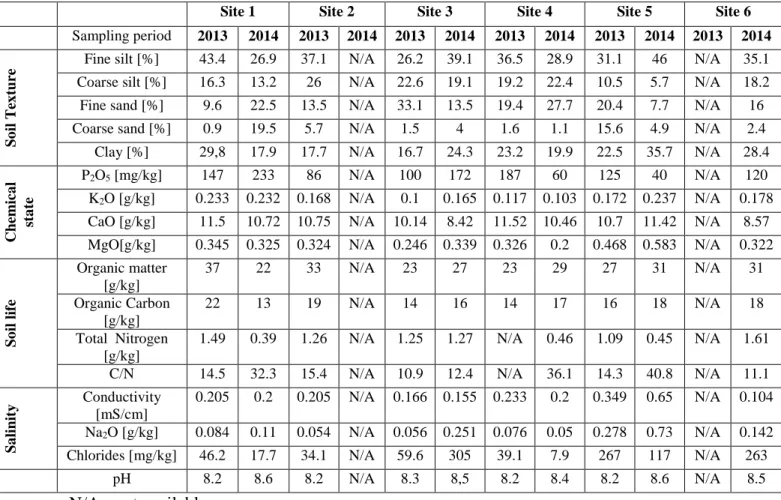
Oligotrophic soil ecosystems can contain a wide variety of bacterial communities. However, these communities vary in composition according to the mineral content and the chemical and physical properties of their environment. This creates a challenge for microbiologists intending to identify population patterns based on ecosystem characteristics. Advances in new technologies improve the identification of bacterial groups, which helps in understanding the role of microbial populations in different biomes. From studies of several sites, we have described the principal bacterial members involved in mineral transformations in the surface layers of nutrient-poor soil ecosystems as well as the bacterial groups present in various oligotrophic ecosystems We also assess factors that could shape the bacterial communities in these ecosystems, to better understand their distribution and their possible roles on mineral soil surfaces.
9 1-Introduction
Soil has been recognized as one of the most diverse microbial habitats on Earth (1, 2). Soils are complex physical and chemical environments, where heterogeneous mixtures of minerals and nutrients, water, organic matter and biological species interact (3). Soil microorganisms play a fundamental role as principal drivers of biogeochemical cycles, through activities such as nutrient acquisition and cycling of nitrogen and carbon, and through their roles in soil formation and in soil plant and animal health (4, 5). The development of new sequencing technologies has enabled researchers in microbial ecology to understand how ecosystem components and microbes interact. Altogether, microbial populations, which participate in soil chemical cycling, are important for predicting future dynamics and consequences of global environmental changes (6, 7, 8). The principal biological fluxes of the major elements of life (H, C, N, S, O and P) are driven by microorganisms, although in the case of P, volcanic activities and rock weathering are also significant contributors. These elements are transformed by redox cycles, where a chemical element is reduced or oxidized in a series of abiotic or biotic steps (6). The known principal bacteria that play preponderant roles in nutrient cycles are members of the genera Nitrosomonas and Nitrobacter (nitrification), Thiobacillus (sulfur and iron oxidation), Rhizobium and Frankia (N2 fixation), Bacillus and Clostridium
(carbon cycling) and Caulobacter and Pseudomonas (manganese oxidation) (9, 10).
Terrestrial soil ecosystems are of many types, such as deserts (11), forests (12), caves
(13) and grasslands (14). These ecosystems differ in nutrient availability and
composition, soil structure and composition and species distribution. They are subject to interventions that change the original soil structure, such as agriculture and climate (15). The consequent alterations in soil physicochemical properties can influence microbial community composition and diversity (16, 5). The main factors influencing soil structure are both abiotic - pH, organic matter content, H2O content, O2 concentration, temperature
10
- and biotic - plants and animals as well as microbe-microbe and microbe-organism interactions (17, 18).
Here, we review the communities of soil-associated bacteria and their potential functions in terrestrial ecosystems, with a special focus on oligotrophic and/or hostile habitats. The term “oligotrophy” pertains to life in low-nutrient habitats (poor food) and describes a wide range of environments, including terrestrial, aquatic and aerial ecosystems (19, 20). Oligotrophic environments are characterized by limited supply of nutrients (carbon, nitrogen and phosphorus) and H2O (19, 21), and are distributed all over the Earth’s
11 2-Advances in microbial identification
Microbiology as a discipline began in the 17th century with the invention by Antonie Van Leuwenhoek of the first microscope. It allowed him to observe single-celled organisms, which he named “animalcules”. His contributions were essential for the later identification and classification of microorganisms, and thus crucial for research in microbiology (22).
The Earth is estimated to contain approximately 4-6 x 1030 prokaryotic cells (23). Microorganisms constitute approximately 60% of the Earth’s biomass, with microbial cell numbers estimated to be approximately 1.2 x 1029 in aquatic environments and 4-5 x 1030 in terrestrial ecosystems (24).
In nature, prokaryotes are key players in most ecological processes and require sources of energy, nutrients and proper physicochemical conditions for growth (25).
Culture based-methods Culture-based approaches for identifying microorganisms was
indispensable for the study of microbial ecology, but it biases estimation of biodiversity, as different microorganisms require different concentrations and types of nutrients and growth conditions and most are not cultivable (26). Over the course of the 20th century, microbiologists designed a wide variety of selective growth conditions by varying pH, nutrient concentration and composition, oxygen gradients and temperature, in an effort to expand the cultivable fraction of microbes. These included in vitro reproductions of natural environments such as sea water and soil (27). However, studies of the taxonomic relationships among cultivated microorganisms based on DNA-DNA hybridization (28) led to the realization that it is difficult to culture the vast majority (>95%) of microorganisms under laboratory conditions (29, 30), owing to ignorance of the factors required for their growth (25).
12
Culture-independent methods Advances in DNA, RNA and protein sequencing
techniques have enhanced the ability to discern phylogenetic relationships among microbes. In the 1970’s, Carl Woese pioneered the study of molecular phylogenetics based on 16S rRNA sequences, with the purpose of reconstructing the tree of life. On this basis, he proposed that life comprises three primary evolutionary domains: Eucarya, Bacteria and Archaea (31, 32).
Norman Pace used recombinant DNA techniques to reveal that the Bacteria domain contains over 40 bacterial divisions (28, 33). Research on the Archaea revealed that this group comprises most known microbial inhabitants (“extremophiles”) of environments generally too hostile for Eucarya and Bacteria, and enlarged our view of habitats compatible with life. (34).
Metagenomics and the 16S rRNA gene as molecular marker The earliest sequencing
methods, based on chemical cleavage and synthesis termination (35, 36), have been supplanted by techniques based on hybridization and on ligation and cleavage (37).
Such high-throughput sequencing techniques have enabled the development of metagenomics, the derivation of microbial genome sequences from mass sequencing of environmental samples. (38). The collective genome of coexisting microbes (both cultivable and non-cultivable) is sampled from the environment and subsequently sequenced (39, 40). Metagenomics enables derivation of functional gene comparisons, genetic diversity, species composition and interaction with the environment (41, 42, 43,
44, 45), and can lead to discovery of new taxa and genes (46, 47). Several metagenomic
surveys have been performed with the aim of understanding the functioning of ecosystems such as oceans (48, 49, 50), cold and hot deserts (5), soils in France (12), permafrost soils in Alaska (51) and submerged sediments in Brazil (52). Most studies of bacterial and archaeal diversity in environmental samples have used the 16S rRNA gene, a component of the 30S small subunit of prokaryotic ribosomes. This gene is very
13
important in the study of microbial ecology and evolution owing to its universality, its extreme sequence conservation coupled with distinct regions of genetic variability, and the rarity of its transfer between taxa (32, 53, 54, 55). However, the 16S rRNA gene also has limits in taxonomy studies, especially when low sequence divergence restricts distinction between organisms (56) and when variation in 16S rRNA gene copy number distorts abundance estimates (57).
Next Generation DNA Sequencing (NGS) and bioinformatic tools Next generation
sequencing platforms allow analysis of hundreds of environmental samples in a single sequencing run (58).
Various DNA sequencing platforms have been implemented in the past decade. They share three steps: template preparation, sequencing, and data analysis. The different platforms produce different types of data (59, 60). GS FLX pyrosequencing (454 by Roche), the first to use sequencing by synthesis, generates about 200 000 reads (20 Mb) of 110 base-pairs (bp). HiSeq 2000/2500 and MiSeq (Illumina/Solexa), also using sequencing by synthesis, reached approximately 180 million reads yielding about 600 Gb of data (61). SOLiD (Life Technologies), generated more reads than 454 but of only 35 nt in length (62). Newer technology being incorporated by Ion Torrent does not rely on the optical detection of incorporated nucleotides using fluorescence and camera scanning. This platform has lower cost, is smaller and has higher throughput than 454, but as in 454, the sequence templates are generated on a bead or sphere via emulsion PCR (emPCR). Other NGS technologies have been established, such as Helicos BioSciences, using a single molecule detection system and PacBIO (Pacific Biosciences), released in 2010, and these may be ideal for de novo genome assemblies, based on the detection of DNA synthesis by a single DNA polymerase (63).
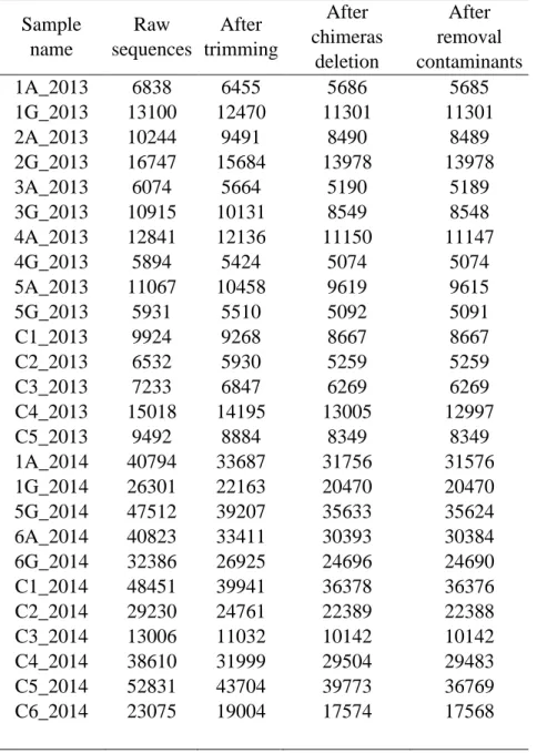
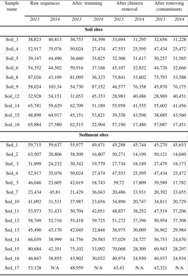
Interpretation of the data generated by NGS technologies requires complex bioinformatics analyses. A typical pipeline process includes sequence cleaning steps,
14
followed by alignment to database reference sequences. The cleaning steps, essential for accurate downstream analysis, involve filtering to remove low quality reads, contaminating sequences and primer sequences (64). Several open source pipeline algorithms are available for these procedures, including Qiime (65) and Mothur (66). The alignment to a known 16S rRNA reference database is extremely important, as this step enables identification of taxa found in each sample from different environments. The databases, SILVA (67), GreenGenes (68) and RDP (69), are the most frequently used for 16S rRNA gene annotation.
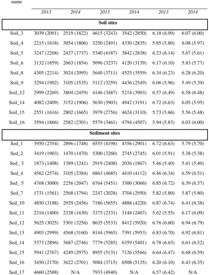
The 16S rRNA should be clustered into OTUs (Operational Taxonomic Units) using a cut-off, generally of 97% (species level at taxonomy identification) sequence identity, with algorithms such as UCLUST (70) or UPARSE (71). This step delineates operational species that then allows calculations of alpha and beta diversity, respectively the number of species in each sample and the degree of dissimilarity among samples (72).
15
3-Bacterial biogeography: Are bacteria distributed ubiquitously?
Biogeography, as applied to microbial ecology, comprises the study of microorganism distribution in space and time and interprets the mechanisms responsible, including selection, drift, dispersal and mutation on richness and composition of microbial communities (73). Endemic taxa are restricted to particular habitats and microorganisms living in extreme or oligotrophic environments appear to be endemic to distinct types of environments (74, 75), and non-randomly distributed in space (73). Taking into account the bacterial diversity at the micro-scale, studies based on the 16S rRNA gene show a large diversity of taxa in a few cm of soil. However, many of these taxa present may not be metabolically active, thus, genes for microbial identification may underestimate the bacterial diversity. Under unfavorable environmental conditions, some elements of bacterial communities may be in a dormant state, and then revive when conditions turn in their favor (76, 77). Different mechanisms can influence the distribution of bacteria, for example, colonization of new environments, in particular in soils where passive transport is limited by physical influences such as pore size and surface particle interactions (73, 78). However, bacterial dispersal is not always a restriction due to the fact that they can colonize different ecosystems via different dispersal mechanisms. In oligotrophic soil environments, diverse physical and chemical characteristics can play a role in the profile of native bacterial populations (79, 80) and are discussed in the next section.
16
4–Principal factors affecting bacterial diversity in surface soils
Microorganisms living in the surface soil of oligotrophic environments are exposed to various abiotic and biotic factors that can selectively influence the survival and resistance of particular groups (12). The factors known to influence bacterial composition in oligotrophic ecosystems include pH, salinity, temperature, plant and animal presence, fertilizer use, soil moisture, and water and nutrient availability. The structure of soils, in particular the size of aggregates, plays an important role in the distribution of bacterial communities (3, 81). For example, a study of mineral composition in artificial soils suggested that quartz, montmorillillonite and charcoal particles can affect the abundance and functional diversity of soil bacterial communities, notably those belonging to the Proteobacteria, Actinobacteria, Firmicutes and Bacteroidetes phyla (82).
Soils can be characterized by different physical and chemical properties, and can contain a unique microbial community based on geographical location (11). The most important known physical factors shaping microbial communities in soils are pH (83) and salinity
(84). The pH is commonly lower on the surface than deeper in the soil, as a result of
interaction with organic matter (85). However, in deserts surface soil, the pH is commonly alkaline, with pH values higher than 8.5 being observed (83). It has been previously shown that phylum composition is influenced by changes in soil pH, particularly for the Acidobacteria, which are commonly found, though not restricted, to low pH soils, and for
the Actinobacteria phyla that are found in soils with high pH in arid or semiarid ecosystems. In contrast, diversity has been reported to be higher in neutral pH value soils
(86). Salinity may decrease bacterial biomass and enzyme activity (87), though some
groups, such as halophilic bacteria, can resist osmotic stress and desiccation (84, 88). Other factors such as temperature, UV radiation, wind, altitude, and nutrients also affect bacterial community composition. Bacteria can confront these challenges through
17
different tolerance strategies such as biofilm formation, stress responses or dormancy (89,
18 5- Biogeochemistry: How soils get mineralized?
Definition: Mineralization in soils is the effect of the dissolution of minerals, which can
influence the physicochemical structure of the soils and, as a consequence, soil fertility
(91). Mineralization occurs by the generation of new rock material, generally by tectonics,
by sedimentation of organic and inorganic particles, and by rock erosion (92).
Composition and processes The minerals in surface soils are the result of weathering and
erosion of rocks exposed on the Earth’s surface. These minerals are classified into three primary types: silicates (granite, basalt and shale), carbonates (limestone and dolomite) and evaporates (halite and gypsum) (93). Silicates compose more than 90% of the Earth’s land surface (94). The chemical weathering of rock is an irreversible process brought about by temperature and pressure changes, precipitation, erosion and runoff (94). Weathering of continental rocks is a major cause of soil mineralization: it determines the formation, evolution, chemical and physical properties and fertility of soil. Weathering of silicate rocks (which increases with temperature and runoff) consumes CO2, an
important factor in climate regulation, and produces chemically mobile elements as Na and Ca and immobile elements such as Al or Fe that are constituents of clays (e.g., Kaolinite) and metallic oxyhydroxides (e.g., goethite) (93). The dissolved material is derived from rock by chemical (i.e., production of secondary minerals) and physical (i.e., breakdown of the rocks) weathering (92, 93). Biotic factors that contribute to soil weathering include plant roots and microorganisms, which, by releasing CO2 and organic
acids, produce an acid environment that improves nutrient uptake and mineral dissolution
(95). The principal parameters controlling chemical weathering in soils are lithology
(nature of the rocks), climate, mechanical erosion, biotic activity and organic material
19
Lithology The major elements present on the Earth’s surface resulting from soil
weathering processes are SiO2, cations (Na, K, Ca and Mg) and anions (HCO3, SO4 and
Cl) (97). Rock properties such as mineralogy, crystal sizes, surface and fracture density can influence the lithology and, as a consequence, chemical weathering (96). The dissolution rates vary among the different mineral types and depend on the soil matrix and the environmental factors of different locations, and are essential for specific microbial group colonization (98, 99).
Climate and Mechanical erosion Local climate effects are often directly related to
temperature and runoff. Depending on the ecosystem (i.e., tropical, mountainous, etc.) temperature may influence weathering and its fluctuation may do so through effects on activation energies (100). However, runoff will produce a direct interaction with the rock and, depending on soil characteristics (nature, thickness, porosity and root systems) and rain intensity, will induce soil weathering. Soils are often thick at low elevation and thin at high elevation (93). The role of physical erosion in mineral weathering is also related to the aging of the mineral surface and present-day climatic conditions (101, 102) that may contribute to clay mineralogy and thus the microorganism populations present in these soils. It has been reported that the basalt age is also a factor that can influence the bacterial communities associated with basaltic rock (103).
Organisms and organic matter From a physical perspective, the vegetation and, in
particular, plant root systems can decrease the mechanical erosion of soils because they facilitate water circulation and increase the surface contact between solutions and soil minerals (104). Vegetation also induces the evapotranspiration that can decrease runoff but generate local rainfall (96). Chemically, plants produce organic acids in their root exudates, which promote solubilization of insoluble and immobilized minerals from the deep soil layers (93, 95). Bacteria also help accelerate mineral weathering though the decomposition of litterfall that acidifies the upper soil (104) (Figure 1).
20
21
6- Bacterial adaptations and roles in mineral soil surfaces
Bacterial communities in soils may cause mineral complex transformations such as biological weathering (92). However, determining whether such a process results from activity of specific microbes (105) can be difficult, owing to low cultivability of copiotrophic microorganisms (106). Bacteria interact with minerals by various mechanisms, depending on mineral type, organism and environmental conditions, while mineral composition and bacterial metabolic activity influence mineral solubility, mobility and bioavailability (107). Bacteria promote mineral release from silicates by colonizing the mineral surfaces, where they release hydrogen ions, low molecular-weight organic molecules such as siderophores and organic acids (108). Abiotic and biotic factors can modify the original chemical structure of the soil surface and, as a consequence, influence changes in the taxonomic distribution of bacterial groups present on mineral surfaces (92). The products of dissolution from mineral complexes by bacterial activity depends on bacterial cell wall structure, outer layers and products of secondary metabolism such as exopolymers that can adsorb to minerals such as clays, colloids or oxides (107). Several studies of bacteria associated with minerals report that in silicate mineral weathering processes, the principal dynamics are associated with the production of hydrogen ions, hydroxyl ions or metal chelating metabolic products (98). Different mineral types may be associated with a wide variety of bacterial types, as found by Glesson et al. (109), who demonstrated that distinct bacterial communities present on the surfaces of mineral types are determined principally by the chemical composition of the mineral substrate, suggesting that chemically different substrates are colonize by different bacterial communities, as are different sized particles (110). Ding et al. (82), suggested that bacterial dissemination in artificial soils is not a stochastic process, and that the abundance of bacterial communities is determined by the type of clay mineral, metal oxides and the presence of charcoal. Under aerobic conditions, bacterium-mineral
22
interactions generate hydrogen ions and ligands that contribute to acidolysis, complexolysis and iron immobilization mechanisms of minerals by specific bacterial members (111). Microbes can also contribute to biomineralization forming minerals such as calcium carbonates, silicates, iron oxides and sulfides. Bacterium-mineral interactions may also affect bacterial growth and metabolic activity. The principal mechanisms of bacterial survival in the presence of minerals are redox transformations, productions of proteins and peptides for binding minerals, precipitation and active mineral transport
(112). Energy generation, nutrient acquisition, cell adhesion and biofilm formation can
all respond to toxicity of minerals. Micro-topography, surface composition, surface charge and hydrophobicity can also play an important role in the ecology of bacterial communities associated with mineral surfaces (113). This is particularly the cases with Al and Fe oxides, which are among the most reactive components on the surface of acidic and neutral soils, and play a role in the mineral catalysis of humic substances (114). The principal elements forming inorganic mineral complexes under the influence of bacterial activity are Al, Fe, Si, Mg, Mn, S and P (107). Specific bacterial groups are involved in these geochemical transformations, mainly those that oxidize and reduce iron and manganese and that reduce sulfur and sulfate. (107). The common minerals transformed by bacteria are bauxite, whose major constituents are Al2O3, Fe2O3 and SiO2, where the
weathering is promoted by bacteria that mobilize oxides of aluminum, iron and silicates, and that reduce iron under anaerobic conditions (115). Carbonates, which represent a significant portion of insoluble minerals, are also mineralized by bacterial biofilms. Cyanobacteria participate in global carbon cycling and photosynthesis (116).
Sulfate-reducing bacterial groups have a role in carbonate deposition due the production of extracellular polymeric substances which nucleate carbonate (116, 117). Phosphates can also be found on the surface of mineral soils, and bacteria can solubilize inorganic phosphate complexes (FePO4, AlPO4) by producing organic or mineral acids or by
23
environmental change (115, 118), while their bond structures may be altered by bacterial mechanisms through the release of organic acid products, ligands of cations and acidic polysaccharides (115).
Table 1 Bacterial members associated with inorganic mineral transformations
Bacteria Janthinobacterium spp. Geobacter spp. Gallionella spp. Leptothrix spp. Acidithiobacillus spp. Leptospirillum spp. Sulfolobus spp. Acidianus spp. Paenibacillus spp. Erwinia spp. Pedobacter spp. Chinitophaga spp. Shewanella spp. Apatite 147 Nontronite 152 Biotite, bauxite 150 Biotite 120 Apatite 147
Arthrobacter spp. Apatite, biotite 123, 147
Bacillus spp. Granite, apatite, bentonite 147, 151 Sulfides oxidation 107
Pseudomonas spp. Biotite, phosphate, kaolinite, vermiculite 120
Iron 149
Iron 115
Iron 115
Bacterial members associated to inorganic mineral transformation in surface soils
147 Biotite 111 Biotite, phosphate Sphingomonas spp. Reference Mineral dissolution (or released)
111, 146, 147, 148 Apatite, Biotite
Collimonas spp.
111, 123, 146, 147, 148 Biotite, phosphate, granite, apatite
24
7-Bacteria living in mineralized surface soil ecosystems: Which genera are the most well reported?
Some bacteria transform mineral complexes by various mechanisms. These bacteria interact with mineral complexes in soil surfaces. Ecosystems such as deserts or caves have low levels of nutrients (90, 119) Others, such as forest soils, have high levels of nutrients as well as surface layers composed of a variety of mineral complexes, creating oligotrophic niches for bacterial communities, which depend on mineral weathering for their growth and survival (105). In this section we describe the bacteria that play a role in mineral soil surfaces. The principal elements released by microorganisms from the mineral complex of biotite and granite are Al, Fe, Ni and Si (92, 120). Table 1 is a list of the principal bacterial genera playing a role in mineral transformation, in particular mineral weathering. Different bacteria colonize rock and sand surfaces (121), endholitic and epilithic bacterial communities being different (122). In deep mineral soils, bacteria are principally involved in complexolysis, whereas on the surface they weather minerals by acidolysis (111). Surface soils mostly harbor Burkholderia, Collimonas, Pseudomonas, Bacillus and Arthrobacter genera (see Table 1). Wang et al. (123) studied
the differences in weathering ability of bacteria in upper and deeper soils from red soil in China. Their results suggest that the elements released from mineral surfaces are Fe, Si and Al, and that the bacterial diversity was higher in upper soils than in deeper soils. Some Burkholderia, Bacillus and Lysinibacillus are highly efficient weathering bacteria, while some Burkholderia were the most well reported in mineral transformations
25
8-Bacterial communities of oligotrophic ecosystems: Which bacterial groups predominate?
Many different ecosystems are oligotrophic on the basis of low nutrient availability. These ecosystems are usually represented by soils composed of a complex of mineral elements, for example desert soil crusts, caves, ice and rocks. However, several ecosystems, where the availability of nutrients (in particular C, N, and P) is higher than in oligotrophic ecosystems, can also represent specific niches and be considered as oligotrophic, for example, acid forest soils, grasslands, sandy dunes and other mineralized surface soils. Bacteria living in oligotrophic soil ecosystems adapted to drastic climatic changes are likely to be adapted also to conditions of carbon scarcity, suggesting that the study of these ecosystems may offer an opportunity to discover novel metabolic products, such as proteases (125). Desert ecosystems make up more than 30% of the Earth’s land mass (90) and, because of interest in the impact of micro-organisms on desertification, are one of the most studied oligotrophic ecosystems in relation to its bacterial communities (11, 80, 126). In deserts, surface soils are composed of complex mineral structures formed principally by sandy loam and load sand, which are formed by weathering of the surface soils and have a very low percentage of water retention and thus less vegetation. Sandy structures are formed principally of rocks such as granite, quartz and limestone, as also observed in soils from coastal forest and grasslands (127), where bacterial populations able to weather rock have been described (128). Biological soil crusts (BSC) are complex microbial communities, which colonize intersticial soil surfaces and can be distributed in arid and cold soil ecosystems, representing a niche for various microbial organisms, including both photosynthetic and heterotrophic bacteria
(90). BSC’s also exist in other biomes such as grasslands, forest soils, permafrost soils
and Polar regions (129), where Cyanobacteria, phototrophic microorganisms of high radiation and salt resistance were the first colonizers and thus created niches in which
26
subsequent microorganisms are integrated (130). Certain Cyanobacteria are able to fix atmospheric nitrogen in the presence of CO2 (80). A study of BSC’s in the surface of
pasture soils (131) showed that the soil surface (0-3mm) and bulk soil (3-12mm) are exposed to different light conditions. They also observed that the influence of light on microbial communities was restricted to the soil surface, and that nutrients such as extractable P and K were altered at the soil surface as a result of growth of these phototrophic communities. Numerous studies of bacterial diversity in arid ecosystems (e.g. deserts) have been performed. The most abundant groups observed are members belonging to the Actinobacteria, Proteobacteria and Bacteroidetes phyla (132) while, in lower proportions, members belonging to the Firmicutes, Acidobacteria,
Gemmatimonadetes and Cyanobacteria phyla (11, 133) were also found. However, the
bacterial diversity in surface desert soils differs significantly from that of other terrestrial biomes (5). Bacterial association with minerals from sandy soils have been studied, revealing that members of the Proteobacteria, Actinobacteria and Acidobacteria phyla create better associations with quartz, magnetite and pyroxene, though differences were observed between types of minerals and bacterial types (128). Microbial abundance has been explored, for other non arid-desert oligotrophic ecosystems, such as polar deserts (McMurdo Dry Valley, Antarctica) (134). Here, Acidobacteria and Actinobacteria phyla were present in higher proportions in high pH soils, while Acidobacteria phyla are commonly found in acidic soils (86, 135). However, pH is not the only factor that can influence bacterial composition (85).
Ganzert et al. (136) studied bacterial diversity of permafrost soils in Greenland, and showed that soil bacterial communities depend significantly on soil pH. Members of the Acidobacteria, Bacteroidetes and Proteobacteria phyla were found to predominate in the
27
phyla, whose members predominate at higher soil pH. Proteobacteria phyla were found principally in soils with high nitrogen and carbon availability (137).
Other terrestrial oligotrophic soil ecosystems such as grassland soils (138) have been studied, revealing that oligotrophic and acidophilic organisms such as Acidobacteria and Proteobacteria decrease in abundance with high pH, total C and N, while copiotrophic
and alkaplophilic bacteria (Firmicutes, Chloroflexi, Actinobacteria and Bacteroidetes) are more abundant in soils rich in organic matter and nutrients. In the surface soil of an acid forest ecosystem, similar results were found; Proteobacteria and Acidobacteria phyla were found in low pH soils, suggesting that these bacteria could be a microbial indicator of soil quality improvement (139).
28 9- Conclusions and perspectives
A variety of studies have provided new insights into the bacterial communities present in oligotrophic or hostile conditions, such as those of arid deserts (140), rock surfaces (141), shallow biofilms in mountains (142) and coastal saline sandy soils (143).
The response of bacteria in oligotrophic environments, in particular in surface layers, is the formation of biofilm complexes, which protect the microorganisms from environmental fluctuation (144). Such ecosystems may provide keys for understanding the evolution of these microorganisms under harsh environmental conditions (145).
The study of bacterial communities living in terrestrial surface soil ecosystems is a challenge, owing to the fact that fewer than 5% of bacterial species are cultivable under laboratory conditions (30). Oligotrophic ecosystems are not well characterized, and new technological approaches to bacterial identification will facilitate the study of bacterial communities in different ecosystems subject to varied environmental factors.
29 References
1. Quince, C., Curtis, T. P., & Sloan, W. T. (2008). The rational exploration of microbial diversity. The International Society for Microbial Ecology Journal. 2, 997-1006. 2. Vos, M., Wolf, A. B., Jennings, S. J., & Kowalchuk, G. A. (2013). Micro-scale
determinants of bacterial diversity in soil. FEMS Microbiology Reviews, 37, 936– 954.
3. Hirsch, P. R., Mauchline, T. H., & Clark, I. M. (2010). Culture-independent molecular techniques for soil microbial ecology. Soil Biology and Biochemistry, 42, 878–887. 4. van der Heijden, M.G.A., Barggett, R.D., & van Straalen, N.M. (2008). The unseen
majority : soil microbes as drivers of plant diversity and productivity in terrestrial ecosystems. Ecology letters, 11, 296–310.
5. Fierer, N., Leff, J. W., Adams, B. J., Nielsen, U. N., Thomas, S., Lauber, C. L., et al. (2012). Cross-biome metagenomic analyses of soil microbial communities and their functional attributes. Proceedings of the National Academy of Sciences of the United States of America, 109, 2–7.
6. Falkowski, P. G., Fenchel, T., & Delong, E. F. (2008). The microbial engines that drive earth’s biogeochemical cycles. Microbial Ecology, 320, 1034–1039.
7. Zhao, M., Xue, K., Wang, F., Liu, S., Bai, S., & Sun, B. (2014). Microbial mediation of biogeochemical cycles revealed by simulation of global changes with soil transplant and cropping. The Intenational Society for Microbial Ecology Journal, 8, 2045–2055.
8. Alivisatos, A. P., Blaser, M. J., Brodie, E. L., Chun, M., Dangl, J. L., Donohue, T. J., et al. (2015). A unified initiative to harness Earth’s microbiomes. Policy Forum. Sciencexpress. 10.1126/science.aaa8480.
9. Laanbroek, H.J. (1990). Bacterial cycling of minerals that affect plant growth in waterlogged soils: a review. Aquatic Botany, 38, 109-125.
10. Bush, T., Butler, I. B., Free, A., & Allen, R. J. (2015). Redox regime shifts in microbially mediated biogeochemical cycles. Biogeosciences, 12, 3713–3724. 11. Makhalanyane, T. P., Valverde, A., Gunnigle, E., Frossard, A., Ramond, J., & Cowan,
D. A. (2015). Microbial ecology of hot desert edaphic systems. FEMS Microbiology Reviews, 39, 203–221.
12. Uroz, S., Turpault, M. P., Delaruelle, C., Mareschal, L., & Pierrat, J. (2012). Minerals affect the specific diversity of forest soil bacterial communities. Geomicrobiology Journal, 29, 88–98.
13. Barton, H., Taylor, M., & Pace, N. (2004). Molecular phylogenetic analysis of a bacterial community in an oligotrophic cave environment. Geomicrobiology Journal, 21, 11–20.
30
14. Shinoda, M., Gillies, J. A., Mikami, M., & Shao, Y. (2011). Temperate grasslands as a dust source : Knowledge, uncertainties, and challenges. Aeolian Research, 3, 271– 293.
15. Hobbs, R. J., Higgs, E., & Harris, J. A. (2009). Novel ecosystems : implications for conservation and restoration. Trends in Ecology and Evolution, 24, 599–605. 16. Gerdes, B., Brinkmeyer, R., Dieckmann, G., & Helmke, E. (2005). Influence of crude
oil on changes of bacterial communities in Arctic sea-ice. FEMS Microbiology Ecology, 53, 129–39.
17. Barto, E. K., Alt, F., Oelmann, Y., Wilcke, W., & Rillig, M. C. (2010). Contributions of biotic and abiotic factors to soil aggregation across a land use gradient. Soil Biology and Biochemistry, 42, 2316–2324.
18. Papatheodorou, E. M., Kordatos, H., Kouseras, T., Monokrousos, N., & Menkissoglu-spiroudi, U. (2012). Differential responses of structural and functional aspects of soil microbes and nematodes to abiotic and biotic modifications of the soil environment. Applied Soil Ecology, 61, 26–33.
19. Raven, J. A., Andrews, M., & Quigg, A. (2005). The evolution of oligotrophy : implications for the breeding of crop plants for low input agricultural systems. Annals of Applied Biology, 146, 261–280.
20. Womack, A. M., Bohannan, B. J. M., & Green, J. L. (2010). Biodiversity and biogeography of the atmosphere. Philosophical Transactions of the Royal Society B, 365, 3645–3653.
21. Kociolek, J.P & Stoermer, E. (2009). Oligotrophy : the forgotten end of an ecological spectrum. Acta Botanica Croatia, 68, 465–472.
22. O’Malley, M.A. (2007). The nineteenth century roots of everything is everywhere. Nature Reviews Microbiology, 5, 647-651.
23. Whitman, W. B., Coleman, D. C., & Wiebe, W. J. (1998). Perspective Prokaryotes: The unseen majority. Proceedings of the National Academy of Sciences of the United States of America, 95, 6578–6583.
24. Singh, B. K., Campbell, C. D., Sorenson, S. J., & Zhou, J. (2009). Soil genomics Soil genomics. Nature Reviews Microbiology, 7, 756.
25. Alain, K., & Querellou, J. (2009). Cultivating the uncultured: limits, advances and future challenges. Extremophiles, 13, 583–594.
26. Pham, V. H. T., & Kim, J. (2012). Cultivation of unculturable soil bacteria. Trends in Biotechnology, 30(9), 475–484.
27. Zengler, K, Walcher, M, Clark, G, Haller, I, Toledo, G, Holland, T, Mathur, E.J, Woodnutt, G, Short, J.M & Keller, M. (2005). High-throughput cultivation of microorganisms using microcapsules. Methods in Enzymology, 397, 124–130.
31
28. Hugenholtz, P., & Pace, N. R. (1996). Identifying microbial diversity in the natural environment: a molecular phylogenetic approach. Trends in Biotechnology, 14, 190– 197.
29. Torsvik, V., Goksyr, J., & Daae, F. L. (1990). High diversity in DNA of soil bacteria. Applied and Environmental Microbiology, 56, 782–787.
30. Amann, R. I., W. Ludwig, & K.H. Schleifer. 1995. Phylogenetic identification and in situ detection of individual microbial cells without cultivation. Microbiological Reviews, 59,143–169.
31. Woese, C. R., & Fox, G. E. (1977). Phylogenetic structure of the prokaryotic domain: The primary kingdoms. Proceedings of the National Academy of Sciences of the United States of America, 74, 5088–5090.
32. Woese, C. R. (1987). Bacterial evolution. Microbiological Reviews, 51, 221–271. 33. Pace, N. R. (1997). A molecular view of microbial diversity and the biosphere.
Science, 276, 734–740.
34. Delong, E. F & Pace, N. R. (2001). Environmental diversity of bacteria and archaea. Systematic Biology, 50, 470–478.
35. Sanger, F., & Nicklen, S. (1977). DNA sequencing with chain-terminating. Proceedings of the National Academy of Sciences of the United States of America, 74, 5463–5467.
36. Maxam, A. M., & Gilbert, W. (1977). A new method for sequencing DNA. Proceedings of the National Academy of Sciences of the United States of America, 74, 560–564.
37. Drmanac, R., Labat, I., Brukner, I., & Crkvenjakov, R. (1989). Sequencing of megabase plus DNA by hybridization : Theory of the Method. Genomics, 4, 114– 128.
38. Brenner, S., Johnson, M., Bridgham, J., Golda, G., Lloyd, D. H., Johnson, D, et al. (2000). Gene expression analysis by massively parallel signature sequencing (MPSS) on microbead arrays. Nature Biotechnology, 18, 630–634.
39. Handelsman, J., Rondon, M. R., Brady, S. F., Clardy, J., & Goodman, R. M. (1998). Molecular biological access to the chemistry of unknown soil microbes: a new frontier for natural products. Chemistry and Biology, 5, R245–249.
40. Handelsman, J. (2004). Metagenomics : application of genomics to uncultured microorganisms metagenomics : application of genomics to uncultured microorganisms. Microbiology and Molecular Biology Reviews, 68, 669–685. 41. Schloss, P. D., & Handelsman, J. (2003). Biotechnological prospects from
32
42. Cowan, D., Meyer, Q., Stafford, W., Muyanga, S., & Cameron, R. (2005). Metagenomic gene discovery : past, present and future. Trends in Biotechnology, 23, 321–329.
43. Vieites, J. M., Guazzaroni, M.-E., Beloqui, A., Golyshin, P. N., & Ferrer, M. (2009). Metagenomics approaches in systems microbiology. FEMS Microbiology Reviews, 33, 236–55.
44. Thomas, T., Gilbert, J., & Meyer, F. (2012). Metagenomics - a guide from sampling to data analysis. Microbial Informatics and Experimentation, 2, 1-12.
45. Myrold, D. D., Zeglin, L. H., & Jansson, J. K. (2013). The potential of metagenomic approaches for understanding soil microbial processes. Soil Science Society of America Journal, 78, 3–10.
46. Béjà, O., Aravind, L., Koonin, E. V, Jovanovich, S. B., Cates, C. M., et al. (2000). Bacterial rhodopsin : evidence for a new type of phototrophy in the Sea. Science, 289, 1902–1907.
47. Nicol, G. W., & Schleper, C. (2006). Ammonia-oxidising Crenarchaeota: important players in the nitrogen cycle ? Trends in Microbiology, 14, 207–212.
48. Delong, E. F. (2009). The microbial ocean from genomes to biomes. Nature, 459, 200–206.
49. Williamson, S. J., Allen, L. Z., Lorenzi, H. A., Fadrosh, D. W., Brami, D., Thiagarajan, M., et al. (2012). Metagenomic exploration of viruses throughout the Indian Ocean. PloS One, 7, e42047.
50. Bork, P., Bowler, C., de Vargas, C., Gorsky, G., Karsenti, E., & Wincker, P. (2015). Tara oceans studies plankton at planetary scale. Science, 348, 873–875.
51. Mackelprang, R., Waldrop, M. P., Deangelis, K. M., David, M. M., Chavarria, K. L., Blazewicz, S. J., et al. (2011). Community reveals a rapid response to thaw. Nature, 480, 368–373.
52. Andreote, F., Jiménez, D., Chaves, D., Dias, A. C., et al (2012). The Microbiome of Brazilian mangrove sediments as revealed by metagenomics. PloS One, 7, e38600. 53. Langille, M. G. I., Zaneveld, J., Caporaso, J. G., Mcdonald, D., Knights, D., Reyes, J. A., et al. (2013). Predictive functional profiling of microbial communities using 16S rRNA marker gene sequences. Nature Biotechnology, 31, 814–821.
54. Clarridge, J. E. (2004). Impact of 16S rRNA gene sequence analysis for identification of bacteria on clinical microbiology and infectious diseases. Clinical Microbiology Reviews, 17, 840–862.
55. Tringe, S. G., & Hugenholtz, P. (2008). A renaissance for the pioneering 16S rRNA gene. Current Opinion in Microbiology, 11, 442–446.
56. Janda, J. M., & Abbott, S. L. (2007). 16S rRNA gene sequencing for bacterial identification in the diagnostic laboratory: pluses, perils, and pitfalls. Journal of Clinical Microbiology, 45, 2761–2764.
33
57. Klappenbach, J. A., Dunbar, J. M., & Schmidt, T. M. (2000). rRNA operon copy number reflects ecological strategies of bacteria. Applied and Environmental Microbiology, 66, 1328–1333.
58. Ansorge, W. J. (2009). Next-generation DNA sequencing techniques. New Biotechnology, 25, 195–203.
59. Shendure, J., & Ji, H. (2008). Next-generation DNA sequencing. Nature Biotechnology, 26, 1135–1145.
60. Metzker, M. L. (2010). Sequencing technologies - the next generation. Nature Reviews Genetics, 11, 31–46.
61. Buermans, H. P. J., & Dunnen, J. T. D. (2014). Next generation sequencing technology: advances and applications. Biochimica et Biophysica Acta, 1842, 1932– 1941.
62. Morozova, O., & Marra, M. A. (2008). Genomics applications of next-generation sequencing technologies in functional genomics. Genomics, 92, 255–264.
63. van Dijk, E. L., Jaszczyszyn, Y., & Thermes, C. (2014). Ten years of next-generation sequencing technology. Trends in Genetics, 30, 418-426.
64. Schmieder, R., & Edwards, R. (2011). Fast identification and removal of sequence contamination from genomic and metagenomic datasets. PloS One, 6, e17288. 65. Caporaso, J. G., Kuczynski, J., Stombaugh, J., Bittinger, K., Bushman, F. D., Costello,
E. K., et al. (2010). QIIME allows analysis of high-throughput community sequencing data. Nature Methods, 7, 335–336.
66. Schloss, P. D., Westcott, S. L., Ryabin, T., Hall, J. R., Hartmann, M., Hollister, E. B., et al. (2009). Introducing mothur: open-source, platform-independent, community-supported software for describing and comparing microbial communities. Applied and Environmental Microbiology, 75, 7537–7541.
67. Quast, C., Pruesse, E., Yilmaz, P., Gerken, J., Schweer, T., Glo, F. O., & Yarza, P. (2013). The SILVA ribosomal RNA gene database project: improved data processing and web-based tools. Nucleic Acids Research, 41, 590–596.
68. Desantis, T. Z., Hugenholtz, P., Larsen, N., Rojas, M., Brodie, E. L., Keller, K., et al. (2006). Greengenes, a chimera-checked 16S rRNA gene database and workbench compatible with ARB. Applied and Environmental Microbiology, 72, 5069–5072. 69. Cole, J. R., Wang, Q., Fish, J. A., Chai, B., Mcgarrell, D. M., Sun, Y., et al. (2014).
Ribosomal database project: data and tools for high throughput rRNA analysis. Nucleic Acids Research, 42, 633–642.
70. Edgar, R. C. (2010). Search and clustering orders of magnitude faster than BLAST. Bioinformatics, 26, 2460–2461.
71. Edgar, R. C., Haas, B. J., Clemente, J. C., Quince, C., & Knight, R. (2011). UCHIME improves sensitivity and speed of chimera detection. Bioinformatics, 27, 2194–200.
34
72. Thomas, T., Gilbert, J., & Meyer, F. (2012). Metagenomics - a guide from sampling to data analysis. Microbial Informatics and Experimentation, 2, 1-12.
73. Hanson, C. A., Fuhrman, J. A., Horner-devine, M. C., & Martiny, J. B. H. (2012). Beyond biogeographic patterns : landscape. Nature Reviews Microbiology, 10, 497– 506.
74. Cho, J. C. & Tiedje, J. M. (2000). Biogeography and degree of endemicity of fluorescent Pseudomonas strains in soil. Applied and Environmental Microbiology, 66, 5448-5456.
75. Oakley, B. B., Carbonero, F., van der Gast, C. J., Hawkins, R.J & Purdy, K. J. (2010). Evolutionary divergence and biogeography of sympatric niche-differentiated bacterial populations. International Society for Microbial Ecology Journal, 4, 488-497.
76. Prosser, J. I., Bohannan, B. J. M., Curtis, T. P., Ellis, R. J., Firestone, M. K., Freckleton, R. P., Green, J., et al. (2007). The ecological theory in microbial ecology. Nature Reviews Microbiology, 5, 384-392.
77. Lennon, J. T. & Jones, S. E. (2010). Dormancy contributes to the maintenance of microbial diversity. Proceedings of the National Academy of Sciences of the United States of America, 107, 5881-5886.
78. Martiny, J. B. H., Bohannan, B. J. M., Brown, J. H., Colwell, R. K., Fuhrman, J. A., Green, J. L., et al. (2006). Microbial biogeography : putting microorganisms on the map. Nature Reviews Microbiology, 4, 102–113.
79. An, S., Sin, H. H., & Dubow, M. S. (2015). Modification of atmospheric sand-associated bacterial communities during Asian sandstorms in China and South Korea. Heredity, 114, 460–467.
80. Gombeer, S., Ramond, J., Eckardt, F. D., Seely, M., & Cowan, D. A. (2015). The influence of surface soil physicochemistry on the edaphic bacterial communities in contrasting terrain types of the Central Namib Desert. Geobiology, 13, 494–505. 81. Mummey, D. L., & Stahl, P. D. (2004). Analysis of soil whole- and
inner-microaggregate bacterial communities. Microbial Ecology, 48, 41–50.
82. Ding, G.-C., Piceno, Y. M., Heuer, H., Weinert, N., Dohrmann, A. B., Carrillo, A., et al. (2013). Changes of soil bacterial diversity as a consequence of agricultural land use in a semi-arid ecosystem. PloS One, 8, e59497.
83. Lauber, C. L., Hamady, M., Knight, R., & Fierer, N. (2009). Pyrosequencing-based assessment of soil pH as a predictor of soil bacterial community structure at the continental scale. Applied and Environmental Microbiology, 75, 5111–20.
84. Lozupone, C. a, & Knight, R. (2007). Global patterns in bacterial diversity. Proceedings of the National Academy of Sciences of the United States of America, 104, 11436–40.
35
85. Rousk, J., Baath, E., Brookes, P. C., Lauber, C. L., Lozupone, C., Caporaso, J. G., Knight, R., & Fierer, N. (2010). Soil bacterial and fungal communities across a pH gradient in an arable soil. International Society for Microbial Ecology Journal, 4, 1340-1351.
86. Fierer, N., & Jackson, R. B. (2006). The diversity and biogeography of soil bacterial communities. Proceedings of the National Academy of Sciences of the United States of America, 103, 626–631.
87. Wichern, J., Wichern, F., Joergensen, R. G. (2006). Impact of salinity on soil microbial communities and the decomposition of maize in acidic soils. Geoderma, 137, 100-108.
88. Nemergut, D. R., Schmidt, S. K., Fukami, T., O’Neill, S. P., Bilinski, T. M., Stanish, L. F., Knelman, J. E., Darcy, J. L., et al. (2013). Patterns and processes of microbial community assembly. Microbiology and Molecular Biology Reviews, 77, 342-356. 89. Gorbushina, A. A., Kort, R., Schulte, A., Lazarus, D., Schnetger, B., Brumsack, H-J.,
Broughton, W. J. & Favet, J. (2007). Life in Darwin’s dust: intercontinental transport and survival of microbes in the nineteenth century. Environmental Microbiology, 9, 2911-2922.
90. Pointing, S. B., & Belnap, J. (2012). Microbial colonization and control in dryland systems. Nature Reviews Microbiology, 10, 551-562.
91. Brantley, S. (2008). Kinetics of water-rock interaction, pp. 151-210, Springer. 92. Uroz, S., Kelly, L. C., Turpault, M. P. Lepleux, C., & Frey-Klett, P. (2015). The
mineralosphere concept: mineralogical control of the distribution and function of mineral-associated bacterial communities. Trends in Microbiology, 23, 751–762. 93. Viers, J., Oliva, P., Dandurand, J., Dupre, B., & Vii, P. (2007). Chemical weathering
rates, CO2 consumption, and control parameters deduced from the chemical composition of rivers. Treatise on Geochemistry, 5, 1–25.
94. White, A. F. & Buss, H. L. (2003). Natural weathering of silicate minerals. Treatise on Geochemistry, 5, 133–168.
95. Schoebitz, M., Osman, J., & Ciampi, L. (2013). Effect of immobilized Serratia sp. by spray-drying technology on plant growth and phosphate uptake. Chilean Journal of Agricultural and Animal Science, 29, 111–119.
96. Hartmann, J., Moosdorf, N., Lauerwald, R., Hinderer, M., & West, a. J. (2014). Global chemical weathering and associated p-release - the role of lithology, temperature and soil properties. Chemical Geology, 363, 145–163.
97. Einsele, G. (2000). Sedimentary basins: evolution, facies and sediment Budget, 2nd edition. Berlin: Springer.
98. Rogers, J. R., Bennett, P. C. (2004). Mineral stimulation of subsurface microorganisms: release of limiting nutrients from silicates. Chemical Geology, 203, 91-108.
36
99. Montros, S. N., Skidmore, M., Tranter, M., Kivimaki, A.L., Parkes, R. J. (2012). A microbial driver of chemical weathering in glaciated systems. Geology, 41, 215-218. 100. Moquet, J. S., Crave, A., Viers, J., Seyler, P., Armijos, E., Bourrel, L., et al. (2011). Chemical weathering and atmospheric/soil CO2 uptake in the Andean and Foreland Amazon basins. Chemical Geology, 287, 1–26.
101. Pasquini, A. I., Depetris, P. J., Gaiero, D. M., Probst, J. L. (2005). Material sources, chemical weathering and physical denudation in the Chubut River basin (Patagonia, Argentina): implication for Andean rivers. The Journal of Geology, 113, 451-469. 102. Goldsmith, S. T., Carey, A. E., Johnson, B. M., Welch, S. a., Lyons, W. B.,
McDowell, W. H., & Pigott, J. S. (2010). Stream geochemistry, chemical weathering and CO2 consumption potential of andesitic terrains, Dominica, Lesser Antilles. Geochimica et Cosmochimica Acta, 74, 85–103.
103. Santelli, C. M., Edgcomb, V. P., Bach, W., Edwards, K. J. (2008). The diversity and abundance of bacteria inhabiting seafloor lavas positively correlate with rock alteration. Environmental Microbiology, 11, 86-98.
104. Berner, E.K., Berner, R. A. (2003). Plants and mineral weathering: present and past. Trends in Geochemistry, 5, 169-188.
105. Uroz, S., Calvaruso, C., Turpault, M. P. & Frey-Klett, P. (2009). Mineral weathering by bacteria : ecology, actors and mechanisms. Trends in Microbiology, 17, 378–387. 106. Torsvik, V., & Øvreås, L. (2002). Microbial diversity and function in soil : from
genes to ecosystems. Current Opinion in Microbiology, 5, 240–245.
107. Gadd, G. M. (2010). Metals, minerals and microbes: Geomicrobiology and bioremediation. Microbiology, 156, 609–643.
108. Balland-bolou-bi, C., & Poszwa, A. (2012). Effect of calco-magnesian amendment on the mineral weathering abilities of bacterial communities in acidic and silicate-rich soils. Soil Biology and Biochemistry, 50, 108–117.
109. Gleeson, B. D., Kennedy, N. M., Clipson, N., Melville, K., Gadd, G. M., McDermott, F. P. (2006). Characterization of bacterial community structure on a weathered pegmatitic granite. Microbial Ecology, 51, 526-534.
110. Hemkemeyer, M., Pronk, G., Heister, K., Kögel-Knabner, I., Martens, R., & Tebbe, C. C. (2014). Artificial soil studies reveal domain-specific preferences of microorganisms for the colonisation of different soil minerals and particle size fractions. FEMS Microbiology Ecology, 90, 770–782.
111. Balland, C., Poszwa, A., Leyval, C., & Mustin, C. (2010). Dissolution rates of phyllosilicates as a function of bacterial metabolic diversity. Geochimica et Cosmochimica Acta, 74, 5478–5493.
112. Konhauser, K. (2007). Introduction to Geomicrobiology. Oxford: Blackwell. ISBN: 978-0-632-05454-1
37
113. Carson, J.K., Campbell, L., Rooney, D., Clipson, N., & Gleeson, D. B. (2009). Minerals in soil select distinct bacterial communities in their microhabitats. FEMS Microbiology Ecology, 67, 381-388.
114. Huang, P. M., Wang, M. K & Chiu, C. Y. (2005). Soil mineral-organic matter-microbe interactions: impacts on biogeochemical processes and biodiversity in soils. Pedobiología, 49, 609-635.
115. Ehrlich, H. L. & Newman, D. K. (2009). Geomicrobiology, 5th edition. Boca Raton, FL: CRC Press/Taylor & Francis.
116. Dupraz, C., Reid, R. P., Braissant, O., Decho, A. W., Norman, R. S. & Visscher, P. T. (2009). Processes of carbonate precipitation in modern microbial mats. Earth Science Reviews, 96, 141-162.
117. Tebo, B. M., Johnson, H. A., Mc Carthy, J. K. & Templeton, A. S. (2005). Geomicrobiology of manganese (II) oxidation. Trends in Microbiology, 13, 421-438.
118. Tazaki, K. (2006). Clays, microorganisms and biomineralization. In Handbook of Clay Science, Developments in Clay Science, 1, 477-497.
119. Ortiz, M., Legatzki, A., Neilson, J. W., Fryslie, B., Nelson, W. M., Wing, R. A., Soderlund, C. A., et al. (2014). Making a living while starving in the dark: metagenomic insights into the energy dynamics of a carbonate cave. International Society for Microbial Ecology, 8, 478-491.
120. Ahmed, E., & Holmström, S. J. M. (2015). Microbe-mineral interactions: The impact of surface attachment on mineral weathering and element selectivity by microorganisms. Chemical Geology, 403, 13–23.
121. Certini, G., Campbell, C. C., Edwards, A. C. (2004). Rock fragments in soil support a different microbial community from the fine earth. Soil Biology and Biochemistry, 36, 1119-1128.
122. McNamara, C. J., Perry, IV T. D., Bearce, K. A., Hernandez-Duque, G., Mitchell, R. (2006). Epilithic and endolithic bacterial communities in limestone from a Maya archaeological site. Microbial Ecology, 51, 51-64.
123. Wang, Q., Wang, R. R., He, L. Y., & Lu, J. J. (2014). Changes in weathering effectiveness and community of culturable mineral-weathering bacteria along a soil profile. Biology and Fertility of Soils, 50, 1025–1034.
124. Uroz, S., Ioannidis, P., Lengelle, J., Cébron, A., Morin, E., Buée, M., & Martin, F. (2013). Functional assays and metagenomic analyses reveals differences between the microbial communities inhabiting the soil horizons of a Norway spruce plantation. PloS One, 8, e55929.
125. Neveu, J., Regeard, C., & DuBow, M. S. (2011). Isolation and characterization of two serine proteases from metagenomic libraries of the Gobi and Death Valley deserts. Applied Microbiology and Biotechnology, 91, 635–44.
38
126. Osman, J., de Zelicourt, A., Bisseling, T., Geurts, R., Hirt, H., & Dubow, M. S. (2016). Bacterial rhizosphere biodiversity from several pioneer desert sand plants near Jizan, Saudi Arabia. The Open Conference Proceedings Journal, 7, 70–79. 127. Crawford, R. M. M. (2009). The biology of coastal sand dunes. Annals of Botany,
104, 288.
128. Nishiyama, M., Sugita, R., Otsuka, S., & Senoo, K. (2012). Community structure of bacteria on different types of mineral particles in a sandy soil. Science and Plant Nutrition, 58, 562–567.
129. Schmidt, S. K., Nemergut, D. R., Todd, B. T., Lynch, R. C., Darcy, J. L., Cleveland, C. C., & King, A. J. (2012). A simple method for determining limiting nutrients for photosynthetic crusts. Plant Ecology & Diversity, 5, 513–519.
130. Rossi, F., Olguín, E. J., Diels, L., & De Philippis, R. (2015). Microbial fixation of CO2 in water bodies and in drylands to combat climate change, soil loss and desertification. New Biotechnology, 32, 109–120.
131. Davies, L. O., Schäfer, H., Marshall, S., Bramke, I., Oliver, R. G., & Bending, G. D. (2013). Light structures phototroph, bacterial and fungal communities at the soil surface. PloS One, 8, e69048.
132. Fierer, N., Strickland, M. S., Liptzin, D., Bradford, M. A., & Cleveland, C. C. (2009). Global patterns in belowground communities. Ecology Letters, 12, 1238–1249. 133. Richer, R., Banack, S. A., Metcalf, J. S., Cox, P. A. (2014). The persistence of
cyanobacterial toxins in desert soils. Journal of Arid Environments, 112, 134-139. 134. Geyer, K. M., Altrichter, A. E., Takacs-Vesbach, C. D., Horn, D. J. Van, Gooseff,
M. N., & Barrett, J. E. (2014). Polar desert. FEMS Microbiology Ecology, 89, 490– 494.
135. Cary, S. C., Mcdonald, I. R., Barrett, J. E., & Cowan, D. A. (2010). On the rocks: the microbiology of Antarctic Dry Valley soils. Nature Reviews Microbiology, 8, 129–138.
136. Ganzert, L., Bajerski, F., & Wagner, D. (2014). Permafrost-affected soils of Northeast Greenland. FEMS Microbiology Ecology, 89, 426–441.
137. Fierer, N., Breitbart, M., Nulton, J., Salamon, P., Lozupone, C., Jones, R., et al. (2007). Metagenomic and small-subunit rRNA analyses reveal the genetic diversity of bacteria, archaea, fungi, and viruses in soil. Applied and Environmental Microbiology, 73, 7059–66.
138. Chronákova, A., Schloter-hai, B., Radl, V., Endesfelder, D., Quince, C., Elhottová, D., et al. (2015). Response of archaeal and bacterial soil communities to changes associated with outdoor cattle overwintering. PloS One, 10, e0135627.
139. Clivot, H., Pagnout, C., Aran, D., Devin, S., Bauda, P., & Poupin, P. (2012). Changes in soil bacterial communities following liming of acidified forests. Applied Soil Ecology, 59, 116–123.