HAL Id: tel-01656732
https://tel.archives-ouvertes.fr/tel-01656732
Submitted on 6 Dec 2017
HAL is a multi-disciplinary open access archive for the deposit and dissemination of sci-entific research documents, whether they are pub-lished or not. The documents may come from teaching and research institutions in France or abroad, or from public or private research centers.
L’archive ouverte pluridisciplinaire HAL, est destinée au dépôt et à la diffusion de documents scientifiques de niveau recherche, publiés ou non, émanant des établissements d’enseignement et de recherche français ou étrangers, des laboratoires publics ou privés.
Understanding ribosome binding interactions and
conformational changes of the EngA bacterial GTPase, a
potential target for new antibiotics
Catarina da Silveira Tome
To cite this version:
Catarina da Silveira Tome. Understanding ribosome binding interactions and conformational changes of the EngA bacterial GTPase, a potential target for new antibiotics. Biomolecules [q-bio.BM]. Uni-versité Grenoble Alpes, 2016. English. �NNT : 2016GREAY033�. �tel-01656732�
THÈSE
Pour obtenir le grade de
DOCTEUR DE LA COMMUNAUTÉ UNIVERSITÉ
GRENOBLE ALPES
Spécialité : Physique pour les sciences du vivant
Arrêté ministériel : 25 mai 2016
Présentée par
Catarina DA SILVEIRA TOMÉ
Thèse dirigée par Dominique HOUSSET et codirigée par Jean-Michel JAULTpréparée au sein de l’Institut de Biologie Structurale dans l'École Doctorale de Physique
Intéractions avec le ribosome et
changements conformationnels de la
GTPase bactérienne EngA, une cible
potentielle pour de nouveaux
antibiotiques
Thèse soutenue publiquement le 05/12/2016, devant le jury composé de :
M Franz BRUCKERT
Professeur des universités, Institut Polytechnique de Grenoble, Président
Mme Jacqueline CHERFILS
Directeur de recherche, École Normale Supérieure Paris-Saclay, Rapporteur
Mme Mirjam CZJZEK
Directeur de recherche, Station Biologique de Roscoff, Rapporteur
M Jean-François GUICHOU
Professeur des universités, Centre de Biochimie Structurale, Examinateur
M Dominique HOUSSET
Chercheur CEA, Institut de Biologie Structurale, Directeur de thèse
Understanding ribosome binding interactions and conformational
changes of the EngA bacterial GTPase, a potential target for new
Acknowledgements
I would like to thank the members of my committee, Jacqueline Cherfils, Mirjam Czjzek, Franz Bruckert and Jean-François Guichou, for accepting to evaluate and discuss this work.
I thank my thesis advisors, Dominique Housset and Jean-Michel Jault, for welcoming me to this project. I’m grateful for their guidance and for the support and trust they gave me.
A special thanks to Anne-Emmanuelle Foucher, for her coaching, advice, discussions and companionship.
I want to thank the people who contributed to our project with their expertise: our collaborators from Institut de Biologie Structurale and Université Grenoble-Alpes, the scientists from the HTX Laboratory and ESRF beamlines, and all the members of the IRPAS group for always being available to help me throughout this project.
Finally, I want to express my gratitude to my family and friends with whom I shared these three years. In particular, a special thank you to my parents and Sara for their encouragement and motivation, and Nuno for his constant presence.
Le résumé en français
Le traitement des infections bactériennes est devenu possible au XX
esiècle grâce au
développement des premiers antibiotiques. La découverte de nombreuses nouvelles
familles d’antibiotiques entre les années 1940 et 1970 s’est accompagnée de leur
usage généralisé
ce qui a conduit à l’émergence de phénomènes de résistance à
ces médicaments. Aggravé par le ralentissement du développement de nouveaux
antibiotiques après les années 1980, la résistance aux antibiotiques constitue
aujourd’hui, selon l’OMS, «l’une des plus graves menaces pesant sur la santé
mondiale». Des nouveaux outils envisageant la prévention et le traitement des
infections bactériennes sont actuellement en développement. La prophylaxie (dont
les vaccins), les traitements complémentaires aux antibiotiques (dont les
probiotiques) et les traitements alternatives aux antibiotiques (y compris les
bactériophages et les anticorps monoclonaux) sont parmi les voies thérapeutiques
en développement. La reprise de la recherche sur des nouveaux antibiotiques pour
lesquels les bactéries n’ont pas encore acquis de mécanismes de résistance est
aussi nécessaire. Le point de départ de cette recherche passe par l’identification des
nouvelles cibles moléculaires. Une cible pharmacologique idéale doit comporter trois
caractéristiques principales : être essentielle à la survie cellulaire, être conservée au
sein des bactéries, et être absente chez les eucaryotes. Parmi les machineries
cellulaires ciblées par les antibiotiques aujourd’hui disponibles, le ribosome bactérien
représente environ 40% des cas. Alors que tous ces antibiotiques agissent
directement sur le fonctionnement du ribosome (par interaction avec le ribosome et
inhibition de la synthèse protéique), la voie de biogenèse du ribosome reste toujours
inexplorée.
La biogenèse du ribosome est un processus cellulaire essentiel et complexe, où plus
de cinquante protéines et trois ARN doivent être synthétisés, modifiés, repliés et
assemblés pour former une particule 70S fonctionnelle. En plus des protéines et
ARN ribosomaux, des facteurs d’assemblage non ribosomiques participent à ce
processus pour le faciliter et
l’accélérer. Parmi eux on trouve des endonucléases
responsables par la maturation des ARN, des enzymes responsables des
modifications chimiques des ARN et protéines, des hélicases et chaperonnes qui
jouent un rôle dans le repliement, et des GTPases dont le rôle exact reste encore
méconnu mais qui, chez les bactéries, représentent une grande partie des facteurs
impliqués dans la biogenèse du ribosome.
Les GTPases sont des enzymes qui lient et hydrolysent le GTP. Elles possèdent un
domaine catalytique conservé
– le domaine G – composé d’un feuillet β central
entouré par les hélices α et des boucles. Cinq boucles font partie du site actif et
présentent des motifs conservés, nommés G1 à G5, qui jouent un rôle dans la
fixation et hydrolyse du nucléotide. Grâce à leur flexibilité, ces boucles adoptent
plusieurs conformations en fonction du nucléotide présent dans le site catalytique qui
caractérisent différents états de la GTPase. Notamment, les boucles nommées
Switch I et Switch II, contenant respectivement les motifs G2 et G3, sont en grande
partie responsables de ces changements structuraux. Le cycle catalytique
s’initie
avec la fixation du GTP, qui engendre un changement conformationnel qui permet
l’activation de l’enzyme et l’interaction avec des effecteurs. Les principaux contacts
protéine-ligand présents dans les GTPases se produisent entre la nucléobase et les
motifs G4 et G5, les phosphates α et β du nucléotide et le motif G1, et le phosphate γ
du GTP et les motifs G2 et G3. L’hydrolyse du GTP en GDP entraîne un changement
de conformation de la GTPase, qui passe ainsi de l’état actif à l’état inactif, avec une
faible affinité pour les effecteurs. En particulier, les éléments responsables par la
coordination du phosphate γ du GTP sont perdus, ce qui engendre une
déstabilisation et repositionnement des motifs G2 et G3. Le GDP est finalement
échangé contre du GTP pour que l’enzyme puisse entrer dans un nouveau cycle.
Malgré une structure et un mécanisme catalytique conservés, les GTPases
présentent une grande diversité fonctionnelle. Dans les bactéries, elles participent
dans la réplication du DNA, la division cellulaire, la réponse au stress, les
mécanismes de pathogenèse, la synthèse protéique et l’assemblage du ribosome.
Depuis quelques années, plusieurs GTPases bactériennes essentielles ont été
identifiées comme étant impliquées dans la biogenèse du ribosome.
C’est le cas
d’EngA, identifiée en 2000 et associée à la division cellulaire. Des études ultérieures
ont montré qu’elle interagit de façon nucléotide-dépendante avec le ribosome et que
sa déplétion entraîne l’accumulation de précurseurs ribosomaux, suggérant un rôle
dans
la biogenèse du ribosome. Le caractère essentiel d’EngA chez les bactéries et
son absence chez les humains font de cette enzyme une bonne cible
particularités structurales d’EngA la rendent plus difficile à étudier. En effet, malgré
sa conservation et la présence des motifs caractéristiques de la famille des GTPases,
EngA est la seule GTPase connue à posséder deux domaines G. Dans sa structure,
EngA présente trois domaines : un domaine G N-terminal (nommé GD1) lié par une
boucle à un deuxième domaine G (GD2) et un domaine C-terminal non catalytique
(KH). Ainsi, la présence de deux domaines catalytiques chez EngA apporte une
diversité conformationnelle plus grande. Plusieurs structures d’EngA ont été résolues
par cristallographie et cryo-microscopie électronique et ont suggéré qu’outre les
changements structuraux locaux au niveau des motifs G, typiques des GTPases, des
changements conformationnels au niveau de la structure tertiaire de la protéine
peuvent aussi avoir lieu. Trois conformations différentes d’EngA ont été observées et
associées à des différents états du cycle catalytique. Ces trois structures présentent
une position conservée des domaines GD2 et KH, pendant que le domaine GD1 se
repositionne en fonction de l’étape du cycle. Cependant, les informations structurales
disponibles présentent aussi des aspects peu clairs, notamment l’absence d’une
structure avec du GTP fixé simultanément aux deux domaines G d’EngA ou la faible
résolution des structures de cryo-microscopie électronique. Malgré les données
disponibles actuellement, les événements qui régulent les changements
conformationnels d’EngA, la fixation/hydrolyse du nucléotide et les interactions avec
le ribosome restent encore mal compris.
Notre objectif a donc été de mieux comprendre les mécanismes structuraux,
biochimiques et fonctionnels d’EngA, et de mettre en place un protocole de criblage
de composés inhibiteurs. Pour cela, nous avons combiné des données structurales
en solution et dans l’état cristallin et des données d’interaction, et développé un test
pour identifier des inhibiteurs d’interactions EngA-ribosome.
Des expériences de SAXS (small-angle X-ray scattering), protéolyse limitée,
séquençage N-terminal et spectrométrie de masse ont été réalisées pour étudier
l’effet du nucléotide dans les changements conformationnels d’EngA. Les données
de SAXS ont montré que la protéine dans son état apo ou lié au GDP adopte la
conformation observée par des structures cristallographiques EngA-GDP. Par contre,
un changement structural a été observé en présence d’analogues du GTP,
suggérant que les nucléotides triphosphate sont capables d’induire une nouvelle
conformation. Cependant, cette conformation ne correspond à aucune structure
connue : la conformation active (liée au GTP) d’EngA reste toujours indéterminée. En
outre, ce changement est observé uniquement en présence de hautes
concentrations de nucléotide (10 mM), indiquant une faible affinité de l’enzyme pour
le GTP. Des expériences de protéolyse limitée corroborent un changement de
conformation nucléotide-dépendant : le profil de fragmentation d’EngA par la trypsine
en absence et présence de GDP est identique, alors qu’en présence d’analogues du
GTP la cinétique de protéolyse est modifiée. Ceci suggère que le nucléotide
triphosphate engendre des changements structuraux qui rendent la protéine plus
susceptible à l’action de la protéase. Les fragments issus de la protéolyse ont été
analysés par séquençage N-terminal et spectrométrie de masse. Cinq régions ont
été identifiées, dont une semble contribuer significativement à la différente cinétique
de protéolyse : l’étiquette histidine N-terminale est plus activement clivée en
présence d’analogues du GTP, suggérant un changement de la structure tertiaire
d’EngA, possiblement par un mouvement du GD1, vers une conformation où
l’extrémité N-terminale est plus accessible. Un test ELISA a été développé pour
étudier les interactions entre EngA et le ribosome bactérien. Ces études ont montré
que les changements conformationnels nucléotide-dépendants chez EngA modifient
son affinité pour le ribosome : alors que le GDP déstabilise l’interaction
EngA-ribosome, les analogues du GTP (à 10 mM) la renforcent. La faible affinité qu’EngA
semble avoir pour les nucléotides, de l’ordre du millimolaire, est proche des
concentrations basales intracellulaires de GTP (1–2 mM). Le rôle d’EngA pourrait
alors être la détection des niveaux intracellulaires de nucléotides, de façon à moduler
ses interactions avec le ribosome
– et donc son assemblage – selon l’état
énergétique de la cellule. Des expériences de cryo-microscopie électronique ont
débuté afin de visualiser le complexe EngA-50S. Les données enregistrées sont
encore en train d’être analysées.
Des études cristallographiques ont été menées avec l’objectif de déterminer la
structure de l’état actif d’EngA liée au GTP. Plusieurs cristaux ont diffracté à une
résolution suffisante pour pouvoir résoudre la structure d’EngA. Toutefois, malgré les
nombreuses conditions de cristallisation essayées et les différents groupes d’espace
obtenus, EngA est toujours observée dans sa conformation inactive. Nonobstant,
nous avons pu rassembler quelques nouvelles informations sur EngA. Une analyse
comparative de ces structures suggère une affinité distincte pour les deux domaines
catalytiques : alors que le domaine GD2 présente toujours un nucléotide fixé, des
ions phosphate et sulfate présents dans la solution de cristallisation occupent
facilement
le site catalytique du GD1. Une de ces structures, obtenue à partir d’un
cristal préparé dans l’absence de nucléotide, a révélé la présence d’un GDP lié au
GD2, suggérant une très forte fixation du GDP. D’après cette analyse, le GD2
présenterait une forte affinité pour les nucléotides en opposition au GD1 (qui dans ce
cas contribuerait pour la faible affinité d’EngA observé par SAXS). Ces observations
s’opposent aux données biochimiques présentes dans la littérature qui montrent une
affinité identique pour le GD1 et le GD2 tronqués. Ces résultats contradictoires
pourraient être expliqués par une coopérativité entre les deux domaines dans la
protéine full-length
qui, quand les deux domaines sont étudiés séparément, n’est
plus présente. Une de nos structures a, pour la première fois, montré un analogue du
GTP fixé dans les deux domaines G. L’observation détaillée du site catalytique du
GD1 révèle un résidu Gln en amont du
le motif G3 qui stabilise le phosphate γ du
nucléotide. Ainsi, cette glutamine pourrait avoir un rôle catalytique identique aux
GTPases Ras-like, ce qui impliquerait un mécanisme d’hydrolyse différent pour le
GD1 et le GD2 – pour lequel un tel résidu catalytique n’a pas été identifié. Malgré la
présence d’un analogue du GTP dans les deux domaines, la conformation d’EngA
reste inchangée. Cette conformation inactive d’EngA semble être très stable :
l’analyse de l’empilement cristallin des plusieurs cristaux a montré une interface
conservée entre les molécules symétriques qui est possible uniquement avec cette
conformation. Ainsi, la conformation inactive serait sélectionnée pendant la
nucléation au détriment d’autres conformations possibles. Cette prédisposition
pourrait être modifiée par des mutations qui perturbent ces contacts cristallins.
Le test ELISA a été adapté au criblage de composés afin d’identifier des molécules
capables d’interférer avec les interactions EngA-ribosome. Parallèlement, des essais
de cristallisation ont été réalisés afin de déterminer la structure des complexes
EngA-ligands. Le caractère hydrophobe des composés testés a rendu difficile soit les
essais biochimiques, soit les efforts de cristallisation. Cependant, des données
préliminaires suggèrent que certains composés seraient capables d’interférer avec
les interactions d’EngA. Deux scénarios peuvent être considérés : soit les inhibiteurs
empêchent les interactions EngA-ribosome, soit ils empêchent la dissociation des
deux partenaires. En tous les cas, la fonction d’EngA, et donc l’assemblage du
ribosome, serait compromise. EngA semble donc être une bonne cible thérapeutique
pour le développement de nouveaux antibiotiques. Des efforts pour dévoiler ses
mécanismes sont encore nécessaires. La compréhension de tels mécanismes serait
un pas important pour les travaux futurs visant la lutte contre la résistance aux
antibiotiques.
Contents
Acknowledgements ... 5 Le résumé en français ... 7 List of tables ... 17 List of figures ... 17 Abbreviations ... 21 Context ... 25 INTRODUCTION ... 27 1. Antibiotic resistance ... 29 2. Bacterial Ribosome ... 34 2.1. Structure ... 34 2.2. Protein synthesis ... 372.3. Inhibitors of bacterial translation ... 38
2.3.1. Inhibitors of translation initiation ... 38
2.3.2. Inhibitors of translation elongation ... 39
2.3.3. Inhibitors of translation termination and recycling ... 40
2.4. Ribosome biogenesis ... 40
2.4.1. In vitro assembly mechanisms ... 40
2.4.2. In vivo assembly mechanisms ... 42
2.5. Non-ribosomal proteins involved in ribosome subunit assembly ... 44
3. GTPases ... 46
3.1. Structural and sequence features ... 46
3.2. Guanine nucleotide binding ... 48
3.3. Catalytic cycle ... 49
3.3.1. GAPs, GEFs and GDIs ... 52
3.4. Classification of GTPases ... 54
4. Bacterial ribosome assembly GTPases... 55
4.1. Properties of RA-GTPases ... 55
4.2. Possible roles of GTPases in ribosome assembly... 56
4.3. GTPases involved in ribosome biogenesis ... 58
5. The Bacterial GTPase EngA ... 60
5.1. Tandem GTP-binding domains ... 60
5.2. Functional studies ... 63
5.2.1. Essential nature... 63
5.2.2. Role in ribosome biogenesis ... 63
5.3. Biochemistry ... 66
5.4. Conformational changes ... 68
MATERIAL AND METHODS ... 79
1. Molecular biology methods ... 81
1.1. Vectors for recombinant protein expression ... 81
1.2. Mini-prep for preparation of plasmid DNA ... 81
1.3. Site-directed mutagenesis ... 81
1.4. Preparation of Ca2+-competent E. coli strains ... 83
1.5. Transformation of competent cells ... 83
2. Expression and purification of recombinant EngA ... 83
2.1. Overexpression ... 83
2.2. Purification ... 84
2.3. Determination of enzymatic activity ... 85
3. Expression and purification of recombinant S7 ... 86
3.1. Overexpression ... 86
3.2. Purification ... 87
4. Purification of ribosomes ... 89
5. Methods for protein structural characterisation ... 90
5.1. Small-angle X-ray scattering ... 90
5.2. Limited Proteolysis by trypsin ... 93
5.3. N-terminal sequencing ... 94
5.4. Mass spectrometry ... 94
5.5. Cross-linking assay ... 95
5.6. X-ray crystallography ... 95
5.6.1. Protein crystallisation ... 96
5.6.2. Data collection and processing ... 99
5.7. Cryo-electron microscopy ... 101
6. Methods for protein-protein interactions characterisation ... 102
6.1. ELISA assay ... 102
6.2. Sucrose density gradient ultracentrifugation ... 104
6.3. Surface plasmon resonance ... 105
6.4. Bio-layer interferometry ... 107
7. Methods for inhibitor screening ... 108
7.1. ELISA assay ... 108
7.2. Thermal shift assay ... 108
RESULTS... 111
1. Protein expression and purification ... 113
1.1. Expression and purification of recombinant EngA ... 113
1.2. GTPase activity assay ... 114
1.3. Expression and purification of recombinant S7 ribosomal protein ... 115
2. Analysis of conformational changes ... 119
2.1. Effect of nucleotides on EngA structure ... 119
2.1.1. Small-angle X-ray scattering (SAXS) ... 119
2.1.2. Limited proteolysis by trypsin ... 130
2.1.3. N-terminal sequencing and mass spectrometry analysis ... 132
2.2. Mutants trapping different conformational states ... 141
2.2.1. Generation of mutants ... 141
2.2.2. Purification of EngA mutants ... 142
2.2.3. Cross-linking assay ... 143
2.2.4. Small-angle X-ray scattering (SAXS) ... 145
3. Crystallographic studies ... 149
3.1. Crystallisation goals ... 149
3.2. Crystallisation trials ... 150
3.3. Determination of EngA structures ... 153
3.3.1. Crystallisation with CEB-II-5x inhibitors ... 155
3.3.2. Crystallisation with S7 ... 156
3.3.3. Crystallisation with GTP analogues ... 157
3.4. Crystal packing and contacts analysis ... 159
3.5. Analysis of structural changes ... 162
3.5.1. Structures alignment ... 162
3.5.2. Overall conformation ... 163
3.5.3. Structural analysis: GD2 ... 164
3.5.4. Structural analysis: GD1 ... 167
4. Interaction studies ... 171
4.1 EngA and the bacterial ribosome ... 171
4.1.1 ELISA assay ... 171
4.2 EngA and individual ribosomal subunits ... 173
4.2.1 Sucrose density gradient ultracentrifugation ... 173
4.2.2 ELISA assay ... 174
4.2.3 Bio-layer interferometry (BLI)... 176
4.2.4 Cryo-electron microscopy (cryo-EM) ... 177
4.3 EngA and the small ribosomal subunit protein S7 ... 181
4.3.1 ELISA assay ... 181
4.3.2 SPR ... 182
4.3.3 BLI ... 183
5. Screening of inhibitors ... 185
5.1 Thermal shift assay ... 185
DISCUSSION ... 189
1. High concentrations of GTP analogues trigger conformational changes in EngA and promote ribosome binding ... 191
2. Contrasting affinities of EngA G-domains for nucleotides ... 193
3. In search for the GTP-bound conformation: crystallisation strategies based on crystal packing analysis ... 195
4. Structural features of EngA G-domains: implications in nucleotide binding and hydrolysis ... 197
5. Mutants trapping conformations: insights into the on-/off-states of EngA ... 199
6. Inhibitors of ribosome assembly: targeting EngA function ... 201
6.1. Challenges in screening bioassays ... 201
6.2. Inhibitors of EngA function ... 203
CONCLUSIONS AND PERSPECTIVES ... 207
List of tables
Table 1 Kinetics parameters for EngA orthologs. ... 67
Table 2 Oligonucleotide primers used to generate EngA cysteine mutants. ... 82
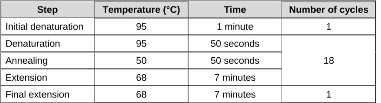
Table 3 Thermocycling conditions used for site-directed mutagenesis of EngA cysteine mutants. ... 82
Table 4 Cryoprotectants and respective concentration used when fishing EngA crystals. ... 100
Table 5 Experimental settings used in BLI experiments. ... 108
Table 6 Overall parameters of His-EngAWT (apo) obtained by SAXS. ... 121
Table 7 Overall parameters obtained by SAXS for His-EngAWT in the absence and presence of different concentrations of nucleotides and fitting analysis with three models. ... 129
Table 8 Trypsin-sensitive regions detected by N-terminal sequencing analysis. ... 133
Table 9 Fragments originated after 15 and 120 minutes of tryptic digestion of apo and 10 mM GMPPCP His-EngAWT detected by mass spectrometry. ... 133
Table 10 Statistics of diffraction data processing and refinement of seven crystal structures of His-EngAWT. ... 154
Table 11 Sample and crystallisation conditions from each crystal described in Table 10. ... 155
Table 12 Hydrogen and salt bonds involved in the interface of each crystal packing. ... 161
Table 13 Rmsd values of the eleven structures of EngA after superposition on the GD2. ... 164
List of figures
Fig. 1 Discovery dates of antibiotic classes... 29Fig. 2 Major targets for the action of antibacterial drugs. ... 32
Fig. 3 Structure of the E. coli 70S ribosome. ... 35
Fig. 4 Structure of the E. coli 50S and 30S ribosomal subunits. ... 36
Fig. 5 Overview of bacterial translation. ... 38
Fig. 6 Assembly maps of bacterial ribosomal subunits. ... 41
Fig. 7 Mechanism of the in vitro assembly of the 30S and 50S subunits. ... 42
Fig. 8 Maturation of ribosomal RNA. ... 43
Fig. 9 Mechanism of the in vivo assembly of the 30S and 50S subunits. ... 44
Fig. 10 Diagram of the G-domain. ... 46
Fig. 11 3D structure of the G-domain from human Ras GTPase. ... 47
Fig. 12 Guanine nucleotide binding site. ... 48
Fig. 13 Nucleoside interactions with GTP-binding proteins. ... 49
Fig. 14 GTPase catalytic cycle. ... 50
Fig. 15 The universal switch mechanism of P-loop GTPases, described as the loaded-spring mechanism. ... 51
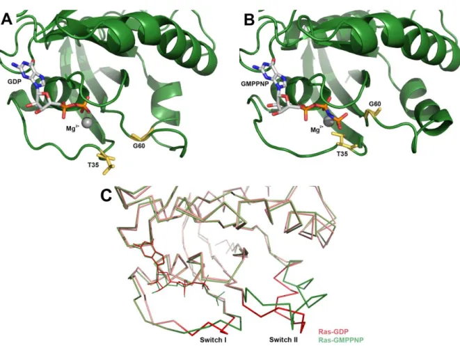
Fig. 16 3D structures of the human Ras GTPase in the GDP-bound and GTP-bound forms. ... 51
Fig. 17 Conventional mechanism of GAP-mediated GTP hydrolysis. ... 53
Fig. 18 3D structure of the G-domain of E. coli GTP-binding protein MnmE, and close view of the catalytic centre. ... 56
Fig. 19 Amino acid alignment of EngA orthologs. ... 61
Fig. 20 Representation of the domain organisation of EngA. ... 62
Fig. 21 3D structure of the KH-domain of E. coli Era and T. maritima EngA. ... 62
Fig. 22 3D structure of the E. coli 30S subunit and the T. maritima ribosomal protein S7. ... 65
Fig. 23 3D structure of T. maritima EngA. ... 69
Fig. 24 Superposition of the 3D structures of GTP-bound Era and phosphate-bound EngA. ... 70
Fig. 25 3D structure of B. subtilis EngA. ... 71
Fig. 26 Superposition of B. subtilis GD1 “off-state” and T. Maritima full-length EngA; Superposition of T. maritima GD1 “on-state” and B. subtilis full-length EngA. ... 72
Fig. 27 3D structure of the E. coli 50S:EngA complex. ... 73
Fig. 28 3D structure of E. coli EngA. ... 74
Fig. 29 Superposition of the 3D structures of EngA in different ligand-bound forms. ... 75
Fig. 30 Representation of the interaction between residues in the His-tag and nickel ions. ... 84
Fig. 31 Representation of the separation of molecules by SEC... 85
Fig. 32 Schematic representation of the PK/LDH coupled assay for determination of the GTPase activity of EngA. ... 86
Fig. 33 Representation of the terminal structure of glutathione immobilised in a sepharose matrix. .... 88
Fig. 34 Structure of three GTPase inhibitors CEB-II-54, CEB-II-55 and CEB-II-56. ... 97
Fig. 35 Schematic representation of the vapour diffusion method by hanging drop and sitting drop. .. 98
Fig. 36 Schematic representation of the microseeding technique. ... 99
Fig. 37 Principle of surface plasmon resonance and representation of a CM5 chip. ... 106
Fig. 38 Representation of a typical TSA response. ... 109
Fig. 39 SDS-PAGE analysis of the expression and IMAC of His-EngAWT. ... 113
Fig. 40 Chromatogram of His-EngAWT purification using a Superdex ® 75 column. ... 114
Fig. 41 GTPase activity assay for EngAWT. ... 115
Fig. 42 SDS-PAGE analysis of the affinity chromatography and size exclusion chromatography of S7-His and GST-S7. ... 116
Fig. 43 Chromatogram of S7 purification using a Resource® S column... 117
Fig. 44 5-20% sucrose gradient profile for dissociated 50S and 30S subunits. ... 118
Fig. 45 Logarithmic plot of the scattering curve for His-EngAWT (apo) measured using the robot sample changer or the eluted fraction from a size exclusion chromatography column. ... 120
Fig. 46 3D structures of different conformations of EngA used for model validation with CRYSOL. .. 122
Fig. 47 Fit of the logarithmic plot of the scattering curve for His-EngAWT (apo) with the crystal structures PDB 4DCU, 1MKY and 3J8G. ... 123
Fig. 48 Distance distribution functions of His-EngAWT (apo) obtained experimentally and of the three models. ... 124
Fig. 49 Fit of the logarithmic plot of the scattering curve for His-EngAWT with 10 mM GMPPNP with the crystal structures PDB 4DCU and 1MKY. ... 126
Fig. 50 Distance distribution functions of His-EngAWT in the absence and in the presence of 1 mM GDP, 10 mM GDP, 1 mM GMPPNP and 10 mM GMPPNP or GMPPCP. ... 127
Fig. 51 Distance distribution functions of His-EngAWT (10 mM GMPPNP) obtained experimentally and of the three models 4DCU, 1MKY and 3J8G. ... 128
Fig. 52 Schematic representation of a Kratky plot for a globular protein, partially folded proteins and
unfolded protein. ... 128
Fig. 53 Normalised Kratky plot from the experimental data of His-EngAWT in the absence and presence of 10 mM GDP and GMPPNP. ... 129
Fig. 54 SDS-PAGE analysis of the tryptic digestion of His-EngAWT in different nucleotide bound-states. ... 131
Fig. 55 Effect of nucleotides on the proteolytic rate of EngA by trypsin. ... 132
Fig. 56 SDS-PAGE from the tryptic digestion of His-EngAWT at times 15 and 120 minutes. ... 134
Fig. 57 Surface representation of the 3D structures of EngA from the PDB files 4DCU, 1MKY and 3J8G. ... 135
Fig. 58 ESI-TOF MS deconvoluted spectra of the tryptic digestion (time 15 minutes) of His-EngAWT in the absence and in the presence of 10 mM GMPPCP. ... 136
Fig. 59 3D structure of GDP-bound EngA with two highlighted regions possibly associated to changes in the protein global conformation triggered by a triphosphate nucleotide. ... 138
Fig. 60 ESI-TOF MS deconvoluted spectra of the tryptic digestion (time 120 minutes) of His-EngAWT in the absence and in the presence of 10 mM GMPPCP. ... 139
Fig. 61 Fragmentation map of tryptic cleavage of His-EngAWT. ... 140
Fig. 62 Representation of the double-cysteine EngA mutants designed to block the 4DCU and 1MKY conformations. ... 142
Fig. 63 GTPase activity assay for EngAY51C/T169C and EngAG139C/V388C. ... 143
Fig. 64 SDS-PAGE analysis of the cross-linking assay for EngAG139C/V388C and EngAY51C/T169C. ... 144
Fig. 65 Logarithmic plot of the scattering curve for cross-linked His-EngAG139C/V388C. ... 146
Fig. 66 Logarithmic plot of the scattering curve for cross-linked His-EngAY51C/T169C. ... 147
Fig. 67 Crystals obtained from the first crystallisation screening of EngA with GTP analogues. ... 151
Fig. 68 Crystals obtained after steps of optimisation of the crystallisation conditions of EngA. ... 151
Fig. 69 Details of the nucleotide-binding site of EngA from Crystals 2 and 3. ... 156
Fig. 70 EngA 3D structure obtained from Crystal 1 superposed with 4DCU and close view of the nucleotide-binding sites of GD1 and GD2. ... 157
Fig. 71 Electron density map of molecule A from the asymmetric unit of Crystals 6 and 7.. ... 158
Fig. 72 Electron density map of molecule B from the asymmetric unit of Crystal 7. ... 159
Fig. 73 Interface between two molecules of 4DCU crystal related by crystallographic symmetry. ... 160
Fig. 74 Comparison of the EngA 3D structures from Crystal 6 and two symmetry related molecules from 4DCU. ... 162
Fig. 75 Superposition of EngA structures 4DCU and 4DCV and our refined structures 1-7. ... 163
Fig. 76 Close view of the GD2-P-loop from structures 4DCU, Crystal 2 and Crystal 3 ... 165
Fig. 77 Alignment of the GD2 of PDB entries 4DCU and 4DCV; Comparison of different GMPPN/CP-bound structures. ... 165
Fig. 78 Close view of the GD2-switch II region of the several GMPPN/CP-bound structures. ... 166
Fig. 79 Close view of the GD2-G4 motif. ... 167
Fig. 80 Close view of the GD1-P-loop in the apo, GDP-bound and pyrophosphate-bound forms. ... 168
Fig. 81 Close view of the GD1-switch II region. ... 168
Fig. 82 Close view of the GD1-G4 motif. ... 169
Fig. 83 Alignment of the GD1 from 4DCU and Crystal 6B. ... 170
Fig. 85 Western-blot analysis of the fractions obtained from centrifugation on a 5-20% sucrose
gradient of a mixture of His-EngAWT and dissociated 70S ribosome. ... 174
Fig. 86 Interaction curves of His-EngAWT and the 50S and the 30S subunit. ... 175
Fig. 87 Kinetics workflow of a BLI experiment... 176
Fig. 88 Sensorgram obtained from immobillisation of His-EngAWT on a HIS1K biosensor. ... 177
Fig. 89 Negative staining images of the 50S subunits. ... 178
Fig. 90 Distribution of the orientations of the particles; One of the analysed images of the complex 50S:EngA; Classification of particles into different classes. ... 179
Fig. 91 Classes obtained after 2D ab initio classification of the 50S:EngA particles. ... 179
Fig. 92 Cryo-EM 3D reconstructions of the 50:EngA complex. ... 180
Fig. 93 Refined cryo-EM 3D reconstructions of the 50:EngA complex. ... 180
Fig. 94 Interaction between His-EngAWT and S7 measured by ELISA assay. ... 182
Fig. 95 Sensorgram obtained for binding of His-EngAWT (apo) to S7-His immobilised in a CM5 chip via amine coupling. ... 183
Fig. 96 Sensorgram obtained for binding of S7 to His-EngAWT (apo) immobilised in a Ni-NTA biosensor. ... 184
Fig. 97 Fitting of the experimental data of the interaction EngA:S7 with the 1:1 binding model ... 184
Fig. 98 TSA profile of His-EngAWT in the presence of GTPase inhibitors. ... 186
Fig. 99 Interaction between His-EngAWT and 70S ribosome by ELISA assay in the presence of CEB-II-5x inhibitors. ... 188
Fig. 100 Contacts in nucleotide-binding site in GD1 and GD2 of B. subtilis GDP-bound EngA. ... 194
Fig. 101 Superposition of the 3D structures of EngA dimers in four crystal forms... 196
Abbreviations
A. thaliana Arabidopsis thaliana
aa amino acid A-site aminoacyl-site AlFx aluminium fluoride AMR antimicrobial resistance AU absorbance unit
B. subtilis Bacillus subtilis
BLI bio-layer interferometry BSA bovine serum albumin
C. crescentus Caulobacter crescentus
CDC Centers for Disease Control and Prevention
CDDEP Center for Disease Dynamics, Economics & Policy
CEB-II-5x chemical library of compounds (CEB-II-54, CEB-II-55, CEB-II-56) CRE carbapenem-resistant Enterobacteriaceae
Dmax maximum dimension
DMSO dimethyl sulfoxide DNA deoxyribonucleic acid
dNTP deoxynucleotide triphosphate DOC deoxycholate
DTT dithiothreitol
E. coli Escherichia coli
E-site exit-site
ECDC European Centre for Disease Prevention and Control EDTA ethylenediaminetetraacetic acid
ELISA enzyme-linked immunosorbent assay EM electron microscopy
FKB ForteBio kinetics buffer GAP GTPase-activating protein
GDI Guanine nucleotide dissociation inhibitor GEF Guanine nucleotide exchange factor GD G-domain
GD1 G-domain 1 GD2 G-domain 2
GDP guanosine diphosphate Glncat catalytic glutamine
GMPPCP/GMPPNP GTP analogues GTP guanosine triphosphate
GST glutathione S-transferase GV EngA variant G139C/V388C
H. influenza Haemophilus influenza
HAS-GTPase hydrophobic amino acid substituted GTPase HBS HEPES-buffered saline
HPLC high-performance liquid chromatography I0 forward scattering intensity
IA interface area
IEC ion exchange chromatography
IMAC immobiliseed metal-affinity chromatography IPTG isopropyl β-D-1-thiogalactopyranoside ITC isothermal titration calorimetry
kcat catalytic constant
ka association rate
KD dissociation constant
KH K-homology KM Michaelis constant
LB Luria broth cell culture medium
LC-ESI-TOF liquid-chromatography electrospray-ionisation time-of-flight LDH lactate dehydrogenase
LLG log likelihood gain
M. smegmatis Mycobacterium smegmatis
Mc main chain
mAb monoclonal antibody mRNA messenger RNA MS mass spectrometry
MRSA methicillin-resistant Staphylococcus aureus MW molecular weight
N. benthamiana Nicotiana benthamiana N. gonorrhoeae Neisseria gonorrhoeae
NAD+/NADH oxidised/reduced nicotinamide adenine dinucleotide OD optical density
(p)ppGpp guanosine (penta)tetraphosphate P-site peptidyl-site
PCR polymerase chain reaction PDB Protein Data Bank
PEG polyethylene glycol PEP phosphoenolpyruvate PK pyruvate kinase
PMSF phenylmethylsulfonyl fluoride PTC peptidyltransferase center PVDF polyvinylidene fluoride r-protein ribosomal protein
RA-GTPase ribosome assembly GTPase Rg Radius of gyration
rmsd root mean square deviation RNA ribonucleic acid
rRNA ribosomal RNA RU response units tRNA transfer RNA
S. aureus Staphylococcus aureus
S. pneumoniae Streptococcus pneumoniae S. typhimurium Salmonella typhimurium
SA specific enzymatic activity SAXS small-angle X-ray scattering SDM site-directed mutagenesis
SDS-PAGE sodium dodecyl sulfate polyacrylamide gel electrophoresis SEC size exclusion chromatography
SIMIBI signal-recognition particle, MinD, BioD related SPR surface plasmon resonance
SwI / SwII switch I / switch II regions
t50 time at which full-length protein is reduced by half upon proteolysis
T. maritima Thermotoga maritima
TCA trichloroacetic acid TFA trifluoroacetic acid Tm melting temperature
TMB 3,3’,5,5’-tetramethylbenzidine TRAFAC translation factor-related TSA thermal shift assay
Vmax maximal velocity
Vp Porod volume
WHO world health organisation WT wild-type
Context
The development of new therapeutic options against bacterial infections has aroused great interest over the last years in the context of drug resistance. In the drug discovery and development pipeline, a long path stands before a molecule can enter clinical trials and lastly be approved. The starting-point in the pursuit of new antibiotics for which bacterial resistance mechanisms do not pre-exist is the identification of novel cellular targets. An ideal drug target should have three main characteristics: play a critical role in an essential metabolic pathway in the bacterial cell; be conserved among pathogens (in the context of the search for broad-spectrum antibiotics); and be absent from the human genome to avoid adverse drug reactions in the human host. Academic research is a key component in the early stages of drug discovery: genetics has been of central importance in the identification of bacterial genes whose products represent potential drug targets, and the structural and biochemical characterisation of such enzymes is a major contributor for the implementation and validation of hit discovery approaches.
Genetics studies in the early 2000s have identified engA as a conserved bacterial gene that is not present in the human genome and whose product is an enzyme essential for cell survival. These initial studies revealed that EngA exhibits characteristics of an ideal drug target and several studies have since contributed to its biochemical and structural characterisation.
EngA is now known to be a GTPase that acts as an assembly factor for the bacterial ribosome. Although the bacterial ribosome is one of the major bacterial targets for the action of antibiotics, the pathway of ribosome assembly has remained unexploited.
The aim of this work was to study EngA as a target for inhibitors of the assembly process. A brief bibliographic summary of what is known on bacterial ribosome biogenesis and EngA is presented. The results from our work and future perspectives are discussed next and will hopefully give some useful insights into the characterisation of EngA as a potential bacterial drug target.
1. Antibiotic resistance
The advent of the first antibiotics, approximately 70 years ago, introduced a period where it became possible to treat bacterial infectious diseases.
The antibiotic era began in the first half of the twentieth century, with the introduction by Paul Ehrlich of the idea that chemical compounds could selectively target disease-causing microorganisms. The discovery of penicillin by Alexander Fleming and the first sulfonamide by Gerhard Domagk and co-workers were the breakthrough for the “golden” antibiotic era, between the 1940s and 1970s, when many new classes of antibiotics were discovered. Antibiotic discovery was one of the major medical advances, contributing significantly to the public (control of endemic and epidemic diseases) and individual health (reduction of morbidity, mortality and increase of life expectancy). However, the discovery rate of novel classes has decreased since the 1980s, with very few new antibiotics being discovered or approved for clinical use since then.
Fig. 1 Discovery dates of classes of antibacterial drugs. While between 1940 and 1980 about 20 new classes of antibiotics were identified from natural substances, no major advances have been made since then. Since the 1980s, most of the approved antibiotics were members of already known classes. Adapted from WHO, 2014.
Along with the introduction of antibiotics came the emergence of resistance in pathogenic bacteria. Antimicrobial resistance (AMR) is the ability of a microorganism to resist to drugs that would normally kill it or limit its growth. Early after penicillin discovery, Fleming warned for the possibility of microbes becoming resistant in case of penicillin underdosage (Nobel Prize Lecture: Penicillin, 1945). Indeed, resistance started to emerge years after the
large-scale production of penicillin. Since then it has been transmitted to many pathogenic bacteria and it concerns all antibiotic classes.
Evolution of resistant strains occurs spontaneously in nature, either by spontaneous mutations or by acquisition of genes (i.e. horizontal gene transfer) from environmental microbiota, and it precedes the introduction of antibiotics (Martínez, 2012). However, since their introduction, the use and misuse of antibiotics has accelerated this process by elimination of susceptible organisms and selection of the resistant ones. A study by Alonso and collaborators reported an increase in P. aeruginosa’s mutation rate by 105 times after a
4-days incubation with tetracyclines (Alonso et al., 1999). In E. coli, a linear relationship is observed between the mutation rate and the antibiotic concentration used (Long et al., 2016).
As a direct consequence of antibacterial resistance, standard treatments become ineffective and infections persist and spread to others, with a huge impact in public health. E. coli is the most common cause of urinary tract infections. E. coli resistance to third generation cephalosporins and fluoroquinolones has attained more than 50% in some countries (WHO, 2014). For such strains, carbapenems remain the only available treatment. Public health implications for such high resistance proportions imply less specificity of treatment and increased adverse-side effects (by the use of broad-range therapy), longer hospital stays, high medical costs and expansion of resistance to last-resort antibiotics (such as carbapenems). Reports of carbapenem-resistant Enterobacteriaceae (CRE) confirm this reality, with a rising number of deaths from untreatable infections caused by CRE E. coli and K. pneumonia all around the world (CDDEP, 2015; WHO, 2014). S. aureus is another common cause of healthcare-associated infections. Over the last decade, methicillin-resistant S. aureus (MRSA) strains, which were initially limited to hospital-acquired infections, now account for more than 80% of community-acquired infections in some countries (CDDEP, 2015).
Facing this situation, international nongovernmental health agencies together with governments and private entities around the world have launched initiatives to stop the spread of antimicrobial resistance. In the occasion of the 2011 World Health Day, dedicated to the combat against drug resistance, the WHO introduced a policy package with critical actions to struggle against the spread of antimicrobial resistance, calling on «all key stakeholders (…) to act and take responsibility for combating antimicrobial resistance». In November 2015, several worldwide activities such as the European Antibiotic Awareness Day, the United States Get Smart About Antibiotics Week, the Canadian Antibiotic Awareness Week and the Australian Antibiotic Awareness Week were simultaneously held in
was addressed for the first time in the United Nations General Assembly to define a coordinated approach across multiple sectors to be adopted and reported in a two-years time.
Despite the global efforts to tackle antimicrobial resistance, this is still a mounting problem that is far from being solved. Upon the 2014 Surveillance Report on Antimicrobial Resistance (WHO, 2014), the WHO stated that «without urgent, coordinated action, the world is heading towards a post-antibiotic era, in which common infections and minor injuries (…) can once again kill».
The development of new initiatives for the treatment of bacterial infections is crucial to stop antibiotic resistance. Alternative therapies to complement antimicrobials are being developed. Therapies that target the host are based on the fact that signs and symptoms upon infection result from a response driven by the host’s immune system. By targeting the host rather than the microbe, immunomodulation can avoid selective pressure for the development of resistance (Hancock et al., 2012). The use of anti-inflammatory monoclonal antibodies (mAbs) is a potential strategy to reduce pathogen-mediated tissue damage and help recovery (Spellberg et al., 2013). Conversely, immune stimulation has been suggested as a way of enhancing antibiotics’ action by promoting an appropriate immune response and is currently in clinical development (Czaplewski et al., 2016). Vaccines have also been in development, although their action is restricted to prophylaxis. mAbs can have a direct antimicrobial mechanism, by binding to microbes, virulence factors or toxins. This strategy is currently in clinical trials (Czaplewski et al., 2016). Their high specificity for their target and the exploitation of mechanisms different from those of antibiotics, make mAbs unlikely to select for mAbs resistance among non-targeted microorganisms or for cross-resistance (Bebbington and Yarranton, 2008; Saylor et al., 2009). Modulation of the microbe growth environment can potentially be done by metal-chelation sequestration of nutrients, so that microbes do not proliferate. Another approach that has reached phase III clinical development is the use of probiotics that compete with microbial growth (Czaplewski et al., 2016; Spellberg et al., 2013). Phage lysins that destroy bacterial cell wall and reduce formation of biofilms can be used in combination with antibiotics. Bacteriophages could eventually replace antibiotics as not only they directly attack bacteria, but also have the ability to replicate and evolve to adapt to the pathogenic strain (Czaplewski et al., 2016).
The return to the investment on new antibiotics is also critical to overcome resistant microbes. Different strategies can be employed in the discovery of new antibiotics. Examples include the exploration of new sources of natural antibiotics, such as marine or extremophilic bacteria, or the search for novel cellular targets (Ventola, 2015). Genetics studies have revealed genes that are critical to the survival of bacteria or that are responsible for virulence
mechanisms. Such genes represent potential new targets for antibiotic design. Identification of bacterial essential enzymes that do not present susceptibility to resistance mechanisms has become an important approach for antibiotic development. Such is the case of the engA gene, now known to encode for an essential bacterial GTPase, which was initially discovered in a study aiming at identifying genes responsible for pilin variation and adaptation in bacteria (Mehr et al., 2000).
This work focuses on the EngA bacterial GTPase as a potential target for antibiotic design. EngA is conserved in bacterial species and essential for cell viability. Since its identification in 2000 by Mehr and collaborators, studies on EngA have revealed a role in bacterial ribosome assembly (Bharat and Brown, 2014; Bharat et al., 2006; Foucher et al., 2012; Hwang and Inouye, 2006; Jeon et al., 2014; Lamb et al., 2007; Schaefer et al., 2006; Tan et al., 2002; Tomar et al., 2009; Zhang et al., 2014). While the ribosome itself is a drug target for many of the available antibiotics (Fig. 2), its biogenesis process has remained unexploited from a pharmacological perspective.
Fig. 2 Major targets for the action of antibacterial drugs. Antibiotics can interfere with the cell wall, mainly by inhibiting the synthesis of peptidoglycan. Others injure the cytoplasmic membrane, leading to the depolarisation and leakage of cell content. Folic acid is essential for the synthesis of nucleic acids in cells and can be inhibited by the action of antibacterials that interfere with its synthesis pathway. Inhibition of nucleic acid synthesis is achieved by antibiotics that interfere with enzymes such as DNA gyrases, topoisomerases or RNA polymerases. Finally, the protein biosynthesis pathway in bacteria is inhibited by drugs that directly bind the ribosome subunits and alter their translational capability. Modified from Servier Image Bank.
The absence of exploitation of the ribosome assembly pathway together with the ubiquitous and essential nature of EngA and the lack of a human ortholog make EngA a promising therapeutic target for the development of new antibacterial agents.
2. Bacterial Ribosome
Ribosomes are ribonucleoprotein cellular machineries responsible for the translation of the genetic code into proteins. These catalytic machines are present in all domains of life, sharing a common structural core that harbours the functional centres, highlighting their importance in the fundamental process of protein biosynthesis (Yusupov, 2014).
2.1. Structure
Since the 1970s, when the shape of the bacterial ribosome and its subunits was for the first time reconstituted by electron microscopy images (Lake, 1976), a great progress has been made on the determination of the ribosome structure. During the last decades, studies on X-ray crystallography and cryo-electron microscopy, along with advances in synchrotron light sources and crystallographic hardware and software, have lead to the elucidation of the ribosome atomic structure (Ban et al., 2000; Schuwirth et al., 2005; Wimberly et al., 2000). With this information, the understanding of protein biosynthesis pathways and antibiotics’ mechanisms of action was made possible.
All ribosomes – prokaryotic, eukaryotic or organellar – consist of two unequal subunits responsible for protein biosynthesis in cells: a large subunit, that contains the catalytic site for peptide-bond formation, and a small subunit, responsible for mRNA binding and decoding. With the exception of the mammalian mitochondria ribosome, that shows a reversed ratio, ribosomes are composed of one-third protein and two-thirds RNA (Ramakrishnan, 2009). The next sections will present a description of the bacterial ribosome, which can be explored as a target for antibacterial drugs. The following references contain information on the structure of chloroplast, mitochondrial and eukaryotic ribosomes, respectively: Agrawal et al., 2011; Greber and Ban, 2016; Yusupova and Yusupov, 2014.
The basic architecture of the 70S ribosome and its subunits is represented in Fig. 3. The 70S ribosome is an asymmetrical assembly of more than 50 proteins and 3 RNA chains, with a ratio by weight of 2:1 RNA:protein. It is built of a small subunit 30S and a large subunit 50S, that associate to form a functional ribosome with a molecular weight of 2.5 MDa, a sedimentation coefficient of 70S and a diameter of ~20 nm (Kaczanowska and Rydén-Aulin, 2007; Nelson and Cox, 2005).
Fig. 3 Structure of the E. coli 70S ribosome. The 50S subunit is represented in green (proteins) and blue (rRNA) and the 30S subunit in yellow (proteins) and orange (rRNA). Image edited in PyMol from PDB file 4V4Q.
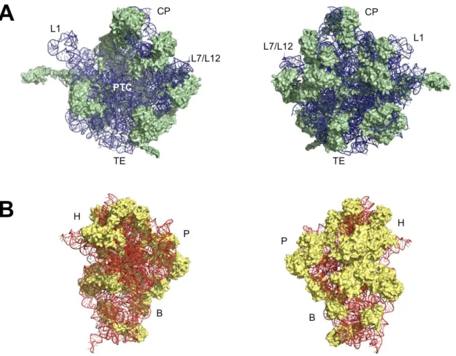
The 50S subunit (1.45 MDa) consists of two rRNA chains, 23S (~2900 nucleotides) and 5S (~120 nucleotides), and 34 r-proteins, L1–L34, that range from 5–30 kDa (Fig. 4A). It is responsible for the catalysis of peptide bonds linking, providing the peptidyltransferase-centre (PTC). Its crown-like shape encompasses a central protuberance, formed by 5S rRNA and associated r-proteins, and two flanking arms: the L1-stalk, formed by r-protein L1 and rRNA, and the L7/L12-stalk, formed by r-proteins L7 and L12 associated with L10, L11 and rRNA (Yusupov, 2014). The PTC is located in the centre of the interface side of the 50S subunit, where peptide bond formation occurs. Just below the PTC, there is the polypeptide exit tunnel of about 100 Å long and 25 Å in diameter that accommodates the nascent peptide (up to 40 amino acids) (Kaczanowska and Rydén-Aulin, 2007).
The 30S subunit (0.85 MDa) contains a single RNA chain 16S (~1500 nucleotides) and 21 r-proteins, S1–S21, that range from 8–60 kDa (Fig. 4B). The small subunit provides the decoding-centre, responsible for decoding mRNA and controlling translation fidelity. The shape of the 30S is segmented into 3 main regions, defined by the secondary structure of 16S rRNA: the body (domain I), formed by the 5’ end; the platform (domain II), formed by the central part of the rRNA; and the head (domain III), formed by the 3’ end. A narrow region between the head and the platform, called neck, forms a cleft where the decoding centre is located (Kaczanowska and Rydén-Aulin, 2007).
Fig. 4 Structure of the E. coli (A) 50S and (B) 30S ribosomal subunits. The left images represent the internal view of the subunits and the images on the right the exterior view. The 50S subunit is represented in green (proteins) and blue (rRNA) and the 30S subunit in yellow (proteins) and orange (rRNA). L1: L1-stalk; L7/L12: L7/L12-stalk; CP: central protuberance; PTC: peptidyltransferase centre; TE: tunnel exit; H: head; P: platform; B: body. Image edited in PyMol from PDB file 4V4Q.
It is at the interface between the two subunits that the functional regions of the ribosome are located. This interface consists mainly of RNA, with no protein within 18 Å of the active site, suggesting that the ribosome is a ribozyme (Yusupov, 2014). While the core of the subunits is made of rRNA chains, r-proteins are located at the surface. Most r-proteins contain a globular domain, which stands at the surface of the subunit, and an extended domain that protrude into the rRNA to stabilise its tertiary structure (Yusupov, 2014).
Association of the 30S and 50S forms a tunnel in their interface through which mRNA passes during translation and where the codons of mRNA interact with the anticodons of the tRNAs. Three tRNA-binding sites are present: aminoacyl (A)-site, peptidyl (P)-site and exit (E)-site. The A-site binds the aminoacyl-tRNA to be presented to the mRNA. The P-site carries the nascent peptide chain attached as a peptidyl-tRNA complex. The E-site harbors deacylated-tRNAs ready to leave the ribosome (Lafontaine and Tollervey, 2001; Schmeing and Ramakrishnan, 2009).
2.2. Protein synthesis
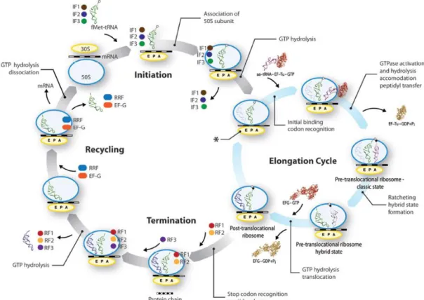
Bacterial translation can be divided into three main stages: initiation, elongation and termination (Fig. 5) (Nelson and Cox, 2005; Schmeing and Ramakrishnan, 2009).
The initiation of polypeptide synthesis in bacteria requires the 30S and 50S subunits, the mRNA coding for the peptide, the fMet-tRNAfMet, three initiation factors (IF-1, IF-2 and IF-3), Mg2+ and GTP. Initiation starts with binding of IF-1 and IF-3 to the 30S subunit, which stimulate the release of mRNA and deacylated tRNA from the previous cycle and prevent association of the 50S subunit. The mRNA binds to the small subunit and the initiation codon (5’)AUG is guided to its correct position at the P-site. This alignment occurs through the interaction between the Shine-Dalgarno sequence in the mRNA, a sequence of 4–9 purine bases (8–18bp) upstream the start codon, and a complementary pyrimidine-rich sequence in the 16S rRNA. The GTP-bound initiation factor IF-2 and the fMet-tRNAfMet bind to the complex, allowing the pairing of the tRNA anticodon and the mRNA start codon. GTP hydrolysis by IF-2 induces the release of the initiation factors and the binding of the 50S subunit, forming a functional 70S ribosome – the initiation complex.
Elongation requires the initiation complex, aminoacyl-tRNAs, three elongation factors (EF-Tu, EF-Ts, and EF-G) and GTP. In the first elongation step, the incoming aminoacyl-tRNA binds to the elongation factor EF-Tu GTP-bound. The resulting aminoacyl-tRNA–EF-Tu–GTP ternary complex binds the A-site of the initiation complex. EF-Tu then hydrolyses GTP and dissociates from the complex, while the aminoacyl end of A-site tRNA moves into the PTC. Decoding is followed by peptide bond formation between the fMet and the new amino acid bound by their tRNAs to the P- and A-sites, respectively. Upon peptide bond formation, the nascent peptide on the P-site is transferred to the aminoacyl-tRNA on the A-site, leaving the P-site deacylated. Hydrolysis of GTP by EF-G catalyses translocation, where the tRNAs and the mRNA move relative to the ribosome, shifting positions of both tRNAs: the deacylated tRNA on the P-site is transferred to the E-site and the dipeptidyl-tRNA on the A-site is transferred to the P-site. The ribosome is now ready for the next elongation cycle and elongation will continue until the ribosome adds the last amino acid coded by the mRNA.
Termination is signalled by the presence of a termination codon (UAA, UAG, UGA). Once a termination codon occupies the A-site, release factors RF-1 or RF-2 will bind, leading to the cleavage and release of the nascent polypeptide from the tRNA in the P-site. RF-3 will bind to the ribosome–RF-1/2 complex and accelerate the dissociation of the release factors. At this stage, the mRNA and the deacylated tRNA on the P-site are released from the ribosome by the recycling factor RRF and EF-G. Finally, the 70S ribosome dissociates into its 30S and 50S subunits to start a new cycle.
An interactive overview of protein translation in bacteria can be found at Ramakrishnan‘s group web page (http://www.mrc-lmb.cam.ac.uk/ribo/homepage/mov_and_overview.html), from the MRC Laboratory of Molecular Biology.
Fig. 5 Overview of bacterial translation. Adapted from Agirrezabala and Frank, 2010.
2.3. Inhibitors of bacterial translation
Protein synthesis is a major cellular target for antibiotic action, with approximately half of the available antibiotics targeting the translation machinery. Antibiotics that inhibit translation act by directly binding to the ribosomal subunits, affecting translational capability. With some exceptions, the functional centres of the ribosome – PTC, polypeptide exit tunnel and tRNA/mRNA interaction sites – constitute the binding sites for antibiotics (Wilson, 2009). A few examples of bacterial translation inhibitors are described below.
2.3.1. Inhibitors of translation initiation
The initiation stage of translation is inhibited by drugs that interfere with the formation of the initiation complex by preventing the binding of one interaction partner. Few antibiotics that inhibit the translation initiation step are available for clinical use.
GE81112 is a synthetic tetrapeptide antibiotic, representing a structurally unique class, still in research, that binds the 30S subunit and overlaps the P-site. GE81112 induces conformational changes in the 16S rRNA that lead to an incorrect binding of fMet-tRNAfMet and mRNA to the 30S subunit, preventing subsequent binding of 50S and progression of the initiation stage (Fabbretti et al., 2016).
G1(MW297) is a synthetic nitrovinylfuran broad-spectrum antibiotic that binds the 30S subunit, overlapping the P-site and preventing binding of the fMet-tRNAfMet (Fabbretti et al., 2012).
GSK1322322 is a synthetic hydrazide and represents a new class of antibiotics, having recently completed phase II clinical trial. It targets peptide deformylase, a bacterial enzyme required for modification of the N-terminus of the nascent polypeptide chain in prokaryotes (Corey et al., 2014).
2.3.2. Inhibitors of translation elongation
Many of the currently available antibiotics target the elongation step of translation.
Tetracyclines are broad-spectrum antibiotics that bind reversibly to the head of the 30S subunit, blocking the binding of aa-tRNAs and preventing addition of amino acids to the nascent peptide.
By irreversibly binding the 30S, aminoglycosides are bactericidal agents that interfere with a loop comprising the decoding site. Aminoglycosides interfere with regions of the 16S rRNA that are responsible for ensuring the binding of a cognate aa-tRNA to the mRNA during decoding. The presence of an aminoglycoside results in misreading and misincorporation of amino acids. Other members of this antibiotic class bind the mRNA in the P-site of the PTC, inhibiting translocation.
Chloramphenicol is a bacteriostatic drug that reversibly binds to the A-site of the PTC on the 50S subunit, overlapping the aminoacyl moiety of aa-tRNAs and inhibiting the peptidyltransferase step. Oxazolidinones, such as linezolid, have a similar mechanism.
Macrolides are bacteriostatic and bactericidal drugs that act by binding to the exit tunnel in the 50S subunit, adjacent to the PTC. This blocks aminoacyl translocation and the progression of the nascent polypeptide chain and also prevents formation of initiation complexes. Ketolides are semi-synthetic macrolides derivatives developed to escape
resistance mechanisms. Structural modification of the sugar moiety renders ketolides poor substrates for efflux pumps and increases ribosome binding affinity (Katzung, 2006).
Lincosamides bind to the PTC in the 50S subunit and overlap with both binding sites of chloramphenicol and macrolides by binding to A-site of the PTC and extending into the exit tunnel. They inhibit peptide growth, translocation of aminoacyl and formation of initiation complexes.
Streptogramines are bactericidal drugs that also bind adjacent sites on the PTC, interfering with binding of tRNA to both A- and P-sites, and on the exit tunnel.
2.3.3. Inhibitors of translation termination and recycling
Fusidic acid is a steroidal antibiotic that binds to EF-G in complex with the ribosome. Binding of fusidic acid allows GTP hydrolysis by EF-G but prevents conformational changes of the GTPase and hence its release from the ribosome.
2.4. Ribosome biogenesis
While numerous antibiotics inhibit the translating ribosome, none is directed towards the pathway of ribosome assembly. The assembly of such complex cellular machineries has been subject of intensive study for the last decades and important steps and factors have been unravelled.
2.4.1. In vitro assembly mechanisms
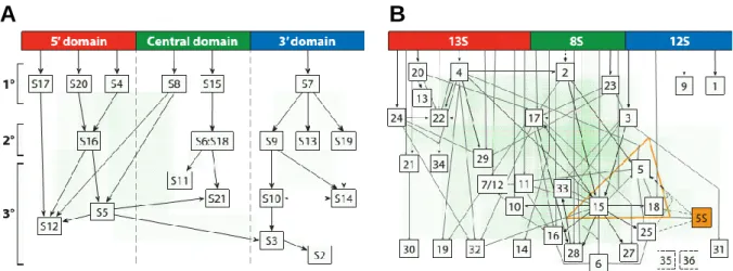
In the late 1960s, work from the laboratories of Masayasu Nomura (Traub and Nomura, 1968) and Knud Nierhaus (Nierhaus and Dohme, 1974) demonstrated that both ribosomal subunits could be broken down into rRNA and r-proteins and reconstituted to functionally active particles in vitro, in an orderly and self-assembly process. The observations from studies on ribosome biogenesis in vitro resulted in assembly maps for the 30S and 50S subunits (Fig. 6).
A
B
Fig. 6 Assembly maps of ribosomal subunits. (A) Nomura assembly map represents the hierarchy of protein binding in the 30S subunit. 16S rRNA is represented in three domains (domain I in red; domain II in green; domain III in blue) and r-proteins are classified as primary (1º), secondary (2º) or tertiary (3º). (B) Nierhaus assembly map represents the hierarchy of protein binding in the 50S subunit. 23S is divided in three sections analogous to the 16S domains. Three r-proteins are responsible for binding of 5S rRNA (orange). Adapted from Shajani et al., 2011.
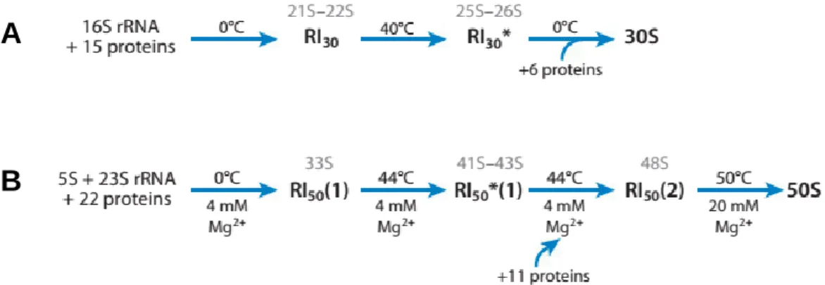
During 30S in vitro assembly, two intermediates are formed (Fig. 7A). Low temperature (0ºC) results in a first intermediate that sediments at 21S–22S. An increase in temperature to 40ºC induces a rearrangement of the particle, forming a second intermediate that sediments at 25S–26S. The final 30S subunit is formed by addition of the remaining proteins after a decrease in temperature (Shajani et al., 2011). The binding of ribosomal proteins to rRNA is an interdependent and hierarchical process, where early binding events dictate binding of late stage proteins. Ribosomal proteins are classified as primary proteins, which bind directly to rRNA, secondary proteins, which depend on primary binders, and tertiary binding proteins, which depend on secondary binders (Kaczanowska and Rydén-Aulin, 2007).
With almost twice the number of proteins and RNA molecules, the assembly of the 50S subunit is more complex and involves the formation of three intermediates, also depending on temperature and ionic conditions (Fig. 7B). The first intermediate (33S) is formed at low temperature and undergoes conformational changes that result in the second intermediate (41S–43S) at high temperature. The transition to the third intermediate (48S) occurs by addition of the remaining proteins. A functional 50S subunit is obtained by further increasing temperature and magnesium concentrations (50ºC, 20 mM MgCl2).