HAL Id: tel-01673289
https://tel.archives-ouvertes.fr/tel-01673289v2
Submitted on 9 Jan 2018HAL is a multi-disciplinary open access archive for the deposit and dissemination of sci-entific research documents, whether they are pub-lished or not. The documents may come from teaching and research institutions in France or abroad, or from public or private research centers.
L’archive ouverte pluridisciplinaire HAL, est destinée au dépôt et à la diffusion de documents scientifiques de niveau recherche, publiés ou non, émanant des établissements d’enseignement et de recherche français ou étrangers, des laboratoires publics ou privés.
Role of XPC in the human cutaneous cancer cells
invasion
Sahar Al-Qaraghuli
To cite this version:
Sahar Al-Qaraghuli. Role of XPC in the human cutaneous cancer cells invasion. Human health and pathology. Université Côte d’Azur, 2017. English. �NNT : 2017AZUR4037�. �tel-01673289v2�
0
ECOLE DOCTORALE DES SCIENCES DE LA VIE ET DE LA SANTE Génétique et physiopathologie des cancers épidermiques
Institut de Recherche sur le Cancer et le Vieillissement de Nice (IRCAN) CNRS UMR 7284 – INSERM U1081 – UNS
Faculté de Médecine 28 avenue de Valombrose 06107 Nice Cedex 02 - France
Thèse de doctorat
Présentée en vue de l’obtention du
grade de docteur en Recherche clinique et thérapeutique de
L’Université Côte d’Azur
par
Sahar Al-Qaraghuli
ROLE OF XPC IN THE HUMAN CUTANEOUS CANCER CELLS
INVASION
Dirigée par Thierry Magnaldo
Soutenue le 28 juin 2017 Devant le jury composé de :
Walid Rachidi Maître de Conférence, Grenoble Rapporteur Hamid Rezvani Chargé de recherche, Bordeaux Rapporteur Marie Françoise Avril PHPH, APHP, Hôpital Cochin, Paris Examinateur Sophie Tatare-Deckert Directrice de recherche, INSERM, C3M Examinateur Thierry Magnaldo Directeur de recherche, CNRS, IRCAN Directeur de thèse Yannick Gache Chargé de recherche, INSERM, IRCAN Invitée
1
ACKNOWLEDGEMENT
First of all, I would like to express my special thanks and sincere gratitude to my advisor, Thierry MAGNALDO for the possibility to work under his supervision, for his patience, motivation, and immense knowledge. Thank you for your great and continuous support during my PhD thesis all this time at the scientific and personal level. I am grateful for you and for your kindness. I have been lucky to have the opportunity to learn from your great experience.
Second, I would like to thank a person with a great heart, Yannick, thank you for your help, kindness, your calls, attention and asking if I am fine and to say “je t’aime” moi aussi je t’aime Yannick.
Thanks Campus France to give me the opportunity to realize my dream by studying in France.
I would also like to thank all the second floor staff, my colleagues where I worked on my PhD thesis. Thank you all to be there for me, for your consult, advices and suggestions, for your encouragement, for your help, feelings, and for participating in all steps of my life during my staying here in France. It is not easy but it is true, I am glad to be there with you Nathalie, Patricia, Heidy, Annie, Valérie, Sabine, David, Jean-Claude and George, love you all.
Other Persons from other floors in the Faculty of Medicine touch greatly my heart. Even if we didn’t work in the same floor, you always were there. I am thankful that you were there for me when I needed, not only for work issues but also for sharing together great and important moments in my life. Thank you Françoise, Jean-Claude Chambard, Zoubir Amri, Valérie Barone, for being awesome “vous êtes des amours, je vous aime”, especially Sabine Scarzello, with her smile, her kindness, her love for all and thinking about all, all the time, I want to say “you are wonderful”.
I would also like to thank my lovely friends, Maria, Sophie, Margot, Manue, Laetitia, with whom I established a solid base for our friendships through all these years. I am glade to share these moments with you, many laughs and tears, many memories! I’ll keep it all the rest of my life. Love you.
Special thanks to the accident that made me met you Dr Matias Winter. I would like to express my gratitude to the doctor that helped me get through after my accident. You are a great person, that helped me to recover, thank you for being kind
2 compassionate and friendly and also I get a job for all these last years. You are doctor but before you are human, and not all the Doctors are human, love you.
I would like to express my gratitude to Doctor Vincent Raimondi. Many thanks to give me the chance to be your employee. I just smile now when I remember our first meeting at Friday 15 Mars 2013. I was very happy for your reception. Furthermore, upon all these years I feel that I am a part of your family, as you take care of me. It is so nice. Love you.
A special thanks to my friends at the “Clinic St. Jean” Pauline, Lucie, Melanie, Isabelle, Pierre, Christian, Sébastien, Thomas, Sabine, Laure, Marie-Laure, Fiona, Nadia. I remember all the moments that we shared together. All the kindness that I felt for you all. Love you.
My destiny carried me to another boat, hospital “Arnault Tzanck”, and to a lot of friends near from to my heart, Elizabeth, Sandrine, Marie, Laurent, Romy, Loetitia, Nadia, Odette, Marie-Christine, Laurence, Aurore, Fabrice, Grande et Petite Isabelle, Assia, Sylvie.
Ada and Ardit my true friends and family, that accompanied me through all my trip in France, and shared with me all the important things in my life. You are my little sister Ada, and you are my brother Ardito. Love you so much.
Thank you for your support, Ikram, Souad and Sadik, thank you for your help and kindness. Thank you for considering me as a member of your family. I feel good because I know that you are here. Love you.
Madame Nathalie, how to say thank you!! You are a great french prof, thank you for your lessons, for your assistance in my life, for your consults and kindness, love you so much Madame.
Thank you to my destiny to make me meet my love here, at Nice, when I lost the idea that it exist!! My love Adel. Thank you for your love and your help. Hope we will have all the rest of our lives together. I love you so much!
Now I want to give a special thanks to my great family whom I love over and over and over, my parents, Dad… I hoped that you could be here now to be proud, but you left me to early… love you Dad. Thanks you my great Mom to gift us all your life. Thank you brothers to be there for me all the time, love you so so so much. Love you my family. Thanks for my daughter, love you dear. I think about you all the time when I am here and very far from you. You are in my heart, one day we will reunite and nothing will separate us anymore. Love you my baby.
3
TABLE OF CONTENTS
INTRODUCTION ... 15
Chapter one: The skin ... 16
I. Anatomy of the skin ... 17
I.1. The epidermis ... 17
I.1.a. Stratum basale ... 19
I.1.b. Stratum spinosum ... 19
I.1.c. Stratum granulosum ... 19
I.1.d. Stratum corneum ... 20
I.1.e. Other cells of the epidermis ... 20
Langerhans cells (LC) ... 22
I.2.The dermal-epidermal junction (DEJ) ... 22
I.3. The dermis ... 24
I.4. The skin appendages... 26
I.4.a. The pilosebaceous follicle... 26
I.4.b. Sweat glands ... 27
Chapter two: Stem cells ... 29
I. Embryonic stem cells (ESCs) ... 29
II. Induced pluripotent stem cells (IPSCs) ... 29
III. Keratinocyte stem cells ex vivo ... 30
IV. Localization of stem cells in vivo ... 32
IV.1. Stem cells in the basal layer ... 32
IV.2. Stem cells of the hair follicle ... 34
Chapter three: Consequences of solar radiations on skin ... 37
I. Effects of UVB and UVC on DNA ... 37
I.1. CPD formation ... 39
I.1. 6-4 PP formation ... 39
II. Analysis of mutations induced by UVB and UVC ... 39
III. Effect of UVA on the skin ... 40
Chapter Four: Skin cancers ... 42
4
I.1. Actinic keratosis (AK) ... 42
I.2. Squamous cell carcinoma in situ (SCCIS) or Bowen’s disease (BD) ... 43
I.3. Invasive SCC (ISSC) ... 43
I.4. Genetics of skin SCC carcinogenesis ... 44
II. Basal cell carcinoma (BCC) ... 45
III. Melanoma ... 47
IV. Cancer and microenvironment ... 48
Chapter five: DNA repair mechanisms and XP-C ... 49
I. The nucleotide excision repair (NER) ... 51
I.1. Recognition of the lesions ... 53
I.1.a. Global genome repair (GGR)... 53
I.1.a.a) XPC role in recognizing DNA damages ... 53
I.1.a.b) RAD23A/B and CENT-2 regulate XPC activity ... 55
I.1.a.c) Role of the DNA damage binding complex (UV-DDB) and ubiquitin ligases in GGR regulation ... 56
I.1.b.The transcription coupled repair (TCR) ... 57
I.1.b.a) Opening of DNA around the lesion in NER ... 59
I.1.b.b) Pre-incision complex assembly ... 59
I.1.b.c) Excision of damaged DNA ... 60
I.1.b.d) Replicative synthesis ... 60
II. Other roles for XPC in the cell ... 61
II.1. XPC and BER ... 61
II.2. Post-translational regulation of XPC ... 62
III. NER and cell cycle ... 63
Chapter six: Syndromes associated with NER defects ... 67
I. Xeroderma pigmentosum ... 67
II. XP heterogeneity ... 69
II.1. “Classical“ XP groups (XP-A to XP-G) ... 69
II.1.a. Life expectancy and tumor development in XP patients ... 69
II.1.b. Ocular damage and neurological disorders ... 71
II.2. “Pure” XP groups of genetic complementation ... 72
II.3. XP- variant ... 73
5
Chapter seven: Signaling pathways ... 76
I. UV and intracellular transduction signal pathways ... 76
II. Receptor tyrosine kinases (RTKs) ... 76
III. Activation of RTKs ... 78
IV. Mitogen-activated protein kinases (MAPK) signaling pathways ... 79
IV.1. Activation of ERK1/2 pathway ... 82
IV.2. Jun N-terminal kinases (JNKs) pathway ... 83
IV.3. P38 pathway ... 86
THESIS OBJECTIVES ... 88
SCIENTIFIC ARTICLE ... 93
... 129
DISCUSSION AND PERSPECTIVES ... 131
I. Characteristics of XP-C fibroblasts ... 132
I. a. XP-C fibroblasts present an aged-like phenotype ... 132
I. b. XP-C fibroblasts express high levels of hepatocyte growth factor (HGF), a growth factor implicated in carcinogenesis ... 133
II. XP-C fibroblasts and carcinogenesis ... 138
II. a. Role of Stromal cells in carcinogenesis ... 138
II. b. SCC13 invasions in XP-C dermal equivalent: is there a leader cell ? .. 141
III. HGF secreted by XP-C fibroblasts activates major pathways in cSCC in vitro. ... 143
III. a. HGF/c-Met structure ... 143
III . b. HGF secreted by XP-C fibroblasts activates major pathways in cSCC in vitro. ... 145
ANNEXES ... 148
ANNEX i ... 149
ANNEX II: Conference publications ... 172
6
LIST OF FIGURES
Figure 1: Skin structure.. ... 16
Figure 2: Schematic architecture of the epidermis. ... 18
Figure 3: Melanization. ... 21
Figure 4: Dermal epidermal junction. ... 23
Figure 5: Histology of the skin.. ... 25
Figure 6: The hair follicule. ... 28
Figure 7: The 3 types of colonies formed by keratinocytes in culture. ... 31
Figure 8: Epidermal proliferative unit (EPU) hypothesis. ... 34
Figure 9 The different compartments of stem cells in the skin.. ... 36
Figure 10: UV penetration in the skin. ... 38
Figure 11: DNA lesions induced by UV... 38
Figure 12: Mutagenesis mechanisms at a cyclobutane pyrimidine dimer (CPD). ... 41
Figure 13: Histologic aspects of squamous Cell Carcinomas and their precursor lesions. ... 43
Figure 14: Histomorphology of BCC variants.. ... 47
Figure 15: Genotoxic stress, DNA damages and repair mechanisms. ... 50
Figure 16: Scheme for the nucleotide excision repair (NER). ... 52
Figure 17: Crystallographic structure of the binding of Rad4 (XPC ortholog in the yeast) to the DNA. ... 54
Figure 18: Hypothetical mechanism of lesion recognition by RAD4.. ... 54
Figure 19 Model for UV-Induced UV-DDB Dependent Ubiquitylation of XPC. ... 58
Figure 20: A model for XPC mediated p53 degradation ... 62
Figure 21: DNA damage response signaling pathway (DDR). ... 65
Figure 22 Activation of ATM by DSBs. ... 66
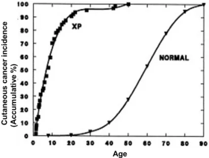
Figure 23: Skin cancer incidence as a function of age. ... 70
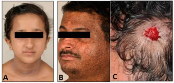
Figure 24 XP-C patients. ... 73
Figure 25: Mutations in the genes involved in NER and the associated syndromes. 75 Figure 26 Human receptor Tyrosine Kinase Families. ... 77
Figure 27 Activation of tyrosine kinase receptor. ... 78
Figure 28: MAPK signaling cascades. ... 80
7
Figure 30 JNK signaling pathway. ... 85
Figure 31 p38 signaling cascades. ... 87
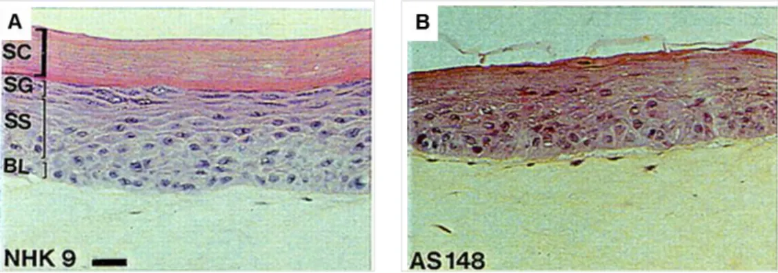
Figure 32: Histology of skin reconstructed in vitro from normal and XP-C keratinocytes on normal dermal equivalent. ... 90
Figure 33 XP-C fibroblasts provoke epidermal invasions in organotypic skin cultures. ... 90
Figure 34: The fibroblasts and their microenvironment. ... 140
Figure 35: Schematic representation of models of carcinoma invasions. ... 143
Figure 36: HGF and their receptor c-Met. ... 144
8
LIST OF TABLES
Table 1: Distribution of extracellular matrix components in dermal layer ... 26
Table 2: XP genes, products and their characters ... 68
Table 3: MAP kinase and their genes. ... 81
9
LIST OF ABBREVIATIONS
5-Mc Methylated cytosine at position C5
6-4 PP 6, 4 pyrimidine-pyrimidone photoproducts
8-oxoG 8-Oxoguanine
AKs Actinic keratosis
ATM Ataxia-telangiectasia mutated
ATR ATM- and Rad3-Related
BrdU Bromodeoxyuridine
BD Bowen’s disease
BCC Basal cell carcinoma
BER Base excision repair
BHD1 β-hairpin domain 1
BM Basement membrane
CAMs Cellular adhesion molecules
CAF Carcinoma associated fibroblasts
CMM Cutaneous metastatic melanoma
CP Committed progenitors
CSF2 Colony stimulating factor 2
CPD Cyclobutane pyrimidine dimers
CS Cockayne syndrome
CAK CDK-activating kinase
10 DDR DNA damage response
DNA Deoxy riboneucleic acid
DSBs Double-stranded DNA breaks
DEJ Dermal-epidermal junction
ECM Extracellular matrix components
EGF Epidermal growth factor
EPU Epidermal proliferation units
ESCs Embryonic stem cells
EYFP Enhanced yellow fluorescent protein FGF7 Fibroblast growth factors 7
GGR Global Genome Repair
HD Hemidesmosomes
HF Hair follicle
HFSC Hair follicle stem cells
HGF Hepatocyte growth factor
HR Homologous Recombination
IFE Interfollicular epidermis
IGF Insulin growth factor
IRS Inner root sheath
IL-1 Interleukin 1
IPSCs Induced pluripotent stem cells
ISCC Invasive SCC
11 LL Lamina lucida
LRCs Label retaining cells
MC Merkel’s cells
MDM2 Mouse double minute 2 homolog protein
MEF Mouse embryonic fibroblasts
MITF Microphtalmia-associated transcription factor
MMPs Matrix metalloproteinase
MMR Mismatch repair mechanism
MED Minimal erythema dose
MM Malignant melanoma
MAPK Mitogen-activated protein kinases
NER Nucleotide excision repair
NHEJ Non-homologous end joining
ORS Outer root sheath
PCNA Proliferating cell nuclear antigen
ROS Reactive oxygen species
RPA Replication protein A
RTKs Receptor tyrosine kinases
SCs Stem cells
SB Suprabasal cells
SCC Squamous-cell carcinoma
SCCIS Squamous cell carcinoma in situ
12 TGFβ Transforming growth factor-β
TCR Transcription Coupled Repair
TDG Thymine DNA glycosylase
TFIIH Transcription factor IIH
TGD Transglutaminase domain
TTD Trichothiodystrophy
UBA Ubiquitin-Associated domains
UBL Ubiquitin-like
VEGF Vascular endothelial growth factor
XRCC1 X‐ray repair cross-complementing protein1
13 RESUME
Le cancer spinocellulaire (CSC) est le cancer métastatique de la peau le plus fréquent chez l’homme. Son étiologie est liée à l'exposition aux stress génotoxiques, notamment les rayonnements ultraviolets (UV). Le xeroderma pigmentosum de type C (C) est une maladie génétique rare caractérisée par l’absence de la protéine XP-C entrainant une déficience dans le mécanisme de la réparation par excision de nucléotides des lésions à l’ADN induites par les UV. Le maintien de ces lésions chez les patients XP-C entraine l’apparition précoce de CSC particulièrement agressifs. Les fibroblastes cutanés XP-C présentent un phénotype proche de celui de fibroblastes associés aux cellules cancéreuses avec une accumulation des espèces réactives de l’oxygène et une surexpression de la métalloprotéinase matricielle 1 (MMP1).
Nous avons approfondi l’étude du phénotype des fibroblastes XP-C et leurs effets sur l’invasion de cellules de carcinomes. Dans des cultures organotypiques de peau, les fibroblastes XP-C favorisent l'invasion des cellules de CSC. De plus, in vitro, la cicatrisation des cellules CSC est plus rapide en présence de surnageants de culture de fibroblastes XP-C et est due à un effet mitogénique des fibroblastes XP-C sur les cellules de CSC dont la proportion la phase G2-M du cycle cellulaire est alors augmentée de 100 %. Nos données montrent que les fibroblastes XP-C surexpriment le facteur de croissance HGF/SF (hepatocytes growth factor/ scatter factor). Le HGF sécrété par les fibroblastes XP-C active son récepteur c-Met et les voies de signalisation p38 et JNK dans les cellules de CSC. Le blocage de HGF entraîne l’inactivation de c-Met, p38 et bloque l'invasion des cellules CSC dans les équivalent de derme de culture de peau organotypiques. Nos résultats ont également montré que les fibroblastes XP-C jouent un rôle de cellules « leader » dans l’invasion des CSC. Mes résultats suggèrent que les fibroblastes XP-C créent un microenvironnement permissif à la prolifération et l'invasion des CSC via la surexpression de HGF. Des thérapies ciblant les fibroblastes XP-C pourraient améliorer le contrôle de l’invasion des CSC chez les patients XP-C.
14 ABSTRACT
Squamous cell carcinoma (SCC) is the most frequent metastatic skin cancer from epithelial origin. In the majority of cases, the etiology of skin SCC cancer is linked to exposure to genotoxic stress, notably ultraviolet radiation (UVR), in the vast of skin cancers. Xeroderma pigmentosum type C (XP-C) is a rare genetic disorder characterized by a severe susceptibility to aggressive SCCs following minimal exposure to UVR, hence compromising the life expectancy of patients. XP-C cells are deficient in the nucleotide excision repair mechanism (NER) of DNA lesions introduced at bipyrimidine sequences upon UVR exposure. Prior results from the laboratory have shown that XP-C dermal fibroblasts constitutively express a phenotype resembling that of stromal fibroblasts associated to cancer cells with accumulation of reactive oxygen species and over expression of matrix metalloproteinase 1 (MMP1).
In this study, we further explored the phenotype of XP-C primary fibroblasts and their effects on the invasion of SCC cells. Our results show that primary XP-C fibroblasts constitutively overexpress hepatocyte growth factor/scatter factor (HGF/SF). In organotypic skin cultures, XP-C fibroblasts promote the invasion of SCC cells. Also, scratch healing of SCC cells was enhanced in the presence of culture supernatants of XP-C fibroblasts through a mitogenic effect connected to increased ratio of SCC cells in the G2-M phase of the cell cycle. Blockage of c-Met activation by blocking HGF antibodies prevented invasiveness of SCC cells within dermal equivalent presumably through the inhibition of p38 or JNK activation by XP-C fibroblasts culture supernatants. Spheroid assay shows that XP-C fibroblasts led SCC invasions. Our data indicated for the first time that absence of XPC in fibroblasts leads to overexpression of cell growth stimulators such as HGF and other factors (MMP1) responsible for the formation of a microenvironment permissive towards SCC cells proliferation and invasion. Moreover, our findings show that XP-C fibroblasts are leaders cells for SCC cells invasion. Therapies targeting XP-C fibroblasts may be considered as a way to control development of aggressive cancer in XP-C patients.
15
INTRODUCTION
16 Chapter one: The skin
The skin is the largest organ of the body and represents 15 % of our body weight. The skin has several important functions. It plays the role of a barrier to protect our body from environmental insults like mechanical constrains, solar radiation, chemical injuries, and bacterial/viral infections (Thomas and Fernandez-Penas 2016). Another role of the skin is to maintain constant the body temperature by regulating fluids exchanges notably through the evaporation of sweat. All these functions are very critical for our wellbeing (Kanitakis 2002).
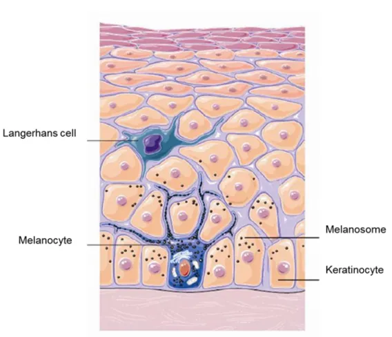
Figure 1: Skin structure. The skin is composed of two compartments, the epidermis in the upper layer and the dermis in the lower layer of the skin. The hypodermis is below the dermis and is composed of fatty acids. The skin contains also the pilosebaceous units and sweats glands.
17 I. Anatomy of the skin
The skin is composed of two compartments: the epidermis and the dermis (figure 1). The epidermis is a multilayered compartment composed of keratinocytes in direct contact with the external environment. The epidermis is renewing continuously within about three weeks through the proliferation/differentiation of the basal keratinocytes layer (Amberg, Holcmann et al. 2015). The skin contains also specialized structures, called “skin appendages” such as the pilosebaceous units and the sweat glands.
The dermis is located in the deeper layer of the skin, just beneath the epidermis to which it provides a physical and nutritive support. It is composed, of a loose connective tissue layers. Deeper in the skin there is the hypodermis which includes adipocytes and is an important source of fat (Luetteke, Phillips et al. 1994, Sibilia, Fleischmann et al. 2000).
I.1. The epidermis
The epidermis is mainly composed of four types of cells: keratinocytes, melanocytes, Langerhans and Merkel cells. The keratinocytes are of ectodermal origin (Sengel and Mauger 1976) and constitute the majority (about 80 %) of epidermal cells. They synthesize the intermediate filaments of keratins ensuring an important structural role (Simpson, Patel et al. 2011, Loschke, Homberg et al. 2016). Histology of human skin sections shows four main types of epidermal layers: the stratum basale (one cell layer), the stratum spinosum, the stratum granulosum and the stratum corneum (several cell layers) (Candi, Schmidt et al. 2005) (Figure 2). Each layer expresses specific markers. The expression and distribution of the biochemical markers specific of each layer of the skin attest of proper skin homeostasis.
18 Figure 2: schematic architecture of the epidermis. The epidermis is composed of four cell layers. The basal proliferating cell layer in the stratum basale is in contact with the dermis through hemidesomsomes and integrin-based adhesions. During keratinocytes differentiation, basal keratinocytes detach from the DEJ (basement membrane) and migrate outwards to form successively the stratum spinosum, the stratum granulosum and the stratum corneum. In each layer a unique cytoarchitecture is elaborated supporting their specific functions (adapted from Simpson et al. 2011).
19 I.1.a. Stratum basale
The stratum basale, also said basal layer, is the deeper layer of the epidermis. Keratinocytes in this layer are brought together with the dermis through a specialized basement membrane which constitutes the central structure of the “dermal-epidermal junction” (DEJ). Some keratinocytes of the basal layer have a high proliferative capacity (Fuchs and Raghavan 2002, Mascre, Dekoninck et al. 2012). Cells in the basal layer of the epidermis remain in contact with the basement membrane through hemidesmosomes and integrin-based adhesions. Intracellular cytoskeleton of basal keratinocytes is made of an extensive network of K5 and K14 keratin filaments (Nelson and Sun 1983). Keratins are essential components of the keratinocytes cytoskeleton in the epidermis. They play a key role in the structure of the epidermis via their interaction with the desmosomes that link two adjacent keratinocytes and the hemidesmosomes that link the basal keratinocytes to the DEJ. When keratinocytes in the basal layer are triggered to differentiate, they lose their proliferative capacity, and detach from the basal layer to fuel the stratum spinosum (Fuchs and Green 1980, Moll, Franke et al. 1982).
I.1.b. Stratum spinosum
In the stratum spinosum, keratinocytes acquire a spindle morphology (Yardley and Goldstein 1976). Spinous cells no longer synthesize the K5/K14 keratins pair and instead synthesize K1/K10 keratins. This transition represents one of the earliest change illustrating triggering of keratinocytes towards terminal differentiation (Fuchs and Green 1980, Rao, Babu et al. 1996). As cells reach the stratum spinosum, they lose some of their lipids and water content. Assembly of intermediate filaments in vitro showed that K1 and K10 keratins self-aggregate more than the K5/K14 keratins, and that they are less soluble. These properties are thought to confer a protective role to suprabasal cells against external constraints and aggressions (Fuchs 1990).
I.1.c. Stratum granulosum
In the stratum granulosum, cells flatten and produce keratohyalingranules. The cells keep their anabolic properties and start their destructive phase to eliminate cytoplasmic organelles as well as nuclei (Hoober and Bernstein 1966, Lavker and
20 Matoltsy 1970). Concentration of intracellular calcium increases, promoting the formation of a dense protein and lipid envelope, called “horny envelop” (cornified envelop). This process is due to the covalent bonding of several proteins including loricrin, involucrin and pro-filaggrin by the specific calcium-dependent membrane bound transglutaminase (Kalinin, Marekov et al. 2001, Candi, Schmidt et al. 2005).
I.1.d. Stratum corneum
Formation of the stratum corneum is the last phase of the differentiation of keratinocytes in the epidermis. It is composed of dead cells called corneocytes. During this terminal step of epidermal differentiation, keratin organize and “line up” together through their interaction with filaggrin, leading to the condensation of keratin filaments into tight bundles (Palmer, Irvine et al. 2006). The profilaggrin present in the keratohyalin granules of the stratum granulosum is converted by proteolysis to mature filaggrin. Filaggrin makes a complex with keratins in the lower layers of the stratum corneum, resulting in morphological flattening of corneocytes. These “polygonal” flat cells lacking a nucleus are linked together by lipids and specific desmosomes called “corneodesmosomes” to form a brick-and-mortar like structure (Plewig 1970). When corneocytes migrate towards the outer skin surface, they change in structure, composition and function. In the deeper layers of the stratum corneum cells are thicker and have high density of keratins (Fuchs and Green 1980). The mechanical behavior of the stratum corneum is influenced by the deeper part of this layer. The outer cells of this layer become highly hydrophobic and their desmosomes undergo proteolytic degradation. Even if the corneocytes are considered as dead cells, they are still fully functional, particularly in the terms of “barrier properties and metabolic functions”. I.1.e. Other cells of the epidermis
Melanocytes are pigmentary cells derived from the neural crest upon embryonic development. They are found in the basal layer of epidermis (Joly-Tonetti, Wibawa et al. 2016). In human’s skin, there is about 1 melanocyte for 36 keratinocytes (Seiberg 2001). Melanocytes extend their dendrites to adjacent keratinocytes of both the basal layer and the suprabasal layers (Yamaguchi, Brenner et al. 2007, Brenner and Hearing 2008) (figure 3). Melanocytes produce pigment molecules called melanins that
21 accumulate in granules called melanosomes. Melanosomes are transferred to keratinocytes to provide skin pigmentation. There are two types of melanins: eumelanin, a brown-black polymer, and pheomelanin, a reddish-yellow hue (Rawles 1947). All human skins contain both types of melanin, but the ratio of eumelanin/pheomelanin determines the skin typology (or, more classically, phototype) (Wakamatsu, Kavanagh et al. 2006). The main role of melanins produced by the melanocytes is to protect skin cells against UV radiations and, hence, development of cutaneous cancers.
Figure 3: Melanization. Adapted from
http://oerpub.github.io/epubjs-demo-book/content/m46060.xhtml. Melanin pigments produced by the melanocytes
accumulate in the melanosome to be transferred at the end to the adjacent keratinocytes.
22 Langerhans cells (LC) are dendritic cells found in the skin (Merad, Ginhoux et al. 2008). These cells are considered as leucocyte cells derived from the bone marrow. LC represent about 3-5 % epidermal cells; LC are linked to keratinocytes through E-cadherin-dependent adherent junctions (Tang, Amagai et al. 1993). LC are considered as the first line of immune defense against infectious pathogens in the skin, notably in the epidermis.
Merkel cells (MC) are neuroendocrine cells principally found in touch sensitive areas such as the fingertips and lips. In the epidermis, MC are present in the basal layer where they interact with nerve fibers crossing the DEJ (Fitzpatrick and Breathnach 1963, Boulais and Misery 2007).
I.2.The dermal-epidermal junction (DEJ)
The dermal-epidermal junction (DEJ) is located between the epidermis and the underlying dermis to tightly attach them together (McMillan, Akiyama et al. 2003). Electron microscopic analysis shows that the DEJ contains three zones (figure 4): 1) The hemidesmosomes (HD) provide a link between the keratin filaments of the basal keratinocytes and the other components of the DEJ thanks to transmembrane integrin α6β4. 2) The lamina lucida (LL) composed of anchoring filaments, such as laminin 332 (laminin 5) and collagen XVII. 3) The lamina densa (LD) composed of a dense network of small adhesion molecules (Nidogen, fibronectin) connecting the anchoring filaments to anchoring fibrils (collagen IV and collagen VII) present in the dermis (Ellison and Garrod 1984). Collagen VII is important in the formation of an extensive network in the superficial dermis, under the DEJ (McMillan, Akiyama et al. 2003). The synthesis and the organization of the components of the DEJ are very important for cell adhesion, migration and signal transduction from the extracellular matrix into the epidermal keratinocytes (Burgeson and Christiano 1997). The DEJ allows the circulation between the dermal and the epidermal compartment of substances like cytokines and growth factors and even, cells (lymphocytes and immune cells) (Timpl and Brown 1996).
23 Figure 4: Dermal epidermal junction. A) Under electron microscopy shows: hemidesmosome (HD), lamina lucida (LL) and lamina densa (LD) (According to Dr. M. Démarchez - Biologie de la peau, http://biologiedelapeau.fr/spip.php?article47&lang=fr). (B) Schema showing the intermolecular interactions between HD and LL/LD. In HD, plectin and BPAG1 link the keratin filaments to the transmembrane collagen XVII and the integrin . Collagen XVII and integrin interact with Laminin 5 anchoring filaments in the LL that in turn binds the anchoring fibrils of Collagen VII and the Laminins 6/10. (According to (McMillan, Akiyama et al. 2003).
24 I.3. The dermis
The dermis is a complex tissue underlying the epidermis. It contains blood and lymphatic vessels, nerves and nerve endings, glands, and, except on glabrous skin, hair follicles (figure 1). The majority of cells populating the dermis are fibroblasts from mesodermal origin (Sengel and Mauger 1976). Dermis varies in thickness as a function of its location in the body ranging from 0.6 mm on the eyelids to 3 mm on the back, palms and soles (Arda, Goksugur et al. 2014). The dermis is composed of two zones: a superficial layer that interdigitates with the epidermis, called the “papillary dermis”, and a deeper zone called “reticular dermis” (figure 5). The papillary dermis is thinner than the reticular dermis and represents about 10 % of the dermis. The density and the proliferative capacity of papillary fibroblasts are higher than reticular fibroblasts (Sorrell, Baber et al. 2004). Papillary and reticular layers are mainly composed of extracellular matrix components (ECM) produced by the fibroblasts (Sorrell, Baber et al. 2004, Driskell, Lichtenberger et al. 2013). They differ in both the composition and the organization of their ECM (table 1).
The dermis is characterized by the loose assembly of collagen and elastin fibers embedded in the extracellular matrix (ECM). In the papillary dermis, “papillary fibers” are arranged vertically and connect to the dermal epidermal junction, while fibers of the reticular dermis, tend to arrange horizontally. Dermal fibroblasts mainly produce collagen type I and III, elastin, structural proteoglycans (versican, decorin), glycoproteins (fibronectin and tenascins) and collagen VII, a major actor of dermis-epidermis junction (table 1).
Dermal fibroblasts act together with epidermal keratinocytes in the organization of the dermis-epidermis junction. They produce collagen IV and VII, two components present in the lamina densa and the anchoring fibrils. Fibroblasts also modulate the activity of keratinocytes in the epidermis by producing growth factors and cytokines. It has been shown that the proliferation and differentiation capacities of keratinocytes are regulated by dermal fibroblasts notably by the expression of the fibroblast growth factor 7 (FGF7) and the colony-stimulating factor 2 (CSF2). FGF7 stimulates keratinocytes proliferation and CSF2 is necessary to their differentiation (Maas-Szabowski, Shimotoyodome et al. 1999, Szabowski, Maas-Szabowski et al. 2000). The expression of these proteins is regulated by interleukin1, a regulatory cytokine 1 expressed by keratinocytes (Carr, Li et al. 2016). Moreover, fibroblasts play important roles in
25 wounds healing and the balanced production of type I and type III collagens (Smola, Stark et al. 1998). The dermis also contains dermal dendritic cells, macrophages and mast cells that represent the resident immune system in the dermis.
Figure 5: Histology of the skin: A) the papillary and the reticular dermis are separated by vascularized rete subpapillare. Dermal papillae interdigitates within the epidermis, scale bar 45 µm. B) Schema of adult human skin. Two different populations of dermal fibroblasts can be cultured from the interfollicular dermis, the papillary fibroblasts cultured from skin located at a depth of 0.3 mm and the reticular fibroblasts cultured from the skin located at 0.7 mm (Sorrell and Caplan 2004).
26 Table 1: Distribution of extracellular matrix components in dermal layer (Sorrell, 2004)
I.4. The skin appendages
Upon embryonic development, embryonic mesodermal stem cells receive signals from ectodermal cells to instruct them to commit to a particular differentiation program and generate a stratified epidermis and skin appendages such as hair follicles and sebaceous glands (Fuchs 2007).
I.4.a. The pilosebaceous follicle
Pilosebaceous follicles are found in the hairy part of the body. They include hair follicles, sebaceous glands and the arrector pili muscle which causes the hair to stand up when it contracts (pilo-arrection). In the dermis, the hair follicles consist of the hair shaft surrounded by an inner root sheath (IRS) and an outer root sheath (ORS) (figure 6-A). In the adult human skin, the ORS is in continuation with the basal layer of the inter-follicular epidermis. At the deep end of the hair follicle, the ORS forms a germinal matrix (hair bulb) where cells proliferate and differentiate to form the different cells of the internal sheaths and the hair shaft (Fuchs 2007). In the mouse, hair follicle stem
27 cells reside in a niche that is located at the bottom of the non-cycling portion of the ORS, called the bulge (Taylor, Lehrer et al. 2000, Oshima, Rochat et al. 2001). Hair follicle stem cells are multipotent cells (Majo, Rochat et al. 2008, Bonfanti, Claudinot et al. 2010). They possess the potential to proliferate to allow hair regeneration, but they possess also the potential to make epidermis and sebaceous glands (Blanpain and Fuchs 2006).
The growth of hair follicles is cyclic and is subdivided in 3 phases (figure 6-B): 1) The anagen phase is an active growth phase of the hair cells in the bulb. 2) In the catagen phase, hair growth stops. The follicle enters a destructive phase, leading to its degeneration; in contrast, the bulge region that contains the stem cells remains intact. 3) The telogen phase is a quiescent phase (Blanpain and Fuchs 2009). In the human hair follicle, the average duration of each phase is 3 years, 3 weeks and 3 months, respectively (Bernard 2006). Under “normal hair conditions” about 85% of follicles are in anagen phase and 15 % in telogen phase. At the end of the telogen phase, the follicles regenerate from the germinative cells and enter a new anagen phase.
Sebaceous glands (SG) are exocrine glands attached to the hair follicle. SG produce and secrete sebum, a liquid rich in lipids. Secretion of sebum into the hair canal helps lubrification and growth of the hair shaft.
I.4.b. Sweat glands
Sweat glands are exocrine glands that play an essential role in thermoregulation. Eccrine sweat glands are localized on the entire surface of the skin but they are particularly numerous on the palms of hands, soles of feet and forehead. The secretion by the sweat glands represent a primary form of refreshing in humans.The secretory portion of these glands is found in the dermis and the excretory duct opens directly at the skin surface. The sweat glands are found in the armpits and perianal areas.They are not significant for cooling in humans, but rather act as scent glands. Their excretory duct opens into hair follicle.
28 Figure 6: the hair follicule. A) Haired skin is composed of a pilosebaceous unit that contains a hair follicle (HF) and its surrounding interfollicular epidermis (IFE), which is responsible for skin barrier function. HFs are composed of different concentric layers of cells. At the base of the HF, the matrix contain proliferative cells and then embark concentric terminal differentiation programs that generate the mature follicle. In mice, hair follicle stem cells (HFSCs) reside in a niche that is located at the bottom of the non-cycling portion of the outer root sheath (ORS), called the bulge. HFs also contain sebaceous glands, which form from and are replenished by resident SCs located in the outer root sheet (ORS). These SCs differentiate to form the sebocytes which release oils that lubricate the hair channel and skin surface following degeneration (Jahoda and Reynolds 2001). B) Schema of hair cycle stages, starting from the first postnatal anagen, hair shaft is growing and arising through the skin surface. Hair follicles progress to the destructive (catagen) phase, during which the lower two-thirds of the follicle undergo apoptosis and regress. The dermal papilla is brought to rest below the bulge-stem-cell compartment, and after the resting (telogen) phase, a critical threshold of activating factors is reached and the stem cells become activated to regrow the hair (Fuchs 2007).
29 Chapter two: Stem cells
“Epidermal” stem cells are located in the epidermal basal layer and in the outer root sheath of the hair follicle. They play important role in the maintenance of skin homeostasis, renewal and hair regeneration through cycling regeneration of hair follicles. Epidermal stem cells also contribute to epidermal regeneration during wound healing after epidermal injuries such as burns, cuts, abrasion or pathology-led epidermal injuries (Blanpain and Fuchs 2006). Stem cells are known for their ability to self-renew to generate additional stem cells, and the capacity to generate progeny that are fated to differentiate into at least one type of highly differentiated descendant (Lajtha 1979).
I. Embryonic stem cells (ESCs)
ESCs are isolated from the inner cells mass (ICM) of blastocyst. They are pluripotent cells that can give rise to all cell types of the body under specific circumstances such as the presence of tissue-specific growth factors. ESCs may then give rise to one or more progenitor cell lineage involved in the functional organization of a given tissue.
II. Induced pluripotent stem cells (IPSCs)
Studies showed that human induced pluripotent stem cells (IPSCs) could be generated from adult somatic cells by introducing genes encoding the transcription factors Oct3/4, Sox2, c-Myc, and Klf4. IPSCs exhibit the morphology and growth properties of ESCs and express ESCs marker genes (Yamanaka and Takahashi 2006). They can be propagated indefinitely and are able to differentiate into virtually every cell type which “theoretically” make them viable for transplantation and experimental disease modeling (Wu, Doorenbos et al. 2016).
To avoid inappropriate recruitment and differentiation triggering, stem cells are thought to be maintained in a very specific microenvironment, called a “niche”. The stem cell niche constitutes a tissular microenvironmental context. In the niche, stem cells are undifferentiated; they self-renew by dividing very rarely to preserve the integrity of their genetic and epigenetic information comprising the program of tissue morphogenesis (Morrison, Shah et al. 1997, Morrison and Spradling 2008).w
30 III. Keratinocyte stem cells ex vivo
A major difficulty in the study of stem cells is to obtain an efficient model for preserving their presence and functionality ex vivo, for example, when taken out from their natural niche. After many failures to reliably isolate living embryonic and somatic stem cells, the idea emerged that development of appropriate culture systems should allow preserving both long-term potential of renewal and stem cells differentiation potential. In 1975, Rheinwald and Green reported that epidermal keratinocytes from a skin biopsy could be grown for a very long-term so long as a feeder layer of lethally irradiated mouse embryonic fibroblasts was seeded few hours prior primary keratinocyte seeding (3T3-J2 fibroblasts are gamma irradiated in order to stop their growth, but not the growth of keratinocytes, and hence play the role of feeder cells) (Rheinwald and Green 1975). In the presence of epidermal growth factor (EGF) keratinocytes can be maintained in culture for more than 100 populations doublings (Rheinwald and Green 1977). Under appropriate conditions, a single stem keratinocyte can give not only the entire epidermis of an individual (≈ 1011 cells), but also of all individuals on the planet (≈ 1021 cells) (Warrick, Garcia et al. 2012).
Under adequate culture conditions, human epidermal keratinocytes clonogenic cells may initiate three types of colonies: the holoclones, the meroclones and the paraclones (Barrandon and Green 1987) (figure 7). The holoclones are large colonies with smooth perimeter. 95 % holoclones progeny is clonogenic. The paraclones are small, terminally differentiating colonies, with a very limited proliferative potential (<15 divisions). Less than 5 % of the cells initiated by a paraclone are themselves clonogenic. The meroclones are medium-sized colonies with irregular contour. The clonogenic potential of their progeny is very heterogeneous, ranging from 5 to 95 %. Along serial propagation, the percentage of meroclones and of paraclones progressively increases at the expense of holoclones. The proportion of epidermal stem cells (holoclones) present in a keratinocytes culture is a good indicator of the quality of the culture.
31 Figure 7: The 3 types of colonies formed by keratinocytes in culture. A) Colony forming efficiency of three different clones isolated from primary cultures of keratinocytes. Holoclones have high proliferative potential and correspond to epidermal stem cells. Paraclones are composed of cells with a limited growth potential (<15 divisions). Meroclones are intermediate and constitute a transitional state between holoclones and paraclones. B) Macroscopic colony types formed by keratinocytes. Twelve days’ colonies were fixed and stained with rhodamine. Note the smooth perimeter of holoclones (a) and the wrinkled perimeter in meroclones (b) (Barrandon and Green 1987).
32 The most important evidence of the presence and the maintenance of stem cells in culture comes from the success of the transplantation of human cultured epithelium autografts on patients with severe burns. Renewal for very long term (over 20 years) of an epidermis with normal characteristics, despite the absence of skin appendages, shows that stem cells in culture were preserved and retained all their potentials of proliferation and differentiation after transplantation (Gallico, O'Connor et al. 1984, Pellegrini, Ranno et al. 1999, Ronfard, Rives et al. 2000).
Ex vivo, reconstructed skin models composed of human keratinocytes growing
on a collagen lattice containing dermal fibroblasts were also developed to reproduce the processes of stratification and epidermal differentiation (Bell, Ehrlich et al. 1981, Asselineau, Bernard et al. 1986). The reconstructed skins can be grafted on the immunodeficient mouse to regenerate human skin in vivo (Demarchez, Hartmann et al. 1992, Del Rio, Larcher et al. 2002). Human skin can also be regenerated in the long term from single layered epithelial sheets grafted onto the immunocompromised mouse. Following the seminal paper of Marcela Del Rio (Del Rio, Larcher et al. 2002), my host laboratory demonstrated that stem keratinocytes can be enriched from mass culture of genetically corrected human primary keratinocytes (Bergoglio, Larcher et al. 2007, Warrick, Garcia et al. 2012).
IV. Localization of stem cells in vivo IV.1. Stem cells in the basal layer
Although the existence of a population of stem cells in the basal layer of the epidermis has been evoked since several years, their number and the precise location of their niche remains a subject of debates. According estimations, epidermal stem cells would represent between 0.1 % and 1 % of keratinocytes present in the basal layer (Schneider, Barland et al. 2003). Recent studies show that there are mutual influences between skin stem cells and their niches (Hsu, Li et al. 2014). Stem cells and their progeny are thought to interact with components of the niche. Stem cells and their progeny can coexist within a niche, suggesting that niche signals alone are not sufficient to maintain ‘stemness’ (Hsu, Pasolli et al. 2011, Sato, van Es et al. 2011). These studies suggest that “there is an exchange in the regulation between the parents (stem cells) and their progeny cells” (Hsu and Fuchs 2012, Hsu, Li et al. 2014).
33 According to first observations described by Potten and his team in the late seventeens, progeny of epidermal stem cell is organized as columns, called epidermal proliferation units (EPU) (Potten 1974). Using retroviral transfer of a tracing gene (beta-galactosidase), it has been shown that each column comprises about ten basal keratinocytes subsequently giving rise to the suprabasal cell layers (Mackenzie 1997). Cells of each EPU are thought to be the progeny of a single stem cell. This progeny would possess a limited proliferative potential and are called transit amplifying cells (TACs). TACs divide several times before leaving the basal layer to embark the stepwise process of differentiation (figure 8) (Jones and Simons 2008, Hsu, Li et al. 2014). The existence of TACs would present two advantages: 1) The rapid regeneration of the skin in case of injury; 2) The limitation of stem cells divisions, which preserves their pool and limits the possibility of genotoxic mutagenesis, and hence cancer initiation.
In 2007, Clayton et al. proposed another model of epidermal stem cells regulation in the mouse. In brief, using cell tracing strategies, the authors suggested that recruitment of stem cells in the basal layer of the mouse tail epidermis i) is stochastic, ii) does not give rise to TACs (Clayton, Doupe et al. 2007). No report generalizing this model to human interfollicular epidermis has been published yet.
34 Figure 8: Epidermal proliferative unit (EPU) hypothesis. a) Self-renewing stem cells (yellow) generate a transit-amplifying (TA) cell population (purple), which undergoes symmetric division (TA1 to TA3) before their terminal differentiation into post-mitotic (PM) cells (blue). These PM cells migrate from the basal layer and become suprabasal (SB) cells (green). b) Schema showing the spatial organization of cells in EPUs. c) EPU model in a clonal labelling experiment (Jones and Simons 2008).
IV.2. Stem cells of the hair follicle
One of the first methods used to determine the location of stem cells in vivo was based on the use of nucleotide analogs such as bromodeoxyuridine (BrdU) or [3H] thymidine. After peritoneal injection, these DNA precursors are incorporated in the DNA of dividing cells during replication. After a few cell divisions, the marker is rapidly diluted and is no longer detectable in the progeny of stem cells. Based on the fact that stem cells do not divide frequently, a population called «label retaining cells" (LRCs), presumably corresponding to stem cells was identified.
In 1990, Cotsarelis et al. showed that the higher number of LRCs of skin reside in the bulge of the mouse hair follicle. The bulge is a region of the outer root sheath beneath the sebaceous gland and the pili arrector muscle. Only few LRCs are observed in the interfollicular murine of epidermis (Cotsarelis, Sun et al. 1990). Using the rat vibrissal follicle as a model, Kobayashi, reported that clonogenic assays showed
35 that most clonogenic cells (keratinocyte colony-forming cells) are clustered in the bulge. By microdissection of rat vibrissal follicles, the authors reported that about 95 % of clonogenic cell are concentrated in the bulge area. In contrast, less than 5 % clonogenic cells were found located in the matrix area of the follicle (lower part of HF, sometimes called “the bulb”) (Kobayashi, Rochat et al. 1993). The same team showed that clonogenic cells of the human hair follicle are also concentrated in a region below the midpoint of the hair follicle and outside the hair bulb, in the site of insertion of arrector muscles; however it is important to notice that the morphology of the human hair follicle does not include a “bulge like” region (Rochat, Kobayashi et al. 1994).
Later, it has been shown that cells of the bulge are multipotent stem cells with high clonogenic capacity and capable to regenerate the different compartment of the pilosebaceous follicle (Taylor, Lehrer et al. 2000, Blanpain, Lowry et al. 2004, Claudinot, Nicolas et al. 2005). During wound healing, some keratinocyte located in the bulge region participate to regeneration of the intrefollicular epidermis. However, under normal conditions, stem keratinocytes located in the bulge do not seem to participate to homeostasic renewal of the interfollicular epidermis (Ito, Liu et al. 2005, Levy, Lindon et al. 2005) (figure 9).
36 Figure 9 The different compartments of stem cells in the skin. a) Epidermal stem cells of the hair follicle (HF), sebaceous gland (SG) or interfollicular epidermis (IFE) are interchangeable and functionally equivalent. b) The stem cell of the IFE are located in the basal layer and allow permanent renewal of the epidermis. The progenitors of the sebaceous glands (SG) are responsible for the physiological renewal of sebocytes. The HF stem cells form the cells of the outer root sheath (ORS) and inner root sheath (IRS) (Owens and Watt 2003).
37 Chapter three: Consequences of solar radiations on skin
Exposure of skin to sun radiations leads to many types of biological responses, including immunosuppression, glycation of ECM, peroxidation of lipids and proteins. Schematically, the first category of “immediate” short-term responses to sunlight exposure include suntan (for moderate exposure) as sunburn and erythema (for high UV dose exposure). The second one is a long-term response, characterized by photoaging and the development of skin cancers.
Solar UV are classified into three categories depending of their wavelengths: UVC (200 - 280 nm), UVB (280 - 320 nm), and UVA (320 - 400 nm) (Cerutti and Trump 1991). UVA (95 %) and UVB (5 %) of the UV spectrum reaching the earth’s surface, respectively. UVC are absorbed by the ozone layer of the stratosphere avoiding them to reach the Earth. However, due to the depletion of the ozone in certain regions of the world, sunlight reaching the surface of the Earth tend to be enriched in UVB and UVC radiations (Lloyd 1993). In the human skin, the epidermis and the superficial dermis “absorb” UVB due to their high energy; UVA are less energetic than UVB and penetrate more deeply into the skin (figure 10). UVB are about ∼ 10,000 times more carcinogenic than UVA. UVB cause damages in DNA and initiate, and promote skin cancer progression (Bowden 2004).
I. Effects of UVB and UVC on DNA
In the human body, 1013 cells receive about 104 DNA lesions every day (Lindahl and Barnes 2000). Accumulation of these lesions is mutagenic. DNA aromatic bases pyrimidines (cytosine (C) or thymine (T)) absorb photons energy at wavelengths ranging from 230 to 290 nm (i.e. from UVC to UVB wavelengths, respectively). The absorption of UV photons in the DNA gives rise to dimeric photoproducts between adjacent pyrimidine bases on the same DNA strand. Two types of bulky DNA lesions that introduce a majXPor DNA distortion are produced: cyclobutane pyrimidine dimers (CPD) and 6, 4 pyrimidine-pyrimidone photoproducts (6-4 PP) (figure 11).
38 Figure 10: UV penetration in the skin. UVA penetrate deeply into the dermis, while UVB is absorbed in the epidermis and the surface of the dermis. UVC are absorbed by the ozone layer which prevent it these wavelengths to reach the Earth surface.
Figure 11: DNA lesions induced by UV. CPD is obtained by the formation of covalent linkage between C5 and C6 atoms in the adjacent pyrimidines on the same DNA strand. 6-4 PP is formed by founding covalent linkage between C6 and C4 atoms between the two adjacent pyrimidines on the same DNA strand (Yokoyama and Mizutani 2014).
39 I.1. CPD formation
CPD lesions are formed by a covalent link between the C5 and C6 atoms of two adjacent pyrimidine bases (figure 11). Irradiation of cultured cells with UVB leads to the production of about 1-5.10-4 lesions / kbp / J.m2 (Perdiz, Grof et al. 2000, Douki and Cadet 2001). CPD lesions distort the DNA double helix of about 7 to 10° compared to its initial conformation (Kim, Patel et al. 1995, Miaskiewicz and Ornstein 1996). It has also been shown that methylated cytosine at position C5 (5-mC) in CpG promoter regions absorb UV lengths of 273 nm (UVB radiation) (Drouin and Therrien 1997) and thus promote CPD formation in genetic regulatory regions (Pfeifer, You et al. 2005) (figure 12).
I.1. 6-4 PP formation
6-4 PP lesions result from the formation of a covalent bridge between the C6 and C4 carbons of two adjacent pyrimidines on the same DNA strand (Figure 11). 6-4 PP lesions are much more distorting than CPD lesions. They lead to a 44 ° distortion of the DNA (Kim, Patel et al. 1995). The ratio of the number of CPD and 6-4 PP formed for a given UVB dose is estimated between 8 : 1 (Perdiz, Grof et al. 2000) and 3 : 1 respectively (Douki and Cadet 2001). The influence of cytosine methylation on the formation of 6-4 PP has not been clearly established (Pfeifer, Drouin et al. 1991, Mitchell 2000).
CPDs are repaired slower than 6-4 PP (Thomas, Okumoto et al. 1989, Mitchell, Brash et al. 1990). This difference in repair is thought to be related to the highest DNA distortion induced by 6-4 PP lesions which facilitate their recognition by the DNA repair mechanisms (Szymkowski, Lawrence et al. 1993, Kim, Patel et al. 1995, Sugasawa 2010). The CPDs persist longer in DNA and are responsible for at least 80 % of the UVB-induced mutation (You, Lee et al. 2001).
II. Analysis of mutations induced by UVB and UVC
Studies on mutations induced by UVC and UVB radiations demonstrate that, from yeast to mammalian cells, the majority of UV mutations observed in cells are C → T or CC → TT transitions formed at adjacent pyrimidines (figure 12) (Armstrong and Kunz 1990, Pfeifer, You et al. 2005). These mutations are a “signature” of the mutagenesis induced by UVB / UVC radiations (Hutchinson 1994, Brash 2015).
40 III. Effect of UVA on the skin
DNA bases weakly absorb UVA but UVA can activate photosensitizers in the skin such as melanin, porphyrins and riboflavin. Continuous exposure to sunlight generates reactive oxygen species (ROS), organic free radicals and other toxic products (Wondrak, Jacobson et al. 2006, Ridley, Whiteside et al. 2009).
Accumulation of ROS leads to direct damages of cellular components, including lipid peroxidation, protein oxidation and nucleic acids alteration. These damages may induce both cell death and malignant transformation (Bragado, Armesilla et al. 2007, Liu, Chen et al. 2008).
The modification in skin structural proteins is accompanied by depletion of anti-oxidant enzymes such as catalase and superoxide dismutase (Bernerd and Asselineau 1998, Sander, Chang et al. 2002). Ex vivo studies have shown that photooxidative stresses lead to the induction of MMPs which trigger the extracellular matrix proteins (ECM). The induction of MMPs leads to the degradation of the collagen fibres present in the ECM (Dalle Carbonare and Pathak 1992, Adams 2000, Frechet, Warrick et al. 2008). UVA can lead to indirect DNA damages by generating CPD and 8-oxoG lesions due to ROS generation (Douki, Reynaud-Angelin et al. 2003). To attenuate the effect of UV exposure, cells have several mechanisms in place, including antioxidants, DNA repair, and stress signalling pathways (Cadet, Delatour et al. 1999, Birben, Sahiner et al. 2012).
41 Figure 12: Mutagenesis mechanisms at a cyclobutane pyrimidine dimer (CPD). Scheme shows, 5′-CmCG is particularly prone to CPD formation after irradiation due to the presence of a 5-methylecytosine base (mC). Bypass of the CPD by an error-prone DNA polymerase could produce a C to T or CC to TT mutation. Deamination may occur within the CPD and may affect the methylcytosine alone or the 5-methylcytosine and cytosine (double deamination). During subsequent DNA replication, polymerase η will introduce a C to T or a CC to TT tandem transition mutation (Pfeifer, You et al. 2005).
42 Chapter Four: Skin cancers
In the Human, 80 % tumors are from epithelial origin (DePinho 2000). The high proliferative potential of epidermal skin cells and the exposition to genotoxic agents, notably UV, both contribute to the development of cutaneous tumors during ageing. There are three types of skin cancers: basal cell carcinoma (BCC) and squamous-cell carcinoma (SCC) that arise from keratinocytes, and melanoma derived from melanocytes. SCC and BCC represent about 20 % and 80 % of nonmelanoma skin cancers, respectively (Kwa, Campana et al. 1992, Brellier, Marionnet et al. 2004, Amberg, Holcmann et al. 2015). Ultraviolet exposure is the major etiological agent of these types of cancers (Fears and Scotto 1983, Preston and Stern 1992).
I. Squamous cell carcinoma (SCC)
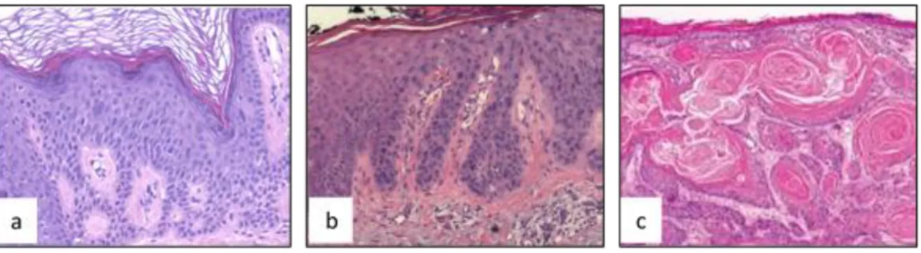
SCCs are associated with significant risk of metastasis. Over 250,000 new cases of SCCs appear in US annually. The average incidence rates of SCC in UK was 22.65/100 000 person-years (Lomas, Leonardi-Bee et al. 2012). In addition of factors as age, sex and skin color, two or more painful sun exposures may increase the risk of SCCs skin cancer about 3-fold in Australian population (Green and Battistutta 1990). SCCs development is regarded as a multistep process starting from a precursor lesion called actinic keratosis (AKs). In about 5 % cases, AKs may develop as squamous cell carcinoma in situ (SCCIS) or Bowen’s disease, invasive SCC (ISCC) and metastatic SCC (figure 13), while 95 % spontaneously regress (Yanofsky, Mercer et al. 2011, Ratushny, Gober et al. 2012, Chen, Halliday et al. 2013).
I.1. Actinic keratosis (AK)
AK is a scaly hyperkeratosis lesion which develops in skin areas exposed to sun notably the face, ears, and neck (Salasche 2000, Rogers, Weinstock et al. 2010). Histological analysis of AKs shows atypical keratinocytes with enlarged, irregular, and hyperchromatic nuclei (Arciniegas, Carrillo et al. 2015). Abnormal growth of AKs disrupts cell differentiation and results in a thickening of the stratum corneum (Cockerell 2000). AK is a form of intra-epidermal keratinocytic neoplasia. The risk of progression of AK to SCCIS or ISCC has been estimated from 7784 AKs lesions; it is
43 0.60 % at 1 year, 1.5 % at 2 years, 2.2 % at 3 years, 2.6 % at 4 years, and 3.5 % at 5 years (Criscione, Weinstock et al. 2009).
Figure 13: Histologic aspects of squamous Cell Carcinomas and their precursor lesions. a) Actinic keratosis. b) SCC in situ (SCCIS). c) Invasive squamous cell carcinoma (ISCC) (Yanofsky, Mercer et al. 2011, Chen, Halliday et al. 2013).
I.2. Squamous cell carcinoma in situ (SCCIS) or Bowen’s disease (BD)
Patients with SCCIS have scaly red lesions in sun-exposed areas. SCCIS is considered as the early stage of the intra-epidermal form of SCC. Histological analysis of SCCIS shows thatkeratinocytes become pale with abnormal differentiation features and atypical nuclei, contributing to parakeratotic traits (Maione, Errichetti et al. 2016). SCCIS do not invade the dermis (Chen, Halliday et al. 2013) (figure 13). About 3–5 % untreated SCCIS develop into invasive SCCs (Cox, Eedy et al. 2007).
I.3. Invasive SCC (ISSC)
About 70-80 % ISCC is found in photo-exposed areas. ISCC incidence increases in photo-exposed areas (Iversen and Tretli 1999). Histologically, early stage ISCCs look like AK lesion; AK may however be distinguished from ISCCs by the presence of invasive cells passing through the basement membrane into the dermis. In the later stages of invasion, carcinoma cells are characterized by the formation of nests of atypical tumor cells in the dermis (figure 13) (Kelder, Ebrahimi et al. 2012, Chen, Halliday et al. 2013). Cells in this stage exhibit a clinical and histological spectrum of aggressiveness and metastatic potential, depending on tumor thickness and depth of invasion (Dinehart, Nelson-Adesokan et al. 1997).
44 I.4. Genetics of skin SCC carcinogenesis
The TP53 gene is considered as the “Guardian of the genome” due to its key role in the control of cycle and apoptosis. TP53 is the most frequently mutated gene in human cancers (Olivier, Eeles et al. 2002) and disruption of the normal p53 function is generally accepted as an important step in human carcinogenesis (Greenblatt, Bennett et al. 1994, Ziegler, Jonason et al. 1994). The analysis of p53 mutations in skin and cutaneous SCC have been extensively studied in human and murine tumors including skin and other organs such as lung, colon and liver.
In the epidermis, p53 is constitutively expressed in small quantities. UV radiation induces p53 stabilization after phosphorylation by ATR and p38 protein kinases allowing its contribution to repair processes of DNA damages and/or the induction of apoptosis (Campbell, Quinn et al. 1993, Hall, McKee et al. 1993). In sun-exposed skin, numerous clones/clusters of p53 immunoreactive keratinocytes have been identified (Urano, Oura et al. 1992, Ponten, Berne et al. 1995, Jonason, Kunala et al. 1996, Ren, Ahmadian et al. 1997). DNA sequencing of these clones have shown that 30-70 % harbor a TP53-mutated gene (Ren, Ponten et al. 1996, Ponten, Berg et al. 1997, Tabata, Nagano et al. 1999). Furthermore, TP53 is mutated in 75-80 % AK lesions. The presence of TP53 gene mutations in SCCIS and SCC has been found to be 40 %, and 41-90 %, respectively. These results suggest that AK lesions have acquired the genetic mutations prior becoming cutaneous SCC (Ziegler, Jonason et al. 1994, Ortonne 2002). These results also indicate that p53 loss is important to skin SCC progression (Campbell, Quinn et al. 1993, Ren, Ponten et al. 1996, Ziegler, Jonason et al. 1996, Ren, Ahmadian et al. 1997). The vast majority of p53 mutations found in AK, SCCIS, and SCC most often display the typical UV (C > T or CC > TT) signature, thus being indicative of UV-radiation-induced mutations.
Several other mutations in tumor suppressor genes or oncogenes, such as RAS or p16 (CDKN2A), have been implicated in carcinogenesis. Ras is a member of GTPase proteins family encoded by RAS oncogenes. Ras activity activates wide a set of downstream effectors pathways involved in cell growth, differentiation and cell survival. Mutations of RAS oncogenes have been reported to occur in 10-40 % non-melanoma skin cancers (SCC and BCC) (Ananthaswamy and Pierceall 1990). Data from the Catalog Of Somatic Mutations In Cancer (COSMIC; Sanger Institute) indicate
45 that 21 % cutaneous SCC harbor activating RAS mutations (9 % Hras, 7 % Nras, 5 % Kras) (Bamford, Dawson et al. 2004).
p16 is a tumor suppressor protein, which contributes to the regulation of cell cycle by inhibiting entry in the S phase. p16 is downregulated in high numbers of tumors and is frequently dysfunctional in SCCs (Mortier, Marchetti et al. 2002, Pacifico, Goldberg et al. 2008, Romagosa, Simonetti et al. 2011). Studies by Hodges showed that p16 is expressed at higher levels in AK and SCCI than in SCC (Hodges and Smoller 2002, Salama, Mahmood et al. 2003) suggesting that disruption of the p16 gene is associated with AKs progression to SCCs.
AK also often shows frequent loss of heterozygosity (LOH) in p16 in chromosomes (3p, 9p, 9q, 13q, 17p and 17q) (Rehman, Takata et al. 1996). p16 LOH in AK leads to uncontrolled G1 to S transition, then promoting the progression of AK premalignant lesions to SCCs (Rehman, Quinn et al. 1994, Mortier, Marchetti et al. 2002, Yoo, Cho et al. 2004).
II. Basal cell carcinoma (BCC)
Basal cell carcinoma (BCC) is the most common malignancy in humans and its incidence has increased rapidly over the last 50 years. The rising incidence of BCC is probably due to increased sun exposures and longevity of the population (Tourli, Langner et al. 2016). BCC are thought to be the consequence of acute UV sun burns in early age (Rubin, 2005). BCC are predominantly diagnosed in sun exposed areas as head, neck, and back of the hands (Rubin, Chen et al. 2005), but they can also be found in sun-protected skin (Crowson and Magro 1996). The risk of BCC is higher in elderly persons and in persons with fair skin with blonde/reddish hair (Green and Battistutta 1990).
BCC is thought to arise from epidermal basal keratinocytes (Preston and Stern 1992) and has been histologically subdivided into several subtypes by Crowson in 2006: The “indolent-growth” BCCs that include superficial BCC and nodular BCC, and “aggressive-growth” BCCs that include sclerosing BCC, infiltrative BCC, and metatypical BCC (Crowson 2006). Superficial BCC represents 10-15 % of BCCs (http://www.med.muni.cz/biomedjournal/pdf/2006/05/261_270.pdf). Superficial BCC is characterized by a proliferation of atypical basaloid cells that invade the dermis and