HAL Id: tel-03168655
https://tel.archives-ouvertes.fr/tel-03168655
Submitted on 14 Mar 2021
HAL is a multi-disciplinary open access archive for the deposit and dissemination of sci-entific research documents, whether they are pub-lished or not. The documents may come from teaching and research institutions in France or abroad, or from public or private research centers.
L’archive ouverte pluridisciplinaire HAL, est destinée au dépôt et à la diffusion de documents scientifiques de niveau recherche, publiés ou non, émanant des établissements d’enseignement et de recherche français ou étrangers, des laboratoires publics ou privés.
Unraveling the physiopathological actions of the newly
discovered Aη peptides in the brain
Maria Mensch
To cite this version:
Maria Mensch. Unraveling the physiopathological actions of the newly discovered Aη peptides in the brain. Molecular biology. Université Côte d’Azur, 2020. English. �NNT : 2020COAZ6001�. �tel-03168655�
Étude du rôle pathophysiologique des
peptides Aη récemment découverts
dans le cerveau
Maria MENSCH
Institut de Pharmacologie Moléculaire et Cellulaire
Présentée en vue de l’obtention du grade de docteur en
Sciences de la Vie et de la Santé d’Université Côte d’Azur
Dirigée par : Dr Hélène MARIE Soutenue le : 13 mars 2020
Devant le jury, composé de :
Dr Yoon, CHO, MC/HDR,
CNRS UMR 5287-Laboratoire INCIA (Bordeaux), Rapportrice
Dr Thierry AMÉDÉE, DR,
CNRS UMR 5297-Laboratoire IINS (Bordeaux) Rapporteur
Dr Corinne, BEURRIER, CR
CNRS UMR 7288, IBDM (Marseille), Examinatrice
Dr Laurent GIVALOIS, CR
CNRS UMR 1198, MMDN (Montpellier), Examinateur
Dr Stéphane MARTIN, DR
CNRS UMR 7275, IPMC (Valbonne), Président du jury
Dr Hélène MARIE, DR
CNRS UMR7275, IPMC (Valbonne), Directrice de thèse
Étude du rôle pathophysiologique des
peptides Aη récemment découverts
dans le cerveau
Unraveling the physiopathological actions of the newly
discovered Aη peptides in the brain
Jury :
Président du jury
Dr Stéphane MARTIN, DR CNRS UMR 7275, IPMC (Valbonne) Rapporteurs
Dr Thierry AMÉDÉE, DR, CNRS UMR 5297-Laboratoire IINS (Bordeaux) Dr Yoon, CHO, MC/HDR, CNRS UMR 5287-Laboratoire INCIA (Bordeaux) Examinateurs
Dr Corinne, BEURRIER, CR CNRS UMR 7288, IBDM (Marseille) Dr Laurent GIVALOIS, CR CNRS UMR 1198, MMDN (Montpellier)
Titre : Étude du rôle pathophysiologique des peptides Aη récemment découverts dans le cerveau
Résumé :
L’implication de la protéine précurseur de l’Amyloïde (APP) est bien établie dans la pathologie Alzheimer, une des maladies neurodégénératives la plus étendue à travers le monde. Ces 30 dernières années de nombreuses études focalisent sur cette pathologie mais le progrès dans la compréhension de son étiologie et les cures possibles restent cependant limitées. Toutes les études concernant la forme familiale et les cibles potentielles, soulignent l’importance d’APP et des fragments issus de son clivage.
Déchiffrer le rôle des différents fragments d’APP dans la fonction synaptique et leurs effets comportementaux est crucial dans la compréhension de l’étiologie de cette pathologie. En 2015, Willem et al, ont décrit une nouvelle voie de clivage produisant le peptide Amyloid-η (Aη). Ils ont pu démontrer que le peptide Aη-, dans sa forme la plus longue, produit par le clivage de l’η-secretase et α-secretase, alimente des propriétés bioactives. Appliqué dans l’hippocampe ex vivo, il diminue la potentialisation à long terme (PLT) et in vivo il réduit les ondes calciques. Au-delà de ces observations initiales, ma thèse intitulée « Étude du rôle pathophysiologique des peptides Aη récemment
découverts dans le cerveau » focalise sur le rôle d’Aη dans différents paramètres de la plasticité synaptique excitatrice et les comportements associés.
Nous avons testé les effets d’une augmentation aigue et chronique d’Aƞ-α, en administrant de l’Aη-α synthétique (M108) de manière aigue sur des tranches d’hippocampe ex vivo et via l’analyse de nouvelles lignées de souris transgéniques MISEPA2 et MISEPA4 sur-exprimant l’Aη-α. Nos résultats montrent l’impact d’Aη-α sur la plasticité synaptique à des concentrations nano-molaires en induisant une dépression à long terme (DLT), tandis que la plasticité présynaptique à court terme et la
transmission synaptique basale sont inchangées.
Puis, afin d’élucider les effets aigus et chroniques d’un taux élevé d’Aη-α sur la cognition, nous avons mené une série de tests mnésiques. L’analyse des lignées transgéniques révèlent aucun déficit majeur de mémoire, bien que des altérations subtiles soient notables lors d’analyses individuelles. De plus des injections intracérébrales aigues de M108 chez des souris in vivo ne montrent pas de déficit de mémoire. Nous concluons que, bien que la plasticité synaptique excitatrice hippocampique soit clairement impactée par des taux élevés d’Aη-α, aucun phénotype comportemental majeur ne semble apparent.
En parallèle, nous avons étudié la plasticité synaptique et le comportement dans un nouveau modèle de souris Knock-out APPΔEta, dans lequel le clivage d’APP par la η-secretase est absent. Dans cette lignée la DLT ne peut pas être induite, mais l’application aigue de M108 rétablit la DLT. Ces données révèlent donc un rôle crucial de l’Aη-α dans ce mécanisme de plasticité synaptique. De plus, les souris APPΔEta montrent un déficit en mémoire spatiale dans le test de la piscine de Morris et une baisse de l’anxiété dans le test d’openfield et les boites claires obscures, indiquant que le clivage d’APP par η-secretase est nécessaire dans les fonctions cognitives.
En conclusion, nos résultats contribuent à la compréhension du rôle d’Aη-α dans la physiopathologie, mettant en avant son rôle essentiel dans la plasticité synaptique et la cognition.
Title : Unraveling the physiopathological actions of the newly discovered Aη peptides in the brain
Abstract :
The amyloid precursor protein (APP) is well known by its association with Alzheimer's disease (AD), the most common neurodegenerative disease worldwide. Despite intense research focusing on AD over the last 30 years, the progress in understanding its etiology and finding a cure has been limited. Thus far, all data gathered regarding the genetic causes of familiar AD, the progression of the disease, and potential therapeutic targets for AD, highlight the importance of APP and its cleavage products. Deciphering the role of the different APP fragments in synaptic function and behavior is crucial to understand AD etiology fully. In 2015, Willem et al., described a new APP processing pathway producing amyloid-η (Aη) peptides. They could demonstrate that the Aη-α peptide, the longest form of Aη produced by η-secretase and α-secretase cleavage, harbors bioactive properties. Applied on the hippocampus ex vivo it lowers long-term potentiation (LTP) and in vivo it lowers calcium wave activity. Going beyond these initial observations, my thesis, "Unraveling the physiopathological role of the newly discovered Aη peptides in the brain" focused on further identifying Aƞ-α actions on different parameters of excitatory synaptic plasticity and associated behavioral outputs.
We tested the effects of acute and chronic elevations of Aƞ-α, employing acute application of synthetic Aƞ-α (M108) on hippocampal slices ex vivo or via the analysis of new transgenic mouse lines
MISEPA2 and MISEPA4 overexpressing Aƞ-α, respectively. Our results show that Aη-α impacts synaptic plasticity at low nanomolar concentrations and shifts plasticity towards long-term depression (LTD), while it does not perturb pre-synaptic short-term plasticity or basal synaptic transmission. Next, to unravel the effects of both acute and chronic elevated Aƞ-α levels on cognition, we performed a series of memory-dependent behavioral tests. Analysis of the transgenic mouse lines indicated no major memory impairments, although subtle alterations were noticeable upon individual testing and analysis paradigms. Also, an acute injection of M108 in the brain in vivo did not correlate with significant memory loss. We conclude that, while hippocampal excitatory synaptic plasticity is clearly impacted by elevated Aη-α levels, this cellular phenotype failed to robustly translate into alterations of behavioral output thus far.
In parallel, we went on to investigate the effects on synaptic plasticity and behavior caused by the absence of APP processed by ƞ-secretase in a novel knock-out APPΔEta mice line. In this mouse line LTD could not be induced, but acute M108 application rescued this phenotype. These data reveal a crucial role of Aη-α in this synaptic plasticity mechanism. Additionally, APPΔEta mice exhibited
impaired spatial memory in MWM task and reduced anxiety in the Open field and Light-Dark box tests, indicating that this APP cleavage is necessary for cognitive functions.
In conclusion, our results advanced the understanding of the physio-pathological role of Aƞ in the brain, highlighting an essential function in excitatory synaptic plasticity and cognition.
Université Côte d’Azur, École Doctorale 85 Science de la Vie et de la Santé
Institut de Pharmacologie Moléculaire et Cellulaire, UMR 7275, CNRS/UCA
Thèse de doctorat
Présentée en vue de l’obtention du titre de
Docteur en Sciences de la Vie
Spécialité : Interactions Moléculaires et Cellulaires
Soutenue par :
Maria MENSCH
Étude du rôle pathophysiologique des peptides Aη récemment
découverts dans le cerveau
“
Unraveling the physiopathological actions of the newly discovered
Aη peptides in the brain”
Thèse dirigée par le Dr Hélène MARIE
Soutenue à Sophia-Antipolis le 13 mars 2020
Devant le jury composé de :
Dr Yoon, CHO, MC/HDR, CNRS UMR 5287-Laboratoire INCIA (Bordeaux) Rapporteur Dr Thierry AMÉDÉE, DR, CNRS UMR 5297-Laboratoire IINS (Bordeaux) Rapporteur
Dr Corinne, BEURRIER, CR CNRS UMR 7288, IBDM (Marseille) Examinateur
Dr Laurent GIVALOIS, CR CNRS UMR 1198, MMDN (Montpellier) Examinateur
Université Côte d’Azur, École Doctorale 85 Science de la Vie et de la Santé
Institut de Pharmacologie Moléculaire et Cellulaire, UMR 7275, CNRS/UCA
Ph.D. Dissertation
Submitted for the degree of
Doctor of Philosophy
Specialization: Molecular and Cellular Interactions
Presented by:
Maria MENSCH
Unraveling the physiopathological actions of the newly
discovered Aη peptides in the brain
Thesis supervised by Dr Hélène MARIE
Defended in Sophia-Antipolis 13 March 2020
In front of a committee composed of:
Dr Yoon, CHO, MC/HDR, CNRS UMR 5287-Laboratoire INCIA (Bordeaux) Reviewer
Dr Thierry AMÉDÉE, DR, CNRS UMR 5297-Laboratoire IINS (Bordeaux) Reviewer
Dr Corinne, BEURRIER, CR CNRS UMR 7288, IBDM (Marseille) Examiner
Dr Laurent GIVALOIS, CR CNRS UMR 1198, MMDN (Montpellier) Examiner
Dr Stéphane MARTIN, DR CNRS UMR 7275, IPMC (Valbonne) President of committee
Dedication
To Elfi,
For her support, her advice and her love.
She always knew the best is yet to come.
Für Elfi,
Für ihre Unterstützung, ihren Rat und ihre Liebe.
Geduld mein Kind, das ist erst der Anfang.
Acknowledgements
I want to thank Hélène Marie for allowing me to work as a PHD student in her laboratory. Furthermore, I thank all former and present members of the Marie & Barik team for their support and encouragement. Thank you, Carine Gandin, for your Western blots and open ear. A special thank goes to Ingrid Bethus and Paula Pousinha, who both helped me advancing in their fields and were always willing to answer any question I came up with.
Thank you to Yishan Ma and Jade Dunot, two students I had the pleasure to work with and train during their stay in the Marie & Barik laboratory and whose helping hands were greatly appreciated.
The Labex SIGNALIFE program, I want to thank for the opportunity to do a PhD in Nice. In this sense I especially want to thank Dr. Konstanze Beck, for organizing the great seminars, assistance with bureaucracy and most of all for introducing me to my fellow SIGNALIFE PhDs. Christina, Aiden, Torsten, Renan, Gosia, Meng, and Monse, thank you for all the little adventures outside the lab and long nights throughout the last three years.
A special thanks goes out to my lunch crew: Loïc, who was there from the start and willing to discuss some crazy theories over the years. Katharina, who became a good friend and who always had an open ear and a helping hand. Ligia, who always stays calm in the storm and supportive. Paula and Joanna, who joined the group last but have become a vital part of the group and whose company I wouldn’t want to miss anymore.
A special thank goes out to Marta and Gaurav, for everything. You two helped me in so many ways. I will always remember our early morning rides in the bus, the deep conversations, and your advice.
Danke auch an Angi und Kathi, ihr zwei seit schon so lange ein Teil meiner Reise und ich bin dankbar immer auf euch zählen zu können. Auch wenn wir uns nicht oft sehen, ihr versteht mich und schafft es immer das die Sonne nach einem Gespräch mit euch doch hervorschaut. Eure Freundschaft bedeutet mir viel.
Ich bedanke mich auch bei meiner Familie. Papa, dafür das ich dich zu jeder Tages- und Nachtzeit anrufen kann, du mich unterstützt, mir Ratschläge gibst und mich aber auch meinen eigenen Weg gehen lässt. Martin und Frida, ich bin dankbar euch zwei meine Geschwister nennen zu dürfen. Mit euch aufzuwachsen hat mich zu dem Besserwisser gemacht, der ich heute bin. Eure Zuversicht und unsere Gespräche möchte ich nicht missen. Danke auch an Anette, für deine Ratschläge, Hilfe und Lunchpakete. Ein spezieller Dank geht an Charlotte, du bist mein großes Glück und motivierst mich jeden Tag aufs Neue besser zu sein.
Last but not least, I want to shout out a massive thank you to Tom, my partner in crime, you have always been there by my side, supported me throughout the way, had my back and believed in me. Thank you for listening, discussing, your help with the graphics, staying up
Summary
The amyloid precursor protein (APP) is well known by its association with Alzheimer's disease (AD), the most common neurodegenerative disease worldwide. Despite intense research focusing on AD over the last 30 years, the progress in understanding its etiology and finding a cure has been limited. Thus far, all data gathered regarding the genetic causes of familiar AD, the progression of the disease, and potential therapeutic targets for AD, highlight the importance of APP and its cleavage products.
Deciphering the role of the different APP fragments in synaptic function and behavior is crucial to understand AD etiology fully. In 2015, Willem et al., described a new APP processing pathway producing amyloid-η (Aη) peptides. They could demonstrate that the Aη-α peptide, the longest form of Aη produced by η-secretase and α-secretase cleavage, harbors bioactive properties. Applied on the hippocampus ex vivo it lowers long-term potentiation (LTP) and in vivo it lowers calcium wave activity. Going beyond these initial observations, my thesis, "Unraveling the physiopathological role of the newly discovered Aη peptides in the brain" focused on further identifying Aƞ-α actions on different parameters of excitatory synaptic plasticity and associated behavioral outputs.
We tested the effects of acute and chronic elevations of Aƞ-α, employing acute application of synthetic Aƞ-α (M108) on hippocampal slices ex vivo or via the analysis of new transgenic mouse lines MISEPA2 and MISEPA4 overexpressing Aƞ-α, respectively. Our results show that Aη-α impacts synaptic plasticity at low nanomolar concentrations and shifts plasticity towards long-term depression (LTD), while it does not perturb pre-synaptic short-term plasticity or basal synaptic transmission.
Next, to unravel the effects of both acute and chronic elevated Aƞ-α levels on cognition, we performed a series of memory-dependent behavioral tests. Analysis of the transgenic mouse lines indicated no major memory impairments, although subtle alterations were noticeable upon individual testing and analysis paradigms. Also, an acute injection of M108 in the brain in vivo did not correlate with significant memory loss. We conclude that, while hippocampal excitatory synaptic plasticity is clearly impacted by elevated Aη-α levels, this cellular phenotype failed to robustly translate into alterations of behavioral output thus far.
In parallel, we went on to investigate the effects on synaptic plasticity and behavior caused by the absence of APP processed by ƞ-secretase in a novel knock-out APPΔEta mice line. In this mouse line LTD could not be induced, but acute M108 application restored this phenotype. These data reveal a crucial role of Aη-α in this synaptic plasticity mechanism. Additionally, APPΔEta mice exhibited impaired spatial memory in MWM task and reduced anxiety in the Open field and Light-Dark box tests, indicating that this APP cleavage is necessary for cognitive functions.
In conclusion, our results advanced the understanding of the physio-pathological role of Aƞ in the brain, highlighting an essential function in excitatory synaptic plasticity and cognition
.
Résume
L’implication de la protéine précurseur de l’Amyloïde (APP) est bien établie dans la pathologie Alzheimer, une des maladies neurodégénératives la plus étendue à travers le monde. Ces 30 dernières années de nombreuses études focalisent sur cette pathologie mais le progrès dans la compréhension de son étiologie et les cures possibles restent cependant limitées. Toutes les études concernant la forme familiale et les cibles potentielles, soulignent l’importance d’APP et des fragments issus de son clivage.
Déchiffrer le rôle des différents fragments d’APP dans la fonction synaptique et leurs effets comportementaux est crucial dans la compréhension de l’étiologie de cette pathologie. En 2015, Willem et al, ont décrit une nouvelle voie de clivage produisant le peptide Amyloid-η (Aη). Ils ont pu démontrer que le peptide Aη−α, dans sa forme la plus longue, produit par le clivage de l’η-secretase et α-secretase, alimente des propriétés bioactives. Appliqué dans l’hippocampe
ex vivo, il diminue la potentialisation à long terme (PLT) et in vivo il réduit les ondes calciques.
Au-delà de ces observations initiales, ma thèse intitulée « Étude du rôle pathophysiologique des peptides Aη récemment découverts dans le cerveau » focalise sur le rôle d’Aη dans différents paramètres de la plasticité synaptique excitatrice et les comportements associés.
Nous avons testé les effets d’une augmentation aigue et chronique d’Aƞ-α, en administrant de l’Aη-α synthétique (M108) de manière aigue sur des tranches d’hippocampe ex vivo et via l’analyse de nouvelles lignées de souris transgéniques MISEPA2 et MISEPA4 sur-exprimant l’Aη-α. Nos résultats montrent l’impact d’Aη-α sur la plasticité synaptique à des concentrations nano-molaires en induisant une dépression à long terme (DLT), tandis que la plasticité présynaptique à court terme et la transmission synaptique basale sont inchangées.
Puis, afin d’élucider les effets aigus et chroniques d’un taux élevé d’Aη-α sur la cognition, nous avons mené une série de tests mnésiques. L’analyse des lignées transgéniques révèlent aucun déficit majeur de mémoire, bien que des altérations subtiles soient notables lors d’analyses individuelles. De plus des injections intracérébrales aigues de M108 chez des souris in vivo ne montrent pas de déficit de mémoire. Nous concluons que, bien que la plasticité synaptique excitatrice hippocampique soit clairement impactée par des taux élevés d’Aη-α, aucun phénotype comportemental majeur ne semble apparent.
En parallèle, nous avons étudié la plasticité synaptique et le comportement dans un nouveau modèle de souris Knock-out APPΔEta, dans lequel le clivage d’APP par la η-secretase est absent. Dans cette lignée la DLT ne peut pas être induite, mais l’application aigue de M108 rétablit la DLT. Ces données révèlent donc un rôle crucial de l’Aη-α dans ce mécanisme de plasticité synaptique. De plus, les souris APPΔEta montrent un déficit en mémoire spatiale dans le test de la piscine de Morris et une baisse de l’anxiété dans le test d’openfield et les boites claires obscures, indiquant que le clivage d’APP par η-secretase est nécessaire dans les fonctions cognitives.
En conclusion, nos résultats contribuent à la compréhension du rôle d’Aη-α dans la physiopathologie, mettant en avant son rôle essentiel dans la plasticité synaptique et la cognition.
Table of Contents
Summary ... I Index of Figures ... XI Index of Tables... XV Abbreviations ... XVII 1. Introduction ... 3Alzheimer’s Disease and Amyloid Precursor Protein processing ... 3
1.1.1 The Amyloid Precursor Protein (APP) ... 6
1.1.2 Amyloidogenic Pathway ... 8 1.1.2.1 ß-Secretase ... 8 1.1.2.2 Amyloid ß ... 9 1.1.3 Non-Amyloidogenic Pathway ... 12 1.1.3.1 α -Secretase ... 12 1.1.3.2 P3 ... 13 1.1.4 γ-Secretase ... 13 1.1.5 η-Secretase Pathway ... 15 1.1.5.1 η -Secretase ... 16
1.1.5.2 Amyloid η-α and Aη-β ... 18
1.1.6 Soluble APP ... 18 1.1.6.1 sAPP-α ... 19 1.1.6.2 sAPPβ ... 19 1.1.6.3 sAPP-η ... 19 1.1.7 Carboxyl-Terminal Fragment ... 20 1.1.7.1 AICD ... 20
1.1.8 Other APP Processing Pathways ... 21
The Hippocampus ... 23
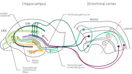
1.2.1 Anatomy of the Hippocampus ... 23
1.2.2 Functional Role of the Hippocampus ... 26
1.2.3 Hippocampus and Memory ... 27
1.2.3.1 Definition of Memory ... 27
1.2.3.2 Memory formation ... 29
1.2.3.3 Working memory ... 29
1.2.4 Spatial Memory ... 29
1.2.5 Anxiety ... 30
Methods to Study Physio-Pathological Role of Proteins ... 33 1.3.1 Electrophysiology ... 33 1.3.1.1 The Synapse... 34 1.3.1.1.1 Glutamate ... 36 1.3.1.1.1.1 mGluRs ... 37 1.3.1.1.1.2 iGluR ... 37 1.3.1.1.2 AMPAR ... 37 1.3.1.1.3 NMDAR... 41
1.3.1.2 Long-Term Synaptic Plasticity ... 44
1.3.1.2.1 LTP ... 45
1.3.1.2.1.1 Mechanism of LTP ... 45
1.3.1.2.2 LTD ... 46
1.3.1.2.2.1 Mechanisms of LTD ... 46
1.3.1.3 Short-Term Synaptic Plasticity... 47
1.3.1.3.1 Facilitation ... 47
1.3.1.3.2 Depression ... 48
1.3.1.4 Synaptic plasticity reflects behavioral outcome ... 49
1.3.2 Behavioral Studies ... 51
1.3.2.1 Experimental Design ... 51
1.3.2.1.1 Husbandry ... 51
1.3.2.2 Behavior ... 52
1.3.2.2.1 Spatial Navigation Tasks ... 52
1.3.2.2.2 Aversive Learning ... 52
1.3.2.2.3 Recognition Memory ... 53
1.3.2.2.4 Anxiety ... 53
2. Objectives ... 57
3. Material and Methods ... 61
Animal Model ... 61
3.1.1 Acute Effects of Aη on Synaptic Plasticity in Electrophysiology Field Recordings ... 61
3.1.2 Acute Effects of Synthetic Aη-α Injections into the CA1 Hippocampal Region or Lateral Ventricle ... 61
3.1.3 MISEPA2: A Transgenic Mouse Line Overexpressing Aη-α in the Brain ... 61
3.1.4 MISEPA4: A Transgenic Mouse Line Overexpressing Aη-α in the Brain with an Elevated Expression Level Compared to MISEPA2 Line ... 62
3.1.5 APPΔEta: A Mouse Model Without η-Secretase Processing of APP due to Deletion the Enzymatic Recognition Site on APP ... 62
3.1.1 Immunofluorescence and Western Blot Verify Expression of Aη-α in MISEPA2 Mice and Absence of Aη-α in APPΔEta Mice ... 64
3.1.2 Housing Conditions ... 65
3.1.3 Genotyping ... 65
3.1.3.1 Lysis ... 65
3.1.4 Polymerase-Chain Reaction Protocol for MISEPA2 and MISEPA4 Mouse Line ... 66
3.1.5 Polymerase-Chain Reaction Protocol for APPΔEta Mouse Line ... 67
3.1.6 Gel Electrophoresis ... 68
Electrophysiology ... 70
3.2.1 Peptides ... 70
3.2.2 Solutions ... 71
3.2.3 Harvesting and Slicing of Mice Hippocampi ... 72
3.2.4 Rig Set-Up ... 73
3.2.5 Field Recordings ... 73
3.2.5.1 Long-Term Synaptic Plasticity Recordings ... 73
3.2.5.2 Short-Term Synaptic Plasticity Recordings ... 74
3.2.5.2.1 Paired-Pulse Ratio ... 74
3.2.5.2.2 Synaptic Fatigue ... 74
3.2.5.2.3 Input/ Output... 74
Acute M108 Injection ... 75
3.3.1 Surgery ... 75
3.3.2 Injection Volume and Concentration ... 76
3.3.3 Verification of Injection Site and Distribution of M108 ... 77
3.3.3.1 Microscopy imaging for injection side in bilateral hippocampal injections... 77
3.3.3.2 Blue Evans Staining to Verify Correct Placement of Canula Guides and Distribution after Injection ... 77
3.3.3.2.1 Perfusion Surgery ... 77
3.3.3.2.2 Perfusion ... 78
3.3.3.3 Western Blot ... 79
3.3.3.3.1 Brain Harvesting and Storage ... 79
3.3.3.3.2 RIPA Extraction Protocol... 79
3.3.3.3.3 Immunoblotting ... 80
Behavioral Testing ... 83
3.4.1 Experimental Design for MISEPA2 and MISEPA4 Lines ... 83
3.4.2 Experimental Design of M108 Injected Mice Submitted to Behavioral Tasks ... 84
3.4.3 Experimental Design for APPΔEta Mice ... 85
3.4.4 Morris Water Maze ... 85
3.4.5 Novel Object Recognition ... 87
3.4.6 Contextual Fear Conditioning ... 88
3.4.7 T-Maze ... 91
3.4.7.1 Injection of M108 During T-Maze Testing ... 92
3.4.7.2 Familiar versus New Arm ... 92
3.4.7.2.1 Alterations of the T-Maze Task for Testing APPΔEta Mice ... 93
3.4.7.3 Forced Alternation ... 93
3.4.8 Open Field ... 94
3.4.9 Light-Dark Box ... 95
3.4.10 3-Chambers Social Interaction Task ... 96
3.4.11 Actimeter ... 97
Statistical Analysis ... 98
3.5.1 Electrophysiology ... 98
3.5.2 Behavioral Testing ... 98
4. Results ... 99
Consequences of Elevated Aƞ Levels on Synaptic Plasticity and Behavior ... 103
4.1.1 Effect of Acute Increase of Aη-α Levels on Synaptic Function and Behavior ... 103
4.1.1.1 Impact of Acutely Elevated Aƞ-α Levels on Synaptic Plasticity ... 103
4.1.1.1.1 Aƞ-α, the secreted APP fragment processed by ƞ- and α-secretases, acutely modulates post-synaptic plasticity mechanisms shifting the balance towards depression of post-synaptic strength. ... 105
4.1.1.2 Impact of Acute in vivo Injection of M108 into the Brain ... 125
4.1.1.2.1 Optimization of Protocol for in vivo Delivery of M108 Peptide ... 125
4.1.1.2.2 Presence of M108 in the Hippocampus Post-Injection Confirmed via Blue Evans Dye and Western Blot ... 126
4.1.1.2.3 Impact of Acute Injection of M108 into the Hippocampus on CFC ... 129
4.1.1.2.3.1 Acute Injection of M108 Prior to Conditioning Session in CFC Does Not Impact Memory Formation but Increases Memory Extinction During Secondary Downstream Retrieval ... 129
4.1.1.2.3.2 Contextual Memory Formation Is Not Impacted by Acute in vivo M108 Injection Irrespective of Time-point of Injection ... 130
4.1.1.2.4 Effect of Acute Injection of M108 into the Right Lateral Ventricle on Performance in T-Maze ... 131
4.1.1.2.4.1 Times of Injections Rather Than Delay of Retrieval Leads to Performance Impairment in M108 Injected Mice in a Familiar versus New Arm T-maze Task ... 131
4.1.1.2.4.2 Performance in M108 Injected Mice Is Not Impaired in a Forced Alternation T-Maze ... 134
4.1.1.3 Discussion ... 135
4.1.2 Effect of Chronic Enrichment of Aη-α Levels on Synaptic Function and Behavior ... 139
4.1.2.1 Impact of Chronically Elevated Aƞ-α Levels in a MISEPA2 Mouse Line on Synaptic Plasticity and Behavior ... 139
4.1.2.1.1 Influence of Chronically Elevated Aƞ-α Levels in a MISEPA2 Mouse Line on Synaptic Plasticity .... 139 4.1.2.1.1.1 Impaired LTP in MISEPA2 Mice ... 139 4.1.2.1.1.2 Short-Term Synaptic Plasticity and Basal Transmission Unaltered in
MISEPA2 Mice ... 140 4.1.2.1.2 Influence of Chronically Elevated Aη-α Levels in a MISEPA2 Mouse Line on Behavior ... 143
4.1.2.1.2.1 No Impairment of Performance in a Spatial Memory Dependent MWM Task for MISEPA2 Mice ... 143 4.1.2.1.2.2 Indication of External Factors Influencing Contextual Memory of MISEPA2
MICE in CFC ... 144 4.1.2.1.2.3 Unaltered Diurnal Activity in MISEPA2 Mice ... 145 4.1.2.2 Impact of Chronically Elevated Aƞ-α Levels in a MISEPA4 Mouse Line on Synaptic Plasticity
and Behavior ... 147 4.1.2.2.1 Synaptic Plasticity in MISEPA4 Mouse Line ... 147 4.1.2.2.1.1 LTP is Normal in MISEPA4 Mice ... 147 4.1.2.2.1.2 Impaired Basal Transmission in MISEPA4 Mice but No Alterations in Short-Term Synaptic Plasticity ... 148 4.1.2.2.2 Impact of Chronically Elevated Aη-α Levels in a MISEPA4 Mouse Line on Behavior ... 149 4.1.2.2.2.1 No Effect on Recognition Memory in NOR for MISEPA4 Mice ... 149 4.1.2.2.2.2 No Impairment of Performance in a Spatial Memory Dependent MWM Task for MISEPA4 Mice ... 149 4.1.2.2.2.3 Insufficient CFC Task Set-Up to Examine an Effect on Contextual Learning for MISEPA4 Mice ... 150 4.1.2.2.2.4 Normal Diurnal Activity in MISEPA4 Mice ... 151 4.1.2.3 Discussion ... 153
Consequences of Inhibition of the APP Processing η-Secretase Pathway on Synaptic Plasticity and Behavior ... 159
4.2.1 Impact of Deficiency in η-Secretase Processed APP in an APPΔEta Mouse Line on Synaptic Plasticity ... 159 4.2.1.1 No Alterations of LTP in APPΔEta Mice ... 159 4.2.1.2 Deficiency in ƞ-Secretase Processed APP Prevents LTD, a Phenotype Rescued by Acute
Application of M108 ... 160 4.2.1.3 Short-Term Synaptic Plasticity and Basal Transmission are Normal APPΔEta Mice ... 161 4.2.2 Impact of Loss of η-Secretase-dependent Cleavage of APP on Behavior ... 163 4.2.2.1 Indication of Reduced Anxiety for HOMO in Open Field ... 163 4.2.2.2 Indication of Reduced Anxiety for APPΔEta Mice in the Light-Dark Box ... 164 4.2.2.3 Regular Social Interaction Observed for APPΔEta Mice in the 3-Chambers Social Interaction Task ... 164 4.2.2.4 Impaired Spatial Memory in HOMO in T-Maze Task ... 165 4.2.2.5 APPΔEta Mice Display Loss of Spatial Memory in the MWM... 167 4.2.2.6 APPΔEta Mice Display Normal Contextual Fear Memory ... 168
4.2.2.7 Diurnal Activity is Altered in HOMO Mice (Preliminary Data) ... 169 4.2.3 Discussion ... 171
5. General Discussion and Perspectives ... 177
Synaptic Plasticity, Under Acute and Chronic elevated Aη-α Conditions ... 177 Behavioral Studies, Under Acute and Chronic elevated Aη-α Conditions ... 178 Comparing the Acute and Chronic elevated Aη-α Conditions ... 180 Depletion of Aη-α levels ... 181 APPΔEta and Alternatives: A Comparison ... 182 Comparing the Outcomes of Aη-α Level Modification ... 183 Probing the η-Secretase Pathway: Prospective Approaches ... 184
6. Conclusion ... 189 Bibliography ... XXI Supplementary ... XLVIII
Index of Figures
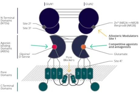
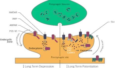
Figure 1 Illustration of the three main APP processing pathways discussed in this Thesis. ... 5 Figure 2 Illustration of APP cleavage site of η-secretase. ... 16 Figure 3 Cross section through the hippocampus. ... 23 Figure 4 Hippocampal Circuitry. ... 25 Figure 5 Basic anatomy of the hippocampus. ... 26 Figure 6 The two different pathways of information processing in the hippocampus. ... 26 Figure 7 Different forms of memory, their subcategories and the brain structures associated with them. ... 27 Figure 8 The tripartite synapse. ... 34 Figure 9 Excitatory glutamatergic synapse. ... 36 Figure 10 Composition of subunits of the iGluR. ... 37 Figure 11 Depending on AMPAR composition and post-translational modifications they are added or removed from the post-synaptic density during LTD and LTP. ... 38 Figure 12 Schematic of AMPAR structure. ... 39 Figure 13 Schematic NMDAR structure and the different binding sites for ligands. ... 42 Figure 14 NMDAR subunit diversity and expression pattern throughout development. ... 43 Figure 15 The two main forms of long-term plasticity LTD and LTP. ... 45 Figure 16 Schematic illustration of facilitation. ... 48 Figure 17 Schematic illustration of depression. ... 48 Figure 18 Representation of the pSec.2A plasmid expressing Aη-α. ... 62 Figure 19 APPΔEta mice harbor the endogenous APP protein with deletion of ƞ-secretase recognition site. ... 63 Figure 20 Immunofluorescence images confirming expression of Aƞ-α in different brain regions. ... 64 Figure 21 Western blots confirming absence of endogenous Aƞ-α in APPΔEta mice and presence of recombinant human Aƞ-α in MISEPA2 mice. ... 65 Figure 22 Gel showing the bands to be expected by the MISEP PCR samples. ... 69 Figure 23 Band options for the APPΔEta PCR samples. ... 69 Figure 24 Field recordings at the hippocampal CA3-CA1synapse were performed to investigate alterations in synaptic plasticity in our experiments. ... 70 Figure 25 The two different types, single and bilateral, cannula guides used for injections of M108. ... 76
Figure 27 Timeline of the MISEPA2 behavioral battery testing... 84 Figure 28 Timeline of MISEPA4 behavioral battery testing. ... 84 Figure 29 Timeline of M108 behavioral testing. ... 84 Figure 30 Timeline of APPΔEta behavioral battery testing. ... 85 Figure 31 Timeline of the Morris Water Maze experiment. ... 86 Figure 32 The three stages of Novel Object Recognition. ... 88 Figure 33 Contextual Fear Conditioning consisting of two stages. ... 89 Figure 34 Contextual Fear Conditioning protocol for M108. ... 90 Figure 35 Contextual Fear Conditioning protocol for the APPΔEta line. ... 91 Figure 36 T-maze set-up. ... 92 Figure 37 Illustration of the Familiar vs. New arm protocol... 93 Figure 38 The Forced Alternation protocol for M108 injected mice. ... 94 Figure 39 The Open field recorded movement of the mouse for 10 min. ... 95 Figure 40 The Light-Dark box. ... 95 Figure 41 3-Chamber social interaction task. ... 97 Figure 42 Removal of dummies during recovery phase after surgery induces necrosis in brain tissue. ... 126 Figure 43 Injection of blue Evans dye distributes throughout all ventricles. ... 126 Figure 44 Presence of M108 in the hippocampi post-injection into the CA1 region is confirmed via Western blot. ... 127 Figure 45 Distribution and presence of M108 post-injection into the right lateral ventricle was confirmed via Western blot. ... 128 Figure 46 A single acute M108 injection prior to the conditioning session does not perturb memory formation but significantly increases memory extinction. ... 130 Figure 47 Moving the time point of M108 injection to post- conditioning session does not prevent memory formation independent of time-point of Retrieval. ... 130 Figure 48 A 10 min ITI showed no alterations of performance in Retrieval, after acute injection of M108 during the first phase of T-maze. ... 132 Figure 49 A 1-hour ITI T-maze protocol showed a significantly reduced ability in M108 injected mice to identify the new arm. ... 133 Figure 50 A single 1 h ITI T-maze task, with premier injection of M108, shows no performance impairments. ... 134 Figure 51 Forced alternation with 4 trials showing no significant difference between groups, but disability of M108 injected mice to perform above Chance level (50 %). ... 134
Figure 52 LTP is impaired for MISEPA2 mice at the SC-CA1 synapse of hippocampal slices. ... 140 Figure 53 No short-term plasticity deficits observable in hippocampal slices of MISEPA2. 141 Figure 54 transgenic MISEPA2 mouse line displays normal function of spatial learning and memory in MWM. ... 143 Figure 55 Reduced contextual fear memory in transgenic mice line MISEPA2. ... 144 Figure 56 Contextual fear memory increases in MISEPA2 when analyzed automatically. .. 145 Figure 57 MISEPA2 mice show normal circadian activity. ... 146 Figure 58 Normal LTP in MISEPA2 mice at the SC-CA1 synapse of hippocampal slices. . 147 Figure 59 No deficits in short-term plasticity dependent PPR observable, but a significant decrease in basal synaptic transmission in hippocampal slices of MISEPA4. ... 148 Figure 60 MISEPA4 mice show significant exploration time towards novel objects but fails to reach DI criterion. ... 149 Figure 61 MWM testing showed no sign of spatial memory impairment in MISEPA4 mice. ... 150 Figure 62 A single low intensity shock proved too mild to test for contextual memory. ... 151 Figure 63 MISEPA4 mice show normal diurnal activity. ... 151 Figure 64 Normal LTP in HOMO mice at the SC-CA1 synapse of hippocampal slices. ... 160 Figure 65 Addition of M108 rescues LTD in HOMO at the SC-CA1 synapse of hippocampal slices. ... 161 Figure 66 No pre-synaptic short-term plasticity deficits were observable in APPΔEta mice. ... 162 Figure 67 HOMO APPΔEta mice show reduced anxiety in the Open field. ... 163 Figure 68 Light-Dark box test hints toward reduced anxiety in HOMO. ... 164 Figure 69 APPΔEta mice show no impairment in sociability in the 3-chambers social interaction test paradigm. ... 165 Figure 70 A 10 min ITI familiar vs new arm T-maze test indicates a quick wear off for novelty in HOMO. ... 166 Figure 71 HOMO display impairments in the 1 h ITI T-maze task. ... 166 Figure 72 HOMO show impairment in retrieval of platform location during Probe in the MWM test. 167 Figure 73 A two-day CFC experiment followed by an extinction paradigm revealed no impairments in contextual memory or memory extinction in HOMO. ... 168 Figure 74 Diurnal activity is altered in APPΔEta mice. ... 169 Figure 75 Illustrating Aη-α as a neuromodulator crucial for LTD... 183
Index of Tables
Table 1 Deletion of 41 amino acids (blue) containing the ƞ-secretase recognition site (orange) on APP. ... 63 Table 2 Comparing MISEPA2 and MISEPA4 and their respective Aη-α expression levels in different brain areas. ... 64 Table 3 content of the MISEP PCR master mix. ... 67 Table 4 Primer sequence of Myosin and MISEP used in the PCR master mix ... 67 Table 5 content of the APPΔEta PCR master mix ... 68 Table 6 Sequences of Primers used in APPΔEta PCR master mix ... 68 Table 7 Amino acid sequence of synthetic proteins ordered... 70 Table 8 Ingredient list of the cutting solution with their respective concentration ... 71 Table 9 aCSFI solutions ingredient list with their respective concentration ... 71 Table 10 aCSFII solutions ingredients list with their concentration. ... 72 Table 11 Time points to sacrifice mice post-injection of M108. ... 79 Table 12 composition of RIPA buffer ... 80 Table 13 Loading buffer composition ... 81 Table 14 Blocking solution composition ... 81 Table 15 Separation Gel and Spacer gel recipe ... 81
Abbreviations
A
Aβ Amyloid β protein
Aβ17-42 p3
Aη-α Amyloid η-α protein
Aη-β Amyloid η-β protein
ABD agonist-binding domain
aCSF artificial cerebrospinal fluid
AD Alzheimer’s disease
ADAM disintegrin and
metalloproteinase
ADAM10 disintegrin and
metalloproteinase 10, α-secretase
AMPAR
α-amino-3-hydroxy-5- methyl-4-isoxazole-propionate receptor
ANOVA analysis of variance
AP anteroposterior
APP Amyloid precursor protein
APPLP1 Amyloid precursor like
protein 1
APPLP2 Amyloid precursor like
protein 2
ASP2 aspartyl protease 2
B
BACE1 beta-site amyloid precursor
protein cleaving enzyme 1, β-secretase C C83 see CTF-α C99 see CTF-β CA Cornu Ammonis CAMKII
Calcium/calmodulin-dependent protein kinase II
CFC contextual fear conditioning
CHO-cells Chinese hamster ovary cells
CNO clozapine-N-oxide
CP-AMPAR Ca2+ permeable AMPAR
CSF cerebrospinal fluid
CTF carboxy-terminal fragment
CTF-α C-terminal APP α fragment
CTF-β C-terminal APP β fragment
CTF-η C-terminal APP η fragment
D
DI discrimination index
DMSO dimethyl sulfoxide
dNTP deoxynucleotide mix
DREADD designer receptors
exclusively activated by designer drugs
DTT Dithiothreitol
DV dorsoventral
E
E east
EDTA Ethylenediaminetetraacetic
acid
EPSP excitatory post-synaptic
potential
F
fAD familiar or early-onset AD
fEPSP field excitatory post synaptic potential
FV fiber volley
G
GABA γ-aminobutyric acid
GABABR1 γ-aminobutyric acid type B receptor subunit
GluA AMPAR subunit
GluA2L long-tailed GluA2
GluA4s short-tailed GluA4
GluN NMDAR subunit
GLUT plasma membrane glucose
transporter g gram H HEPES 4-(2-hydroxyethyl)-1-piperazineethanesulfonic acid HET heterozygote HOMO homozygote HRP horseradish peroxidase Hz Hertz I Ig Immunoglobulin
iGluR ionotropic glutamate
receptor
I/O input/output
i.p. intraperitoneal
ISI inter stimulus interval
ITI inter trial interval
K
K kappa
kb kilo base
kDa kilo Dalton
kg kilogram
L
LTD long-term depression
LTP long-term potentiation
LxWxH length x width x height
lux luminous flux per unit
M
M2 re-entrant pore loop
M92 synthetic Amyloid η-α
protein
M108 synthetic Amyloid η-β
protein
memapsin-2 membrane-associated aspartic protease 2
mg milligram
mGluR metabotropic glutamate
receptor ML mediolateral mM millimolar MMP metalloproteinase MT5-MMP membrane-type 5 metalloproteinase, η-secretase mV millivolt
MWM Morris water maze
µgr microgram µl microliter N N north nM nanomolar NMDAR N-methyl-D-aspartate receptor ng nanogram
NOR novel object recognition
NTD amino-terminal domain
NTF amino-terminal fragment
P
P postnatal day
PB phosphate buffer
PBS phosphate buffered saline
PCR polymerase chain reaction
PFA formaldehyde pH potential of hydrogen PPR paired-pulse ratio PS1 presenilin 1 PS2 presenilin 2 PSD post-synaptic density PSD-95 a neuron-specific PSD protein R RM repeated measured
rtTA reverse tetracycline
transactivator
S
S south
sAD sporadic or late-onset AD
sAPP-α soluble APP α fragment
sAPP-β soluble APP β fragment
sAPP-η soluble APP η fragment
SDS-PAGE sodium dodecyl sulfate-polyacrylamide gel
S.E.M. standard error of mean
T
TARP transmembrane AMPAR
regulatory protein
TBST Tris buffered saline with
Tween 20
TCA tricarboxylic acid cycle
TM transmembrane
V
V volt
W
W west
1. Introduction
Alzheimer’s Disease and Amyloid Precursor Protein processing
Alzheimer's Disease (AD) is a neurodegenerative disease familiar by name to a wide audience, scientists and lay-people alike. It was first described in 1906 at the 37th meeting of the Southwest German Psychiatrists in Tübingen by Alois Alzheimer (Alzheimer, 1906). He described symptoms of dementia in his patient Auguste Deter and the post-mortem discovery of insoluble extra- and intracellular proteinaceous deposits in her brain.
However, AD received only minor attention post-discovery until 1976 when Robert Katzman made the connection between AD and "senile dementia" identifying the disease and its progression as the leading cause of death in the USA (Katzman, 1976).
AD is a neurodegenerative disease mainly affecting the older population, as age is one of the main risk factors. It is a progressive disease starting on the cognitive level with a decline in episodic memory, spreading to affect short-term memory, and finally, affecting procedural memory. The disease is defined by selective neuronal loss and atrophy at some medial temporal lobe structures, which makes it distinct from other dementia (Puzzo, Lee, Palmeri, Calabrese, and Arancio, 2014). The different types of memory and the role the hippocampus plays in its formation are described in more detail in the hippocampus section of the introduction.
Since 1976 research focused its investigation on identifying the cause of AD. On the cellular level, we have the two hallmarks of AD, a) the amyloidogenic plaques and b) neurofibrillary tangles, an aggregation of hyperphosphorylated cytosolic tau. In 1985 Masters and colleagues revealed amyloid β (Aβ), a protein discovered the previous year by Glenner and Wong, as the
major component of the extracellular plaques described by Alzheimer (Glenner and Wong, 1984; Masters et al., 1985). The subsequent identification of the amyloid precursor protein
(APP) as the Aβ producing protein and the linkage of mutations promoting Aβ production and
familiar AD, led to the formulation of the amyloid cascade hypothesis (Goate et al., 1991; J. Hardy and Higgins, 1992; John Hardy and Allsop, 1991; Kang et al., 1987; Tanzi et al., 1987). These discoveries directed research efforts to focus on the pathological processing of APP. As research progressed, several other factors contributing to AD have been discovered. Mutations on Presenilin, a protein involved in APP processing have also been linked to AD and we know about several risk-factor genes such as harboring the gene encoding the Apolipoprotein E (APOE) isoform APOE4, a protein involved in lipid metabolism and transportation (Naj and
Schellenberg, 2017; Sherrington et al., 1995; Van Cauwenberghe, Van Broeckhoven, and Sleegers, 2016). Today, we have about twelve theories for AD genesis developing and adapting throughout the years trying to explain the cause and associated processes in AD. These theories cover a vast range of fields, ranging from the vascular hypothesis, to the traumatic brain injury hypothesis, to the amyloid hypothesis, the most widely accepted theory.
Despite significant efforts in the preceding decades to this date Alzheimer's disease remains incurable. At the moment there exist 5 FDA approved drugs which help minimize the symptoms in AD. These drugs also cause severe side effects. They can be divided into two groups: four of them target the cholinergic system, while Memantine, the fifth drug, acts as an N-methyl-D-aspartate receptor (NMDAR) antagonist.
As mentioned before, the plaques first described by Alois Alzheimer in 1906 consist mainly of Aß, a protein resulting from APP processing via the amyloidogenic pathway (Alzheimer, 1906) (see Figure 1). Thus, the main attention of research was focused on this pathway in pathological conditions, the enzymes involved, and the cleavage products. It is therefore not surprising that the physiological role of APP and the amyloidogenic pathway remain elusive. Due to the focus on amyloidogenic processing of APP, knowledge about the non-amyloidogenic pathway, the η -secretase pathway and their products is even more limited.
Figure 1 Illustration of the three main APP processing pathways discussed in this Thesis. In the middle is the full-length APP, which can be processed by α-, ß- or η -secretase initiating three different pathways. On the left are the well-known non-amyloidogenic pathway and non-amyloidogenic pathway. The non-non-amyloidogenic pathway on the top is initiated by cleavage of APP through α-secretase creating sAPP-α and CTF-α. The latter can be further processed by γ-secretase releasing p3 and AICD. The amyloidogenic pathway shown below, is driven by cleavage of APP by ß-secretase, producing sAPP-ß and CTF-ß. CTF-ß is further cleaved by γ-secretase producing Aß and AICD. Shown on the right is the η -secretase pathway. In this pathway APP cleavage by η -secretase produces sAPP-η and CTF-η. CTF-η is then further processed by either α-secretase generating Aη-α or by ß-secretase producing Aη-ß. Adapted from (Ludewig and Korte, 2017).
Lately, the scope of research on APP also opened up towards new processing pathways and identifying secretases involved in these pathways. The η-secretase pathway is an example of these efforts to shed more light on APP processing. This pathway was discovered and first described in 2015 by two independent teams (Haizhi Wang et al., 2015; Willem et al., 2015). In their work, the teams identified η-secretase as an enzyme able to cleave APP leading to the discovery of a novel secretion pathway. Two of the products of this pathway, Aη-α and Aη-ß, and their physiopathological role, have been the focus of this thesis. We aimed to investigate the physiopathological role of these newly discovered Aeta peptides including studies focusing on their ability to alter synaptic plasticity as well as behavior. The following chapter will introduce the main pathways known in APP processing and describe the different proteins involved in these pathways.
1.1.1 The Amyloid Precursor Protein (APP)
The APP, together with the amyloid precursor protein-like protein (APPLP) 1 and APPLP2, constitute a highly conserved protein family (Bhadbhade and Cheng, 2012). Various knock-out models of each of the proteins and combinations have shown that they can replace each other to a certain degree. The knock-out of APPLP2, for example, does not alter the phenotype of mice, indicating that APP and APPLP1 together can replace it. However, the knock-out of APP, APPLP1, and APP/APPLP1 leads to physiological changes in mice, reducing their body weight, grip strength, locomotor activity, and increases age-associated memory deficits. The triple knock-out APP/APPLP1/APPLP2 and double knock-outs APP/APPLP2 and APPLP1/APPLP2 are lethal, demonstrating the importance of this family of proteins and the limits of in vivo substitution of the functions of individual proteins by other family members (Heber et al., 2000; Z. W. Li et al., 1996; Müller et al., 1994; Senechal, Kelly, and Dev, 2008; The Human Protein Atlas, 2018; Von Koch et al., 1997; H. Zheng et al., 1995).
While the expression of APPLP1 is limited to the brain, APP and APPLP2 are expressed ubiquitously. These proteins are type 1 transmembrane proteins resembling cell-surface receptors. Their extracellular domain at the N-terminal represents nearly 90 % of the total protein mass. The C-terminal, on the other hand, consists of a 47 amino acid short cytoplasmic domain connected to the N-terminal via a single membrane-spanning domain (Bhadbhade and Cheng, 2012). It is at the C-domain where the family members have the highest sequence similarity, with motifs involved in different functions of the proteins (Gralle and Ferreira, 2007; H. Zheng and Koo, 2011).
For example, some of the implied functions of these proteins are cell-cell adhesion and migration, synaptogenesis, and synaptic plasticity, indicating the importance of these proteins in brain development and maturation (Gralle and Ferreira, 2007; Kedikian et al., 2010; Korte, Herrmann, Zhang, and Draguhn, 2011; Nalivaeva and Turner, 2013; Octave, Pierrot, Ferao Santos, Nalivaeva, and Turner, 2013; Shariati and De Strooper, 2013; Soba et al., 2005; Sosa et al., 2017; Zhang, Thompson, Zhang, and Xu, 2011; H. Zheng and Koo, 2011).
As APP is the only family member containing the Aβ domain, thus considered highly crucial in AD, it has been by far the most studied member of the family (J. Hardy and Higgins, 1992; John Hardy, 2017; John Hardy and Allsop, 1991; Raphaëlle Pardossi-Piquard et al., 2005). Several studies suggest an essential role for APP in synaptogenesis, spine formation, regulation of intracellular calcium homeostasis, signaling, and in synaptic vesicle and transmitter release. Resembling a receptor, as mentioned above, several binding proteins have been being proposed,
However, to this date, knowledge about the physiological role of APP is poorly understood due to the complex splicing, proteolysis, co-localization, and similarity to its other family members and proteins generated by APP processing (Gralle and Ferreira, 2007; Kedikian et al., 2010; Korte et al., 2011; Nalivaeva and Turner, 2013; Octave et al., 2013; Shariati and De Strooper, 2013; Soba et al., 2005; Sosa et al., 2017; Zhang et al., 2011; H. Zheng and Koo, 2011). APP has eight isoforms, among which the following three are the most common ones: APP695,
APP752, and APP770 derived by alternative splicing of exon 7 and exon 8 on chromosome 21
(Cappai, 2014; J. C. Phillips, 2019; Rupert Sandbrink, Masters, and Beyreuther, 1994). Out of the three isoforms APP695 is the main isoform in the brain and mainly expressed in neurons,
while the other two forms primarily express in glial cells like astrocytes (Belyaev et al., 2010; Bhadbhade and Cheng, 2012; De Silva et al., 1997; Matsui et al., 2007; Nalivaeva and Turner, 2013; Puig and Combs, 2013; R. Sandbrink, Masters, and Beyreuther, 1996; Simons et al., 1996; Tanaka et al., 1988). Interestingly, this distribution seems altered in AD as an increase in APP770 mRNA correlating with a decrease of APP695 mRNA can be observed (Cappai, 2014).
The location on chromosome 21 first described in 1987 correlates with increased cases of AD for people with trisomy 21, which have a third copy of this chromosome. Additionally, point mutations in the APP gene correspond with familiar AD (fAD), the rarer form of AD comprising about 1-5 % of all AD cases (J. Hardy and Higgins, 1992; John Hardy and Allsop, 1991; Petronis, 1999; Selkoe and Hardy, 2016; St. George-Hyslop et al., 1987).
Synthesis and processing of APP is a highly regulated process starting in the endoplasmic reticulum. The synthesized APP continues its journey through the Golgi apparatus to finally reach the cell membrane via the constitutive secretion pathway. Throughout this process, APP undergoes post-translational modifications such as O- and N-glycosylation, ubiquitination, phosphorylation, and tyrosine sulfation. About 10 % of the produced APP reaches the plasma membrane mainly at the axons and dendrites via anterograde transportation, while the majority is stored in the Golgi apparatus in a ready state (Kaether, Skehel, and Dotti, 2000; Kins, Lauther, Szodorai, and Beyreuther, 2006; Plácido et al., 2014). Once at the plasma membrane APP can undergo either endocytosis or is proteolytically processed following down one of the different pathways explained more in detail later: a) non-amyloidogenic, b) amyloidogenic, or c) η-secretase pathway (see Figure 1).
If undergoing endocytosis, this process happens due to an internalization motif near the C-terminal and the APP protein goes then either to endosomes, with parts of it recycled to the cell surface, and Golgi apparatus or is degraded in lysosomes (Bhadbhade and Cheng, 2012; De Strooper and Annaert, 2000; Gouras, Willén, and Faideau, 2014; Haass, Kaether, Thinakaran,
and Sisodia, 2012; Haass, Koo, Mellon, Hung, and Selkoe, 1992; Koo et al., 1990; Lorenzen et al., 2010; Marquez-Sterling, Lo, Sisodia, and Koo, 1997; Schmidt, Subkhangulova, and Willnow, 2017).
When the APP does not undergo endocytosis, it is proteolytically processed, following either one of the pathways further discussed below. Different studies have shown that these pathways stand in direct competition with each other, thus blocking one, strengthens the others. Also, colocalization of APP with secretases like disintegrin and metalloproteinase 10 (ADAM10) or beta-site amyloid precursor protein cleaving enzyme (BACE1) in the vesicles predetermines the following proteolysis pathway of APP (Das et al., 2015; Haass et al., 2012; Szodorai et al., 2009). However, it seems that external factors can influence APP processing too. For example, a study indicates that cholesterol depletion prevents the processing of APP through the amyloidogenic pathway (Kerridge, Belyaev, Nalivaeva, and Turner, 2014).
1.1.2 Amyloidogenic Pathway
As mentioned above, the amyloidogenic pathway gained the greatest attention among all pathways involved in APP processing due to its association to AD. As shown in Figure 1, the pathway is mediated by ß-secretase, which splices the APP protein at its recognition site and leads to the secretion of a soluble APP ß fragment (sAPP-ß) and the membrane-anchored APP ß carboxyl-terminal fragment (CTF-ß), also known as C99.
The C99 can then be further cleaved by the γ-secretase complex, which is also involved in the non-amyloidogenic pathway, leading to the secretion of Aß and an APP intracellular domain (AICD) (Haass et al., 2012; Turner, O’Connor, Tate, and Abraham, 2003).
Each of these products, sAPP-ß, C99, Aß, and AICD, serve different roles, some of them having the ability to alter synaptic plasticity, gene expression, or even the production of APP itself (see below).
1.1.2.1 ß-Secretase
The ß-secretase is the enzyme initiating the amyloidogenic pathway of APP processing. Several proteins can fulfill the role of ß-secretase, but in 1999 the BACE1 also known as membrane-associated aspartic protease 2 (memapsin-2) or aspartyl protease 2 (ASP2) was identified to act as the main secretase (Vassar et al., 1999, 2014; H. Zheng and Koo, 2011).
The highest concentration of BACE1 mRNA is in the cortex, while the CA1-CA3 hippocampal region also harbors high concentrations of BACE1. Looking at the neuronal level, the majority of BACE1 concentrations in a healthy state are in pre-synaptic neuronal terminals. However,
after stress and inflammatory conditions, and with aging BACE1 is also found in astrocytes. This accumulation in astrocytes is also encountered in AD brains of humans and animal models, with an overall increase of BACE1 levels of 50 % compared to healthy state (Cai et al., 2001; Hartlage-Rübsamen et al., 2003; Kamenetz et al., 2003; Roßner et al., 2005; Tamagno, Guglielmotto, Monteleone, and Tabaton, 2012; Hui Wang et al., 2010; Hui Wang, Megill, Wong, Kirkwood, and Lee, 2014).
BACE1 is a type 1 transmembrane protease consisting of 501 amino acids with two active site motifs, which are necessary for its correct function. The maturation of BACE1 occurs in the Golgi apparatus via the removal of a short pro-domain. To increase stability, BACE1 can undergo post-translational modifications like phosphorylation or N-glycosylation before it is transported to its final destination via secretory vesicles. The activity of BACE1 is pH-dependent, reaching the maximum activity under acidic conditions. Therefore, BACE1 mainly resides in subcellular compartments like endosomes and lysosomes. As it follows a similar trafficking route to APP, it can be co-localized in neuronal endosomes and cleaves APP there, giving rise to sAPP-ß and C99 (Cole and Vassar, 2008; Das et al., 2013; Huse et al., 2002; Vassar, 2001; Vassar et al., 2014). Interestingly, the co-localization of ß-secretase and APP depends on both of them internalizing separately and meeting at the early endosomes, producing about 60-70 % of total Aß in this intracellular compartment. The trafficking to the plasma membrane is highly regulated, and production of ß-secretase depends on neuronal activity (Das et al., 2013; Niederst, Reyna, and Goldstein, 2015; Prabhu et al., 2012; Sannerud et al., 2011; Schneider et al., 2008).
It is important to note, that while the ß-secretase is the initiating enzyme for the amyloidogenic pathway, it also can cleave other proteins, such as neuregulin 1 and sialyl-transferase ST6Gal I, and fulfills a physiological role. It seems that BACE1 is essential for myelination of peripheral nerves, as indicated by hypomyelination and aberrant axonal segregation in knock-out models (Kitazume et al., 2001; Willem et al., 2006).
Furthermore, several studies focusing on blocking BACE1 action on APP or lowering global levels as a therapeutic measurement to prevent AD progression have had disappointing results, and none has surpassed phase III trials so far (Karran, Mercken, and Strooper, 2011; Panza, Lozupone, Logroscino, and Imbimbo, 2019; Vassar et al., 2014).
1.1.2.2 Amyloid ß
The Aß is a protein created by cleavage of the C99 fragment at APP 672D by γ-secretase, which is one of the cleavage products of APP processing via ß-secretase as mentioned above. Due to the dependency of Aß cleavage on these two secretases, the main production happens in the
early endosomes. It can vary in length from 39-42 amino acids, with the two main forms being Aß40 and Aß42. The Aß monomers are about 4 kilo Dalton (kDa) and described to have
neurotrophic and neuroprotective properties, involved in neural progenitor cell proliferation and synaptic transmission (Abramov et al., 2009; Chasseigneaux and Allinquant, 2012; Giuffrida et al., 2010; J. C. Phillips, 2019; Puzzo et al., 2008, 2011; Rivera, García-González, Khrestchatisky, and Baranger, 2019). Thus, the endogenous Aß can enhance release probability at synapses, with excitatory synapses being the most sensitive to changes in endogenous Aß levels (Abramov et al., 2009).
The production and release of Aß correlate positively with neuronal and synaptic activity. Interestingly, sleep deprivation also seems to enhance Aß levels, with the time spent asleep being the critical factor rather than dark/light exposure (Kang et al., 2009).
This protein exists not only in monomers but can aggregate, forming dimers, trimers, oligomers, fibrils, and eventually form plaques, one of the hallmarks of AD (Alzheimer, 1906; Rivera et al., 2019; Stelzmann, Norman Schnitzlein, and Reed Murtagh, 1995). Notably, while plaques are an indicator, they are neither the cause nor needed for AD, and may also found in healthy brains post-mortem. Indeed, recent studies identify that Aß dimers and trimers as likely to represent the more toxic forms of this peptide (Herrup, 2015; Lesné et al., 2013).
In a healthy brain, Aß40 is the more common form, whereas, in AD brains, the ratio shifts
towards Aß42, the form more prone to aggregate (Rivera et al., 2019). The aggregation of fibrils
to form plaques depends on their hydrophobic side chains. Fibrils possess residues that form a double-horseshoe-like cross-beta-2 sheet burying these hydrophobic chains. Numerous mutations of familiar AD are adjacent to the Aß N-terminals and influence the side chains' ability to cover the hydrophobic side chains and increase the aggregation abilities of Aß (J. C. Phillips, 2019).
The importance of Aß in AD was first described in 1984 by Glenner, whom earlier that year isolated Aß from meningeal vessels of AD cases and later could link the protein to a trisomy 21 case. People with trisomy 21 nearly always develop AD, as the additional chromosome 21 copy enhances the Aß burden (Glenner and Wong, 1984).
The amyloidogenic plaque composition and undeniable connection between Aß and AD led to the formulation of the amyloid cascade hypothesis (J. Hardy and Higgins, 1992; John Hardy and Allsop, 1991). This hypothesis declared the plaques responsible for the cognitive decline observed in AD patients, initiating an ongoing effort to unravel Aß's role as an originator of the disease.
Although fAD, where genetic mutations increase Aß levels and cause an early onset of the disease, comprises only about 5 % of all AD cases, they show a high phenotypic similarity with sporadic or late-onset AD (sAD), which affects the aging population. These similarities suggest that the mechanism involved in fAD applies to sAD paving the foundation of transgenic mouse models of AD (Puzzo et al., 2014).
The PDAPP mouse line was the first one developed, overexpressing human APP containing a fAD mutation, which shifts the APP processing towards the more extended Aß peptide variant (Games et al., 1995). Similarly, the famous Tg2576 mouse line contains two point mutations on their overexpressed human APP, which are mutations of the Swedish fAD and increase overall levels of Aß (Hsiao et al., 1996). These mouse lines develop many characteristics of AD, like amyloidogenic plaques, loss of synapses, and synaptic and cognitive deficits. Moreover, when combining the APP mutations with a mutation on the presenilin 1 (PS1) gene, like in the double transgenic APP/PS1 mice, the AD pathology course progresses quicker, starting symptoms around 3 months old (Holcomb et al., 1998; Puzzo et al., 2014). The 5xFAD mice express a total of five mutations, three fAD mutations on APP and two PS1 mutations, highly accelerating AD pathology (Crouzin et al., 2013; Puzzo et al., 2014).
However, these transgenic mouse models lack tau pathology, the other hallmark of AD. The 3xTgAPP mouse line addresses this by harboring APP and PS1 mutations and expressing mutant tau too, producing the neurofibrillary tangles and neurodegeneration (Chambon, Wegener, Gravius, and Danysz, 2011; Oddo et al., 2003; Puzzo et al., 2014). All these transgenic mouse models overexpress the APP, thus making it difficult to distinguish between the effects of elevated Aβ and APP contributing to the phenotype. In 2016 Saito et al. presented human mutant APP knock-in mice, which express APP normally and show a developing Aβ pathology more resembling the disease progression (Masuda et al., 2016; Saito et al., 2014). However, while these knock-in mice lines are nowadays used in numerous laboratories worldwide, their abilities to model AD more accurate than overexpressing transgenic mice remains debatable (Huang et al., 2015; Jacob et al., 2019; Mehla et al., 2019).
The usage of these transgenic mouse lines advanced the understanding of AD, and they continue to be valuable tools. However, their creation was based on the hypothesis that the plaques are determining the cognitive decline. Today we know that it is not the plaques but rather the soluble Aß levels that correlate with the cognitive impairment. As mentioned above, before plaque formation, Aß exits in soluble form as monomers, dimers, oligomers, globules, and fibrils, all of which could be the toxic formation contributing to the cognitive decline. Thus, the amyloid hypothesis evolved to posit that soluble Aßs are responsible for AD. Indeed
electrophysiology studies have shown that small soluble Aß oligomers are capable of impairing synaptic plasticity, reducing long-term potentiation (LTP), and are linked to neurotoxicity (Chambon et al., 2011; S. Li et al., 2009; Shankar et al., 2008). Similar effects have also been proven by studies using globular and fibrillar Aß (Kayed et al., 2009; Shankar et al., 2007). Synaptic plasticity is essential for memory formation. The reduction in LTP observed through acute application of these peptides on hippocampal slices, implies a connection to the deficits observed in AD. To test this hypothesis, a new kind of animal model emerged, mice with intracerebroventricular injections or infusions of Aß peptides. The injection allows the study of AD pathology without the necessity of plaques. Today, we know that already picomolar concentrations of Aß are sufficient to impair different forms of synaptic plasticity (LTP and long-term depression (LTD)) as well as learning and memory performance in mice (Canet et al., 2020; Doran et al., 2017; Meunier, Villard, Givalois, and Maurice, 2013; Morley et al., 2010; Puzzo et al., 2008; Puzzo, Arancio, and Puzzo, 2013; Toombs, n.d.; Wiseman et al., 2018).
1.1.3 Non-Amyloidogenic Pathway
The second well-established pathway in APP processing is the non-amyloidogenic pathway, which mediates the cleavage of APP within the Aß region by α-secretase (Figure 1). This cleavage generates a sAPP-α fragment for extracellular secretion and CTF-α (C83). The C83, similar to the amyloidogenic pathway CTF-ß, can then be further processed by γ-secretase generating AICD and p3 (Haass et al., 2012; Turner et al., 2003).
Exploring the physiological role of the non-amyloidogenic pathway has not been a major focus of research. Most attention focused on the pathway’s potential use in curing AD. The amyloidogenic pathway competes with the others to process APP. Thus, enhancement of this non-amyloidogenic pathway decreases the amyloidogenic APP processing and, therefore, Aß aggregation.
1.1.3.1 α -Secretase
Different members of disintegrin and metalloproteinase (ADAM) family, which are type 1 transmembrane proteins, can act as α -secretase, of which ADAM10 and ADAM17 are the most common. ADAM17 is not involved in constitutive APP cleavage but plays a role in regulated APP cleavage. Thus, ADAM10 acts as the main α-secretase, mostly found in post-synaptic regions of excitatory synapses. Blocking ADAM10 reduces overall sAPP-α production by up to 90 %, highlighting its importance for non-amyloidogenic processing of APP (Christensen et