HAL Id: hal-02880625
https://hal.archives-ouvertes.fr/hal-02880625
Submitted on 25 Jun 2020
HAL is a multi-disciplinary open access
archive for the deposit and dissemination of
sci-entific research documents, whether they are
pub-lished or not. The documents may come from
teaching and research institutions in France or
abroad, or from public or private research centers.
L’archive ouverte pluridisciplinaire HAL, est
destinée au dépôt et à la diffusion de documents
scientifiques de niveau recherche, publiés ou non,
émanant des établissements d’enseignement et de
recherche français ou étrangers, des laboratoires
publics ou privés.
Wiskott-Aldrich Syndrome Protein (N-WASP) with
Actin and Arp2/3 Complex
Jean-François Gaucher, Chloé Maugé, Dominique Didry, Berengère Guichard,
Louis Renault, Marie-France Carlier
To cite this version:
Jean-François Gaucher, Chloé Maugé, Dominique Didry, Berengère Guichard, Louis Renault, et al..
Interactions of Isolated C-terminal Fragments of Neural Wiskott-Aldrich Syndrome Protein (N-WASP)
with Actin and Arp2/3 Complex. Journal of Biological Chemistry, American Society for Biochemistry
and Molecular Biology, 2012, 287 (41), pp.34646-34659. �10.1074/jbc.M112.394361�. �hal-02880625�
Interactions of Isolated C-terminal Fragments of Neural
Wiskott-Aldrich Syndrome Protein (N-WASP) with Actin
and Arp2/3 Complex
Received for publication, June 26, 2012, and in revised form, July 23, 2012 Published, JBC Papers in Press, July 30, 2012, DOI 10.1074/jbc.M112.394361 Jean-Franc¸ois Gaucher‡, Chloe´ Mauge´§, Dominique Didry§, Be´renge`re Guichard§, Louis Renault§,
and Marie-France Carlier§1
From the‡Laboratoire de Cristallographie et RMN Biologiques CNRS UMR 8015, Faculte´ de Pharmacie, Universite´ Paris Descartes,
Sorbonne Paris Cite´, 4 avenue de l’Observatoire, 75006 Paris and the§Cytoskeleton Dynamics and Motility Group, Laboratoire
d’Enzymologie et Biochimie Structurales, CNRS UPR 3082, 1 Avenue de la Terrasse, 91198 Gif-sur-Yvette Cedex, France Background:VCA in WASP proteins binds Arp2/3 complex and actin via a variable number of WH2 domains.
Results:WH2 repeats in V, VC, and VCA of N-WASP bind actin in 1:2 complexes; VC-actin crystallizes as a 1:1 complex, and the VCA䡠actin䡠Arp2/3 complex has 1:1:1 stoichiometry.
Conclusion:Data reconcile the studies of VCA-actin-Arp2/3.
Significance:Data provide insight in filament branching.
Wiskott-Aldrich syndrome proteins (WASP) are a family of proteins that all catalyze actin filament branching with the Arp2/3 complex in a variety of actin-based motile processes. The constitutively active C-terminal domain, called VCA, har-bors one or more WASP homology 2 (WH2) domains that bind G-actin, whereas the CA extension binds the Arp2/3 complex. The VCA䡠actin䡠Arp2/3 entity associates with a mother filament to form a branched junction from which a daughter filament is initiated. The number and function of WH2-bound actin(s) in the branching process are not known, and the stoichiometry of the VCA䡠actin䡠Arp2/3 complex is debated. We have expressed the tandem WH2 repeats of N-WASP, either alone (V) or asso-ciated with the C (VC) and CA (VCA) extensions. We analyzed the structure of actin in complex with V, VC, and VCA using protein crystallography and hydrodynamic and spectrofluori-metric methods. The partial crystal structure of the VC䡠actin 1:1 complex shows two actins in the asymmetric unit with extensive actin-actin contacts. In solution, each of the two WH2 domains in V, VC, and VCA binds G-actin in 1:2 complexes that participate in barbed end assembly. V, VC, and VCA enhance barbed end depo-lymerization like profilin but neither nucleate nor sever filaments, in contrast with other WH2 repeats. VCA binds the Arp2/3 com-plex in a 1:1 comcom-plex even in the presence of a large excess of VCA. VCA䡠Arp2/3 binds one actin in a latrunculin A-sensitive fashion, in a 1:1:1 complex, indicating that binding of the second actin to VCA is weakened in the ternary complex.
The WASP2/Scar/WAVE family of proteins counts many
members, all of which stimulate actin polymerization in a den-dritic array of actin filaments that deform membranes in a large number of motile processes (1–3). These processes include the extension of the lamellipodium of migrating cells, as well as phagocytosis, dendritic spine activity, immune synapse forma-tion, spatial organization of the Golgi, and endosomal vesicle scission (4 –12). WASP proteins work as membrane-bound, stimulus-responsive enzymes that catalyze cycles of filament branching and share the Arp2/3 complex as a substrate (13, 14). All proteins of the WASP family use the same molecular mechanism to multiply filaments by branching, albeit with vari-able efficiencies (15–17). The conserved, constitutively active C-terminal moiety of the WASP enzymes, conventionally called VCA, is made of one or two WH2 (WASP homology 2) domains (“V” referring to either a single or multiple WH2 domains in Fig. 1), followed by a connector domain (“C”) and an acidic short extension (“A”). In the VCA region, the WH2 domain(s) bind G-actin, whereas the CA domain binds Arp2/3 complex. The resulting VCA䡠actin䡠Arp2/3 ternary complex (in which the shortened notation Arp2/3 refers to the Arp2/3 com-plex) associates with a filament to form the branched junction. Binding of G-actin to VCA is generally required for the branch-ing reaction (18), although a basic branchbranch-ing activity of yeast Arp2/3 complex alone has been detected (19). The Arp2/3 complex is incorporated at the branched junction at each cata-lytic cycle and thus penetrates the network at the same pace as actin, by insertional polymerization (20 –22).
How the morphology of the dendritic array develops and perpetuates at the leading edge and how protrusive force is produced against the membrane tightly depend on the molec-1Recipient of Ligue Nationale Contre le Cancer E´quipe Labellise´e 2010 –2013,
European Research Council Advanced Grant “Forcefulactin” 249982, fund-ing from the European Community Seventh Framework Programme FP7/ 2007–2013 under Grant Agreement 241548, and an Agence Nationale pour la Recherche, Programme Physique et Chimie du Vivant 2006 –2009 “Motilactin.” To whom correspondence should be addressed. Tel.: 33-01-69-82-34-65; E-mail: carlier@lebs.cnrs-gif.fr.
The atomic coordinates and structure factors (code2VCP) have been deposited in the Protein Data Bank, Research Collaboratory for Structural Bioinformatics, Rutgers University, New Brunswick, NJ (http://www.rcsb.org/).
2The abbreviations used are: WASP, Wiskott-Aldrich syndrome protein;
Arp2/3 complex, actin-related proteins 2 and 3 containing complex; WH2, WASP homology 2; SCAR, suppressor of c-AMP receptor; WAVE, WASP fam-ily Verprolin homologous protein; AEDANS, acetyl-N⬘-(5 sulfo-1-naphthyl)-ethylenediamine; PEG, polyethylene glycol; C and A refer to the connector and acidic regions of N-WASP; ASA, accessible surface area.
by guest on October 1, 2017
http://www.jbc.org/
ular mechanism of filament branching. However, despite extensive structural and biochemical studies, this issue remains elusive. The structural organization and stoichiometry of the active VCA䡠actin䡠Arp2/3 complex (called “branching com-plex”), its mode of binding to a filament, the nature of elemen-tary steps and transients in the branching process, the fate of all protein partners in the branching reaction, and the possible requirement of additional G-actin molecules to nucleate a daughter filament are missing pieces of the puzzle (23–28). Among these questions, the functional role of the actin-WH2 interaction(s) in the enzymatic branching activity is not understood.
WH2 domains (first found in WASP/WAVE proteins) are small ubiquitous actin-binding modules found as single or repeated domains in many signaling modular proteins (29). They fold like thymosin4 upon binding to actin. As single domains, they either sequester G-actin or act like profilin, i.e. make a complex with ATP䡠G-actin that participates in filament elongation at barbed ends exclusively (30, 31). As WH2 tandem repeats and/or in association with adjacent motifs/domains interacting with actin or actin-binding proteins, they display multifunctional regulation of actin assembly (29, 31, 32). In contrast to other WASP/SCAR/WAVE family members, the VCA domains of N-WASP, WHAMM, and JMY contain tan-dem repeats of WH2 domains (3). The VCA domain of N-WASP displays a higher efficiency of branching than WASP or WAVE VCA (15). The WH2 repeats of JMY confer to the protein an additional nucleating activity (17). The structures of the complexes of actin with the larger VC and VCA domains of N-WASP are not known. The isolated C domain of SCAR VCA binds G-actin like a WH2 domain using its N-terminal␣-helix (33). The VC domain of N-WASP binds the Arp2/3 complex via the C domain and branches filaments with a lower efficiency than VCA (13). The CA domain binds the Arp2/3 complex at two sites of different affinities (27), and two Alexa 488-labeled VCAs co-sediment with Arp2/3 in analytical centrifugation studies (26).
In a first step toward understanding the general molecular mechanism for filament branching by WASP proteins, in par-ticular the functional role of the association of actin to their WH2 domain(s) in the branching reaction, we describe here the crystal structure of the VC moiety of N-WASP in complex with ATP-G-actin, in the absence of either DNase I, gelsolin, or latrunculin A. In parallel, we analyze the binding of V, VC, and VCA to G-actin in solution and compare the functional prop-erties of the complexes in filament assembly. Although only one of the two WH2 domains of N-WASP binds actin in the crystal structure of VC-actin, in the isolated V, VC, and VCA domains of N-WASP these two WH2 domains form 1:1 (VA) and 1:2 (VA2) complexes with G-actin (A) in solution, which partici-pate in barbed end assembly like profilin-actin. Despite their binding two actin molecules, V, VC and VCA do not nucleate nor sever filaments. Arp2/3 complex binds VCA in a 1:1 plex and forms with VCA and actin a single 1:1:1 ternary com-plex. Thus, binding of the second actin to VCA is abolished in the ternary VCA䡠actin䡠Arp2/3 complex.
EXPERIMENTAL PROCEDURES
Proteins and Peptides—Actin was purified from rabbit skele-tal muscle and was isolated as a monomeric CaATP-G-actin form by gel filtration in G buffer (5 mMTris/HCl, pH 7.8, 1 mM
dithiothreitol, 0.2 mMATP, 0.1 mMCaCl2, and 0.01% NaN3).
Actin was pyrenyl-labeled. Spectrin-actin seeds were prepared from human erythrocytes (34). The Arp2/3 complex from bovine brain, full-length recombinant human gelsolin, and the VCA domain of human neural Wiskott-Aldrich syndrome pro-tein (N-WASP) were purified as described previously (35).
From a DNA fragment encoding human N-WASP VCA(389 –505), two C-terminal His6 recombinant peptides VC(392– 484) and V(392– 458) were cloned and expressed as follows. The peptides were amplified by PCR using nucleotides carrying adaptors including either NdeI or XmaI cutting sites, respectively, VC-XmaI-3⬘ (5⬘-tccccccgggTCAAGAATGAA-TGGCTTTGCTCCT-3⬘) and VC-NdeI-5⬘ (5⬘-ggaattccatatgc- atcatcatcatcatcattatccaagatcttacCCTTCTGATGGGGACCA-TCAGG-3⬘); V1-NdeI-5⬘ and V2-XmaI-3⬘ (5⬘-tccccccgggtcat-ggtgtagactcttggccatc-3⬘). After restriction enzyme digestion, each gene was cloned under the control of an arabinose-induc-ible promoter in the expression vector pBAD33-GFP (36). Plas-mids were expressed in Escherichia coli strain BL21. Cultures were grown at 310 K in LB medium containing 25g䡠ml⫺1 chloramphenicol until A600 nm⫽ 1.2. Cells were induced by the
addition of 10 mMarabinose and harvested 1 h post-induction.
They were centrifuged twice for 20 min at 10,000⫻ g and frozen at 193 K. The cell pellet was resuspended in 20 mMTris/HCl,
pH 8.0, 200 mMNaCl, 5 mMEDTA, 1 mMPMSF. Cells were
lysed with a French press at 69 megapascals. His6-tagged
pep-tides were isolated on 15 ml of nickel-Sepharose 6 Fast Flow (Amersham Biosciences) (loading solution: 90 ml of 100 mM
Tris/HCl, pH 8.9, 1M NaCl, 5 mMimidazole, 0.1% N-lauryl
sarcosyl), washed with 60 ml of solution A (20 mMTris/HCl, pH
8.0, 1MNaCl, 5 mMimidazole), and eluted by a 100-ml linear
gradient of imidazole (50 – 400 mM) at the flow rate of 2
ml䡠min⫺1. The fractions containing the peptide were pulled and exchanged into C buffer (25 mMNaOH/HOAc, pH 4.3, 1
mMEDTA, 25 mMNaCl, 5 mMDTT). Peptide solution was loaded on a Source 15S (Amersham Biosciences) and eluted by a linear gradient of NaCl (5 mMto 1M). Purity of peptides was
checked by SDS-PAGE and isoelectric focusing.
Molar Extinction Coefficient Determination—The 280-nm molar extinction coefficient of the peptides VC and V was measured by the titration of the Cys-431 free sulfhydryl group by Ellman’s reagent (Pierce) (E280⫽ 3180M⫺1䡠cm⫺1) and from
lyophilization of 3.79 mg of VC (E280⫽ 2830M⫺1䡠cm⫺1). These
values are in reasonable agreement with the value of E280⫽ 2980M⫺1䡠cm⫺1calculated according to Pace et al. (37).
Analytical Size-exclusion Chromatography—Size-exclusion chromatography of VC- or VCA-incubated mixtures with G-actin was carried out using a Superdex 200 10/300 GL col-umn (Fig. 1A). The colcol-umn was calibrated using standard glob-ular proteins. Isocratic elution was carried out at 0.3 ml/min in (5 mMTris, pH 7.5, 100 mMNaCl, 0.1 mMCaCl2, 0.1 mMATP,
1 mMDTT). Elutions were monitored by absorbance at 280 nm
and analyzed by densitometry on SDS-PAGE.
by guest on October 1, 2017
http://www.jbc.org/
Crystallization and Data Collection—Solutions of CaATP-G-actin and N-WASP-peptide (in a 1:2 actin/peptide molar ratio) were concentrated to 180Mof actin in 5 mMTris/HCl, pH 7.4, 0.2 mMATP, 20MCaCl2, 20 mM
tris(2-carboxyethyl)-phosphine, 0.01% NaN3.
Crystals were obtained by the vapor-diffusion method at 278 K, under argon, in 3-l drops containing a 2:1 mixture of pro-tein solution and precipitant, 11.5% (w/v) polyethylene glycol 8000 (Sigma), 8.5% TacsimateTM(Hampton Research), 100 m
M
Hepes, pH 7.0. After 3 weeks of growth, the reservoirs were changed to 13.8% (w/v) PEG 8000, 12.5% TacsimateTM, 100 mM
Hepes, pH 7.0. Crystals were soaked in 16% (w/v) PEG 8000, 12,5% TacsimateTM, 100 m
MHepes, pH 7.0, 15% ethylene
gly-col, before flash freezing in liquid nitrogen. Crystals with simi-lar shapes were also obtained using either 150 mMsodium
cit-rate and 9% PEG 8000, 500 mMsodium formate and 11% PEG
8000, 200 mMsodium malonate and 12% PEG 8000, or 400 mM
sodium tartrate and 10.5% PEG 8000.
Structure Determination and Refinement—Data were col-lected at 100 K at beamline ID14-4 and ID23-2 at the European Synchrotron Radiation Facility in Grenoble, France. We obtained diffraction patterns of the two complexes VC䡠G-actin and V䡠G-actin. All of them are isomorphous, but only the first one was of high enough quality to collect data. Data were indexed and scaled in the programs MOSFLM (38) and SCALA (39). The apparent point group was P622, but according to Padilla and Yeates (40), we detected a merohedral twin with a twinning factor of 0.432. Molec-ular replacement using AMoRe (41) was carried out with the start-ing model 2A3Z of actin alone (42) in all P622 enantiomorph groups. Two sets of two highly contrasted solutions of rotation and of translation functions were obtained in the P6122 group. They
correspond to two molecules per asymmetric unit and to the same solution rotated along the twin law.
These solutions were also obtained using PHASER (43) but turning off the crash test. Indeed, the twin resulted in a clash between molecules related by the 2-fold axis perpendicular to the crystallographic 6-fold axis in the P6122 super group.
Therefore, we concluded that the true space group was P61with a twinning axis in the 110 direction and two molecules A and B per asymmetric unit. The solvent content is 76%, and the Mat-thews coefficient is evaluated to 5.2 Å3/dalton. Molecules A and
B are related to the other by a noncrystallographic true 2-fold axis perpendicular to the c axis and at an angle of⫺24.4° with the a axis in the a, b plane. In addition to this contact, analysis of the crystal packing showed that molecules A and B are each organized along the 61axis in a similar manner. The contacts of each molecule of the asymmetric unit with their neighbors along the 61axis are
identical; consequently, the structure can be refined with strict noncrystallographic symmetry constraints.
The model of actin was refined in CNS (44) with the hemi-hedral twinning procedure. We applied first a rigid-body refinement with eight bodies corresponding to the four sub-domains of each actin molecule of the asymmetric unit (twinned Rfact⫽ 0.386 twinned Rfree⫽ 0.390). It was followed by simulated annealing and by energy minimization applying a strict NCS constraint. Manual rebuilding was facilitated because the region linked to molecule A by the twin law con-tained only solvent. So the electron map was unusually clear for
such a twinned structure. We had to rebuild essentially three zones as follows: first, the 10 C-terminal missing residues at the C terminus part of the protein, which were absent in the initial model; second, the D-loop, and third, the VC peptide. Bulk solvent correction was applied, and two restrained and grouped
B-factors per residue were refined. Final statistics are twinned
Rfact⫽ 0.275 and twinned Rfree⫽ 0.331.
Analysis of the Fluorescence Data for Binding of V and VC to AEDANS-Actin—The observed fluorescence of AEDANS-ac-tin was expressed as the sum of the contributions of all species of AEDANS-actin in free and liganded states, as shown in Equa-tions 1 and 2.
Fobs⫽ f0[A]⫹ f1关VA兴 ⫹ 2f2关VA2兴 (Eq. 1)
F0⫽ f0关Atot兴 (Eq. 2)
where f0, f1, and f2represent the intrinsic fluorescence of free AEDANS䡠actin (A), of VA and VA2complex, respectively (f2
refers to the intrinsic fluorescence of actin averaged over the two G-actin molecules in VA2), and [Atot] represents the total
concentration of actin. The saturation of actin by V or VC, Y, was expressed by the relative extent of fluorescence change shown in Equation 3,
Y⫽共关Atot兴 ⫺ 关A兴兲/关Atot兴 ⫽ (Fobs⫺ F0)/共Fmin⫺ F0兲 ⫽ 共关Vtot兴K2
⫹ 2关Vtot兴䡠关A兴K1K2)/{[A兴2⫹ K2关A兴共1 ⫹ 2K1关Vtot兴兲 ⫹ K2共K1
⫹ 关Vtot兴兲} (Eq. 3)
where [Vtot]⫽ [V] ⫹ [VA] ⫹ [VA2].
The fluorescence anisotropy of AEDANS-actin in the pres-ence of V or VC was expressed as the sum of the contributions of A, VA and VA2species as in Equation 4.
rmes⫽ (r0[A]⫹ r1[VA]⫹ r2[VA2])/关Atot兴 (Eq. 4)
in which r0, r1, and r2represent the anisotropies of AEDANS bound to free actin and to actin in VA and VA2complexes,
respectively. Because of the cubic nature of the equations expressing the saturation of actin by V and VC, the values of K1 and K2were determined by manual adjustment of theoretical
curves that were generated as follows using the Excel software. Values of [V] were incremented and used to calculate the cor-responding values of [A], [VA], and [VA2], derived as shown in
Equations 5 and 6. [A]⫽ {⫺(K1K2 ⫹ [V]K2)⫹ ((K1K2⫹ [V]K2)2⫹8 [Atot]䡠关V兴K1K2) 1/ 2}/(4[V]) (Eq. 5) 关VA兴 ⫽ 关V兴䡠关A兴/K1 关VA2兴 ⫽ 关V兴䡠关A兴2/(K1K2)
关Vtot兴 ⫽ 关V兴共1 ⫹ (关A兴/K1兲 ⫹ 共[A兴 2)/K
1K2) (Eq. 6)
The fluorescence intensity data and the anisotropy data (Fig. 2) were analyzed in a global fashion, to find a set of values of K1and K2that best fitted the data.
Analysis of the Seeded Filament Growth Rates—In the pres-ence of V or VC, if only free actin participates in filament
by guest on October 1, 2017
http://www.jbc.org/
gation (pointed end growth), the rate of filament elongation from seeds is expressed as shown in Equations 7 and 8,
Vel⫽ kP⫹关F
0兴共关A兴 ⫺ 关AcP兴兲 (Eq. 7)
In the absence of V or VC,
Vel⫽ V0 ⫽ kP⫹关F0兴共关Atot兴 ⫺ 关 AC P兴兲
(Eq. 8) where [Atot] is the total concentration of G-actin in the growth
assay; k⫹P the on rate constant for G-actin to the filament
pointed end and AC
P is the critical concentration for actin
assembly at pointed ends. The data were expressed as normal-ized rates of elongation as in Equation 9.
Vel/V0⫽ (关A兴 ⫺ 关AcP兴)/共关Atot兴 ⫺ 关AC P兴兲
(Eq. 9) The iteration method described above was used to calculate [V0], [A], and Vel and to compute simulated curves. The best fit
to the experimental curves was searched by manual adjustment of K1and K2.
If VA and VA2complexes participate in barbed end growth
and V caps barbed ends with an equilibrium dissociation con-stant KF, then the normalized elongation rate was described by
Equation 10 (in which the critical concentrations for barbed end assembly of VA and VA2have been neglected).
Vel/V0 ⫽ 兵KF关F0兴/(KF⫹关V兴兲}䡠{kB⫹(关A兴 ⫺ Ac B)⫹ k
⫹ ⬘B (关VA兴 ⫹
2关VA2兴)}/ {kB⫹关F0兴䡠(关Atot兴 ⫺ Ac B)}
(Eq. 10) As described above, the values of [V] were incremented, leading to calculated values of [A], [VA], [VA2], and [V0]. Theoretical curves of Vel/V0were generated. The values of the rate
con-stants for association of VA or VA2to barbed ends (k⬘ B
) were assumed to be identical. Both k⫹⬘Band KFwere adjusted
manu-ally for the best fit to the data.
RESULTS
Design and Production of the V and VC Fragments of N-WASP—Two His6-tagged constructions of fragments were derived from the VCA domain of human N-WASP (Fig. 1) (35). A V construct included the two WH2 domains starting from residue 392 to 458. The VC construct (residues 392– 484) included the two WH2 domains and the C region.
Binding of G-actin to the V, VC, and VCA Regions of N-WASP—The complexes of V, VC, and VCA constructs with G-actin were analyzed using hydrodynamic and spectroscopic methods. Gel filtration analysis, at protein concentrations in the 10⫺5–10⫺6Mrange and near physiological ionic strength,
provided evidence for the binding of two G-actin molecules to either V, VC, or VCA domains of N-WASP (Fig. 2A). The same complex of Stokes radius of⬃42 Å was eluted from the column when the loaded sample contained 7MVC and either 21, 14,
or 7MG-actin. Similarly, the VCA䡠actin complex eluted with a Stokes radius of⬃47 Å. The Stokes radii of these complexes are larger than the Stokes radius of actin (30 Å) or of known 1:1 WH2䡠ATP䡠G-actin complexes obtained with Cobl constructs of 8 – 8.9 kDa containing a single WH2 domain (34 –35 Å), indi-cating that two actin molecules are bound to V, VC, and VCA. This stoichiometry was confirmed by densitometric analysis of the peak fractions on SDS-PAGE (Fig. 2B).
The interaction between G-actin and the WH2 domains of N-WASP was then monitored using the fluorescence of actin as a probe. The fluorescence of AEDANS-actin was decreased by 39⫾ 2% upon binding either V or VC at low ionic strength (G-buffer), as was observed upon bind-ing of thymosin4 (45), the WH2 domains of Spire (46) and Cordon-Bleu (47). This result is consistent with the similar binding of the N-terminal amphipathic␣-helix of -thymo-sin and WH2 domains to the shear zone between actin sub-domains 1 and 3 at the barbed face of the actin monomer. Identical fluorescence titration curves were obtained with V and VC (Fig. 2C). The binding was tight enough to exclude the binding of a single actin molecule. Actually all data points fall well above the titration curve expected for a tight 1:1 complex, and up to 80% of the saturation level are on a straight line corresponding to a 1:2 V䡠actin complex. There-fore, actin binds to each of the two WH2 domains of V and VC in a VA2 complex, consistent with the gel filtration
results. The superimposable binding curves obtained with V and VC indicate that in VC the C region does not bind a third G-actin molecule. In contrast, the isolated C region of human Scar has been reported to bind G-actin (33).
The stable fluorescence plateau reached at saturation of AEDANS-actin by V or VC suggests that the fluorescence of AEDANS-actin is quenched identically in possible 1:1 and 1:2 FIGURE 1. Domain organization of WASP family proteins and design of N-WASP peptides. Modular structures found in WASP, N-WASP, WHAMM, and JMY (17) contain the following: WH1, WASP homology region 1; B, basic region; GBD, GTPase-binding domain; NLS, nuclear localization sequence; PRD/P, pro-rich domain; WMD, WHAMM membrane interaction domain; W, WH2 domain; C, connector region; A, acidic domain. His6-tagged peptides including both WH2
domains (WW, referred as V), VC, and VCA peptides were designed.
by guest on October 1, 2017
http://www.jbc.org/
complexes of V or VC with actin. Therefore, the anisotropy of fluorescence of AEDANS-actin, which is expected to differ in VA and VA2complexes due to their different sizes, was used to
provide complementary information on the distribution of actin in various complexes. Fluorescence anisotropy titrations of AEDANS-actin by either V or VC show a biphasic shape (Fig. 2D). Upon addition of increasing amounts of V or VC, the ani-sotropy of AEDANS-actin first increased from 0.100 to 0.115⫾
0.001 and then decreased to a final lower value of 0.106⫾ 0.001. These data indicate that a large VA2complex, characterized by
a high anisotropy value (r⫽ 0.117), is formed at V/actin ratios lower than 1:2, although when V or VC are in excess over G-ac-tin, the smaller 1:1 VA complex (r⫽ 0.106), predominates. Data in Fig. 2, C and D, were analyzed as described in under “Exper-imental Procedures” (Equations 3–10), using the following sim-ple (sequential) binding scheme which implicitly assumes that
by guest on October 1, 2017
http://www.jbc.org/
actin binds identically (K1) to either WH2 domain in VA com-plex and with a lower affinity (K2) to the available WH2 domain
in VA, giving VA2. (Note that three equilibrium dissociation
constants would be required if a square isoenergetic binding scheme was used.)
V⫹ A7VAK1
VA⫹ A7VA2K2
SCHEME 1
The global analysis of the fluorescence intensity and anisotropy binding curves in Fig. 1 was consistent with values of 0.02⫾ 0.02 and 0.3⫾ 0.1Mfor K1and K2,respectively, in G-buffer. Note that
these two values are low enough to account for the apparent coop-erative binding recorded in gel filtration experiments performed at higher protein concentrations. The distribution of actin in VA and VA2complexes as a function of the total concentration of V is also
shown in Fig. 2, C and D. The curve in Fig. 2D was calculated using the values of K1and K2derived from Fig. 2C, and values of 0.117
and 0.106 for the anisotropies of VA2and VA complexes. The
generated curve fits reasonably well the experimental data, which show appreciable scatter.
To evaluate the robustness of the relative values of K1and K2,
the fit to the anisotropy curves was performed assuming differ-ent values of the anisotropy of VA and VA2. A good fit can also
be obtained with K1⫽ 0.08Mand K2⫽ 0.3 M, imposing
anisotropy values of 0.106 and 0.125 for VA and VA2
respec-tively. Overall the two sets of values are in reasonable agree-ment. As a matter of comparison, anisotropy values of 0.157 and 0.125 were found, respectively, for complexes of AEDANS䡠actin with Spire, in which four actins are bound to the four WH2 repeats (46) and with a fragment of Cordon-Bleu, in which two actins are bound to two WH2 repeats (47).
Identical titration curves and values of binding constants were obtained under physiological ionic conditions (MgATP-actin in 0.1MKCl, 1 mMMgCl2, and 0.2 mMEGTA). Hence,
within experimental error, the VA and VA2complexes are as
stable in polymerization buffer as in G buffer. The presence of 25Mlatrunculin A only very slightly decreased the stability of
the complexes (data not shown), as observed previously on Spire䡠actin complex (46).
The fluorescence of AEDANS-actin was quenched by 37⫾ 2% upon binding VCA, but the titration curves were very dif-ferent from those obtained with V and VC and showed no straightforward evidence for a 2:1 molar ratio in the complex,
due to an overall low affinity of actin to VCA. (Fig. 2C). No overshoot in fluorescence anisotropy of AEDANS-actin was detectable either in the range of concentrations of VCA lower than AEDANS-actin. Consistent with fluorescence intensity measurements, the anisotropy increased slightly upon addition of VCA from 0.100⫾ 0.001 up to a single higher limit value of 0.105⫾ 0.002, indicating a small change in mobility of actin. The resulting large scatter of the data did not allow us to derive a significant equilibrium constant (data not shown). Either actin binds more weakly to the two WH2 domains when the acidic region was present or actin binds to one WH2 and its binding to the second WH2 on VCA was too weak to be detected. In conclusion, the additional C-terminal acidic region (A) of N-WASP has a strong influence on the affinity of actin for the WH2 domains; however, a major 1:2 complex is formed with unlabeled actin at concentrations above micromolar (Fig. 2A). The actin binding modes of the WH2 domains of V, VC, and VCA of N-WASP are summarized in Fig. 2E.
Crystal Structure of VC䡠Actin Complex—Crystals of
Ca-ATP䡠G-actin䡠N-WASP peptide complexes were obtained only with a 2-fold excess of peptide over G-actin, to avoid actin polymerization induced by the high concentrations of protein and salts used in crystallization solutions. Both V and VC fragments of N-WASP co-crystallized in a 1:1 com-plex with Ca-ATP䡠G-actin in identical conditions and gave isomorphous crystals, but only the VC䡠actin complex yielded data of high enough quality at 3.2 Å resolution. The crystals were merohedrally twinned; nevertheless, the structure was solved by molecular replacement starting from the crystal structure of actin䡠DNase I䡠WASP WH2 (PDB code 2A3Z) and was refined using CNS with the hemihedral twinning procedure (twinned Rfact⫽ 0.275 and twinned Rfree⫽ 0.331)
(Table 1).
The VC䡠actin complex crystallizes with two molecules per asymmetric unit (Fig. 3A), which are related to each other by a true 2-fold noncrystallographic axis. The main surface contact lies near the D-loop, which is buried at the contact of residues 87–99 and 127 on subdomain I of the second molecule of the asymmetric unit. The total accessible surface area (ASA) buried at the interface between the two actin molecules of the asym-metric unit is 1835 Å2for the two actin molecules together (48).
This value lies within the limits of the “standard size” (1600⫾ 400 Å2) of total buried ASA observed in protein-protein
recog-nition sites (49). Qualitatively, the buried ASA includes hydro-gen bonds and hydrophobic interactions with two hydrophobic residues Val-45 and Tyr-91, which are almost entirely buried in FIGURE 2. Actin binds to each of the two WH2 domains of N-WASP in the isolated V, VC, and VCA fragments. A, analysis of the complexes of VC or VCA with G-actin on size-exclusion chromatography and SDS-PAGE. Samples at indicated molar ratios containing 7MVC or VCA with either 21 (red, blue), 14 (cyan), 11 (orange), or 7M(green) G-actin were loaded and eluted on a Superdex 200 10⫻ 300 column at near-physiological ionic strength. The elution of G-actin alone
(5M) is shown in black. B, SDS-PAGE analysis of the saturated eluted complex of VCA and G-actin (red curve in A). The peak fraction (lanes f) was co-electro-phoresed with protein markers (lanes a), VCA, and actin standards (lanes b– e). The densitometric analysis indicates a VCA/actin molar ratio of 1:2 in the peak fraction. A similar 1:2 VC/actin molar ratio is obtained in the peak fraction of the saturated eluted VC䡠actin complex (data not shown). C, fluorescence titration curves of AEDANS-actin by the V, VC. and VCA fragments of N-WASP. AEDANS-G-actin (1.5M) was titrated by V (blue), VC (red), or VCA (green closed circles). The resulting quenching of AEDANS fluorescence is normalized to represent the saturation of actin by the peptides. The curves are calculated assuming the formation of VA and VA2complexes (see text) with values of 0.02 and 0.3Mfor K1and K2, and a K1value of 1.2Mfor a putative single 1:1 complex of VCA with
actin. The equivalence point (black dashed line) at⬃0.75MV for 1.5MAEDANS-G-actin indicates a 1:2 VC/A stoichiometry. Inset, fraction of actin in VA (red) and VA2(blue) complexes in the conditions of the main frame. D, fluorescence anisotropy titration curves of AEDANS-actin (1.5M) by V (yellow, orange, and
green closed symbols) and VC (red and blue closed symbols and blue and green open symbols). Independent experiments are superimposed. The continuous red line is calculated according to Equation 4 and using anisotropy values of 0.1, 0.106, and 0.117 for G-actin, VA, and VA2complexes respectively, and values of K1
and K2as in C. Inset, fraction of actin in VA (red) and VA2(blue) complexes. E, scheme for binding of the V, VC, and VCA fragments of N-WASP to G-actin.
by guest on October 1, 2017
http://www.jbc.org/
the interface. For comparison, the other ASA buried between symmetry-related molecules in the crystal packing are com-posed between 280 and 665 Å2for each pair contact.
The N-WASP VC peptide is tightly bound to actin. Whether actin binds the first or the second of the two consecutive WH2 domains of N-WASP, which share 13 identical residues among 18, cannot be discriminated on the 3.2 Å electron density map. However, the distance between the N and C termini of the nearest symmetry-related WH2 is larger than 48 Å, which excludes the possibility that the two WH2 bound to the two actins in the asymmetric unit represent the consecutive (10 residue apart) WH2 domains of a single VC. Hence, the crystal structure corresponds to VC䡠actin 1:1 complex. The sequence of the second WH2 domain (residues 433– 451) fits the non-crystallographic symmetry averaged electronic density better for the long side chain of residues Arg-434, Asp-435, and Ile-445 and for the short side chain of residues Ser-449 and Ala-451 than the corresponding sequence 405– 423 in the first WH2 domain. The sequence 433– 451 was therefore refined on both molecules A and B, leaving the connector (C) domain and the first WH2 domain disordered.
In the VC䡠actin complex, VC shares the fold of WH2 domains of WASP/WIP/WAVE/MIM in their complexes with actin䡠DNase I (42, 50); the N-terminal part of WH2 forms an amphiphilic ␣-helix with its hydrophobic side buried in the groove between subdomains 1 and 3 of actin. The C terminus moiety (Gly-444 –Ala-451) is in an extended conformation. In
the LKKT-related sequence, Lys-445 is in the vicinity of but does not interact directly with Asp-24 of actin. Similarly, Lys-445 and -446 in WASP and at homologous positions in WIP and MIM do not interact with actin directly. A total ASA of 1640 Å2is buried at the contact between actin and the second
WH2 domain. The calculated free energy of solvation is⫺12.4 kcal/mol (48). The hydrophobic residues Leu-434, Leu-435, Ile-438, and Leu-444 are entirely buried, and Ile-442 and Val-447 are partially buried.
Two main differences with the structure of actin䡠DNase I䡠WASP WH2 complex (Protein Data Bank code 2A3Z) are noticed. First, the actin D-loop (residues 41–54) adopts an ␣-helix conformation in the VC䡠actin complex, as observed in the ADP-TMR-actin structure (Protein Data Bank code 1J6Z) (51). Because ATP is seen bound to actin in the VC䡠actin com-plex, we conclude that the D-loop can adopt several conforma-tions (␣-helix, antiparallel -turn, extended, or unstructured loop), independently of the nature of the bound nucleotide, as pointed out by Rould et al. (52). The helix of the D-loop is further stabilized by contacts with the second molecule of the asymmetric unit, suggesting that the conformation of the D-loop is governed by crystal packing. As compared with the ADP-TMR-actin structure, however, in VC䡠actin the residues of the subdomain 2 are rotated by 12° in a rigid-body motion (Fig. 3B) (DynDom) (53). The rotation of subdomain 2 thus appears independent of the D-loop secondary structure and of the neighboring contacts and could correlate with Pirelease
TABLE 1
Data collection and refinement statistics
Data collection
Space group P61
Wavelength 0.97551 Å
Resolution 48.3 to 3.2 Å (3.37 to 3.20 Å)a
Unit cell parameters a⫽ b ⫽ 136.24 c ⫽ 205.41 Å
Unique reflections 35,056 Redundancy 3.5 (2.3) Completeness 98.5% (95.0%) Average I/(I) 8.8 (2.3) Rmerge b 12.0% (40.0%)
Yeates具兩L兩典 and 具兩L2兩典c 0.458 and 0.284
Estimated Twin fraction (␣) 0.432 Refinement
Resolution range 20.0 to 3.2 Å (3.31–3.20 Å) Unique reflections 32,407 (2,338)
No. of protein atoms 6070 No. of heteroatoms 86 No. of solvent atoms 0 Root mean square bond deviation 0.011 Å
Root mean square angle deviation 1.614° Twinned Rwork
d 27.5% (29.6%)
Twinned Rfree
e 33.1% (32.5%)
Mean temperature factor 28.6 Å2
Ramachandran plot
Favored 81.6% Allowed 18.0% Generous 0.3% Disallowed 0.1%
aFigures in parentheses apply to the highest resolution shell. bRmerge⫽ 兺
h兺i兩I(h,i) ⫺ 具I(h)典兩/兺h兺iI(h,i), where I(h,i) is the intensity of the ith
observation of reflection h, and具I(h)典 is the average intensity of redundant measurements of reflection h.
cYeates具兩L兩典untwinned ⫽ 0.500 and perfectly twinned ⫽ 0.375 and 具L2典
un-twinned⫽ 0.333 and perfectly twinned ⫽ 0.200.
dTwinned R
work⫽ 兺h兩Fobs⫺((1 ⫺␣)Icalc(h1)⫹␣ Icalc(h2))1/2兩/兺hFobsfor Fⱖ
3(F) with I(h1) and I(h2) reflections related by the twinning operation but not
by crystallographic symmetry.
eTwinned R
free⫽ 兺h兩Fobs⫺ ((1 ⫺␣)Icalc(h1)⫹␣ Icalc(h2))1/2兩/兺hFobsfor F
ⱖ3(F) for 5% of the reserved reflections.
FIGURE 3. Crystal structure of the 1:1 VC䡠actin complex. A, representation of the Ca-ATP䡠G-actin䡠N-WASP WH2 complex asymmetric unit. The actin mol-ecules A and B of the asymmetric unit (yellow and pale green) are related by a noncrystallographic 2-fold axis, nearly normal to the figure. The D-loops (green and brick) are folded in␣-helices stabilized by extensive contacts with the symmetric molecule. VC peptides (ball and stick) are shown in 2Fo⫺ Fc electron density map (1,2). B, Ca-ADP䡠G-actin䡠rhodamine structure (1J6Z, blue) is superimposed onto the molecule A (red) of the asymmetric unit (root mean square fit on the 5–31 and 73–372 main chain). A ribbon representation of the VC peptide (green) is shown. As compared with ADP-actin, the sub-domain 2 rotates forward by 12° around the hinge axis (orange spheres). The ␣-helix Asn-78 to Arg-95 of subdomain 3 is translated by 0.8 Å along its axis
(orange and marine blue). by guest on October 1, 2017
http://www.jbc.org/
(51), which would promote a shift of the Asn-12–Val-17 hair-pin toward the␥-phosphate position in ADP-actin as compared with the present ATP-bound actin䡠WH2structure. The main
consequences are a shift of the hydrogen-bonded main chain Val-30,-Ser-33 and the reorganization of the “sensor loop” Ile-71–His-73. The subdomain 2 (Ser-33–Glu-72) rotates around the axis represented in Fig. 3B, and the␣-helix Asn-78–Arg-95 is translated by 0.8 Å along its axis toward the N terminus. The second difference between VC䡠actin and actin-DNase I䡠WASP WH2 concerns the structure of the 10 C-terminal residues of actin, which are disordered in actin-DNase I䡠WASP WH2, although they appear clearly defined in the Fourier difference map of the VC䡠actin structure. The refined structure of the C terminus converged to the amphiphilic helix observed in the majority of actin structures.
Functional Analysis of the V and VC Regions of N-WASp in Actin Assembly—The complexes of V, VC, and VCA with G-ac-tin behaved very similarly in kinetic and steady state measure-ments of actin assembly in F-buffer. Their functional properties were investigated in detail because they are likely involved in the overall mechanism of filament branching with Arp2/3 complex.
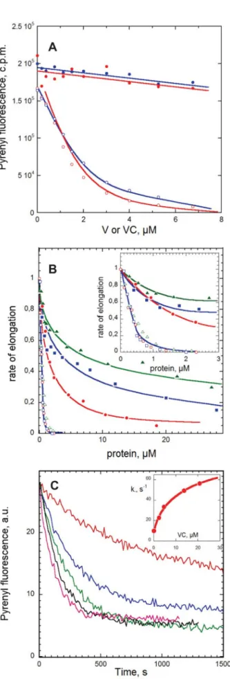
Preliminary assays showed that like profilin both V and VC inhibited spontaneous actin assembly even at substoichiomet-ric amounts, conditions favoring formation of VA2complexes. Hence, in contrast with the tandem repeats of WH2 domains of JMY (17), the two adjacent WH2 domains of N-WASP bind two actins but do not nucleate actin assembly.
The effect of V and VC on F-actin assembled at steady state was then analyzed (Fig. 4A). Superimposable curves were obtained with V and VC. The slopes of the linear declines in the amount of F-actin as a function of the total amount of V or VC, indicating sequestration of G-actin, were much steeper when barbed ends were capped, whereas insignificant depolymeriza-tion was observed when barbed ends were free. In the simple case of a canonical sequestering agent S making a nonpolymer-izable 1:1 complex with G-actin, like T4, the relative values of these slopes are determined by the values of the critical concen-trations for actin assembly in filaments (AC), with free and
capped barbed ends, respectively, and the value of the equilib-rium dissociation constant KSfor the complex SA of actin with the sequestering agent, as described by Equation 11,
[SA]⫽关S0兴䡠AC/(AC⫹ KS) (Eq. 11)
The situation is more complex with V and VC because both
FIGURE 4. Functional properties of V, VC, and VCA in actin assembly. A, effects of V and VC on the amount of F-actin assembled at steady state. Actin (10% pyrenyl-labeled, 3.8M) was polymerized with (open symbols) or with-out (closed symbols) 13 nMgelsolin and incubated overnight in the presence
of V (blue curves) or VC (red curves) at the indicated concentrations. Note that when barbed ends are capped by gelsolin, F-actin is totally depolymerized by addition of less than 1 molar eq V or VC, consistent with sequestration of actin in high affinity VA2complexes. In contrast, V and VC fail to significantly
depo-lymerize F-actin when barbed ends are free. B, effects of V, VC, and VCA on barbed and pointed end growth rates. Initial rates of barbed end (closed sym-bols) and pointed end (open symsym-bols) growth was measured in the presence of 2.5MMgATP-G-actin and the indicated amounts of V (blue curves), VC (red curves), and VCA (green curves). Normalized values of the growth rates are shown. Inset, expanded graph in the 0 –3Mpeptide range. C, increase in barbed end depolymerization rate by VC. Shown are time courses of depo-lymerization of pre-assembled F-actin (2.5M) induced by 40-fold dilution in polymerization buffer containing VC at the following concentrations: 0 (red), 2.3M(blue), 4.6M(green), 13.8M(black), and 20.8M(magenta). Inset, plot of the initial depolymerization rate constant versus VC concentration.
by guest on October 1, 2017
http://www.jbc.org/
make VA and VA2complexes. The possible sequestration of actin in these complexes therefore was expressed as shown in Equations 12–14.
[Vtot]⫽关V兴䡠(1 ⫹ 关A兴/K1 ⫹ 关A兴2/K1K2) (Eq. 12)
[Aseq]⫽关VA兴 ⫹ 2关VA2兴 (Eq. 13)
[Vtot]/关Aseq兴 ⫽ (K1K2 ⫹ 关A兴K2⫹ [A]2)/(关A兴K2⫹ 2[A]2)
(Eq. 14) The concentration of sequestered actin at a given total concen-tration of V or VC, [Aseq], was experimentally determined when
barbed ends were free or gelsolin-capped and was incorporated in Equation 14, using values of [A] equal to the values of the critical concentrations at either the barbed end (0.1M) or the
pointed end (0.6M). This procedure generated a unique set of
values for K1(3.4M) and K2(0.18M) that could satisfy the
data at both barbed and pointed ends in Fig. 4A in terms of sequestration of actin in VA and VA2complexes. These values
of K1and K2are significantly higher than and incompatible with
those derived from titration curves in G- or F-buffers (0.02 and 0.3M, respectively). Thus, a pure sequestration activity of V
and VC is inconsistent with the data. One should note that VA and VA2 are not equally represented at steady state when barbed ends are free or capped, because the ratio [VA2]/[VA]⫽
[A]/K2depends on the critical concentration [A]. Using values
of 0.1 and 0.6Mfor [A] at steady state when barbed ends are
free and capped, respectively, one finds that VA2is 6-fold more
abundant than VA when barbed ends are capped than when barbed ends are free. In summary, steady state measurements strongly suggest that like profilin, V and VC do not behave as
bona fidesequesterers when barbed ends are free.
To understand how V and VC affect barbed end and pointed end assembly dynamics, the effect of V and VC on the rate of barbed end or pointed end growth was examined (Fig. 4B). Both V and VC inhibited pointed end growth very efficiently. Total inhibition was reached at a substoichiometric molar ratio of V or VC with respect to G-actin, consistent with the sequestra-tion of G-actin in a tight predominant VA2complex. The
pre-dominance of VA2in this concentration range was confirmed by calculating the distribution of VA and VA2at different total
concentrations of V or VC, using values of binding constants derived from the titration assays.
The effects of V and VC on barbed end growth rate were more complex. The rate of barbed end growth at 2.5 M
MgATP-G-actin decreased in a biphasic fashion with increas-ing concentrations of V or VC. A steep initial 40% decrease in rate was observed in the range of low concentrations of V or VC (0 to 3M) in which total inhibition of growth at pointed ends
was recorded (Fig. 4B, inset). The partial inhibition of barbed end growth was followed, at a higher concentration of V or VC (5–50M), by a much less steep decrease in rate. These data
indicate that VA and VA2complexes can productively associate with barbed ends like profilin-actin; however, our data do not allow accurate determination of the kinetic parameters for barbed end assembly from VA and VA2specifically. The slower decline in rate at a high concentration of V or VC, although the
calculated concentration of VA complex remains constant, suggests that V and VC may also cap barbed ends with very low affinity to prevent their growth fed by VA. The same behavior at barbed ends and the same inhibition at very high concentra-tions were observed with VCA (green curve in Fig. 4B). The experimental data were well accounted for by a model (Equa-tion 10 under “Experimental Procedures”) in which VA and VA2support barbed end growth with on rate constants (k⫹B) of
5.5, 4, and 6.5M⫺1䡠s⫺1for V, VC, and VCA, respectively, and
V, VC, and VCA cap barbed ends with low affinity (KF⫽ 15, 5,
and 20M, respectively).
The evidence for barbed end growth from the complexes of actin with V, VC, and VCA implies that these WH2 domains interact with barbed ends, at least transiently. To further address this point, the effect of V, VC, and VCA on dilution-induced depolymerization of actin filaments was assayed (Fig. 4C). For V, VC, and VCA, all three increased the rate of barbed end dissociation up to a maximum of 7-fold, in a concentration-dependent behavior consistent with V and VC binding to barbed ends with a Kdof 5⫾ 2M. In contrast, neither V nor VC
affected the rate of pointed end depolymerization (data not shown). Hence, the increase in barbed end depolymerization rate is not due to severing of the filaments, but it results from specific binding of the WH2 domains of V or VC to barbed ends, causing faster release of the V䡠actin complex than actin. A similar effect has been reported for profilin (54, 55) and for the WH2 domains of Cordon-Bleu, which exhibit functional simi-larity to profilin (47).
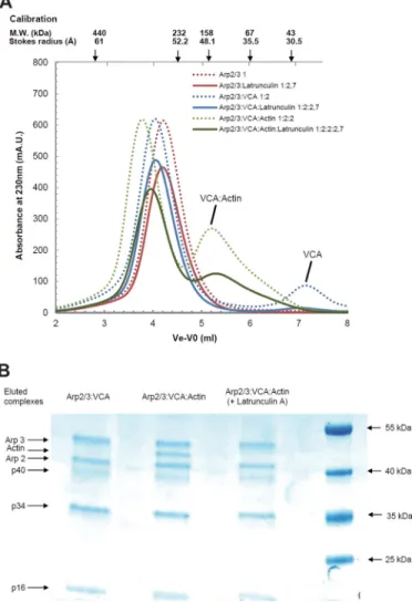
Analysis of the Complexes Formed between VCA, Actin, and Arp2/3 Complex—Branching of filaments by VCA occurs by association of a branching complex of VCA with actin and Arp2/3 complex with a filament. The Arp2/3/VCA/actin stoi-chiometry of the branching complex (in which the shortened notation Arp2/3 refers to Arp2/3 complex) is not well known and has been debated recently (25–27). In small angle x-ray scattering solution studies of Arp2/3 in complex with a covalent (N-WASP)VCA䡠actin 1:1 complex, the V region binds actin; the C region binds the barbed face of Arp2 (25); and the A region binds the small subunit p21 of the Arp2/3 complex (56). How-ever, recent reports proposed that Arp2/3 complex was active when bound to two WASP VCA domains. One study (26) showed binding of two Alexa 488-labeled VCAs to Arp2/3 complex, and the other study (27) demonstrated that the Arp2/3 complex made a 1:2 complex with two molecules of the isolated CA domain. Our data show that the WH2 domains of N-WASP interact with G-actin with an affinity that appears weakened by the acidic (A) C-terminal region, in the isolated VCA state. Upon interaction with Arp2/3 complex, the con-tacts made by the acidic region with subunits of the Arp2/3 complex may neutralize this region and restore enhanced strength of binding of the two WH2 domains of VCA in an unknown fashion. In summary, if VCA can bind two actin mol-ecules and if two VCA molmol-ecules bind to Arp2/3, the Arp2/ 3䡠VCA䡠actin may potentially display up to 1:2:4 stoichiometry. The noncovalent complexes of Arp2/3 complex with unla-beled VCA and actin were characterized by size-exclusion chromatography (Fig. 5). The Stokes radius of the Arp2/3 com-plex increased from 51 Å to a maximum of 52 Å upon addition
by guest on October 1, 2017
http://www.jbc.org/
of VCA, at various VCA䡠Arp2/3 complex molar ratios in the load. A peak corresponding to free VCA appeared as soon as the VCA䡠Arp2/3 complex molar ratio in the load was higher than 1,
indicating that a single 1:1 VCA䡠Arp2/3 complex species was formed (Fig. 5A). Further addition of G-actin in increasing amounts in the load led to a single faster migrating species of FIGURE 5. Gel filtration analysis of the complexes of Arp2/3 complex with VCA and actin. A, samples containing Arp2/3 complex alone (3M) or supple-mented with VCA at indicated molar ratios were loaded and eluted on a Superdex 200 10⫻ 300 column at physiological ionic strength. B, elution patterns of samples containing Arp2/3 complex alone or supplemented with VCA and actin at indicated ratios. Conditions as in A. C, SDS-PAGE analysis of the eluted VCA䡠actin䡠Arp2/3 ternary complex and standards consisting of pre-mixed actin and Arp2/3 in a 1:1 molar ratio and at the indicated concentrations, from which the molar ratio of actin Arp2 and p34 in the eluted complex was derived. Standards and samples were electrophoresed on the same gel to establish the molar composition of VCA䡠actin䡠Arp2/3 complex rigorously. D, hydrodynamic radius and apparent molecular weight of the eluted complexes derived from the size-exclusion chromatography calibration with known protein standards. Stokes radius values are within 10% standard deviation.
by guest on October 1, 2017
http://www.jbc.org/
Stokes radius at 54 Å, consistent with a 1:1:1 VCA䡠actin䡠Arp2/3 complex. The theoretical molecular mass of 266 kDa, assuming a globular shape, is reasonably consistent with the calculated molecular mass of 275 kDa for a 1:1:1 complex. The molar ratio was confirmed both by SDS-PAGE analysis of the content in actin and the Arp2/3 complex in the fractions of the eluted complex, and by the appearance of an actin peak at actin䡠Arp2/3 complex molar ratios higher than 1 in the loaded samples (Fig. 5, B–D). No evidence was observed for a second binding site of VCA to Arp2/3 complex in these experiments nor for binding of two actins to the V moiety of VCA when the CA region was engaged in contacts with Arp2/3 complex. When the chromatography of samples of Arp2/3, actin and VCA was performed in the presence of latrunculin A, the elu-tion profiles of Arp2/3 complex and of VCA䡠Arp2/3 complex were unchanged, but a complex could no longer be detected between actin and VCA䡠Arp2/3 complex (Fig. 6). Because latrunculin A does not prevent binding of VCA to actin, this
result indicates that the pointed face of VCA-bound actin in the ternary complex interacts with components of Arp2/3 complex in a manner incompatible with latrunculin A binding.
DISCUSSION
The analysis of the structural/functional interaction of the two adjacent WH2 domains of N-WASP in the V, VC, and VCA domains and the characterization of the ternary VCA䡠 actin䡠Arp2/3 complex provides new insight into the multifunc-tionality of WH2 repeats and its use in the filament branching reaction.
Each of the two WH2 domains of N-WASP binds one G-ac-tin molecule, with an affinity that is lowered by the presence of the acidic C-terminal extension (VCA). Both VA and VA2 com-plexes are formed from V, VC, and VCA, with affinities in the 107 M⫺1range, and are observed in solution in proportions depending on the concentrations of actin and V or VC or VCA. The fact that the affinity of actin is lowered by the acidic region A in VCA may have an interesting physiological implication, a weak affinity facilitating the dissociation of actin from VCA in the branching process.
In the crystal structure of VC䡠actin 1:1 complex, two VC-actin molecules form the asymmetric unit. The D-loop adopts an␣-helix conformation and forms the major part of the inter-face between the two actins. This contact differs from the actin-actin contacts in the filament. The apolar buried surface, calcu-lated according to Bahadur and Chakrabarti (57) was 989 Å2,
and the fraction of buried atoms was 0.366. Despite the low free energy of solvation, estimated to⫺4.4 kcal/mol, and of the low resolution and high value of the B factor of residues in the D loop, these figures are suggestive of possible functional interac-tions (48, 57). Whether similar contacts might occur in nature, for instance in the structural organization of the branch junc-tion, in which VCA transiently binds G-actin, the Arp2 and Arp3 subunits of the Arp2/3 complex and the mother filament, is an issue raised by these data.
The present results reinforce the view that the protein envi-ronment of the WH2 domains plays a determinant role in their multifunctionality. The two WH2 domains of N-WASP are not nucleators of actin filaments, even when they are present in polymerizing actin solutions at concentrations such that VA2is
the dominant complex. Therefore, two adjacent actin-bound WH2 domains do not necessarily generate a nucleation activity. This conclusion is in full agreement with the observation that the two adjacent WH2 domains of Cordon-Bleu do not nucle-ate actin in the absence of the N-terminally adjacent lysine-rich K region (47). Conversely, filament nucleation by WH2 TABLE 2
Characteristic parameters of the V, VC and VCA fragments of N-WASP for G-actin binding and filament barbed end assembly
V VC VCA Profilina K1,M 0.02 0.02 0.1 a K2,M 0.3 0.3 K⫹B ,M⫺1䡠s⫺1 5.5 4 6.5 7a KF 15 5 20 20 b,c koff(ADP-actin), s⫺1 70 70 70 b,c aSee Ref. 70. bSee Ref. 54. cSee Ref. 71.
FIGURE 6. Gel filtration analysis of the complexes of Arp2/3 complex with
VCA and actin with latrunculin A. A, samples containing Arp2/3 complex
alone or supplemented with VCA and actin at the indicated ratios and with latrunculin A were loaded and eluted on a Superdex 200 10⫻ 300 column at physiological ionic strength. The dotted lines refer to elution patterns observed without latrunculin A (reproduced from Fig. 5). B, SDS-PAGE analy-sis of the VCA䡠Arp2/3 complex and VCA䡠actin䡠Arp2/3 ternary complex eluted with latrunculin A. (Note absence of actin in eluted complex.) For the electro-phoretic positions of actin, some subunits of the Arp2/3 complex and protein molecular weight standards are indicated.
by guest on October 1, 2017
http://www.jbc.org/
domains does not require two adjacent WH2 domains, because the first WH2 domain of Cordon-Bleu, flanked by the K region nucleates as well as two consecutive domains. Similarly, the C-terminal dimerization domain of the pathogen protein VopL weakly nucleates actin, and nucleation is enhanced by addi-tional WH2 domains, which by themselves do not nucleate (58, 59). A general mechanism of “facilitated spontaneous nucle-ation” of actin filaments by WH2 domains, differing from the initial template mechanism (60 – 62), has been proposed, in which binding of a WH2 domain to an actin monomer enhances its electrostatic interaction with another actin mon-omer to form a pre-nucleus actin dimer (47, 63). Thus, WH2 domains that harbor appropriate sequence elements to decrease the electronegativity of G-actin lower the free energy of nucleation associated with elementary steps of the regular actin nucleation pathway. The WH2 domains of N-WASP do not harbor this peculiar property; therefore, in the absence of the Arp2/3 complex they do not nucleate actin either in isolated state or in the context of the activated full-length N-WASP protein in which the VCA C-terminal domain is exposed. Bind-ing of the Arp2/3 complex is not sufficient to confer a nucleat-ing activity to VCA, because the association with a mother fil-ament is required for nucleation of a daughter filfil-ament (64, 65). However, binding of putative ligands to N-WASP or to WAVE might confer an actin-nucleating activity to the WH2 domain by modifying its electrostatic environment. The nucleating activity then might synergize with the Arp2/3 complex-depen-dent branching activity and enhance the potency of the WASP protein to generate new barbed ends.
In contrast with other WH2 tandem repeats like Spire or Cordon-Bleu (46, 47), V and VC do not sever filaments. The absence of severing activity correlates with the absence of nucleating activity.
V, VC, and VCA share two properties with profilin and a few other WH2/-thymosin domains. First, their complexes with MgATP-G-actin participate in barbed end growth (Table 2). Second, in dilution-induced depolymerization assays, in which the core ADP-F-actin subunits dissociate endwise, their interaction with the barbed ends enhances by 7-fold the dissociation of MgADP-F-actin subunits. These data bring growing evidence for a variety of functional inter-actions of WH2 domains with barbed ends, including the capture of barbed ends by the WH2 domain of N-WASP (66), the capping of barbed ends by Spire WH2 domains (46, 67), and the tracking of barbed ends by the WH2 domain of VASP (68, 69). Table 3 summarizes the diverse multifunctionality patterns reported on various WH2 repeats. Domain swap-ping and engineering of chimeric or mutated WH2 repeats
will allow us to design customized WH2 domains and to modulate at will the overall biological function of the pro-teins in which they are inserted.
Filament branching using the Arp2/3 complex is a property shared by all WASP family proteins despite their variable num-ber of WH2 domains, indicating that one actin bound to one WH2 domain interacts with the Arp2/3 complex in a function-ally conserved fashion in the branching process. In the (N-WASP)VCA䡠Arp2/3䡠actin complex, G-actin is probably bound to the second WH2 domain of N-WASP, in a position homolog to WASP WH2. A supplementary WH2 domain of N-WASP was reported to increase the efficiency of branching (15). Assuming that the WH2 domain adjacent to CA binds actin in VCA䡠Arp2/3䡠actin complex, binding of the first (N-ter-minal) WH2 domain of (N-WASP)VCA to another actin mol-ecule in the branching process might account for the enhanced branching efficiency. Such a barbed end tracking function, comforted by other data (66), might participate in the protru-sive activity of dendritic actin arrays.
Only a 1:1 complex of VCA with the Arp2/3 complex is iso-lated using unmodified proteins in a wide range of molar ratios. This result contrasts with the reported association of two Alexa 488-labeled VCA molecules to the Arp2/3 complex (26) and with the binding of fission yeast CA to two sites on the Arp2/3 complex, albeit with very different affinities (27). The present data do not favor the view that branching of filaments requires the binding of two WASP molecules to the Arp2/3 complex. The difference between the various results may be due to the labeling of VCA or to the difference in reactivity of CA versus VCA and of yeast versus bovine proteins. However, the present evidence for single noncovalent 1:1 VCA䡠Arp2/3 complex and 1:1:1 VCA䡠actin䡠Arp2/3 complexes is in agreement with the small angle x-ray scattering solution structure of VCA䡠actin䡠 Arp2/3 in which VCA and actin were covalently linked (25). Although two actin molecules bind to the VCA domain of N-WASP irrespective of the presence of latrunculin A, actin does not bind to the 1:1 VCA䡠Arp2/3 complex in the presence of latrunculin A, suggesting that new latrunculin-sensitive con-tacts are established between actin and Arp2 in the ternary branching complex, also consistent with solution studies (25). The fact that one single branching enzyme (VCA) is bound to its substrates Arp2/3 complex and actin simplifies the possible molecular mechanisms for filament branching and the possible binding modes of the VCA䡠actin䡠Arp2/3 branching complex with a filament. What minimum size F-actin unit can function-ally interact with the VCA䡠actin䡠Arp2/3 complex is the next challenging issue.
TABLE 3
Compared multifunctionality of various WH2 domains in single module or tandem repeats in actin assembly
Symbols used are as follows:⫹, ⫺, and ? indicate that the property has been observed, not observed, or not tested, respectively. Protein (WH2 no.)
G-actin (ATP, ADP) sequestration Barbed end elongation Filament nucleation Filament severing Barbed end capping Barbed end tracking N-WASP (2) ⫺ ⫹ ⫺ ⫺ ⫺ ⫹ ActA (1) ⫺ ⫹ ⫺ ⫺ ⫺ ⫺ VASP (1) ⫺ ⫹ ⫹ ⫺ ⫺ ⫹ INF2 (1⫹ FH2) ⫺ ? ⫹ ⫹ ? ?
Spire (4) ⫹ (ATP, ADP) ⫺ ⫹ ⫹ ⫹ ⫺
Cordon-Bleu (3) ⫹ (ADP) ⫹ ⫹ ⫹ ⫺ ⫺
by guest on October 1, 2017
http://www.jbc.org/
REFERENCES
1. Rottner, K., Ha¨nisch, J., and Campellone, K. G. (2010) WASH, WHAMM, and JMY. Regulation of Arp2/3 complex and beyond. Trends Cell Biol. 20, 650 – 661
2. Veltman, D. M., and Insall, R. H. (2010) WASP family proteins. Their evolution and its physiological implications. Mol. Biol. Cell 21, 2880 –2893 3. Kollmar, M., Lbik, D., and Enge, S. (2012) Evolution of the eukaryotic ARP2/3 activators of the WASP family. WASP, WAVE, WASH, and WHAMM, and the proposed new family members WAWH and WAML. BMC Res. Notes 5,88
4. Svitkina, T. M., and Borisy, G. G. (1999) Arp2/3 complex and actin depo-lymerizing factor/cofilin in dendritic organization and treadmilling of ac-tin filament array in lamellipodia. J. Cell Biol. 145, 1009 –1026
5. Takenawa, T., and Suetsugu, S. (2007) The WASP-WAVE protein net-work. Connecting the membrane to the cytoskeleton. Nat. Rev. Mol. Cell Biol. 8,37– 48
6. Tsuboi, S., and Meerloo, J. (2007) Wiskott-Aldrich syndrome protein is a key regulator of the phagocytic cup formation in macrophages. J. Biol. Chem. 282,34194 –34203
7. Huang, Y., and Burkhardt, J. K. (2007) T-cell receptor-dependent actin regulatory mechanisms. J. Cell Sci. 120, 723–730
8. Wegner, A. M., Nebhan, C. A., Hu, L., Majumdar, D., Meier, K. M., Weaver, A. M., and Webb, D. J. (2008) N-WASP and the Arp2/3 complex are critical regulators of actin in the development of dendritic spines and synapses. J. Biol. Chem. 283, 15912–15920
9. Campellone, K. G., Webb, N. J., Znameroski, E. A., and Welch, M. D. (2008) WHAMM is an Arp2/3 complex activator that binds microtubules and functions in ER to Golgi transport. Cell 134, 148 –161
10. Derivery, E., Sousa, C., Gautier, J. J., Lombard, B., Loew, D., and Gautreau, A. (2009) The Arp2/3 activator WASH controls the fission of endosomes through a large multiprotein complex. Dev. Cell. 17, 712–723
11. Jia, D., Gomez, T. S., Metlagel, Z., Umetani, J., Otwinowski, Z., Rosen, M. K., and Billadeau, D. D. (2010) WASH and WAVE actin regulators of the Wiskott-Aldrich syndrome protein (WASP) family are controlled by analogous structurally related complexes. Proc. Natl. Acad. Sci. U.S.A.
107,10442–10447
12. Suraneni, P., Rubinstein, B., Unruh, J. R., Durnin, M., Hanein, D., and Li, R. (2012) The Arp2/3 complex is required for lamellipodia extension and directional fibroblast cell migration. J. Cell Biol. 197, 239 –251
13. Delatour, V., Helfer, E., Didry, D., Leˆ, K. H., Gaucher, J. F., Carlier, M. F., and Romet-Lemonne, G. (2008) Arp2/3 controls the motile behavior of N-WASP-functionalized GUVs and modulates N-WASP surface distri-bution by mediating transient links with actin filaments. Biophys. J. 94, 4890 – 4905
14. Weisswange, I., Newsome, T. P., Schleich, S., and Way, M. (2009) The rate of N-WASP exchange limits the extent of ARP2/3-complex-dependent actin-based motility. Nature 458, 87–91
15. Yamaguchi, H., Miki, H., Suetsugu, S., Ma, L., Kirschner, M. W., and Tak-enawa, T. (2000) Two tandem verprolin homology domains are necessary for a strong activation of Arp2/3 complex-induced actin polymerization and induction of microspike formation by N-WASP. Proc. Natl. Acad. Sci. U.S.A. 91,12631–12636
16. Zalevsky, J., Lempert, L., Kranitz, H., and Mullins, R. D. (2001) Different WASP family proteins stimulate different Arp2/3 complex-dependent ac-tin-nucleating activities. Curr. Biol. 11, 1903–1913
17. Zuchero, J. B., Coutts, A. S., Quinlan, M. E., Thangue, N. B., and Mullins, R. D. (2009) p53-cofactor JMY is a multifunctional actin nucleation factor. Nat Cell Biol. 11,451– 459
18. Higgs, H. N., and Pollard, T. D. (2001) Regulation of actin filament net-work formation through ARP2/3 complex. Activation by a diverse array of proteins. Annu. Rev. Biochem. 70, 649 – 676
19. Wen, K. K., and Rubenstein, P. A. (2005) Acceleration of yeast actin po-lymerization by yeast Arp2/3 complex does not require and Arp2/3 com-plex activating protein. J. Biol. Chem. 280, 24168 –24174
20. Wiesner, S., Helfer, E., Didry, D., Ducouret, G., Lafuma, F., Carlier, M. F., and Pantaloni, D. (2003) A biomimetic motility assay provides insight into the mechanism of actin-based motility. J. Cell Biol. 160, 387–398
21. Iwasa, J. H., and Mullins, R. D. (2007) Spatial and temporal relationships between actin filament nucleation, capping, and disassembly. Curr. Biol.
17,395– 406
22. Lai, F. P., Szczodrak, M., Block, J., Faix, J., Breitsprecher, D., Mannherz, H. G., Stradal, T. E., Dunn, G. A., Small, J. V., and Rottner, K. (2008) Arp2/3 complex interactions and actin network turnover in lamellipodia. EMBO J. 27,982–992
23. Rouiller, I., Xu, X. P., Amann, K. J., Egile, C., Nickell, S., Nicastro, D., Li, R., Pollard, T. D., Volkmann, N., and Hanein, D. (2008) The structural basis of actin filament branching by the Arp2/3 complex. J. Cell Biol. 180, 887– 895
24. Beltzner, C. C., and Pollard, T. D. (2008) Pathway of actin filament branch formation by Arp2/3 complex. J. Biol. Chem. 283, 7135–7144
25. Boczkowska, M., Rebowski, G., Petoukhov, M. V., Hayes, D. B., Svergun, D. I., and Dominguez, R. (2008) X-ray scattering study of activated Arp2/3 complex with bound actin-WCA. Structure 16, 695–704
26. Padrick, S. B., Doolittle, L. K., Brautigam, C. A., King, D. S., and Rosen, M. K. (2011) Arp2/3 complex is bound and activated by two WASP pro-teins. Proc. Natl. Acad. Sci. U.S.A. 108, E472–E479
27. Ti, S. C., Jurgenson, C. T., Nolen, B. J., and Pollard, T. D. (2011) Structural and biochemical characterization of two binding sites for nucleation-pro-moting factor WASp-VCA on Arp2/3 complex. Proc. Natl. Acad. Sci. U.S.A. 108,E463–E471
28. Xu, X. P., Rouiller, I., Slaughter, B. D., Egile, C., Kim, E., Unruh, J. R., Fan, X., Pollard, T. D., Li, R., Hanein, D., and Volkmann, N. (2012) Three-dimensional reconstructions of Arp2/3 complex with bound nucleation promoting factors. EMBO J. 31, 236 –247
29. Dominguez, R. (2009) Actin filament nucleation and elongation factors. Structure-function relationships. Crit. Rev. Biochem. Mol. Biol. 44, 351–366
30. Hertzog, M., van Heijenoort, C., Didry, D., Gaudier, M., Coutant, J., Gi-gant, B., Didelot, G., Pre´at, T., Knossow, M., Guittet, E., and Carlier, M. F. (2004) The-thymosin/WH2 domain; structural basis for the switch from inhibition to promotion of actin assembly. Cell 117, 611– 623
31. Renault, L., Bugyi, B., and Carlier, M. F. (2008) Spire and Cordon-bleu. Multifunctional regulators of actin dynamics. Trends Cell Biol. 18, 494 –504
32. Carlier, M. F., Husson, C., Renault, L., and Didry, D. (2011) Control of actin assembly by the WH2 domains and their multifunctional tandem repeats in Spire and Cordon-Bleu. Int. Rev. Cell Mol. Biol. 290, 55– 85 33. Kelly, A. E., Kranitz, H., Do¨tsch, V., and Mullins, R. D. (2006) Actin
bind-ing to the central domain of WASP/Scar proteins plays a critical role in the activation of the Arp2/3 complex. J. Biol. Chem. 281, 10589 –10597 34. Casella, J. F., Barron-Casella, E. A., and Torres, M. A. (1995) Quantitation
of Cap Z in conventional actin preparations and methods for further pu-rification of actin. Cell Motil. Cytoskeleton 30, 164 –170
35. Egile, C., Loisel, T. P., Laurent, V., Li, R., Pantaloni, D., Sansonetti, P. J., and Carlier, M. F. (1999) Activation of the CDC42 effector N-WASP by the Shigella flexneri IcsA protein promotes actin nucleation by Arp2/3 com-plex and bacterial actin-based motility. J. Cell Biol. 146, 1319 –1332 36. Guzman, L. M., Belin, D., Carson, M. J., and Beckwith, J. (1995) Tight
regulation, modulation, and high level expression by vectors containing the arabinose PBAD promoter. J. Bacteriol. 177, 4121– 4130
37. Pace, C. N., Vajdos, F., Fee, L., Grimsley, G., and Gray, T. (1995) How to measure and predict the molar absorption coefficient of a protein. Protein Sci. 4,2411–2423
38. Leslie, A. G., and Powell, H. R. (2007) in Evolving Methods for Macromo-lecular Crystallography(Read, R. J., and Sussman, J. L., eds) Vol. 245, pp. 41– 45, Springer NATO Science Series, New York
39. Evans, P. (2006) Scaling and assessment of data quality. Acta Crystallogr. D Biol. Crystallogr. 62,72– 82
40. Padilla, J. E., and Yeates, T. O. (2003) A statistic for local intensity differ-ences. Robustness to anisotropy and pseudo-centering and utility for de-tecting twinning. Acta Crystallogr. D Biol. Crystallogr. 59, 1124 –1130 41. Navaza, J. (1994) AMoRe. An automated package for molecular
replace-ment. Acta Crystallogr. A 50, 157–163
42. Chereau, D., Kerff, F., Graceffa, P., Grabarek, Z., Langsetmo, K., and Dominguez, R. (2005) Actin-bound structures of Wiskott-Aldrich
by guest on October 1, 2017
http://www.jbc.org/