HAL Id: inserm-00990045
https://www.hal.inserm.fr/inserm-00990045
Submitted on 13 May 2014
HAL is a multi-disciplinary open access
archive for the deposit and dissemination of
sci-entific research documents, whether they are
pub-lished or not. The documents may come from
teaching and research institutions in France or
abroad, or from public or private research centers.
L’archive ouverte pluridisciplinaire HAL, est
destinée au dépôt et à la diffusion de documents
scientifiques de niveau recherche, publiés ou non,
émanant des établissements d’enseignement et de
recherche français ou étrangers, des laboratoires
publics ou privés.
Mycobacterial infection of breast prosthesis - a
conservative treatment: a case report
David Atallah, Nadine El Kassis, George Araj, Marwan Nasr, Roy Nasnas,
Nicolas Veziris, Dolla Sarkis
To cite this version:
David Atallah, Nadine El Kassis, George Araj, Marwan Nasr, Roy Nasnas, et al.. Mycobacterial
infection of breast prosthesis - a conservative treatment: a case report. BMC Infectious Diseases,
BioMed Central, 2014, 14 (1), pp.238. �inserm-00990045�
C A S E R E P O R T
Open Access
Mycobacterial infection of breast prosthesis – a
conservative treatment: a case report
David Atallah
1*, Nadine El Kassis
1, George Araj
2, Marwan Nasr
3, Roy Nasnas
4, Nicolas Veziris
5,6,7and Dolla Sarkis
8Abstract
Background: Bacterial infection is a well-known risk of breast implant surgery. It is typically caused by bacterial skin flora, specifically Staphylococcus aureus and the coagulase negative staphylococci. There have been infrequent reports of breast implant infection caused by the atypical mycobacteria, of which Mycobacterium canariasense not yet reported in the literature.
Case presentation: This report summarizes the case of a female patient who underwent mastectomy followed by bilateral breast augmentation and presented approximately three years later with clinical evidence of infected breast prosthesis by Mycobacterium canariasense. One year after thoroughly follow-up, appropriate antibiotherapy and the change of the infected prosthesis, the patient presented no signs of reinfection.
Conclusion: Our case demonstrates that Mycobacterium canariasense should be considered as a new potential cause of infected breast prosthesis.
Keywords: Antibiotherapy, Conservative treatment, Mastectomy, Mycobacterium canariasense, Prosthesis, Sparing
Background
Breast implant-associated bacterial infections occur in 2.0 to 2.5% of cosmetic cases and up to 20% of reconstructive cases [1]. Infections caused by mycobacteria are uncom-mon, but are being increasingly reported [2-5]. However, breast implant infections caused by Mycobacterium canar-iasenseare atypical and not yet reported in the literature.
We report here the case of a female patient who underwent mastectomy followed by bilateral breast aug-mentation and presented approximately three years later with clinical evidence of infected breast prosthesis by Mycobacterium canariasense. The complexities estab-lished in the diagnosis and need for a thorough and accur-ate microbiologic evaluation, as well as the management strategies are summarized in the case report and discus-sion that ensue.
Case presentation
A 44-year old healthy woman was operated on May 2006, of bilateral mastectomy for an invasive lobular car-cinoma and associated in situ lobular carcar-cinoma in the
left breast. On the left side, a skin sparing radical modi-fied mastectomy was performed, and on the right side a prophylactic nipple sparing simple mastectomy was done. Mastectomy was followed by bilateral breast aug-mentation with Mentor prosthesis (Mentor Worldwide LLC, California, USA) filled with 150 mL saline serum, the prosthesis was inserted behind the pectoral muscles.
Prophylactic antibiotherapy with amoxicillin and cla-vulanate 2 g/day was prescribed for 10 days. The patient received later on, chemotherapy and radiotherapy treat-ment. Two years later, she had her Mentor® prosthesis changed with a similar new one filled with 110 mL of gel and 215 cc of saline serum for unsatisfactory esthetic results. Valves were inserted on the lateral thoracic wall. Augmentin® (amoxicillin and clavulanate) was given intravenously 30 minutes before surgery and then was continued for 10 days.
As a follow-up, she was seeing her plastic surgeon on a regular basis and her prosthesis were progressively filled with saline serum through the valves. Her postop-erative course was uneventful until January 2009, when she presented with edema and redness of her right breast after valve ablation, without pus drainage nor fever. She was treated with empiric antibiotherapy (Augmentin® 2 g/ day and ciprofloxacin 500 mg twice daily) for 10 days and
* Correspondence:david.atallah@gmail.com
1Department of Gynecology and Obstetrics, Hôtel-Dieu de France University
Hospital, Saint Joseph University, Beirut, Lebanon
Full list of author information is available at the end of the article
© 2014 Atallah et al.; licensee BioMed Central Ltd. This is an Open Access article distributed under the terms of the Creative Commons Attribution License (http://creativecommons.org/licenses/by/2.0), which permits unrestricted use, distribution, and reproduction in any medium, provided the original work is properly credited. The Creative Commons Public Domain Dedication waiver (http://creativecommons.org/publicdomain/zero/1.0/) applies to the data made available in this article, unless otherwise stated.
received local care. Her serum white blood cell count was 6,000 (neutrophils of 81%). Consequently, since she had no improvement of her symptoms, she underwent re-moval of her right breast implant with capsulectomy and massive irrigation with bacitracin and 9 liters of saline serum, then new prosthesis re-implantation.
Serous fluid drained from her right breast was sent to culture, as well as the capsule and the whole prosthesis. Gram stain, aerobic and anaerobic cultures of the fluid, prosthesis, and breast tissue returned negative. Histo-pathological examination of the breast capsule revealed none specific inflammatory changes without identifying any germ. Fungal culture returned negative as well after 40 days of incubation. Furthermore, acid-fast stain of the fluid drained from the right breast, debridement tissue, and the prosthesis returned negative. Acid-fast culture of the fluid, and of the resected right breast tissue were also negative, while acid-fast culture of the ablated pros-thesis were positive, it revealed the presence of an acid-fast bacilli resembling to Mycobacterium tuberculosis. A swab culture of the periprosthetic space – the space be-tween the prosthesis and the breast wall – as well as a
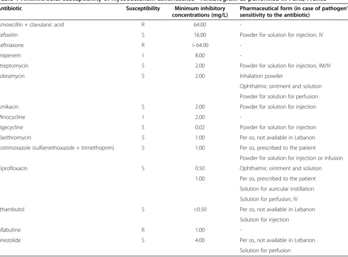
definitive mycobacterial culture of the unidentified patho-gen isolated from the prosthesis in the initial laboratory were sent to the National Reference Center on Mycobac-teria of Pitié-Salpêtrière hospital in Paris, France. The anti-microbial susceptibility testing of the pathogen were obtained from Rapid Growing Mycobacteria Plate Format (RAPMYCO) Sensititre plates in Mueller-Hinton medium incubated for 7 days. The identification was done by se-quencing the gene of the Heat Shock Protein (HSP) and revealed the presence of M. canariasense in the prosthesis and the peri-prosthetic space.
Subsequently, the patient received post-operatively empirical antibiotherapy including vancomycin 1 g every 12 hours, Tienam® (imipinem/cilastatin) 500 mg every 6 hours, and Tavanic® (levofloxacin) 500 mg/day, for 10 days. She was then discharged on oral tritherapy: cip-rofloxacin, Bactrim® (sulfamethoxazole and trimetho-prim) and clindamycin for 6 months, after the culture revealed the presence of an atypical Mycobacterium. The first two antibiotics were prescribed based on the anti-biogram’s results (Table 1). However, clindamycin was prescribed by the infectious disease specialist without
Table 1 Antimicrobial susceptibility of Mycobacterium canariasense - Antibiogram as performed in Paris, France
Antibiotic Susceptibility Minimum inhibitory concentrations (mg/L)
Pharmaceutical form (in case of pathogen’s sensitivity to the antibiotic)
Amoxicillin + clavulanic acid R 64.00
-Cefoxitin S 16.00 Powder for solution for injection, IV Ceftriaxone R > 64.00
-Imipenem I 8.00
-Streptomycin S 2.00 Powder for solution for injection, IM/IV Tobramycin S 2.00 Inhalation powder
Ophthalmic ointment and solution Powder for solution for perfusion Amikacin S 2.00 Powder for solution for injection
Minocycline I 2.00
-Tigecycline S 0.02 Powder for solution for injection Clarithromycin S 1.00 Per os, not available in Lebanon Cotrimoxazole (sulfamethoxazole + trimethoprim) S 1.00 Per os, prescribed to the patient
Powder for solution for injection or infusion Ciprofloxacin S 0.50 Ophthalmic ointment and solution
1.00 Per os, prescribed to the patient Solution for auricular instillation Solution for perfusion, IV Ethambutol S <0.50 Per os, not available in Lebanon
Solution for injection
Rifabutine R 1.00
-Linezolide S 4.00 Per os, not available in Lebanon Solution for perfusion
I: intermediate; IM: intramuscular; IV: intravenous; R: resistant; S: sensitive.
Atallah et al. BMC Infectious Diseases 2014, 14:238 Page 2 of 4 http://www.biomedcentral.com/1471-2334/14/238
performing clindamycin susceptibility testing. The patient was last seen in March 2014. She was symptom-free, with-out signs of infection and achieved a satisfactory psycho-logical, psychosocial and esthetic results.
Conclusions
Breast prostheses are being increasingly used, both for cosmetic and reconstructive purposes [3]. However, peri-prosthetic infection is perhaps the most feared and least understood complication of these procedures [3]. Al-though bacteria such as Staphylococcus aureus, coagulase-negative staphylococci and mycobacteria such as Myco-bacterium fortuitumcomplex are the most common cause of surgical site infections [3,4,6], this case highlights the emergence of atypical and unusual M. canariasense as pathogen associated with breast implant infections. Our review of the literature has produced no similar breast im-plant infection.
M. canariasense is a member of the rapidly growing, non-pigmented and atypical mycobacterium group. The characterization of this mycobacterium, in 2004, was based on a cluster of strains isolated from blood cultures via indwelling catheters from patients with probable noso-comial infection in the Canary Islands, Spain [7,8]. It was also isolated from respiratory sources but the pulmonary infection was deemed possible to doubtful in a patient pri-mary diagnosed with cancer [9].
Regarding management of breast implants’ infections, although prospective studies are lacking, published ports recommend that the mammary prosthesis be re-moved, with earlier removal favored to prevent implant extrusion and tissue contracture [10]. Besides, the best conservative treatment of the implant seems to be the as-sociation of implant change, irrigations and antibiotherapy [11]. These procedures were substantially performed in our case.
Although the cause of breast implant infections might not be truly understood, the challenge is how to treat them appropriately. Classic teaching holds that peripros-thetic infection mandates implant removal and delayed reinsertion after the infection clears [12]. However, the first report of immediate salvage of infected breast pros-theses after complete mastectomy for cancer was by Yii and Khoo in 2003 [13]. Similarly, a more recent study by Chun et al. revealed that eight (100%) patients had posi-tive outcomes following the immediate salvage [12]. In the latter case series, the author reported that the cul-tures of the periprosthetic fluid of the infected implants were positive for Staphylococcus aureus in three patients and Enterococcus faecalis in one patient. A fifth patient was positive for Staphylococcus epidermidis [12]. In our case, given that the patient insisted to conserve her es-thetic results, she was given the choice of delayed or im-mediate re-implantation. At that stage of time, the causal
agent of the infection was not yet identified as a rapidly growing mycobacteria. In close, she decided to undergo the one-stage re-implantation although she was aware of the risk of infection recurrence. Consequently, early and aggressive surgical intervention resulted in successful immediate implant salvage. It is worthy to note that de-layed re-implantation may have major psychological im-plications for the patient. To many women suffering with breast cancer, breast reconstruction is a critical compo-nent in the recovery process. Explantation halts the recon-struction process for upward of 6 months. In this context, one of the goals of the immediate re-implantation is to improve women’s psychological well-being.
On the other side, despite the continuous evolution of mycobacterial taxonomy which may represent a source of confusion for laboratories and clinicians, and which may not be identified by conventional procedures, we were able in our settings to identify the atypical M. canariasense.In fact, definitive diagnosis of mycobacter-ial breast implant infection requires demonstration of the organism from the periprosthetic site. Thus, speci-mens must be submitted for acid-fast stains fungal and mycobacterial culture, in addition to standard analyses, including Gram-stain, bacterial aerobic and anaerobic culture, and histopathology [3]. In this case, it was only after multiple specimens of the fluid drained from the right breast, debridement tissue, and the prosthesis were sterile on routine bacterial and fungal cultures, that the possibility of an atypical infection such as a mycobac-terial process was considered. Subsequent specimens submitted for mycobacterial culture from the ablated prosthesis identified the causative pathogen, M. canaria-sense. Therefore, we prescribed a prolonged course of anti-mycobacterial therapy that allowed for eradication of the infection.
Moreover, empiric antimicrobial therapy was started in our case pending isolation and susceptibility testing re-sults. Of note, in the study of Xiang et al., the results of the antimicrobial susceptibility of M. canariasense showed that this agent is sensitive to amikacin, imipe-nem, ciprofloxacin and trimethoprim-sulfamethoxazole. It is intermediately susceptible to cefoxitin. However, it is resistant to clarithromycin and minocycline [9]. With this knowledge, our patient was discharged on ciproflox-acin, clindamycin and sulfamethoxazole-trimethoprim, after the culture revealed the presence of M. canariasense. We stress the point that clindamycin was empirically prescribed by the infectious disease specialist although nontuberculous mycobacteria (NTM) generally are not susceptible to this agent. He aimed to cover a large spectrum of infectious agents due to the non-conventional diagnosis and management of our patient. In the end, the treatment provided anti-bacterial coverage for the re-implanted prosthesis for 6 months.
In conclusion, the theme of this article is to share a successful one-stage replacement of infected breast pros-thesis by atypical NTM after mastectomy reconstruction. The surgical and pharmacological management of the patient was successful given the very good esthetic re-sults in the patient. Our case also reminds us that a high index of suspicion for unusual pathogens, such as the atypical mycobacteria, is necessary when considering infections that do not improve despite seemingly ap-propriate management. Moreover, it demonstrates that M. canariasense should be considered as a new po-tential cause of infected breast prosthesis.
Consent
Written informed consent was obtained from the patient for publication of this case report. A copy of the written consent is available for review by the Editor-in-Chief of this journal.
Abbreviations
HSP:Heat shock protein; I: Intermediate; IM: Intramuscular; IV: Intravenous; M. canariasense: Mycobacterium canariasense; R: Resistant; RAPMYCO: Rapid growing mycobacteria plate format; S: Sensitive.
Competing interests
The authors declare that they have no competing interests. Authors’ contributions
DA, MN and RN participated in the surgical treatment and follow-up of the patient. DS and NV participated in the microbiological investigations. DA, NK and GA performed a literature review. DA wrote the article. NK and GA participated in the writing of article. All authors read and approved the final manuscript.
Author details
1Department of Gynecology and Obstetrics, Hôtel-Dieu de France University
Hospital, Saint Joseph University, Beirut, Lebanon.2Department of Pathology and Laboratory Medicine, American University of Beirut Medical Center, Beirut, Lebanon.3Department of Plastic Surgery, Hôtel-Dieu de France University Hospital, Saint Joseph University, Beirut, Lebanon.4Department of
Infectious Diseases, Hôtel-Dieu de France University Hospital, Saint Joseph University, Beirut, Lebanon.5Sorbonne University, Université Pierre et Marie
Curie-Paris 6, CR7, Centre d’Immunologie et des Maladies Infectieuses (CIMI), team E13 (Bacteriology), 75013 Paris, France.6INSERM, U1135, Centre
d’Immunologie et des Maladies Infectieuses (CIMI), team E13 (Bacteriology), 75013 Paris, France.7AP-HP, Hôpital Pitié-Salpêtrière, Centre National de
Référence des Mycobactéries et de la Résistance des Mycobactéries aux Antituberculeux, Bactériologie-Hygiène, 75013 Paris, France.8Faculty of
Pharmacy, Saint Joseph University, Beirut, Lebanon.
Received: 17 December 2013 Accepted: 25 April 2014 Published: 5 May 2014
References
1. Feldman EM, Kontoyiannis DP, Sharabi SE, Lee E, Kaufman Y, Heller L: Breast implant infections: is cefazolin enough? Plast Reconstr Surg 2010, 126:779–785.
2. Lizaso D, García M, Aguirre A, Esposto A: Breast implant infection by Mycobacterium fortuitum in a patient with systemic lupus erythematosus. Rev Chilena Infectol 2011, 28:474–478. Article in Spanish.
3. Vinh DC, Rendina A, Turner R, Embil JM: Breast implant infection with Mycobacterium fortuitum group: report of case and review. J Infect 2006, 52:e63–e67.
4. Betal D, Macneill FA: Chronic breast abscess due to Mycobacterium fortuitum: a case report. J Med Case Rep 2011, 5:188.
5. Boettcher AK, Bengtson BP, Farber ST, Ford RD: Breast infections with atypical mycobacteria following reduction mammaplasty. Aesthet Surg J 2010, 30:542–548.
6. Olsen MA, Lefta M, Dietz JR, Brandt KE, Aft R, Matthews R, Mayfield J, Fraser VJ: Risk factors for surgical site infection after major breast operation. J Am Coll Surg 2008, 207:326–335.
7. Jimenez MS, Campos-Herrero MI, Garcia D, Luquin M, Herrera L, Garcia MJ: Mycobacterium canariasense sp nov. Int J Syst Evo Microbiol 2004, 54:1729–1734.
8. Campos-Herrero MI, García D, Figuerola A, Suárez P, Campo C, Garcíam MJ: Bacteremia caused by the novel species Mycobacterium canariasense. Eur J Clin Microbiol Infect Dis 2006, 25:58–60.
9. Xiang YH, Dé I, Jacobson KL: Rapidly growing mycobacteria. Clinical and microbiologic studies of 115 cases. Am J Clin Pathol 2007, 128:612–621. 10. Haiavy J, Tobin H: Mycobacterium fortuitum infection in prosthetic breast
implants. Plast Reconstr Surg 2002, 109:2124–2128.
11. Laveaux C, Pauchot J, Loury J, Leroy J, Tropet Y: Acute periprosthetic infection after aesthetic breast augmentation. Report of three cases of implant “salvage”. Proposal of a standardized protocol of care. Ann Chir Plast Esthet 2009, 54:358–364. Article in French.
12. Chun JK, Schulman MR: The infected breast prosthesis after mastectomy reconstruction: successful salvage of nine implants in eight consecutive patients. Plast Reconstr Surg 2007, 120:581–589.
13. Yii NW, Khoo C: Salvage of infected expander prostheses in breast reconstruction. Plast Reconstr Surg 2003, 111:1087–1092.
doi:10.1186/1471-2334-14-238
Cite this article as: Atallah et al.: Mycobacterial infection of breast prosthesis – a conservative treatment: a case report. BMC Infectious Diseases 2014 14:238.
Submit your next manuscript to BioMed Central and take full advantage of:
• Convenient online submission
• Thorough peer review
• No space constraints or color figure charges
• Immediate publication on acceptance
• Inclusion in PubMed, CAS, Scopus and Google Scholar
• Research which is freely available for redistribution
Submit your manuscript at www.biomedcentral.com/submit
Atallah et al. BMC Infectious Diseases 2014, 14:238 Page 4 of 4 http://www.biomedcentral.com/1471-2334/14/238