HAL Id: hal-02735847
https://hal-univ-rennes1.archives-ouvertes.fr/hal-02735847
Submitted on 2 Jun 2020
HAL is a multi-disciplinary open access
archive for the deposit and dissemination of
sci-entific research documents, whether they are
pub-lished or not. The documents may come from
teaching and research institutions in France or
abroad, or from public or private research centers.
L’archive ouverte pluridisciplinaire HAL, est
destinée au dépôt et à la diffusion de documents
scientifiques de niveau recherche, publiés ou non,
émanant des établissements d’enseignement et de
recherche français ou étrangers, des laboratoires
publics ou privés.
Myeloperoxidase and related biomarkers are suggestive
footprints of endothelial microvascular inflammation in
HFpEF patients
Camilla Hage, Erik Michaelsson, Bengt Kull, Tasso Miliotis, Sara Svedlund,
Cecilia Linde, Erwan Donal, Jean-Claude Daubert, Li-Ming Gan, Lars H.
Lund
To cite this version:
Camilla Hage, Erik Michaelsson, Bengt Kull, Tasso Miliotis, Sara Svedlund, et al.. Myeloperoxidase
and related biomarkers are suggestive footprints of endothelial microvascular inflammation in HFpEF
patients. ESC Heart Failure, Wiley, 2020, 7 (4), pp.1534-1546. �10.1002/ehf2.12700�. �hal-02735847�
Myeloperoxidase and related biomarkers are
suggestive footprints of endothelial microvascular
in
flammation in HFpEF patients
Camilla Hage
1,2*, Erik Michaëlsson
3, Bengt Kull
3, Tasso Miliotis
3, Sara Svedlund
4, Cecilia Linde
2, Erwan Donal
5,
Jean-Claude Daubert
5, Li-Ming Gan
3,6,7and Lars H. Lund
1,21Heart and Vascular Theme, Heart Failure Section, Karolinska University Hospital, SE-171 76, Stockholm, Sweden;2Department of Medicine, Cardiology Unit, Karolinska
Institutet, Stockholm, Sweden;3Research and Early Development Cardiovascular Renal and Metabolism, BioPharmaceuticals R&D, AstraZeneca, Gothenburg, Sweden;
4Department of Clinical Physiology, Sahlgrenska University Hospital, Gothenburg, Sweden;5Département de Cardiologie and CIC-IT U804, Centre Hospitalier Universitaire de
Rennes, Rennes, France;6Department of Molecular and Clinical Medicine, Sahlgrenska Academy, University of Gothenburg, Gothenburg, Sweden;7Department of Cardiology, Sahlgrenska University Hospital, Gothenburg, Sweden
Abstract
Aims In heart failure (HF) with preserved ejection fraction (HFpEF), microvascular inflammation is proposed as an underlying mechanism. Myeloperoxidase (MPO) is associated with vascular dysfunction and prognosis in congestive HF.
Methods and results MPO, MPO-related biomarkers, and echocardiography were assessed in86 patients, 4–8 weeks after presentation with acute HF (EF≥ 45%), and in 46 healthy controls. Patients were followed up for median 579 days (Q1;Q3 276;1178) regarding the composite endpoint all-cause mortality or HF hospitalization. Patients were 73 years old, 51% were female, EF was64% (Q1;Q3 58;68), E/e′ was ratio 10.8 (8.3;14.0), and left atrial volume index (LAVI) was 43 mL/m2(38;52). Controls were60 (57;62) years old (vs. patients; P < 0.001), 24% were female (P = 0.005), and left ventricular EF was 63% (59;66; P = 0.790). MPO was increased in HFpEF compared with controls, 101 (81;132) vs. 86 (74;101 ng/mL, P = 0.015), as was uric acid369 (314;439) vs. 289 (252;328 μmol/L, P < 0.001), calprotectin, asymmetric dimethyl arginine (ADMA), and sym-metric dimethyl arginine (SDMA), while arginine was decreased. MPO correlated with uric acid (r =0.26; P = 0.016). In patients with E/e′ > 14, uric acid and SDMA were elevated (421 vs. 344 μM, P = 0.012; 0.54 vs. 0.47 μM, P = 0.039, respectively), and MPO was121 vs. 98 ng/mL (P = 0.090). The ratios of arginine/ADMA (112 vs. 162; P < 0.001) and ADMA/SDMA (1.36 vs. 1.17;
P =0.002) were decreased in HFpEF patients, suggesting reduced NO availability and increased enzymatic clearance of ADMA,
respectively. Uric acid independently predicted the endpoint [hazard ratio (HR)3.76 (95% CI 1.19–11.85; P = 0.024)] but not MPO [HR1.48 (95% CI 0.70–3.14; P = 0.304)] or the other biomarkers.
Conclusions In HFpEF, MPO-dependent oxidative stress reflected by uric acid and calprotectin is increased, and SDMA is as-sociated with diastolic dysfunction and uric acid with outcome. This suggests microvascular neutrophil involvement mirroring endothelial dysfunction, a central component of the HFpEF syndrome and a potential treatment target.
Keywords Myeloperoxidase; Microvascular inflammation; Endothelial dysfunction; Heart failure with preserved ejection fraction; Prognosis
Received:3 October 2019; Revised: 16 March 2020; Accepted: 18 March 2020
*Correspondence to: Camilla Hage, Heart and Vascular Theme, Heart Failure Section, Karolinska University Hospital, SE-171 76 Stockholm, Sweden. Tel: +46 8 517 792 82; Fax: +46 8 34 49 64. Email: camilla.hage@sll.se
Introduction
The diagnosis of heart failure (HF) is associated with either re-duced ejection fraction (HFrEF) or preserved ejection fraction (HFpEF). The latter is a heterogeneous group of patients
making up nearly 50% of the HF population1 and with a 65% mortality rate 5 years after hospitalization, making the prognosis as ominous as that of HFrEF.1 Despite improve-ments in understanding the underlying disease mechanisms in HFrEF, many of the mechanisms in HFpEF remain
unknown, and the syndrome has been referred to as the greatest unmet need in cardiovascular medicine.2
In HFpEF, non-cardiac co-morbidities such as diabetes, obesity, hypertension, and chronic kidney disease are common3and have the ability to induce systemic in flamma-tion proposed to cause microvascular endothelial dysfunc-tion, in the myocardium as well as in the periphery.4,5
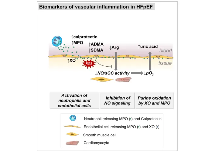
Myeloperoxidase (MPO) is a leukocyte-derived heme pro-tein associated with vascular dysfunction and a prognostic biomarker in cardiovascular disease, including congestive HF. MPO catalyses the formation of hypochlorous acid, among other reactive oxidants. In cardiovascular disease, this is suggested to occur in the subendothelial glycocalyx, where MPO is electrostatically trapped to proteoglycans causing vas-cular dysfunction mediated by direct and indirect oxidation of nitric oxide (NO).6This may result in hypoxia, ATP depletion, and increased purine catabolism and accumulation of uric acid.7
MPO may also affect vascular function by lowering avail-ability of the NO-substrate arginine through symmetric di-methyl arginine (SDMA) and asymmetric didi-methyl arginine (ADMA).8 In cardiovascular disease and HF, MPO and calprotectin, also released in response to oxidative stress, are both involved in amplifying the inflammatory response.9,10
Elevated levels of MPO have been reported in patients with chronic HFrEF compared with controls and correlate with disease severity as defined by New York Heart Associa-tion (NYHA) class, N-terminal brain natriuretic peptide (NT-proBNP), and echocardiographic measurements of systolic and diastolic function11–13and remodelling14after myocardial infarction. Further, increasing levels of MPO are associated with mortality in patients with HFrEF13and acute HF.15
The aim of the present study was to investigate MPO-related oxidative stress through biomarkers reflecting neutrophil involvement (calprotectin), tissue hypoxia (uric acid), and NO availability (arginine, ADMA, SDMA), by com-paring concentrations with those of healthy controls, associa-tions with markers of HF and mortality, and HF hospitalization in patients with HFpEF.
Methods
Patients and controls
The Karolinska Rennes (KaRen) was a prospective observa-tional multicentre study characterizing patients with HFpEF.16 The biochemistry programme recruited86 patients present-ing to hospital with acute signs and symptoms of HF accord-ing to the Framaccord-ingham criteria, NT-proBNP> 300 ng/L, and a left ventricular EF (LVEF)≥ 45% between 21 May 2007 and 29 December2011. Blood sampling and echocardiography were
performed at stable follow-up 4–8 weeks after enrolment. Patients were followed up until30 September 2012 for the composite outcome time to death from any cause or first hospitalization due to HF.
At the follow-up visit, blood samples were collected in EDTA tubes in a fasting condition in the morning and centri-fuged, and plasma was stored in aliquots in 70°C until analysis.
Healthy controls were consecutively recruited by newspa-per advertisement. Controls were free of cardiovascular symptoms, clinical signs of ongoing disease, ongoing medica-tion, and structural heart disease on echocardiography, and all had normal resting electrocardiograms.
Quanti
fication of biomarkers
Plasma MPO and calprotectin concentrations were deter-mined by ELISA (BioLegend, San Diego, CA; and BMA Biomed-icals, Augst, Switzerland, respectively). Uric acid, L-arginine, ADMA, and SDMA were quantified by isotope dilution liquid chromatography coupled to tandem mass spectrometry.17,18 NT-proBNP was quantified by Elecsys electrochemiluminescence “sandwich” immunoassay, proBNPII (Roche Diagnostics, Bromma, Sweden) with a lower detection limit of5 ng/L, and interassay coefficients of varia-tion of≤20%.
Estimated glomerularfiltration rate (eGFR) was calculated according to the CKD-EPI creatinine equation.19
The echocardiographic assessment was performed on a ViVid 7 echo-platform (GE VingMed, Horten, Norway) and analysed in core centre Hôpital Pontchaillou, Rennes, France. Examinations were interpreted once and measure-ments were performed three times and averaged by a sonog-rapher (E. D.) blinded to the clinical history of the patient.
Structural heart disease was assessed as left atrial (LA) vol-ume index (LAVI) calculated as LA volvol-ume in mL divided by body surface area in m2and categorized as>34 mL/m2. Left ventricular hypertrophy (LVH) was assessed as left ventricular mass index and dichotomized as left ventricular mass divided by body surface area (>95 g/m2in women and>116 g/m2in men) according to the European Society of Cardiology (ESC) guidelines.20 Diastolic dysfunction was assessed as E′ and E/e′ ratio and categorized as average E/e′ ratio > 14 accord-ing to the American Society of Echocardiography and the Eu-ropean Association of Cardiovascular Imaging guidelines.21
Statistics
Continuous variables are expressed as median and quartiles (Q1;Q3) and differences between groups determined by the Wilcoxon rank sum test. Categorical variables are expressed as numbers and percentages and analysed using Fisher’s
exact test. Correlations between plasma MPO and echocar-diographic measurements of cardiac function were deter-mined using Spearman’s correlation coefficient. Correlations between MPO and clinical and biochemical characteristics in-cluded in Table 1 were performed, as they may influence MPO levels and/or outcome.
Kaplan–Meier analyses and Cox proportional hazards models were used to analyse MPO and uric acid as a predic-tors of outcome and presented as hazard ratio (HR) and95% confidence interval (CI). The same variables as in the correla-tion analyses were used as covariates in multiple Cox regres-sion models. The final multiple model included NT-proBNP, age, and gender.
Owing to non-normal distribution, all biomarkers were log-transformed prior to analysis. All P-values were two-sided, and statistical significance was set at 0.05. All statistical analyses were performed using SAS software version9.4 (SAS Institute, Cary, NC, USA).
Ethics
The KaRen study was conducted according to the Interna-tional Conference on Harmonisation and Good Clinical Prac-tice guidelines, and the investigation conforms to the principles outlined in the Declaration of Helsinki. The bio-chemistry substudy was approved by the ethical review board at Karolinska Institutet and the controls by the Local Ethics Committee in Gothenburg. Written and oral in-formed consent was obtained from all patients and controls prior to enrolment.
Results
The characteristics of all 86 patients with HFpEF are pre-sented in Table1. The median age of patients was 73 years, and51% were female. LVEF was 64% (Q1;Q3 58;68), E/e′ ratio 10.8 (8.3;14.0), and LAVI 43 mL/m2 (38;52). A proportion of
23% had E/e′ > 15, 67% had E′ < 9, 89% had LAVI> 34 mL/m2, and58% had LVH. The 46 healthy controls were 24% women (vs. patients; P-value = 0.005) and 60 (57;62) years of age, and the body mass index (BMI) was 24.4 (22.9;26.0) kg/m2(both vs. patients; P-value< 0.001).
LVEF was63% (59;66; P-value = 0.790).
Biomarker concentration
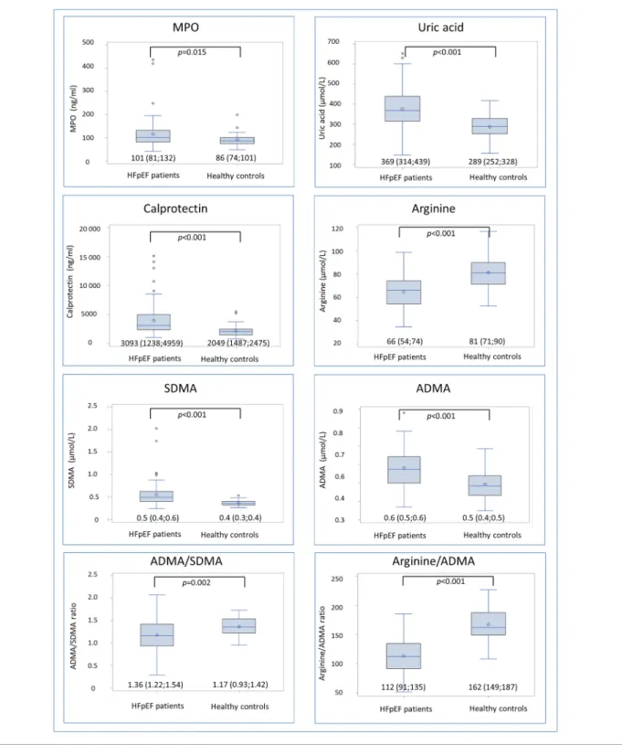
Concentrations of biomarkers in HFpEF patients and controls are depicted in Figure1.
Concentrations of all biomarkers differed in HFpEF patients compared with controls, showing higher plasma concentra-tions of MPO [101 (81;132) vs. 86 (74;101) ng/mL;
P-Table 1 Baseline characteristics in the 86 patients in Karolinska
Rennes
Variable HFpEF patients (n = 86)
Patient history Age (years) 73 (66;79) Gender (female) 44 (51%) Medical history Smoking 45 (52%) Hypertension 68 (79%) COPD 14 (16%)
Diabetes mellitus type 2 27 (31%)
Coronary heart disease 29 (34%)
Stroke 9 (10%)
Atrialfibrillation 49 (57%)
NYHA class I 19 (22%)
NYHA class II 47 (55%)
NYHA class III 20 (23%)
Measurements
Weight (kg) 83.5 (72;98)
BMI (kg/m2) 28.5 (25.0;32.9)
Systolic blood pressure (mmHg) 140 (90;210) Diastolic blood pressure (mmHg) 80 (70;85)
Heart rate (b.p.m.) 70 (60;80) Treatment ARB 28 (33%) ACE-inhibitor 42 (49%) Thiazide diuretics 14 (16%) Potassium-sparing diuretics 18 (21%) Loop diuretics 63 (73%)
Calcium channel blocker 27 (31%)
Beta-blocker 69 (80%) Anticoagulants 47 (55%) Antiplatelet 29 (34%) Statins 38 (44%) Nitroglycerine 12 (14%) Glucose-lowering medication 17 (20%) Pacemaker 20 (23%) ECHO parameters LVEF (%) 64 (58; 68) LAVI (mL/m2) 44 (38; 52) LA volume (mL) 86.5 (75; 104)
Left ventricular mass index (g/m2) 114 (95;142)
Male 125 (102;157) Female 109 (94;136) LVEDd (mm) 47 (43;53) E/A ratio 1.3 (0.9;2.1) E/e′ ratio 10.8 (8.3;14.0) E′ 8.0 (7.0;10.0) IVRT (diastole) 94 (77;113) Mitral VTI 23 (16;30)
E-wave deceleration time (ms) 203 (156;228) Biochemistry
NT-proBNP (ng/L) 1000 (469;2330)
Glucose fasting (mmol/L) 5.6 (5.1;7.5)
Creatinine (μmol/L) 84 (73;107)
eGFR (mL/min/1.73 m2) 70 (53;85)
Haemoglobin (g/L) 131 (122;142)
White blood cells count (x 109cells/L) 7.2 (5.6;8.5) ACE, angiotensin converting enzyme; ARB, angiotensin II receptor blocker; BMI, body mass index; COPD, chronic obstructive pulmo-nary disease; eGFR, estimated glomerular filtration rate; IVRT, isovolumic relaxation time; LAVI, left atrial volume index; LVEDd, left ventricular end-diastolic diameter; LVEF, left ventricular ejec-tion fracejec-tion; NT-proBNP, N-terminal brain natriuretic peptide; NYHA class, New York Heart Association class; VTI, velocity-time integral.
Continuous variables are presented as median and lower and up-per quartiles (Q1;Q3) and categorical variables as numbers (n) and percentages (%).
value = 0.015], uric acid, ADMA, SDMA, and calprotectin, while concentrations of arginine were lower. The ratios be-tween arginine and ADMA and bebe-tween ADMA and SDMA
were decreased in HFpEF patients, suggesting reduced NO availability and increased enzymatic clearance of ADMA, respectively.
Figure 1 Concentrations of biomarkers in heart failure with preserved ejection fraction (HFpEF) patients and healthy controls presented as boxplots
displaying interquartile range (IQR), median, mean (diamond), and outliers (circle). Whiskers represent maximum observation within 1.5 IQR above the 75th percentile.
Correlations between biomarkers and clinical and
echo characteristics
NYHA class correlated with uric acid (r =0.22; P-value = 0.045), SDMA (r =0.33; value = 0.002), calprotectin (r = 0.34; P-value = 0.001) and negatively with ADMA/SDMA ratio (r = 0.30; P-value = 0.005). Further, BMI correlated with MPO (r = 0.29; value = 0.007), uric acid (r = 0.39; P-value< 0.001), and calprotectin (r = 0.38; P-value < 0.001). Hypertension correlated with MPO (r =0.26; P-value = 0.015), uric acid (r =0.26; P-value = 0.016), and calprotectin (r = 0.32;
P-value =0.002).
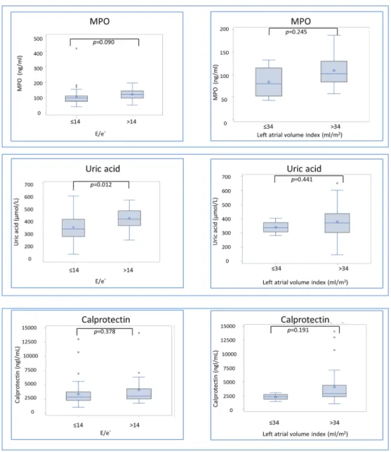
Figure2 depicts difference in biomarker concentration
ac-cording to presence of diastolic dysfunction, reflected as E/e′ ratio > 14, and structural heart disease, assessed as LAVI> 34 mL/m2. Patients tend to have higher diastolic dys-function MPO [121 (97;144) vs. 98 (77;112) ng/mL, P-value =0.090], and they had elevated concentrations of uric acid [421 (370;486) vs. 344 (284;420) μM, P-value = 0.012] and SDMA [0.54 (0.44;0.70) vs. 0.47 (0.38;0.52) μM, P-value = 0.039]. In patients with structural heart disease, arginine/ADMA ratio was lower [116 (103;133) vs. 155 (137;176), P-value = 0.014], whereas the other biomarkers were not significantly different (Figure2).
Figure 2 Biomarker concentrations according to presence of structural heart disease and diastolic dysfunction in heart failure with preserved ejection
fraction (HFpEF) patients. Presented as boxplots displaying interquartile range (IQR), median, mean (diamond), and outliers (circle). Whiskers represent maximum observation within 1.5 IQR above the 75th percentile.
Correlation between biomarkers
In Figure 3, bivariate correlations between MPO and bio-markers display correlations with uric acid (r = 0.22; P-value = 0.038) and calprotectin (r = 0.34; P-value = 0.001). There was an inverse correlation with arginine/ADMA, but it did not reach statistical significance (r = 0.18; P-value = 0.09). Although not displayed in Figure3, uric acid was found to be correlated with calprotectin (r =0.34; P-value = 0.001) and negatively correlated with arginine (r = 0.27; P-value = 0.012), the arginine/ADMA ratio (r = 0.37;
P-value < 0.001), and the ADMA/SDMA ratio (r = 0.34; P-value = 0.001). NT-proBNP correlated with uric acid (r =0.24; P-value = 0.025) and SDMA (r = 0.31; P-value = 0.004) and negatively correlated with arginine (r = 0.22; P-value = 0.041) and arginine/ADMA ratio (r = 0.37; P-value < 0.001) and ADMA/SDMA ratio (r = 0.22; P-value =0.047). eGFR correlated with uric acid (r = 0.36; P-value < 0.001), calprotectin (r = 0.43; P-value < 0.001), SDMA (r = 0.76; P-value < 0.001), arginine/ADMA ratio (r = 0.27; P-value = 0.012), and ADMA/SDMA ratio (r = 0.69; P-value < 0.001).
Prognosis
Median follow-up time was 579 days (Q1;Q3 276;1178). No patient was lost to follow-up. The composite endpoint of HF hospitalization or all-cause death occurred in 36 patients, and11 patients died during follow-up.
As shown in Figure4A and Table 2, increasing concentra-tions of MPO did not predict the composite endpoint in unad-justed analysis [HR1.48 (95% CI 0.70–3.14; P-value = 0.304)].
Figure4B displays that uric acid above median was
associ-ated with decreased survival and predicted the composite endpoint in univariable analysis [HR 4.79 (95% CI 1.55– 14.82); P-value = 0.007].
Uric acid remained as a significant predictor when adjusting for NT-proBNP, age, and gender [HR 3.76 (95% CI 1.19–11.85); P-value = 0.024]. Calprotectin, ADMA, SDMA, or arginine did not predict the composite endpoint (Table 2).
Discussion
In HFpEF, we found higher plasma concentrations of MPO and biomarkers, reflecting inflammation and neutrophil
involvement (Figure 5). MPO-dependent oxidative stress may be reflected by uric acid and calprotectin. NO availability and endothelial dysfunction reflected by SDMA were associ-ated with diastolic dysfunction and arginine/ADMA ratio with structural remodelling. Uric acid was a significant prognostic predictor, which may mirror MPO activity contributing to en-dothelial microvascular dysfunction. This implies MPO-dependent oxidative stress as a component of the HFpEF syndrome.
In our cohort of well-characterized HFpEF patients, we found higher levels of MPO and related biomarkers com-pared with those of healthy controls. Increased concentra-tions of MPO compared with those of controls have previously been described in HFrEF11and in HF populations including HFpEF patients, but the latter have been in minor-ity and not separately studied.12 Recently, it was also re-ported that S100A8 (one of the components of calprotectin) is elevated in plasma of HFpEF patients inde-pendent of symptomatic severity.22 Interestingly, the au-thors could demonstrate adverse effects on calcium handling, as recovery of the spontaneous Ca2+transient fol-lowing each depolarizing pulse was considerably slower in the presence of rS100A8. This suggests a causative potential of calprotectin in HFpEF pathophysiology.
Figure 3 Correlations between myeloperoxidase and uric acid, N-terminal brain natriuretic peptide (NT-proBNP), calprotectin (neutrophil biomarker)
arginine, symmetric dimethyl arginine (SDMA), and asymmetric dimethyl arginine (ADMA) (NO availability and endothelial function) in heart failure with preserved ejection fraction (HFpEF) patients.
Correlations with measurements of diastolic
function and structural heart disease
In HFrEF and acute decompensated HF (ADHF), respectively, MPO concentration is not correlated23and weakly correlated with NYHA class.24 Also, in the present study, NYHA class did not correlate with MPO concentration. However, a correlation was found between NYHA class and uric acid, demonstrating that this latter marker as a reflector of functional status in HFpEF. Further, we found that uric acid was associated with diastolic dysfunction, while MPO trended to be elevated in patients with E/e′ > 14 but did not reach
significant difference. This is in line with findings in ADHF where no association between deteriorating diastolic stage and MPO was found24 but is in contrast to HFrEF where MPO has been associated with deteriorating diastolic stage.13 There are data suggesting MPO as an important part of the structural remodelling of the myocardium. For example, it may be involved in the pathophysiology of atrial fibrillation through atrial accumulation of MPO accompanied by aug-mented fibrosis as demonstrated in mice and humans with atrial fibrillation.25 This suggests that MPO potentially may be of importance also for the development of HFpEF where atrialfibrillation and fibrosis are major components.
Figure 4 Kaplan–Meier curves displaying cumulative survival free from the composite endpoint in heart failure with preserved ejection fraction
(HFpEF) patients below vs. above median concentration of (A) myeloperoxidase (MPO) and (B) uric acid.
Table 2 Associations between biomarkers and the composite outcome in the 86 heart failure with preserved ejection fraction patients
Parameter
All-cause mortality or HF hospitalization (n = 36) unadjusted
All-cause mortality or HF hospitalization (n = 36)
(adjusted age, gender, and NT-proBNP)
n/events Hazard ratio 95% CI P-value Hazard ratio 95% CI P-value
MPO 86/36 1.48 0.70–3.14 0.304 1.23 0.56–2.74 0.606
MPO (adjusted age) 86/36 1.51 0.71–3.23 0.286
MPO (adjusted gender) 86/36 1.40 0.65–3.01 0.386
MPO (adjusted eGFR) 84/36 1.36 0.66–2.82 0.403
MPO (adjusted NT-proBNP) 85/36 1.21 0.56–2.62 0.634
Uric acid 86/36 4.79 1.55–14.83 0.007 3.74 1.19–11.75 0.024 NT-proBNP 85/36 1.49 1.05–2.12 0.027 1.49a 1.02-2.17 0.038 Arginine 86/36 0.70 0.17–2.60 0.562 1.12 0.28–4.44 0.874 SDMA 86/36 1.39 0.65–2.95 0.399 1.08 0.48–2.42 0.859 Calprotectin 86/36 1.38 0.74–2.24 0.374 1.09 0.61–1.94 0.767 ADMA 86/36 1.34 0.16–11.08 0.789 0.91 0.11–7.86 0.929 ADMA/SDMA ratio 86/36 0.73 0.33–1.61 0.436 0.92 0.41–2.05 0.833 Arginine/ADMA ratio 86/36 0.63 0.18–2.26 0.477 1.18 0.28–5.06 0.821
Importance of coronary microvascular
dysfunction in heart failure with preserved
ejection fraction
Both MPO and uric acid correlated with the inflammatory protein calprotectin, which, similar to MPO, is released from neutrophils. This association, and also with arginine and arginine/ADMA, suggests a correlation between neutrophil involvement and reduced NO production. Also ADMA has been suggested to regulate MPO release from neutrophils, and MPO oxidative activity has been shown to inhibit dimethylarginine dimethylaminohydrolase activity, the en-zyme metabolizing ADMA.8In the current study, we did not find correlations supporting these mechanisms, but there was a weak correlation between MPO and ADMA, potentially confounded by the impact of eGFR on ADMA.
The elevated calprotectin concentrations in our HFpEF pa-tients correlated with NYHA class and hypertension. Calprotectin binds to the RAGE receptor (receptor for the ad-vanced glycation end-products) and toll-like receptor4 (TLR4) shown be important for endotoxin-induced dysfunction of
the cardiomyocytes.26Further, there was an association be-tween MPO, uric acid, and calprotectin and BMI linked to un-derlying co-morbidities such as hypertension and diabetes. These conditions have been proposed as drivers of the HFpEF disease initiating endothelial dysfunction and microvascular inflammation.4,27In support of this hypothesis, we have re-cently revealed that75% of patients with HFpEF do have cor-onary microvascular dysfunction assessed as depressed coronaryflow reserve, which also was associated with endo-thelial dysfunction.28
Uric acid as a re
flector of myeloperoxidase
In endothelial dysfunction, there is a depletion of NO as a consequence of oxidative stress. It is not clear if oxidative stress or MPO elevation initiates the process, but MPO may be an important contributor. When exposed to oxidative stress, human endocardial endothelial cells express MPO.29 As activated MPO consumes NO, this causes protein chlorina-tion or nitrachlorina-tion, eventually leading to tissue damage and subsequent remodulation. This has been demonstrated inFigure 5 Kaplan–Meier curves displaying cumulative survival free from the composite endpoint in heart failure with preserved ejection fraction
chronic HF (mean EF27%) where MPO was associated with the cytoplasmic protein heart-type fatty acid-binding protein reflecting myocardial damage.30
It is important to stress that the current study investigated the correlation between systemic plasma levels of MPO and other biomarkers and features of HFpEF, and it is not known how well the plasma concentration of MPO reflects the enzy-matic activity in the tissue, such as in the vascular wall. Possi-bly, only a portion of the circulating MPO is a result of activation and degranulation of neutrophils because MPO is also constitutively secreted by neutrophils.31A more plausi-ble role of the plasma MPO pool is that it feeds the vascula-ture with MPO that is trapped, endocytosed, and bound to proteoglycans in the subendothelial compartment, where it may be enzymatically active.32 Accordingly, biomarkers representing the tissue activity of MPO would possibly better reflect the contribution of MPO to HFpEF pathophysiology. Although the link between MPO, vascular dysfunction, and uric acid is yet to be proven, uric acid may represent such a biomarker. Uric acid is accumulated in hypoxic situations, be-cause of a net increase in ATP consumption and increased pu-rine catabolism by local (endothelial) xanthine oxidase (XO) that generates xanthine and uric acid, along with the reactive oxygen species superoxide anion and hydrogen peroxide.33 Notably, MPO is also enzymatically involved in this process, by oxidizing xanthine into uric acid,34as well as uric acid into radicals that form adducts on proteins35inhibition of MPO.
In HF, inhibition of oxidative stress related to purine oxida-tion appears interesting, but XO inhibioxida-tion investigated in clin-ical trials in HFrEF improvement of clinclin-ical outcomes and prognosis has so far been unsuccessful.36,37 However, it may be more relevant to try this concept in HFpEF where ox-idative stress and microvascular endothelial dysfunction are suggested as fundamental parts of the pathophysiology and development of the disease.4,27
Prognosis
MPO has been demonstrated as a prognostic predictor in HFrEF and ADHF.13,15 In HFpEF, prognostic implications of MPO have not previously been studied; however, in an HF co-hort including79 (28%) patients with HFpEF, diastolic vs. sys-tolic HF did not influence the prognostic value of MPO.12We could not confirm MPO as a prognostic predictor in HFpEF; however, uric acid possibly reflecting MPO activity was associ-ated with outcome. Elevation of uric acid is a well-known pre-dictor of mortality in HFrEF38also demonstrated in HFpEF.39,40
Limitations
This is a relatively small single cohort study, and there is therefore a potential lack of power. The potential roles of
MPO and uric acid as prognostic markers should be investi-gated in larger HFpEF studies. There was a difference in age and gender between the patients and the healthy controls. Further, the protocol did not require structural heart disease or diastolic dysfunction as it was designed prior to the2012 and2016 ESC guidelines; however, 94% of the study popula-tion did comply with the present criteria of HFpEF as recom-mended by ESC 2016 guidelines.41 We analysed several biomarkers as reflectors of inflammation, endothelial func-tion, and oxidative stress. We do, however, acknowledge that additional biomarkers such as 4-hydroxy-2-nonenal or 3-nitrotyrosine would provide even more evidence of oxida-tive and nitrosaoxida-tive stress. Drugs lowering uric acid levels such as allopurinol may have influenced the results but were not registered.
Conclusions
In HFpEF, we found higher concentrations of MPO and bio-markers reflecting inflammation and neutrophil involvement. MPO-dependent oxidative stress may be reflected by uric acid and calprotectin. NO availability and endothelial dysfunc-tion reflected by SDMA were associated with diastolic dys-function and arginine/ADMA ratio with structural remodelling. Uric acid was a significant prognostic predictor, which may mirror MPO activity contributing to endothelial microvascular dysfunction. This may suggest MPO-dependent oxidative stress and xanthine oxidase as components in the development of the HFpEF syndrome in-troducing potential future treatment targets.
Acknowledgements
The authors wish to thank Kambiz Shahgaldi and Maria Westerlind for echocardiogram assessments and Gunilla Förstedt at Karolinska University hospital for blood sampling, laboratory analysis, and patient care.
Con
flict of Interest
There are no specific conflicts of interest related to this study, but as the findings may be associated with HF drugs or de-vices and in ongoing or future trials, we disclose the follow-ing: C. H.: consulting fees from Novartis and speaker and honoraria from MSD and Roche; L. H. L.: research grants and speaker and honoraria from AstraZeneca, consulting hon-oraria from Novartis and St Jude Medical, and research grants from Boston Scientific; C. L.: principal investigator of RE-VERSE, a CRT study sponsored by Medtronic research grants, speaker honoraria, and consulting fees from Medtronic and
speaker honoraria and consulting fees from St. Jude Medical; E. D.: speaker honoraria and consulting fees from Novartis and AstraZeneca; J. C. D.: research grants, speaker honoraria, and consulting fees from Medtronic and St Jude Medical. E. M., B. K., T. M., and L. M. G. are employees of AstraZeneca, which has an MPO inhibitor in development. All authors had full access to all the data and have participated in the work and agreed with the content of the article. The authors take full responsibility for its integrity and the data analysis.
Funding
The work was supported by grants from Fédération Française de Cardiologie/Société Française de Cardiologie, France; Medtronic Bakken Research Center, Maastricht, The Netherlands; Center for Gender Medicine, Karolinska Institutet, Stockholm, Sweden (to C. H.); the Swedish Re-search Council (grant2013-23897-104604-23); Swedish Heart Lung Foundation (grants20080409 and 20100419); and the Stockholm County Council (grants 00556-2009 and 20110120) (L. H. L.).
References
1. Owan TE, Hodge DO, Herges RM, Jacobsen SJ, Roger VL, Redfield MM. Trends in prevalence and outcome of heart failure with preserved ejection fraction. N Engl J Med 2006; 355: 251–259.
2. Butler J, Fonarow GC, Zile MR, Lam CS, Roessig L, Schelbert EB, Shah SJ, Ahmed A, Bonow RO, Cleland JG, Cody RJ, Chioncel O, Collins SP, Dunnmon P, Filippatos G, Lefkowitz MP, Marti CN, McMurray J, Misselwitz F, Nodari S, O’Connor C, Pfeffer MA, Pieske B, Pitt B, Rosano G, Sabbah HN, Senni M, Solomon SD, Stockbridge N, Teerlink JR, Georgiopoulou VV, Gheorghiade M. Developing therapies for heart failure with preserved ejec-tion fracejec-tion: current state and future directions. JACC Heart Fail 2014; 2: 97–112.
3. Ather S, Chan W, Bozkurt B, Aguilar D, Ramasubbu K, Zachariah AA, Wehrens XH, Deswal A. Impact of noncardiac co-morbidities on morbidity and mortality in a predominantly male population with heart failure and preserved versus reduced ejection fraction. J Am Coll
Cardiol 2012;59: 998–1005.
4. Paulus WJ, Tschope C. A novel para-digm for heart failure with preserved ejection fraction: comorbidities drive myocardial dysfunction and remodeling through coronary microvascular endo-thelial inflammation. J Am Coll Cardiol 2013;62: 263–271.
5. Franssen C, Chen S, Unger A, Korkmaz HI, De Keulenaer GW, Tschöpe C, Leite-Moreira AF, Musters R, Niessen HW, Linke WA, Paulus WJ. Myocardial microvascular inflammatory endothelial activation in heart failure with pre-served ejection fraction. JACC: Heart
Failure 2016;4: 312–324.
6. Eiserich JP, Baldus S, Brennan ML, Ma W, Zhang C, Tousson A, Castro L, Lusis AJ, Nauseef WM, White CR, Freeman
BA. Myeloperoxidase, a
leukocyte-derived vascular no oxidase.
Science 2002;296: 2391–2394.
7. Leyva F, Chua TP, Anker SD, Coats AJ. Uric acid in chronic heart failure: a mea-sure of the anaerobic threshold.
Metabo-lism 1998;47: 1156–1159.
8. von Leitner EC, Klinke A, Atzler D, Slo-cum JL, Lund N, Kielstein JT, Maas R, Schmidt-Haupt R, Pekarova M, Hellwinkel O, Tsikas D. Pathogenic cycle between the endogenous nitric oxide synthase inhibitor asymmetrical
dimethylarginine and the
leukocyte-derived hemoprotein myeloperoxidase. Circulation 2011; 124: 2735–2745.
9. Lau D, Mollnau H, Eiserich JP, Freeman BA, Daiber A, Gehling UM, Brummer J, Rudolph V, Munzel T, Heitzer T, Meinertz T, Baldus S. Myeloperoxidase mediates neutrophil activation by association with cd11b/cd18 integrins.
Proc Natl Acad Sci U S A 2005; 102:
431–436
10. Wang S, Song R, Wang Z, Jing Z, Wang S, Ma J. S100a8/a9 in inflammation.
Front Immunol 2018;9: 1298.
11. Tang WH, Brennan ML, Philip K, Tong W, Mann S, Van Lente F, Hazen SL. Plasma myeloperoxidase levels in pa-tients with chronic heart failure. Am J
Cardiol 2006;98: 796–799.
12. Michowitz Y, Kisil S, Guzner-Gur H, Rubinstein A, Wexler D, Sheps D, Keren G, George J. Usefulness of serum myeloperoxidase in prediction of mor-tality in patients with severe heart fail-ure. Isr Med Assoc J 2008;10: 884–888. 13. Tang WH, Tong W, Troughton RW, Mar-tin MG, Shrestha K, Borowski A, Jasper S, Hazen SL, Klein AL. Prognostic value and echocardiographic determinants of plasma myeloperoxidase levels in chronic heart failure. J Am Coll Cardiol 2007;49: 2364–2370.
14. Askari AT, Brennan M-L, Zhou X, Drinko J, Morehead A, Thomas JD, Topol EJ, Hazen SL, Penn MS. Myeloperoxidase and plasminogen activator inhibitor 1 play a central role in ventricular remod-eling after myocardial infarction. J Exp
Med 2003;197: 615–624.
15. Reichlin T, Socrates T, Egli P, Potocki M, Breidthardt T, Arenja N, Meissner J, Noveanu M, Reiter M, Twerenbold R, Schaub N, Buser A, Mueller C. Use of myeloperoxidase for risk stratification in acute heart failure. Clin Chem 2010; 56: 944–951.
16. Donal E, Lund LH, Linde C, Edner M, La-fitte S, Persson H, Bauer F, Ohrvik J, Ennezat PV, Hage C, Löfman I, Juilliere Y, Logeart D, Derumeaux G, Gueret P, Daubert JC. Rationale and design of the Karolinska-Rennes (KaRen) prospec-tive study of dyssynchrony in heart failure with preserved ejection fraction.
Eur J Heart Fail 2009;11: 198–204.
17. Schwedhelm E, Tan-Andresen J, Maas R, Riederer U, Schulze F, Boger RH. Liquid chromatography–tandem mass spec-trometry method for the analysis of asymmetric dimethylarginine in human plasma. Clin Chem 2005; 51: 1268–1271.
18. Luo X, Cai N, Cheng Z. Determination of uric acid in plasma by LC-MS/MS and its application to an efficacy evaluation of recombinant urate oxidase. Anal Sci 2013;29: 709–713.
19. Levey AS, Stevens LA, Schmid CH, Zhang YL, Castro AF 3rd, Feldman HI, Kusek JW, Eggers P, Van Lente F, Greene T, Coresh J. A new equation to estimate glomerular filtration rate. Ann Intern
Med 2009;150: 604–612.
20. McMurray JJ, Adamopoulos S, Anker SD, Auricchio A, Bohm M, Dickstein K, Falk V, Filippatos G, Fonseca C, Gomez-Sanchez MA, Jaarsma T, Køber L, Lip GY, Maggioni AP, Parkhomenko A, Pieske BM, Popescu BA, Rønnevik PK, Rutten FH, Schwitter J, Seferovic P, Stepinska J, Trindade PT, Voors AA, Zannad F, Zeiher A. Esc guidelines for the diagnosis and treatment of acute and chronic heart failure 2012: The task force for the diagnosis and treatment of acute and chronic heart failure 2012 of the European Society of Cardiology. De-veloped in collaboration with the Heart
Failure Association (HFA) of the ESC.
Eur Heart J 2012;33: 1787–1847.
21. Nagueh SF, Smiseth OA, Appleton CP, Byrd BF 3rd, Dokainish H, Edvardsen T, Flachskampf FA, Gillebert TC, Klein AL, Lancellotti P, Marino P, Oh JK, Popescu BA, Waggoner AD. Recommendations for the evaluation of left ventricular dia-stolic function by echocardiography: an update from the American Society of Echocardiography and the European As-sociation of Cardiovascular Imaging. J
Am Soc Echocardiogr 2016;29: 277–314.
22. Raphael R, Purushotham D, Gastonguay C, Chesnik MA, Kwok WM, Wu HE, Shah SJ, Mirza SP, Strande JL. Combin-ing patient proteomics and in vitro car-diomyocyte phenotype testing to identify potential mediators of heart failure with preserved ejection fraction.
J Transl Med 2016;14: 18.
23. Reina-Couto M, Carvalho J, Valente MJ, Vale L, Afonso J, Carvalho F, Bettencourt P, Sousa T, Albino-Teixeira A. Impaired resolution of inflammation in human chronic heart failure. Eur J
Clin Invest 2014;44: 527–538.
24. Shah KB, Kop WJ, Christenson RH, Diercks DB, Kuo D, Henderson S, Han-son K, Mehra MR, deFilippi C. Lack of di-agnostic and prognostic utility of circulating plasma myeloperoxidase concentrations in patients presenting with dyspnea. Clin Chem 2009; 55: 59–67.
25. Rudolph V, Andrie RP, Rudolph TK, Friedrichs K, Klinke A, Hirsch-Hoffmann B, Schwoerer AP, Lau D, Fu X, Klingel K, Sydow K. Myeloperoxidase acts as a profibrotic mediator of atrial fibrillation.
Nat Med 2010;16: 470–474.
26. Kruzliak P, Novak J, Novak M, Fodor GJ. Role of calprotectin in cardiometabolic diseases. Cytokine Growth Factor Rev 2014;25: 67–75.
27. Lam CS, Lund LH. Microvascular endo-thelial dysfunction in heart failure with preserved ejection fraction. Heart 2016; 102: 257–259.
28. Shah SJ, Lam CSP, Svedlund S, Saraste A, Hage C, Tan RS, Beussink-Nelson L, Ljung Faxén U, Fermer ML, Broberg MA, Gan LM, Lund LH. Prevalence and
correlates of coronary microvascular dysfunction in heart failure with pre-served ejection fraction: PROMIS-HFpEF. Eur Heart J 2018; 39: 3439–3450.
29. La Rocca G, Di Stefano A, Eleuteri E, Anzalone R, Magno F, Corrao S, Loria T, Martorana A, Di Gangi C, Colombo M, Sansone F. Oxidative stress induces myeloperoxidase expression in endocar-dial endothelial cells from patients with chronic heart failure. Basic Res Cardiol 2009;104: 307–320.
30. Gedikli O, Kiris A, Hosoglu Y, Karahan C, Kaplan S. Serum myeloperoxidase level is associated with heart-type fatty acid-binding protein but not troponin T in patients with chronic heart failure.
Med Princ Pract 2015;24: 42–46.
31. Hansson M, Olsson I, Nauseef WM. Bio-synthesis, processing, and sorting of hu-man myeloperoxidase. Arch Biochem
Biophys 2006;445: 214–224.
32. Baldus S, Eiserich JP, Mani A, Castro L, Figueroa M, Chumley P, Ma W, Tousson A, White CR, Bullard DC, Brennan ML, Lusis AJ, Moore KP, Freeman BA. Endo-thelial transcytosis of myeloperoxidase confers specificity to vascular ECM pro-teins as targets of tyrosine nitration. J
Clin Investig 2001;108: 1759–1770.
33. Cantu-Medellin N, Kelley EE. Xanthine oxidoreductase-catalyzed reactive spe-cies generation: a process in critical need of reevaluation. Redox Biol 2013; 1: 353–358.
34. Stamp LK, Turner R, Khalilova IS, Zhang M, Drake J, Forbes LV, Kettle AJ. Myeloperoxidase and oxidation of uric acid in gout: implications for the clinical consequences of hyperuricaemia.
Rheu-matology (Oxford) 2014; 53:
1958–1965.
35. Turner R, Brennan SO, Ashby LV, Dickerhof N, Hamzah MR, Pearson JF, Stamp LK, Kettle AJ. Conjugation of urate-derived electrophiles to proteins during normal metabolism and in flam-mation. J Biol Chem 2018; 293: 19886–19898.
36. Hare JM, Mangal B, Brown J, Fisher C Jr, Freudenberger R, Colucci WS, Mann DL, Liu P, Givertz MM, Schwarz RP,
OPT-CHF Investigators. Impact of oxypurinol in patients with symptomatic heart failure. Results of the OPT-CHF study. J Am Coll Cardiol 2008; 51: 2301–2309.
37. Givertz MM, Anstrom KJ, Redfield MM, Deswal A, Haddad H, Butler J, Tang WH, Dunlap ME, LeWinter M, Mann DL, Felker GM, O’Connor CM, Goldsmith SR, Ofili EO, Saltzberg MT, Margulies KB, Cappola TP, Konstam MA, Semigran MJ, McNulty S, Lee KL, Shah MR, Hernandez AF, NHLBI Heart Failure Clinical Research Network. Effects of xanthine oxidase inhibition in hyperuricemic heart failure patients: the xanthine oxidase inhibition for hyperuricemic heart failure patients (EX-ACT-HF) study. Circulation 2015; 131: 1763–1771.
38. Tamariz L, Harzand A, Palacio A, Verma S, Jones J, Hare J. Uric acid as a predic-tor of all-cause mortality in heart fail-ure: a meta-analysis. Congest Heart Fail 2011;17: 25–30.
39. Shimizu T, Yoshihisa A, Kanno Y, Takiguchi M, Sato A, Miura S, Nakamura Y, Yamauchi H, Owada T, Abe S, Sato T, Suzuki S, Oikawa M, Yamaki T, Sugimoto K, Kunii H, Nakazato K, Suzuki H, Saitoh SI, Takeishi Y. Relationship of hyperurice-mia with mortality in heart failure pa-tients with preserved ejection fraction.
Am J Physiol Heart Circ Physiol 2015;
309: H1123–H1129
40. Manzano L, Babalis D, Roughton M, Shibata M, Anker SD, Ghio S, van Veldhuisen DJ, Cohen-Solal A, Coats AJ, Poole-Wilson PPA, Flather MD. Pre-dictors of clinical outcomes in elderly pa-tients with heart failure. Eur J Heart Fail 2011;13: 528–536
41. Persson H, Donal E, Lund LH, Matan D, Oger E, Hage C, Daubert JC, Linde C, Ka-Ren Investigators. Importance of struc-tural heart disease and diastolic dysfunction in heart failure with pre-served ejection fraction assessed accord-ing to the ESC guidelines—a substudy in the Ka (Karolinska) Ren (Rennes) study.