HAL Id: hal-01285776
https://hal.sorbonne-universite.fr/hal-01285776
Submitted on 9 Mar 2016
HAL is a multi-disciplinary open access archive for the deposit and dissemination of sci-entific research documents, whether they are pub-lished or not. The documents may come from teaching and research institutions in France or abroad, or from public or private research centers.
L’archive ouverte pluridisciplinaire HAL, est destinée au dépôt et à la diffusion de documents scientifiques de niveau recherche, publiés ou non, émanant des établissements d’enseignement et de recherche français ou étrangers, des laboratoires publics ou privés.
Does this patient have VAP?
Jean Chastre, Charles-Edouard Luyt
To cite this version:
Jean Chastre, Charles-Edouard Luyt. Does this patient have VAP?. Intensive Care Medicine, Springer Verlag, 2016, 42 (7), pp.1159-1163. �10.1007/s00134-016-4239-1�. �hal-01285776�
Does this patient have VAP?
Jean Chastre, MD, and Charles-Edouard Luyt, MD, PhD
From the Service de Réanimation Médicale, ICAN, Institute of Cardiometabolism and Nutrition, Groupe Hospitalier Pitié–Salpêtrière, Assistance Publique–Hôpitaux de Paris, Université Pierre et Marie Curie–Paris 6, Paris, France
Corresponding author: Jean Chastre
Mailing address: Jean Chastre, MD, Service de Réanimation Médicale, Institut de Cardiologie, Groupe Hospitalier PitiéSalpêtrière, 47–83, boulevard de l’Hôpital,
75651 Paris Cedex 13, France Phone no.: +33 (0)1 42 16 38 22 Fax no.: +33 (0)1 42 16 38 23 E-mail: [email protected]
Introduction
Ventilator-associated pneumonia (VAP) is the most frequent intensive care unit (ICU)-acquired infection among patients on mechanical ventilation (MV). Because VAP leads to substantial antibiotic use and is associated with increased morbidity, prolonged MV and higher mortality rates, its diagnosis is of paramount importance, with two major objectives [1-2]. First, immediately recognize patients with true VAP versus an extrapulmonary bacterial infection, in order to start effective antibiotics against the causative microorganisms as soon as possible. Second, avoid overusing antibiotics in patients with only proximal airways colonization and no ongoing bacterial infection. Epidemiological results have clearly demonstrated that indiscriminate antimicrobial use in ICU patients can have immediate and long-term consequences, which contribute to the emergence of multiresistant pathogens and increase the risk of serious superinfections [3-5].
Theoretically, VAP diagnosis requires documenting an intense infiltration of neutrophils, fibrinous exudates and cellular debris into the intraalveolar spaces, particularly around terminal bronchioles, caused by infectious agents not present or incubating at MV onset [1, 6]. However, establishing which criteria are really pertinent to diagnosing VAP when histopathological findings are out of reach, as is most often the case in clinical practice, is hampered by the extreme difficulty of confirming or excluding the reality of the lung parenchyma invasion by bacterial
pathogens — i.e., to distinguish between patients with a true pneumonia and those merely colonized or with only some form of tracheobronchitis. VAP is typically identified at the bedside by combining imaging, clinical and laboratory findings that include three criteria: 1) new or progressive persistent radiographic infiltrates; 2) clinical observations suggesting infection, e.g., the new onset of fever, purulent sputum, leukocytosis, increased minute ventilation, arterial oxygenation decline and/or the need for increased vasopressor infusion to maintain blood
pressure; and 3) “positive” microbiological culture results for a potentially pathogenic microorganism isolated from endotracheal aspirates (ETAs), bronchoalveolar lavage fluid (BALF), pleural fluid and/or blood [7]. However, this VAP-case definition is complex, frequently inaccurate and leaves room for subjective interpretation as to whether or not a new or worsening pulmonary infiltrate is present — indeed, the latter remains a prerequisite for VAP diagnosis according to the Centers for Disease Control and Prevention (CDC) criteria since it is the only criterion confirming the involvement of the intraalveolar spaces by the infectious process — and/or for deciding which threshold should be applied to define a “positive” culture when using semi-quantitative or quantitative ETA or BALF cultures, especially for specimens obtained after starting new antibiotics (see Table 1) [1]. Thus, the absence of undisputable “reference standards” continues to fuel controversy about the adequacy and relevance of many studies in this field and have led investigators to describe either other types of lower respiratory tract infection, e.g. ventilator-associated tracheobronchitis (VAT), or even to abandon the concept of VAP and replace it by a new construct that comprises different levels of “ventilator-associated events”, including infection-related ventilator-associated condition (IVAC) [8]. The following case scenario exemplifies some of the difficulties encountered in diagnosing real VAP.
Case Scenario
A 69-year-old man was admitted to the ICU for postoperative cardiogenic shock and multiorgan failure after cardiac surgery requiring high-dose catecholamine IV infusion, venoarterial-extracorporeal membrane oxygenation, continuous renal replacement therapy and MV. He slowly recovered thereafter but still required MV over the following 10 days, when he became febrile (38.3°C), with an elevated white blood cell count
(14,000 per mm3, 83% neutrophils). His endotracheal secretions were somewhat purulent and a slight (<50 mmHg) PaO2/FIO2 blood–gas
deterioration occurred— not requiring any major ventilator modifications for the PEEP level or FIO2. Serum procalcitonin (PCT) concentrations
measured the day infection was suspected and 2 days earlier were 4.5 and 0.6 µg/L, respectively (normal value, <0.5 µg/L). A portable chest radiograph showed bilateral infiltrates in the lower lobes that were confirmed on a CT scan obtained the same day (see Figure 1 in the supplementary appendix). However, no clear-cut progression of the radiographic abnormalities could be discerned upon comparison of chest films obtained during the preceding 5 days and that obtained the day pneumonia was suspected (see Fig. 2 in the supplementary appendix). Before starting any new antibiotics, fiberoptic bronchoscopy with BAL was performed to obtain distal respiratory secretions from a left lower lobe segment visualized during bronchoscopy that had purulent secretions and endobronchial inflammatory lesions. Direct microscopic examination of cytocentrifuged BALF stained with modified Wright–Giemsa stain (Diff-Quik) demonstrated neutrophilic alveolitis with numerous bacilli (see Figure 3 in the supplementary appendix). Two days later, BALF quantitative cultures yielded 106 Serratia marcescens CFU/mL and antimicrobial therapy was adjusted based on the antibiogram.
VAP versus VAT
Because a new or progressive radiographic infiltrate could not be documented with certainty, this patient does not entirely satisfy the current CDC definition of VAP used by many infection-control practitioners. Nonetheless, the likelihood that he has developed true pneumonia is very high based on the clinical and laboratory findings, including increased PCT and BAL results that strongly point to pathogen invasion of the deep
lung compartment. Clinical experience and many studies have confirmed that BAL is an accurate sampling technique for assessing the cellular and acellular components of distal bronchioles and gas-exchange units, enabling characterization of the lesion types present in the lung
parenchyma and the presence or absence of bacteria [1, 9]. Most probably, the infection occurred in preexisting atelectatic/injured lung areas, thereby explaining why no further radiographic abnormalities could be detected. Evidently, immediately after fiberoptic bronchoscopy, such a patient should receive antimicrobial therapy targeting Gram-negative bacilli chosen based on local epidemiology and previously received, if any, antibiotics. Several studies on patients with acute respiratory distress syndrome (ARDS), pulmonary edema and/or atelectasis demonstrated that most VAPs develop in previously injured lung territories, rendering irrelevant a criterion requiring progression of the previously seen
radiological abnormalities to diagnose it in this context [10-11]. Although CT scans as well as lung ultrasonography can confirm the presence of lung infiltrates and whether or not an air bronchogram is present in such patients, they cannot prove the invasion of the lung parenchyma by bacteria, except perhaps in the few cases in which clinical and microbiological observations strongly suggest infection and serial exams are available, documenting the progression of the lung infiltrates to new territories [12].
Yet, some investigators will reject a VAP diagnosis for this patient and classify him as having only VAT, arguing that microorganism invasion of the lung parenchyma was not documented in the absence of visualized progression of radiological abnormalities (see table 1). This rejection is highly problematic for several reasons. First, as indicated above, it is highly unlikely that this patient’s infectious process was confined to the proximal airways and did not involve the airspaces. Thus, diagnosing VAT in that setting is inherently wrong and might lead to inappropriate management if doctors at the bedside were to be falsely reassured by the absence of visible pneumonia. Indeed, the results of a
recent prospective, multicenter, observational study conducted in 114 ICUs in eight countries showed that patients with no visible radiographic progression on chest radiographs but who met the other diagnostic criteria for VAT, including positive microbiological isolation from ETAs (≥105 CFU/mL) or BALFs (≥104 CFU/mL) — i.e., like our case-scenario patient — had longer MV durations and ICU lengths of stay, compared to patients with no ventilator-associated lower respiratory tract infection, confirming that VAT, as defined in that study, overlaps substantially with VAP and affects patients’ outcomes [13].
Second, not classifying this patient and others fulfilling the same criteria as VAP will impact the ICU infection-surveillance program: artificially decreasing the visibility and severity of ventilator-associated infections by avoiding VAP diagnosis, while continuing to heavily treat patients with antibiotics. Several investigators have demonstrated that relying too much on the interpretation of chest films, usually
retrospectively, consistently underestimated the VAP incidence compared with clinical criteria combined with quantitative cultures, hence
underreporting the acquired-pneumonia rate during MV [14]. Even though 25% of American ICUs are now reporting a zero VAP rate, those very low rates have never been associated with corresponding reports of decreased antibiotic use or mortality. Third, no consensual VAT definition exists. Some physicians, as indicated above, require a very high bacterial burden in the airways to retain the VAT diagnosis, applying the same cutoffs as those used to define VAP (≥105 CFU/mL for ETAs and ≥104 CFU/mL for BALFs), while others accept any culture positivity, even low or very low bacterial counts. Indeed, the definition applied runs the risk of opening Pandora’s box of antibiotic overuse: when the VAT cut-off is high, VAP and VAT patients will overlap considerably because of the non-specificity of radiographic findings and their interpretation, and, when that threshold is low or not used, VAT and only proximal airways-colonization diagnoses could be blurred [15].
VAP versus IVAC
In September 2011, the CDC proposed abandoning the conventional VAP definition and creating new constructs VAC and IVAC, using routine objective clinical data, readily amenable to electronic data capture [8]. The aim was to identify patients with strongly deteriorating respiratory status after a period of stability or improvement and eliminate some non-specific prerequisites, such as abnormal chest radiographs, that are likely to exclude many patients with pneumonia. Specifically, VAC diagnosis requires an increase of the daily minimum PEEP of ≥3 cm of H2O
and/or the daily minimum FiO2 of ≥20 points sustained for ≥2 days. IVAC requires in addition that some evidence of infection be present, e.g.
abnormal temperature or white blood cell count, and that patients be prescribed a new antibiotic for ≥4 days (Table 1). No specific
microbiological specimens are needed to qualify for IVAC, completely disconnecting the infection diagnosis from the bacterial burden present in the tracheobronchial tree. As found in several investigations, IVAC is clearly associated with higher mortality and longer MV durations than for patients without it [16-17]. However, surveillance for IVAC missed a substantial number of microbiologically documented VAP episodes, particularly when the infection was not sufficiently severe to deteriorate gas exchanges markedly, and, moreover, these symptoms may be observed in many non-VAP diseases, like pulmonary edema, ARDS and/or atelectasis. A growing body of evidence is clearly showing that IVAC and VAP definitions are not interchangeable, targeting different morbid conditions, with different incidences, and attributable morbidities and mortalities. Clearly, when the objective is to decide whether or not an ICU patient should be given antibiotics, the VAC construct cannot
replace the usual VAP definition. This situation was once again highlighted in our case scenario, in which worsening oxygenation was insufficient to qualify for VAC and, thus, IVAC.
Conclusion
No consensus has yet been reached on appropriate diagnostic strategies for VAP. However, although the plain (usually portable) chest film is still an important component in the evaluation of ventilated patients with suspected pneumonia, its interpretation remains speculative for many
patients and the source of divergent notifications. Our proposal would be to delete the requirement to demonstrate a “new” or “progressive” persistent infiltrate on serial chest radiographs from the VAP definition, arguing that many pneumonia acquired during MV occur in a territory already injured and do not immediately progress to new ones. Because BAL harvests cells and secretions from a large area of the lung (~1 million alveoli) and specimens can be microscopically examined immediately after the procedure to verify the presence or absence of
intracellular or extracellular bacteria in the lower respiratory tract, it is particularly well-suited to provide rapid identification of an infection that has reached the intraalveolar spaces and the distal bronchioles. If fiberoptic bronchoscopy with BAL is not available, results of ETA quantitative cultures can be used as an acceptable substitute, provided that a sufficiently high threshold (i.e., ≥105 CFU/mL) is applied to avoid overusing antibiotics in patients with only proximal airway colonization. Whether lung ultrasonography and/or new methods for quantifying the bacterial burden present in the LRT can improve our ability to diagnose VAP or LRT infection requiring antimicrobial therapy in addition to BAL remain elusive and warrant being the focus of future investigations.
References:
1. Chastre J, Fagon JY, (2002) Ventilator-associated pneumonia. Am J Respir Crit Care Med 165: 867-903.
2. Timsit JF, Harbarth S, Carlet J, (2014) De-escalation as a potential way of reducing antibiotic use and antimicrobial resistance in ICU. Intensive Care Med 40: 1580-1582
3. Bretonniere C, Leone M, Milesi C, Allaouchiche B, Armand-Lefevre L, Baldesi O, Bouadma L, Decre D, Figueiredo S, Gauzit R, Guery B, Joram N, Jung B, Lasocki S, Lepape A, Lesage F, Pajot O, Philippart F, Souweine B, Tattevin P, Timsit JF, Vialet R, Zahar JR, Misset B, Bedos JP, (2015) Strategies to reduce curative antibiotic therapy in intensive care units (adult and paediatric). Intensive Care Med 41: 1181-1196
4. Luyt CE, Brechot N, Trouillet JL, Chastre J, (2014) Antibiotic stewardship in the intensive care unit. Crit Care 18: 480
5. Holmes AH, Moore LS, Sundsfjord A, Steinbakk M, Regmi S, Karkey A, Guerin PJ, Piddock LJ, (2015) Understanding the mechanisms and drivers of antimicrobial resistance. Lancet [Epub ahead of print]
6. Tejerina E, Esteban A, Fernandez-Segoviano P, Frutos-Vivar F, Aramburu J, Ballesteros D, Rodriguez-Barbero JM, (2010) Accuracy of clinical definitions of ventilator-associated pneumonia: comparison with autopsy findings. J Crit Care 25: 62-68
7. Horan TC, Andrus M, Dudeck MA, (2008) CDC/NHSN surveillance definition of health care-associated infection and criteria for specific types of infections in the acute care setting. Am J Infect Control 36: 309-332
8. Magill SS, Klompas M, Balk R, Burns SM, Deutschman CS, Diekema D, Fridkin S, Greene L, Guh A, Gutterman D, Hammer B, Henderson D, Hess D, Hill NS, Horan T, Kollef M, Levy M, Septimus E, VanAntwerpen C, Wright D, Lipsett P, (2013) Developing a new, national approach to surveillance for ventilator-associated events*. Crit Care Med 41: 2467-2475
9. Meyer KC, Raghu G, Baughman RP, Brown KK, Costabel U, du Bois RM, Drent M, Haslam PL, Kim DS, Nagai S, Rottoli P, Saltini C, Selman M, Strange C, Wood B, (2012) An official American Thoracic Society clinical practice guideline: the clinical utility of
bronchoalveolar lavage cellular analysis in interstitial lung disease. Am J Respir Crit Care Med 185: 1004-1014 10. Wunderink RG, (2000) Radiologic diagnosis of ventilator-associated pneumonia. Chest 117: 188S-190S
11. Dudeck MA, Horan TC, Peterson KD, Allen-Bridson K, Morrell G, Anttila A, Pollock DA, Edwards JR, (2013) National Healthcare Safety Network report, data summary for 2011, device-associated module. Am J Infect Control 41: 286-300
12. Berlet T, Etter R, Fehr T, Berger D, Sendi P, Merz TM, (2015) Sonographic patterns of lung consolidation in mechanically ventilated patients with and without ventilator-associated pneumonia: a prospective cohort study. J Crit Care 30: 327-333
13. Martin-Loeches I, Povoa P, Rodriguez A, Curcio D, Suarez D, Mira JP, Cordero ML, Lepecq R, Girault C, Candeias C, Seguin P, Paulino C, Messika J, Castro AG, Valles J, Coelho L, Rabello L, Lisboa T, Collins D, Torres A, Salluh J, Nseir S, (2015) Incidence and prognosis of ventilator-associated tracheobronchitis (TAVeM): a multicentre, prospective, observational study. Lancet Respir Med 3: 859-868
14. Skrupky LP, McConnell K, Dallas J, Kollef MH, (2012) A comparison of ventilator-associated pneumonia rates as identified according to the National Healthcare Safety Network and American College of Chest Physicians criteria. Crit Care Med 40: 281-284
15. Price DJ, Sleigh JD, (1970) Control of infection due to Klebsiella aerogenes in a neurosurgical unit by withdrawal of all antibiotics. Lancet 2: 1213-1215
16. Kollef MH, (2013) Ventilator-associated Complications, Including Infection-related Complications: The Way Forward. Crit Care Clin 29: 33-50
17. Muscedere J, Sinuff T, Heyland DK, Dodek PM, Keenan SP, Wood G, Jiang X, Day AG, Laporta D, Klompas M, (2013) The clinical impact and preventability of ventilator-associated conditions in critically ill patients who are mechanically ventilated. Chest 144: 1453-1460
Table: Diagnostic criteria requested for VAP, VAT and IVAC.
VAP VAT (using a strict
definition as defined by Martin-Loeches et al. (see
ref. 13)
VAT (as defined by CDC) IVAC
New or progressive persistent infiltrate on chest radiograph
Present Must NOT be present Must NOT be present Not applicable
Clinical observation suggesting infection
At least two of the following:
‒ New onset of fever
‒ Purulent endotracheal aspirate ‒ Leukocytosis or leucopenia ‒ Increased minute ventilation ‒ Arterial oxygenation decline ‒ Need for increased vasopressor
infusion to maintain blood pressure
At least two of the following:
‒ Body temperature >38.5°C or <36.5°C
‒ Leucocyte count >12 000 cells per μL or <4000 cells per μL,
‒ Purulent endotracheal aspirate
At least two of the following: ‒ Body temperature >38.0°C or <36.0°C ‒ Cough ‒ Rhonchi, wheezing ‒ Leucocyte count >12 000
cells per μL or <4000 cells per μL,
‒ New or increased purulent endotracheal aspirate (moderate-heavy WBC on Gram stain)
Both of the following two criteria within 2 days before or after the onset of worsening oxygenation (PEEP increase of ≥3 cm of H2O and/or FiO2
increase of ≥20 points sustained for ≥2 days):
‒ Body temperature >38°C or <36°C, or leucocyte
count≥12 000 cells per μL or <4000 cells per μL,
‒ AND a new antimicrobial agent(s) is started and continued for ≥4 calendar days
Microbiological culture results
At least one of the following:
‒ Positive blood culture not related to another infection ‒ Positive pleural fluid culture ‒ Positive quantitative culture
from minimally contaminated LRT specimen (e.g., BAL >10⁴ CFU per mL)
‒ Positive quantitative culture
At least one of the following:
‒ Positive quantitative culture from minimally contaminated LRT specimen (e.g., BAL >10⁴ CFU per mL)
‒ Positive quantitative culture from endotracheal aspirate specimen (e.g., ETA >105 CFU per mL)
“Positive” respiratory culture (obtained by deep tracheal aspirate or bronchoscopy) . No thresholds required
Positive respiratory tract Gram stain and/or culture (depending whether qualitative or quantitative cultures were done and their results, the episode is classified as probable or possible VAP)
from endotracheal aspirate specimen (e.g., ETA >105 CFU per mL)
‒ >5% BAL-obtained cells contain intracellular bacteria on direct microscopic exam ‒ Histopathologic exam shows
one of the following:
‒ Abscess formation or foci of consolidation with intense PMN accumulation in bronchioles and alveoli
‒ Positive quantitative culture of lung parenchyma Limitations and drawbacks ‒ A new or progressive persistent pulmonary infiltrate is impossible to demonstrate in many patients with VAP, even using serial chest
radiographs.
‒ Which threshold should be applied to define a “positive” culture when using semi-quantitative or quantitative ETA or BALF cultures, is controversial, especially for specimens obtained after starting new antibiotics.
‒ Such a definition uses essentially the same microbiological criteria as those used for
defining VAP, and only differs by the
interpretation made for the chest radiographs. ‒ It is highly unlikely that
the infectious process in these cases be confined to the proximal airways and do not involve the airspaces.
‒ Not classifying patients fulfilling these criteria as VAP is inherently wrong in most cases and might lead to inappropriate management.
‒ Accepting any ETA culture positivity or using a low threshold for defining a positive result renders difficult to distinguish VAT from proximal airways colonization.
‒ Using such a definition considerably increases the risk of overusing antibiotics.
‒ VAP episodes not sufficiently severe to deteriorate gas exchange for qualifying for VAC are missed, disqualifying such a definition when the objective is for deciding whether or not an ICU patient should receive antibiotics. ‒ The symptoms
qualifying for IVAC may be observed in many non-VAP diseases, like pulmonary edema, ARDS and/or atelectasis.
Supplementary Appendix: Does this patient have VAP?
Jean Chastre, MD and Charles-Edouard Luyt, MD, PhDFig 1 Imaging the day VAP was suspected. A. Chest radiograph. B. Chest CT scan
confirming the presence of bilateral infiltrates.
A
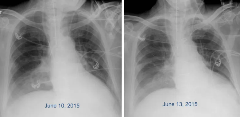
Fig. 2 Chest radiographs obtained during the days preceding suspected pneumonia: no
clear-cut progression of the radiographic abnormalities could be discerned when the earlier films were compared with that obtained on the day pneumonia was suspected (June 15, 2015).
June 15, 2015
Fig. 3 Direct microscopic examination of a cytocentrifuged BALF stained with modified