expression from a subset of methylated promoters
The MIT Faculty has made this article openly available.
Please share
how this access benefits you. Your story matters.
Citation
Haakonsen, Diane L., Andy H. Yuan, and Michael T. Laub. “The
Bacterial Cell Cycle Regulator GcrA Is a σ[superscript 70] Cofactor
That Drives Gene Expression from a Subset of Methylated
Promoters.” Genes & Development 29.21 (2015): 2272–2286.
As Published
http://dx.doi.org/10.1101/gad.270660.115
Publisher
Cold Spring Harbor Laboratory Press
Version
Final published version
Citable link
http://hdl.handle.net/1721.1/105365
Terms of Use
Creative Commons Attribution-NonCommercial 4.0 International
The bacterial cell cycle regulator GcrA is
a
σ
70
cofactor that drives gene expression
from a subset of methylated promoters
Diane L. Haakonsen,
1,2,3Andy H. Yuan,
1and Michael T. Laub
1,21
Department of Biology,2Howard Hughes Medical Institute,3Graduate Program in Microbiology, Massachusetts Institute of Technology, Cambridge, Massachusetts 02139, USA
Cell cycle progression in most organisms requires tightly regulated programs of gene expression. The transcription factors involved typically stimulate gene expression by binding specific DNA sequences in promoters and recruiting RNA polymerase. Here, we found that the essential cell cycle regulator GcrA inCaulobacter crescentus activates the transcription of target genes in a fundamentally different manner. GcrA forms a stable complex with RNA po-lymerase and localizes to almost all activeσ70
-dependent promoters in vivo but activates transcription primarily at promoters harboring certain DNA methylation sites. Whereas most transcription factors that contactσ70interact
with domain 4, GcrA interfaces with domain 2, the region that binds the−10 element during strand separation. Using kinetic analyses and a reconstituted in vitro transcription assay, we demonstrated that GcrA can stabilize RNA polymerase binding and directly stimulate open complex formation to activate transcription. Guided by these studies, we identified a regulon of∼200 genes, providing new insight into the essential functions of GcrA. Collec-tively, our work reveals a new mechanism for transcriptional regulation, and we discuss the potential benefits of activating transcription by promoting RNA polymerase isomerization rather than recruitment exclusively. [Keywords: Caulobacter crescentus; DNA methylation; bacterial transcription; cell cycle]
Supplemental material is available for this article.
Received August 19, 2015; revised version accepted October 8, 2015.
Tightly regulated programs of transcription underlie countless cellular and developmental processes through-out biology. In bacteria, transcription is performed by the multisubunit DNA-dependent RNA polymerase (RNAP). The RNAP core enzyme (composed ofα2ββ′ω subunits) is
capable of RNA synthesis but cannot initiate promoter-specific transcription. Transcription initiation at promot-ers requires aσ factor that associates with the core enzyme to form RNAP holoenzyme (Lee et al. 2012). Bacteria typically encode one primary σ factor, such as σ70 in
Escherichia coli, that is responsible for most transcription during exponential growth and multiple alternativeσ fac-tors that are required for gene expression in different growth or stress conditions (Gruber and Gross 2003). The recognition of conserved promoter elements by the σ subunit leads to binding of RNAP holoenzyme to pro-moters in an initial, typically unstable, closed complex (RPC). Through a series of isomerization steps,σ mediates
strand separation at the−10 element of the promoter, re-sulting in a transcription-competent, stable, open com-plex (RPO) that can initiate RNA synthesis (Saecker
et al. 2011).
In bacteria, the regulation of transcription occurs pri-marily at the level of initiation. The predominant mech-anism underlying transcription activation involves regulators that bind specific DNA sequences upstream of or within the promoter. These canonical transcription factors typically bind DNA independently, recruiting RNAP to target promoters by contacting the α subunit or domain 4 ofσ, which binds the −35 promoter element (Lee et al. 2012). Canonical transcription factors are usual-ly present onusual-ly at promoters that harbor a close match to their consensus binding motif. In contrast, a few regula-tors are known to interact tightly with RNAP and are found at virtually all active promoters (Haugen et al. 2008). These regulators, including E. coli DksA (Paul et al. 2004; Rutherford et al. 2009; Lennon et al. 2012) and Mycobacterium tuberculosis CarD (Stallings et al. 2009; Srivastava et al. 2013), do not affect the initial re-cruitment of RNAP to promoters but instead regulate the transition from the closed to the open complex,
Corresponding author: laub@mit.edu
Article is online at http://www.genesdev.org/cgi/doi/10.1101/gad.270660. 115.
© 2015 Haakonsen et al. This article is distributed exclusively by Cold Spring Harbor Laboratory Press for the first six months after the full-issue publication date (see http://genesdev.cshlp.org/site/misc/terms. xhtml). After six months, it is available under a Creative Commons Li-cense (Attribution-NonCommercial 4.0 International), as described at http://creativecommons.org/licenses/by-nc/4.0/.
although their mechanisms of action are incompletely understood.
Temporal regulation of gene expression is critical for cell cycle progression in the α-proteobacterium Caulo-bacter crescentus. This organism divides asymmetrically, yielding a sessile, replication-competent “stalked” cell and a motile, replication-incompetent “swarmer” cell, which can then differentiate into a stalked cell (Skerker and Laub 2004; Curtis and Brun 2010). Nearly a third of all genes in Caulobacter show cell cycle-dependent ex-pression (Laub et al. 2000; Fang et al. 2013). These patterns of gene expression are driven by several transcription factors. Some are canonical transcription factors that rec-ognize specific DNA sequences within the promoters of target genes. For example, the response regulator CtrA directly binds to and regulates the expression of ∼100 genes, mostly during late stages of the cell cycle (Laub et al. 2002).
Another important but poorly understood cell cycle transcription factor is GcrA, which accumulates during the swarmer-to-stalked cell transition (Holtzendorff et al. 2004). The precise role of GcrA in cell cycle progres-sion remains unclear, and the direct regulon of GcrA is un-known. The first report on GcrA identified ∼125 genes whose expression changed after depleting GcrA but did not distinguish direct and indirect targets (Holtzendorff et al. 2004). More recently, ChIP-seq (chromatin immuno-precipitation [ChIP] combined with deep sequencing) analysis of GcrA was reported, but there was little overlap between the genes showing the highest GcrA promoter occupancy and those originally reported as GcrA-depen-dent, and no consensus binding site was identified, al-though GcrA was shown to bind preferentially to N6
-adenine methylated GANTC sites in vitro (Fioravanti et al. 2013).
GcrA was proposed to activate transcription by bind-ing independently to all GANTC sites and recruitbind-ing RNAP (Fioravanti et al. 2013). However, little data exist to support a canonical recruitment mechanism. Although some GcrA-bound promoters have nearby GANTC sites, GcrA also associates with many promoters lacking meth-ylation sites, and there are many methmeth-ylation sites in the genome without GcrA bound. Moreover, the expression of many genes with methylation sites is unaffected by the loss of GcrA. In short, the mechanism by which GcrA af-fects gene expression is unclear, and the relationship be-tween GcrA and DNA methylation remains ill-defined.
Here, we demonstrate that GcrA forms a stable complex with RNAP holoenzyme through an interaction with the primary σ factor σ73 (hereafterσ70 for consistency with E. coli), associating with nearly allσ70-regulated
promot-ers. However, GcrA does not affect the expression of most genes. Instead, our results show that GcrA primar-ily boosts expression from promoters harboring an extend-ed recognition element containing certain GANTC methylation sites. GcrA interacts with the domain of σ70that binds the−10 element during strand separation,
and we show that GcrA stimulates transcription at pro-moters by increasing both binding of RNAP and open complex formation. Collectively, our findings favor a
model in which GcrA is brought to promoters primarily via its interaction with RNAP holoenzyme containing σ70, stimulating the expression of genes harboring certain
promoter-proximal methylation sites. Unlike many ca-nonical transcription factors, GcrA does not bind only to target promoters and simply recruit RNAP. Informed by these mechanistic studies, we combined ChIP-seq and expression profiling to identify the direct GcrA regu-lon and define a more precise role for GcrA in cell cycle progression.
Results
GcrA colocalizes genome-wide with RNAP holoenzyme containingσ70
To map the genome-wide binding profile of GcrA, we per-formed ChIP-seq using an anti-Flag antibody on cells pro-ducing GcrA with a 3xFlag tag at its C terminus from either the native gcrA promoter or a xylose-inducible pro-moter. These two strains produced GcrA at approximately wild-type levels and were morphologically similar to the wild type. The ChIP profiles were highly correlated (R = 0.97) (Supplemental Fig. S1A,B) and similar to a pro-file produced with a polyclonal antibody (Murray et al. 2013). Comparison of our GcrA-3xFlag ChIP profiles with a control profile generated for cells expressing un-tagged GcrA indicated widespread enrichment of GcrA-3xFlag across the Caulobacter genome (Supplemental Fig. S1C). GcrA was found primarily in promoter regions, with enrichment above background at >500 different intergenic locations.
To determine whether the promoter regions bound by GcrA were transcriptionally active, we generated ChIP-seq profiles of RNAP andσ70using an anti-Flag antibody
on cells producing a 3xFlag-tagged variant of theβ′ sub-unit of RNAP and an anti-σ70antibody on wild-type cells,
respectively. The profiles for RNAP andσ70each had
strik-ing overall similarity to the GcrA profile (Fig. 1A–C; Sup-plemental Fig. S1D,E). In general, the σ70 ChIP signal
showed high correlation with the GcrA and RNAP ChIP signals; i.e., promoters with highσ70enrichment tended
to also have high GcrA enrichment (Supplemental Fig. S1E), with some exceptions discussed below. However, RNAP was found in promoters and within genes, whereas σ70and GcrA were found almost exclusively in promoters
(Fig. 1B). Collectively, the ChIP-seq profiles indicate that GcrA is localized to virtually all activeσ70-dependent
pro-moters. Taken together, our data suggest that (1)σ70may be the primary determinant of where GcrA binds across the genome, and (2) GcrA associates with RNAP but only during transcription initiation (Fig. 1B).
To further test whether GcrA associates specifically with σ70-dependent promoters, we treated cells for 30 min with rifampicin, which traps RNAP at promoters (Campbell et al. 2001). We then performed ChIP-seq for RNAP (β′ subunit), GcrA, σ70, σ54, and σ32. The latter
two proteins are alternativeσ factors that recognize differ-ent promoter consensus sequences. As expected, RNAP was found almost exclusively in intergenic promoter
regions. A comparison with the other ChIP profiles for rifampicin-treated cells indicated that GcrA localized to promoters at whichσ70, but notσ54orσ32, was present
(Fig. 1D,E). These findings demonstrate that GcrA pro-moter association in vivo is specific toσ70-dependent
pro-moters, suggesting that GcrA may be targeted to these promoters by interacting withσ70. Additionally, we noted
that, in rifampicin-treated cells, GcrA occupied promoters that had not exhibited significant occupancy by GcrA, RNAP, orσ70in untreated cells (Supplemental Fig. S1F),
indicating that GcrA has an even more widespread bind-ing pattern than previously appreciated.
GcrA forms a stable complex with RNAP holoenzyme containingσ70
To test whether GcrA is recruited to promoters through a direct interaction with theσ70-containing RNAP holoen-zyme, we sought to determine whether GcrA and RNAP holoenzyme interact. First, we mixed Caulobacter cell
Figure 1. GcrA colocalizes genome-wide with RNAP holoenzyme containingσ70. (A) ChIP-seq profiles of GcrA (ΔgcrA, P
xyl-gcrA-3xFlag)
(red) and the RNAPβ′subunit (rpoC::rpoC-3xFlag) (blue) across the whole genome. ChIP-seq signals were normalized in reads per million
(RPM). (B) ChIP signals for GcrA, RNAP, andσ70at three genomic regions. Black marks below genes represent locations of GANTC
meth-ylation sites. (C ) Normalized ChIP-seq signals for GcrA, RNAP, andσ70at active promoters (see the Supplemental Material) over a 1-kb
range around the promoter and sorted based onσ70signal. Color bars indicate enrichment in RPM. (D) Normalized ChIP-seq signals for the
factors indicated from rifampicin-treated cells. (E) ChIP-seq signals for RNAP, GcrA,σ70,σ54, orσ32from rifampicin-treated cells at
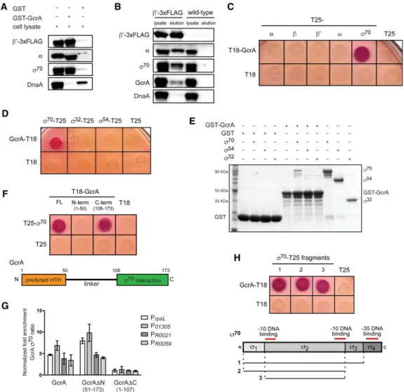
lysate with purified GST-GcrA immobilized on glutathi-one beads and then tested for retention of three subunits of RNAP holoenzyme:α, β′, andσ70. We observed clear
en-richment of each subunit, indicating that RNAP holoen-zyme was pulled down as a complex by GcrA (Fig. 2A). Importantly, the interactions detected were specific, as RNAP holoenzyme subunits were not recovered by GST alone, and GST-GcrA did not pull down other DNA-bind-ing proteins such as DnaA (Fig. 2A). Second, we used an anti-Flag antibody to immunoprecipitate RNAP from cells producing a 3xFlag-tagged variant of theβ′subunit of RNAP; Western blot analysis indicated that GcrA
coimmunoprecipitated with RNAP, as did theα subunit of RNAP andσ70, as expected (Fig. 2B). Taken together, these data indicate that GcrA forms a stable complex with RNAP holoenzyme.
GcrA is brought to promoters through a direct interaction with domain 2 ofσ70
To determine which subunit of RNAP interacts with GcrA, we used a bacterial two-hybrid system based on complementation of the two adenylate cyclase fragments T18 and T25 (Karimova et al. 1998). We fused GcrA to T18
Figure 2. GcrA interacts directly with RNAP throughσ70
. (A) GST-GcrA pull-down eluates were assessed for the presence of RNAP sub-units by immunoblotting. Equal amounts of sample were loaded for each blot. (B) Immunoprecipitatedβ′-3xFlag was assessed for the
pres-ence of coprecipitating RNAP holoenzyme subunits and GcrA by immunoblotting. Equal amounts of sample were loaded for each blot. Identical immunoprecipitation performed on wild-type cells served as a control for specificity. (C) Bacterial two-hybrid analysis. Each RNAP subunit was fused to T25 and tested for interaction with T18-GcrA. Red/purple colonies indicate a positive interaction. (D) Bacte-rial two-hybrid analysis of the interaction between GcrA-T18 andσ70
,σ54
, orσ32
fused to T25. (E) Direct interaction of GST-GcrA andσ70
. Either GST or GST-GcrA was bound to glutathione sepharose beads and then incubated with the indicatedσ factor. Samples were washed, eluted, and then examined by SDS-PAGE and Coomassie staining. (F ) Bacterial two-hybrid analysis of the interaction between the N-ter-minal and C-terN-ter-minal domains of GcrA andσ70
. A schematic of GcrA is shown below. (G) ChIP combined with quantitative PCR (ChIP-qPCR) using an anti-Flag antibody on rifampicin-treated cells expressing GcrA-3xFlag, GcrAΔN-3xFlag, or GcrAΔC-3xFlag from a plasmid in cells depleted of untagged GcrA (ΔgcrA, Pvan-gcrA) for 2 h. Fold enrichment for the GcrA:σ70ratio is relative to a control strain
contain-ing an empty vector. Error bars indicate SD. n = 2 for each of four different promoters. (H) Bacterial two-hybrid analysis of the interaction between GcrA and fragments ofσ70
and each subunit of RNAP holoenzyme to T25 and then coexpressed gene fusion pairs in E. coli. We detected an in-teraction between GcrA andσ70but not the other subunits
of RNAP (Fig. 2C). The interaction was specific toσ70, as we detected no interaction between GcrA and σ54, σ32
(Fig. 2D), or any of the other 16 predicted Caulobacterσ factors (Supplemental Fig. S2A). Although a negative re-sult in the bacterial two-hybrid assay does not completely rule out a potential interaction, these results are consis-tent with our ChIP-seq data (Fig. 1D). We verified that the interaction between GcrA andσ70was direct using
af-finity chromatography in vitro. Again, we observed a sta-ble interaction of purified GST-GcrA withσ70but notσ32
orσ54(Fig. 2E).
To determine which domain of GcrA interacts withσ70,
we tested truncations of GcrA in the bacterial two-hybrid assay. GcrA has a predicted helix–turn–helix at its N ter-minus, connected by a long linker to a C-terminal domain of unknown function. We detected an interaction only between the C-terminal domain of GcrA and σ70 (Fig. 2F). To determine whether this interaction is sufficient to localize GcrA to promoters, we performed ChIP com-bined with quantitative PCR (ChIP-qPCR) using an anti-Flag antibody on cells depleted of full-length GcrA and ex-pressing a 3xFlag-tagged fragment lacking either the N-terminal domain GcrAΔN or the C-terminal domain GcrAΔC. We found that GcrAΔN, but not GcrAΔC, local-izes to promoters withσ70(Fig. 2G), indicating that the N-terminal helix–turn–helix domain is not required to re-cruit GcrA to promoters. These data strongly support a model in which GcrA is brought toσ70promoters
primar-ily via an interaction with RNAP holoenzyme not by binding DNA independently.
To determine which domain ofσ70contacts GcrA, we tested the ability of individual domains ofσ70to interact
with GcrA in the bacterial two-hybrid system. σ70 has four structural domains, with domains 2 and 4 (σ2 and
σ4) involved in binding the −10 and −35 promoter
ele-ments, respectively. Transcriptional activators that inter-act with σ70 typically recruit RNAP to promoters by making contact with σ4(Lee et al. 2012). However, we
found thatσ2, notσ4, was sufficient to mediate an
inter-action with GcrA (Fig. 2H), indicating that GcrA may regulate transcription through an unconventional mecha-nism. To better define the region ofσ2that interacts with
GcrA, we took advantage of the fact that E. coliσ2does not
interact with GcrA (Supplemental Fig. S2B) and tested E. coli–Caulobacter σ2chimeras for interaction with GcrA.
This analysis indicated that GcrA likely interacts with σ2through the twoα helices formed by regions 1.2 and
2.1 as well as the N-terminal and C-terminal ends of the nonconserved region (Supplemental Fig. S2B).
GcrA-binding affinity for methylation sites is sequence-specific
Consistent with GcrA being recruited toσ70-dependent
promoters in vivo through a direct interaction with RNAP holoenzyme, our ChIP-seq studies indicated a strong correlation between the binding profiles of GcrA
andσ70. However, the correlation was not perfect, perhaps
indicating that GcrA exhibits some DNA sequence specif-icity even though promoter association occurs primarily viaσ70. A previous study found that GcrA binds methylat-ed DNA in vitro and proposmethylat-ed that methylatmethylat-ed GANTC sites were required for GcrA binding in vivo (Fioravanti et al. 2013). However, many promoters bound by GcrA in vivo do not have methylation sites, and the enrichment of GcrA at promoters with methylation sites varied signif-icantly (Supplemental Fig. S3A).
To examine the DNA sequence and methylation specif-icity of GcrA, we measured the affinity of GcrA for meth-ylated and unmethmeth-ylated DNA in vitro by filter binding. Using unmethylated probes derived from the ftsZ and mipZpromoters, we measured a Kdof∼0.5 μM
(Supple-mental Fig. S3B). Using methylated PftsZand PmipZprobes,
we observed a 1.4-fold and 1.7-fold reduction in Kd,
respec-tively (Supplemental Fig. S3B). Notably, the Kdof GcrA for
methylated PmipZwas lower than for methylated PftsZ, but
GcrA occupancy in vivo, as judged by ChIP-seq, was sig-nificantly higher at PftsZ(Supplemental Fig. S3C). Thus,
DNA-binding affinity and the presence of a GANTC methylation site are insufficient to explain GcrA enrich-ment at a given promoter.
To identify other sequence elements influencing GcrA binding, we examined the ChIP-seq signals for GcrA, σ70, and RNAP at all 4542 GANTC methylation sites
across the Caulobacter genome. The majority of sites had little or no GcrA bound (Fig. 3A). Of the methylation sites that were bound by GcrA, most were also bound by σ70, indicating that these sites are in or near promoters
(Fig. 3A). However, some methylation sites within genes showed occupancy by GcrA with no enrichment ofσ70;
the enrichment of GcrA at these sites was substantially less than observed at promoters, supporting the notion that the genomic binding of GcrA is determined primarily by its interaction withσ70. Nevertheless, we hypothesized
that intergenic sites bound by GcrA may have sequence elements that strongly favor GcrA binding. MEME analy-sis of 20 such locations revealed a consensus sequence of TGATTCG or, more broadly, YGAKTCK, where Y = C or T and K = G or T (Fig. 3B).
To assess the relevance of this motif, we calculated the average GcrA ChIP signal at all intergenic locations that have one of the 32 possible NGANTCN sequences (Fig. 3C; Supplemental Fig. S3D). This analysis revealed a clear hierarchy (or ranking) of the 32 sequences in terms of GcrA binding, with seven of the eight sequences exhibit-ing the highest GcrA signal included in the consensus YGAKTCK (Fig. 3B,C; Supplemental Fig. S3D). To verify the inferred binding preferences of GcrA, we measured its affinity for a methylated PmipZprobe containing the
third and 12th highest-ranked sequences (TGAGTCG and AGAGTCG) and a probe, called PmipZ(LM)
(low-ranked methylation sites) in which these sequences were mutated to the 30th and 32nd ranked sequences (GGAGTCT and AGATTCA). The four nucleotide substi-tutions in PmipZ(LM) reduced the binding affinity of GcrA
to a level observed when the PmipZprobe was
mutating sites in a probe derived from the cckA promoter (Supplemental Fig. S3E).
To test whether the recognition motif YGAKTCK im-pacts GcrA binding at promoters, we computed the ratio of GcrA:σ70ChIP signal at eachσ70promoter. MEME
anysis of the top 25 promoters revealed a motif (Fig. 3E) al-most identical to that identified above. We also found that promoters containing a methylation site ranked 1–8 (i.e., those closely matching the consensus YGAKTCK) typically had much higher GcrA:σ70ratios than promoters with no methylation site or a low-ranked methylation site (Fig. 3F; Supplemental Fig. S3F). These observations
indicate that the DNA-binding specificity of GcrA iden-tified influences the extent of GcrA occupancy at promot-ers in vivo.
GcrA promotes transcription and RNAP open complex formation at methylated promoters
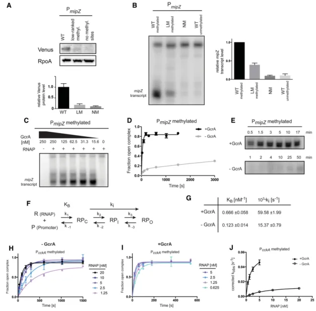
To assess the importance of consensus methylation sites on GcrA-dependent transcription in vivo, we constructed reporters in which a mipZ promoter near the native mipZ chromosomal locus drives expression of the yellow fluo-rescent protein Venus. Using Western blot analysis, we
Figure 3. GcrA-binding affinity for methylation sites is sequence-specific. (A) ChIP-seq signals for GcrA, RNAP, andσ70
in rifampicin-treated cells plotted over 1-kb ranges centered at each of the 4542 GANTC methylation sites in the Caulobacter genome. Signals were capped at 20 RPM and sorted based on the GcrA signal. (B) Identification of a consensus GcrA-binding motif. ChIP signals for GcrA, RNAP, andσ70
for one of the regions used in the analysis are shown. The motif obtained from the 20 selected sequences is depicted. (C) Plot of the average ChIP signal forσ70
versus GcrA at each of 32 possible NGANTCN variants. Analysis was performed on ChIP signals from rifampicin-treated cells and was restricted to nonpromoter regions. One non-NGANTCN site (gray) is shown for comparison (also see Supplemental Fig. S3D). (D) GcrA-binding curves for the probes indicated: methylated PmipZ, unmethylated PmipZ, and methylated
PmipZmutated to have low-ranking methylation sites [PmipZ(LM)]. Sequences surrounding the methylation sites in PmipZand PmipZ(LM)
and their ranks are indicated. Error bars represent SDs. n = 3. (E) Normalized histogram of the GcrA:σ70
ChIP ratio atσ70
promoters and a motif identified in promoters with high ratios. n = 25. (F ) Histograms of the GcrA:σ70
ChIP ratio for promoters lacking methylation sites within 40 base pairs (bp) of the transcription start site (black), containing at least one methylation site ranked 1–8 (green) or 25–32 (orange); ranks were based on C and Supplemental Figure S3D.
found that mutating the high-ranked methylation sites in the mipZ promoter to low-ranked sites reduced expres-sion in vivo by nearly 80%, similar to the effect of elimi-nating the methylation sites completely (Fig. 4A).
We also examined the effects of mutating DNA methyl-ation sites using an in vitro transcription assay with puri-fied Caulobacter RNAP holoenzyme containingσ70and
GcrA. Using the wild-type methylated mipZ promoter
Figure 4. GcrA promotes open complex formation at methylated promoters. (A) Western blot analysis of a Venus reporter expressed from the wild-type PmipZharboring two high-ranked methylation sites, a variant containing low-ranked methylation sites, or a variant with no
methylation sites. (Bottom) Band intensities were quantified. Error bars represent SDs. n = 2. RpoA was used as a loading control. (B) Mul-tiple-round in vitro transcription reactions using the promoter variant indicated as a template. Reactions contained 2 nM purified Cau-lobacterRNAP holoenzyme and 62.5 nM GcrA. (Right) Band intensities were quantified. Error bars represent SDs. n = 2. (C) Multiple-round in vitro transcription reactions using the methylated wild-type mipZ promoter as a template. Reactions contained 2 nM purified CaulobacterRNAP holoenzyme and varying levels of GcrA. (D) Kinetics of open complex formation using 1.25 nM Caulobacter RNAP holoenzyme ±250 nM GcrA. Error bars represent SDs. n = 2. (E) Single-round in vitro transcription assays using a probe containing the methylated wild-type mipZ promoter as a template. Reactions containing 1.25 nM purified Caulobacter RNAP holoenzyme ±250 nM GcrA and DNA probe were preincubated, and nucleotides, along with heparin, were added at the indicated time points. Samples ± GcrA were imaged on the same gel. (F ) Kinetic scheme of transcription initiation highlighting KB(the binding constant of the holoenzyme
to promoter DNA) and ki(the aggregate forward rate constant of isomerization from the closed to the open complex). (G) Summary of
kinetic parameters for GcrA-dependent transcription from methylated PcckA. (H) Kinetics of open complex formation at the methylated
cckApromoter using varying concentrations of Caulobacter RNAP holoenzyme and 1 mM ATP. Error bars represent SDs. n = 2. (I ) Ki-netics of open complex formation at the methylated cckA promoter using varying concentrations of Caulobacter RNAP holoenzyme, 250 nM GcrA, and 1 mM ATP (see the Supplemental Material). Error bars represent SDs. n = 2. (J) Plot of corrected kobsobtained from
each of the curves in H and I versus RNAP concentration and fitted to obtain the constants KBand kireported in G (see the Supplemental
as a template produced a transcript of the expected size (Fig. 4B; Supplemental Fig. S4). Mutating the two high-ranked methylation sites to low-high-ranked sites or eliminat-ing methylation sites dramatically reduced transcription in this assay, consistent with our in vivo reporter studies. Importantly, at the concentration of GcrA used, GcrA alone does not stably bind DNA (Fig. 3D). To gain further insight into the binding affinity of GcrA for RNAP holoen-zyme, we repeated the in vitro transcription assay with varying levels of GcrA. Transcription was maximal at 15 nM, indicating that GcrA’s Kdfor RNAP is <15 nM (Fig.
4C). Higher concentrations of GcrA, at which some DNA binding was observed, seemed to partially inhibit the reaction, possibly by preventing binding of RNAP to the promoter. These data strongly support a model in which GcrA is brought to promoters via a tight interaction with RNAP rather than recruiting RNAP through inde-pendent DNA binding.
To further examine how GcrA impacts transcription, we implemented a filter-binding assay for measuring open complex formation. We incubated purified Caulo-bacterRNAP holoenzyme with a radiolabeled, methylat-ed PmipZprobe in the presence or absence of GcrA and
then isolated open complexes on nitrocellulose filters af-ter competing away closed complexes with heparin. A time course of open complex formation demonstrated that GcrA strongly promotes open complex formation, with maximal levels of open complex forming within 2– 3 min with GcrA while remaining relatively low up to 50 min without GcrA (Fig. 4D). Similarly, we found that GcrA strongly stimulated transcription from the wild-type methylated mipZ promoter in a single-round in vitro transcription assay (Fig. 4E).
The stimulation of open complex formation (Fig. 4D) could indicate that GcrA affects binding of RNAP to pro-moters, isomerization to the open complex, or both (Fig. 4F). To distinguish between these possibilities, we mea-sured the kinetics of open complex formation using a fil-ter-binding assay while titrating RNAP holoenzyme. For these experiments, we used a probe corresponding to the cckA promoter, as the time-course data for this probe were fit well by a single exponential model, enabling us to disentangle the effects of GcrA on RNAP binding and isomerization (see the Supplemental Material). Our re-sults indicated that GcrA increases KB(the binding
cons-tant for RNAP in the closed complex form) by∼5.4-fold and increases ki (the aggregate forward rate constant for
isomerization from the closed to the open complex) by ∼3.9-fold (Fig. 4G–J).
GcrA regulates genes harboring promoter-proximal, near-consensus methylation sites
The composition of the GcrA regulon has been elusive, in large part because ChIP-seq analysis alone is insufficient to pinpoint direct targets, a consequence of the fact that GcrA associates with virtually allσ70-regulated
promot-ers. As a first step to defining the GcrA regulon, we used DNA microarrays to measure RNA levels in synchronized populations of wild-type and GcrA-depleted cells at 30,
45, and 60 min after synchronization, the time window when GcrA normally accumulates to maximal levels (Fig. 5A). We identified 364, 590, and 444 genes at each time point with at least 1.75-fold lower expression in the GcrA-depleted cells relative to wild-type cells (Supple-mental Table S1). In total, GcrA appears to positively reg-ulate at least 774 genes either directly or indirectly.
We hypothesized that promoters directly regulated by GcrA should have high GcrA:σ70ChIP enrichment ratios.
Although GcrA localizes to promoters mainly via σ70, subsequent interaction with methylation sites within tar-get promoters likely stabilizes GcrA binding or increases the efficiency of ChIP. We found that 78% of genes affect-ed by GcrA depletion at the 30-min time point had pro-moters with high GcrA:σ70ratios. The majority of these
genes also had at least one methylation site within 40 base pairs (bp) of their transcription start site (Fig. 5B; Sup-plemental Table S2), consistent with our analysis of the DNA sequence specificity of GcrA.
Combining these observations, we defined the direct GcrA regulon as those genes that had (1) significantly low-er expression levels in GcrA-depleted cells at 30 or 45 min, (2) a high GcrA:σ70ChIP enrichment ratio at their
promot-ers, and (3) a promoter-proximal GANTC site. In total, 140 transcription units encompassing 204 genes satisfied all three criteria (Supplemental Table S3). Notably, most of these genes are cell cycle-regulated (Fig. 5C), with peak ex-pression at or shortly after the time that GcrA levels in-crease (Fig. 5A), even though cell cycle regulation was not one of our criteria. However, using only cell cycle reg-ulation and lower expression levels in GcrA-depleted cells as criteria to define the GcrA regulon would have identi-fied only 64% of the 140 transcription units that satisidenti-fied our three criteria, along with 42 promoters that do not have a high GcrA:σ70ChIP enrichment ratio and
methyl-ation sites (Supplemental Fig. S5B). In sum, our three cri-teria, chosen based on our mechanistic studies, allowed the precise delineation of the direct GcrA regulon.
Some direct targets of GcrA affect the expression of oth-er genes latoth-er in the cell cycle; most of these indirect tar-gets also decrease following depletion of GcrA but lack a promoter-proximal methylation site and have a lower GcrA:σ70ratio. For example, the gene ctrA, which has a
high GcrA:σ70ratio and a high-ranked methylation site, is likely activated directly by GcrA early in S phase. Al-most the entire CtrA regulon is also misregulated in GcrA-depleted cells (Supplemental Fig. S5C); although some may be direct GcrA targets themselves, most are not and instead are indirectly affected via CtrA.
Strikingly, the GANTC sites in promoters called as directly regulated by GcrA were enriched for methylation sites defined above as highly ranked (Fig. 3C; Supplemen-tal Fig. 5D), consistent with our in vitro studies that cer-tain methylation sites better enable GcrA to promote transcription. These methylation sites localize to two re-gions of target promoters: one narrowly centered around −20 and one more widely distributed from +2 to +30 (Fig. 5E). We observed almost no methylation sites around the−10 element, possibly because this region is bound by σ70 and inaccessible to GcrA. The distribution of
methylation sites suggests that GcrA can bind DNA on either side of the −10 element while interacting with domain 2 ofσ70, perhaps as a consequence of its predicted
long unstructured linker (Fig. 2F). Altogether, our data in-dicate that it is not simply the presence of a methylation site that dictates whether a promoter is affected by GcrA, but its precise location and flanking nucleotides.
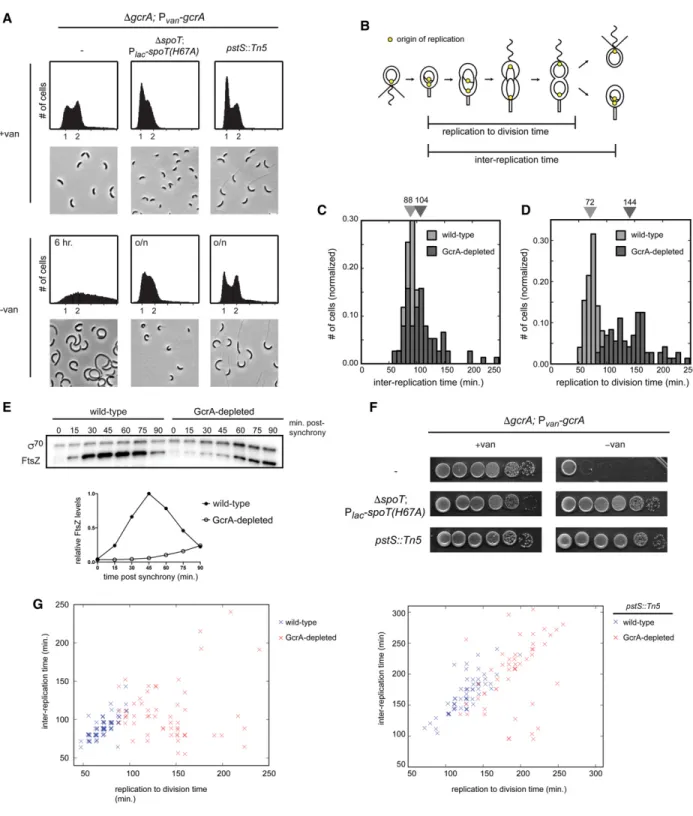
Loss of GcrA delays cytokinesis and desynchronizes DNA replication and cell division
The GcrA regulon spans multiple functional categories (Fig. 5C) and includes several core cell cycle processes: nu-cleotide synthesis, DNA repair, chromosome
organiza-tion and segregaorganiza-tion, and cell division. Several genes involved in activating the master regulator CtrA were also GcrA targets, including cckA, divL, pleC, podJ, and ctrAitself. The regulon did not include components of the replisome, except for dnaE. The functional categories of direct targets suggest that GcrA helps drive expression patterns critical to S phase and progression through the later stages of the cell cycle. Thus, cells lacking gcrA should exhibit pleiotropic defects. To examine the gcrA phenotype, we built a strain in which the only copy of gcrAis driven by a vanillate-inducible promoter (ΔgcrA; Pvan-gcrA). When grown in the absence of vanillate for 6
h, GcrA-depleted cells grew at approximately the same rate as wild type (Supplemental Fig. S6B) but became
Figure 5. GcrA regulates genes with promoter-proximal, near-consensus methylation sites. (A) Western blot for GcrA and RpoA (loading control) during the cell cycle is summarized below. GcrA, CtrA (not shown), and RpoA were imaged on the same blot. (B) Promoters of genes down-regulated in GcrA-depleted cells at each time point indicated were categorized based on their rifampicin-treated GcrA:σ70
ChIP ratio and the presence of a methylation site within 40 bp of the transcription start site. (Right) Distribution at all promoters is given for comparison. (C ) Cell cycle expression patterns of genes directly regulated by GcrA. RNA sequencing data are from Fang et al. (2013). Expression at each time point is relative to a gene’s average expression at all time points. (D) Distribution of methylation site ranks for promoters in the GcrA regulon and genome-wide. (E) Histogram for the locations of methylation sites in GcrA-regulated promoters rel-ative to transcription start sites.
elongated and accumulated extra chromosomes (Fig. 6A; Murray et al. 2013), indicating a cell division defect. To quantify the timing of cell division and DNA replication, we performed time-lapse microscopy of GcrA-depleted cells harboring a fluorescent repressor–operator system that labels origins of replication (Fig. 6B; Supplemental Fig. S6A). We found that the interval between successive rounds of DNA replication was delayed by only∼18% in cells depleted of GcrA (Fig. 6C), whereas the time between DNA replication to cell division increased by 100% (Fig. 6D).
These phenotypic analyses indicated that, in cells lack-ing GcrA, the rate of cell division does not keep pace with the rate of DNA replication. This cell division defect like-ly results from the misregulated expression of many genes; nearly half of the known division genes are positively reg-ulated by GcrA, with MipZ, FtsE, FtsX, FtsN, and FtsZ be-ing direct targets (Supplemental Fig. S5D). Consistent with the defects of GcrA resulting from a deficiency in ex-pressing multiple genes, we found that the ectopic expres-sion of either ftsN or ftsZ could not substantially restore viability to a strain depleted of GcrA in rich medium (Sup-plemental Fig. S6B). Increased expression of these genes may marginally increase viability (Murray et al. 2013), but the phenotype of cells lacking GcrA likely stems from its pleiotropic regulation of gene expression.
Importantly, although cell division occurs less fre-quently in the absence of GcrA, cells do still divide even after GcrA is undetectable (Supplemental Fig. S6C,D). Thus, essential cell division proteins must be transcribed without GcrA but take longer to reach the levels neces-sary for cytokinesis. Quantitative immunoblotting con-firmed that, in GcrA-depleted cells, FtsZ levels do not rise sharply early in the cell cycle as in wild-type cells but do eventually increase (Fig. 6E). These data support a model in which GcrA helps boost the expression of cell division genes so that cells maintain balanced rates of division and DNA replication.
To gain additional insight into the physiological role of GcrA, we sought to identify mutations that restore viabil-ity to cells depleted of GcrA. Using transposon mutagen-esis, we identified a series of insertions in the phosphate import system comprising PstABC and PstS. The pstS:: Tn5mutation almost completely rescued the plating effi-ciency of cells lacking GcrA (Fig. 6F), and these cells ap-peared morphologically similar to cells harboring the pstS::Tn5mutation alone (Fig. 6A).
Why do mutations in the phosphate import system sup-press a gcrA mutant? We suspected that the effect may stem from the fact that pstS mutants grow slowly and are delayed in initiating DNA replication, leading to an in-crease in G1 cells in flow cytometry analysis (Fig. 6A). As noted above, cells lacking GcrA are delayed for cell divi-sion but continue to grow and initiate DNA replication. Hence, the pstS::Tn5, gcrA double mutant has likely re-balanced its rates of growth and DNA replication with rates of cell division; indeed, the intervals between rounds of DNA replication and between replication and division were more closely matched in the pstS::Tn5, gcrA deple-tion strain than in the gcrA depledeple-tion alone (Fig. 6G).
Finally, we wanted to test whether slowing growth and DNA replication in a completely different manner could also rescue the gcrA mutant. We did so by ectopically pro-ducing the signaling molecule (p)ppGpp, which is known to slow cell growth and repress DNA replication (Boutte et al. 2012). We used an IPTG-inducible, hydrolase-defec-tive variant of SpoT, the enzyme responsible for the syn-thesis and hydrolysis of (p)ppGpp in Caulobacter. We observed a nearly complete rescue of plating viability for GcrA-depleted cells expressing this variant of SpoT (Fig. 6F), and the morphology of cells was significantly im-proved in liquid medium compared with the parent GcrA depletion strain (Fig. 6A). Collectively, our data in-dicate that the transcription factor GcrA normally helps cell division and potentially other cellular processes keep pace with both cell growth and DNA replication. Consequently, cells lacking GcrA show an uncoupling (or discoordination) of essential cell cycle events, eventu-ally resulting in lethality. However, mutations that specif-ically slow growth and the rate of replication initiation can largely restore the balance of cell cycle activities, en-abling cells to proliferate without GcrA.
Discussion
Mechanism of transcriptional control by GcrA
Although critical to cell cycle progression in Caulobacter, the mechanism by which GcrA activates transcription was previously unclear, and, consequently, the extent of its regulon was poorly defined. Our results suggest that GcrA is not a canonical transcription activator that drives gene expression primarily by binding DNA and then recruiting RNAP. Instead, our results favor a model in which GcrA forms a stable complex with RNAP through an interaction with the primaryσ factor σ70and
is brought to promoters via this interaction (Fig. 7). Impor-tantly, however, GcrA does not promote transcription from all promoters to which it binds. Rather, GcrA prefer-entially affects promoters harboring certain GANTC methylation sites, with the identity of the flanking nucleotides and the middle nucleotide of the GANTC site determining GcrA’s efficacy as a transcription factor. At such promoters, GcrA may stimulate RNAP binding and open complex formation to activate transcription both in vitro and in vivo. Whether GcrA stimulates tran-scription from a given promoter likely also depends on whether the rate-limiting step for initiation is the step af-fected by GcrA.
Our results argue strongly against a mechanism in which GcrA binds promoters independently and then re-cruits RNAP holoenzyme. First, GcrA forms a stable com-plex with RNAP holoenzyme (Fig. 2A,B), and GcrA is found genome-wide at mostσ70-dependent promoters, in-cluding those lacking methylation sites (Fig. 1C,D; Sup-plemental Fig. S3A). Second, we found that a GcrA variant lacking its DNA-binding domain was still highly enriched at σ70-dependent promoters (Fig. 2G). Finally, we found that GcrA promotes transcription in vitro at low nanomolar concentrations (Fig. 4C), where GcrA
Figure 6. Loss of GcrA delays cytokinesis and desynchronizes DNA replication and cell division. (A) Flow cytometry analysis of chro-mosome content and phase-contrast images of a gcrA depletion strain harboring the additional mutations indicated grown in rich medi-um ± vanillate overnight, except for GcrA-depleted cells that are not viable, for which a 6-h depletion was performed. SpoT(H67A) expression was induced with 500μM IPTG. (B) Schematic of the cell cycle showing the interreplication and replication to division times. (C,D) Normalized histograms of times between successive replication initiations (C) and times between replication and division (D) in wild-type and GcrA-depleted cells. Median values are listed above. (E) Western blot for FtsZ andσ70(loading control) during the cell cycle
for synchronized wild-type and GcrA-depleted cells. FtsZ levels were normalized toσ70and are plotted as a fraction of maximum
expres-sion. (F ) Growth of a gcrA depletion strain harboring the same mutations as in A, assessed by 10-fold serial dilutions on plates ±vanillate. Plates forΔgcrA; Pvan-gcrA;ΔspoT; Plac-spoTH67A also contained 50μM IPTG. (G) Scatter plots of the interreplication time and
replica-tion to division time in wild-type and GcrA-depleted cells (left) and pstS::Tn5-depleted and pstS::Tn5 GcrA-depleted cells (right). Over-layed data points were slightly shifted by addition of a small random number to facilitate visualization.
alone does not stably bind DNA (Fig. 3D), indicating that GcrA’s affinity for RNAP is much tighter than its affinity for DNA. GcrA was found at some nonpromoter regions without RNAP, but the ChIP enrichment values at these sites was substantially smaller than that observed at pro-moters (Fig. 1A; Supplemental Fig. 3C). These observa-tions imply that GcrA binds to promoters together with RNAP holoenzyme rather than independently and prior to RNAP, as previously suggested.
In sum, our results suggest a new model for GcrA as an auxiliary component or cofactor of theσ70-containing RNAP holoenzyme in Caulobacter. GcrA is nearly absent from swarmer cells but is likely very abundant throughout the rest of the cell cycle, with ribosome profiling data (Schrader et al. 2014) indicating synthesis at rates com-parable with other components of RNAP. Moreover, because GcrA has a much tighter affinity for RNAP holo-enzyme (Kd< 15 nM) (Fig. 4C) than for DNA (Kd∼ 270 nM
for highest affinity sites) (Fig. 3D), most GcrA is likely bound to RNAP. GcrA is then brought to promoters via its interaction withσ70, where our kinetic analyses indi-cate that, at least for the model cckA promoter, it can af-fect both KB (the equilibrium binding constant for the
closed complex of RNAP) and ki(the composite rate of
downstream isomerization steps that ultimately result in strand separation). Whether GcrA affects the two steps differently at other promoters is not clear, as the kinetics of open complex formation for some promoters, like PmipZ, did not exhibit the single exponential behavior
needed to elucidate KBand ki.
GcrA’s ability to promote isomerization of RNAP through a direct interaction with region 2 of the house-keeping σ factor distinguishes it from other, previously
characterized transcription factors. Most transcriptional activators bind DNA independently and recruit RNAP to promoters through direct interactions with theα sub-unit or domain 4 ofσ70. Like GcrA, E. coli DksA and M.
tuberculosis CarD affect isomerization from the closed to the open complex, but DksA interacts with theβ and β′subunits, and CarD binds theβ subunit. E. coli Crl binds domain 2 ofσSbut functions differently than GcrA,
pro-moting the association of core RNAP with σS (Banta et al. 2013). In Chlamydia trachomatis, the transcription factor GrgA binds the nonconserved region of domain 2 in the housekeepingσ factor (Bao et al. 2012), but whether GrgA impacts RNAP binding or isomerization is un-known. The actinobacterial protein RbpA also binds domain 2 of a housekeepingσ factor (Tabib-Salazar et al. 2013), but recent data highlighted a role for RbpA in in-creasing affinity of holoenzyme to promoter DNA (Hubin et al. 2015), so its role, if any, in stimulating isomerization to the open complex is uncertain. Thus, to our knowledge, CaulobacterGcrA is the first transcription factor identi-fied that binds region 2 of aσ factor and can promote the closed-to-open complex step of transcription.
GcrA provides a powerful mechanism for controlling gene transcription. By impacting the isomerization step of transcription initiation, GcrA-regulated genes can still be controlled by other, canonical transcription factors, af-fording an opportunity for bona fide combinatorial con-trol. Although GcrA-regulated genes in Caulobacter are generally expressed during S phase, the precise timing of their induction varies, possibly the consequence of regula-tion by other transcripregula-tion factors. Combined regularegula-tion by GcrA and other transcription factors may also increase the dynamic range of expression for target genes, helping
Figure 7. Model for the mechanism of transcription control by and cell cycle functions of GcrA. (A) GcrA forms a stable complex with RNAP through an interaction with domain 2 of the primaryσ factor σ70
and is brought to promoters via this interaction. The presence of a preferred methylation site near the promoter stabilizes GcrA at the promoter and is required for GcrA-dependent stimulation of transcrip-tion. GcrA affects both the binding step and open complex formatranscrip-tion. (B) Summary of the role GcrA plays in driving cell cycle progression by regulating gene expression in S phase.
to switch them from very low to very high states of expres-sion. For instance, the gene ftsZ is activated by DnaA (Hottes et al. 2005) and GcrA (Fig. 6E). In cells depleted of GcrA, ftsZ is still induced but at significantly reduced levels compared with wild-type cells.
N6-adenine methylation-dependent gene regulation N6-adenine methylation by orphan methyltransferases affects many processes in bacteria, including gene expres-sion. The most well characterized example involves theγ-proteobacterial methyltransferase Dam (deoxyade-nosine methyltransferase), which methylates GATC se-quences (Wion and Casadesús 2006). In E. coli, full methylation of the agn43 promoter by Dam activates transcription by preventing binding of the repressor OxyR until DNA replication produces hemimethylated promoters (Haagmans and van der Woude 2000). Similar-ly, binding of the leucine-responsive regulatory protein Lrp to the traJ promoter in the Salmonella enterica viru-lence plasmid is blocked when a GATC site is fully meth-ylated. After DNA replication, Lrp can bind to one of the newly replicated hemimethylated plasmids (Camacho and Casadesús 2005). For both OxyR and Lrp, methyla-tion-dependent binding occurs at only a limited number of promoters. In contrast, N6-adenine methylation
ap-pears to promote the GcrA-dependent activation of most, if not all, target promoters.
In Caulobacter, the role of CcrM-dependent N6-adenine
methylation at GANTC sites in gene regulation has been poorly understood. GANTC sites occur preferentially in intergenic regions, and GcrA was suggested to bind meth-ylated GANTC sites in vitro (Fioravanti et al. 2013). How-ever, GcrA associates with many promoters lacking GANTC sites, and the expression of many genes with promoter-proximal GANTC sites is not affected in cells lacking GcrA. Our results now clarify the relationship between GcrA, CcrM, and promoter methylation. We showed that GcrA is found at virtually allσ70-dependent
promoters but primarily affects only promoters contain-ing certain methylation sites, not any GANTC (Fig. 3). In particular, the two bases flanking a GANTC site and the middle“N” base strongly influenced GcrA binding and GcrA-dependent activation both in vitro and in vivo. Whether N6-adenine methylation affects GcrA binding while RNAP is in the open or the closed complex remains to be determined. Promoter methylation strongly affected GcrA-dependent transcription both in vitro and in vivo (Fig. 4A,B) but had only a modest (approximately twofold) effect on binding to dsDNA (Fig. 3D; Supplemental Fig. S3E), perhaps indicating an effect after strand separation. Further structural studies of GcrA will help reveal how methylation impacts DNA binding and transcription.
Whether GcrA’s transcriptional activity differs for fully versus hemimethylated promoters is also not yet clear. CcrM is cell cycle-regulated and accumulates only after DNA replication is nearly complete (Wright et al. 1996). Thus, many GANTC sites, particularly those close to the origin, remain hemimethylated for significant periods of time after passage of the replication fork. The ctrA P1
promoter, which is GcrA-regulated, was postulated to be more active in the hemimethylated state such that ctrA expression was coupled to passage of the replication fork (Reisenauer and Shapiro 2002). However, the induction of ctrA P1 transcription may simply be driven by the cell cycle-dependent accumulation of GcrA (Fig. 7). More-over, the constitutive production of CcrM, leading to con-stitutive methylation, only affects the expression of a few cell cycle genes (Gonzalez et al. 2014). Thus, a role for changes in DNA methylation as a“trigger” for GcrA-de-pendent gene expression remains uncertain.
Role of GcrA in theCaulobacter cell cycle
Collectively, our findings on GcrA also force a significant reconsideration of its role in the Caulobacter cell cycle. First, because GcrA does not promote transcription at all of the promoters to which it binds, ChIP-based studies alone are inherently unable to accurately define GcrA tar-get genes. By developing a mechanistic understanding of GcrA, we were able to then better delineate the set of genes whose transcription is affected by GcrA. These findings in-dicate that GcrA directly affects at least 204 genes, with many additional indirect targets (Fig. 5). These genes par-ticipate in numerous cell cycle processes, many of which help cells progress through S phase, including nucleotide synthesis, DNA repair, chromosome organization and seg-regation, and cell division. Notably, however, and in con-trast to prior suggestions, the GcrA regulon does not include dnaA or, with one exception, core components of the replisome. Consistent with this finding, cells lack-ing GcrA continue to periodically initiate new rounds of DNA replication, often without an intervening cell divi-sion (Fig. 6). A similar phenomenon occurs with mutants in the CtrA phosphorylation pathway (Jonas et al. 2011). Together, these results underscore the fact that oscilla-tions in DnaA (and DNA replication) occur independently of CtrA and GcrA, meaning that these three cell cycle reg-ulators do not comprise a single genetic oscillator.
Second, GcrA-regulated genes are still expressed in cells depleted of GcrA, albeit at lower rates, leading to a delay in processes such as cell division, not a complete disrup-tion. The magnitude of expression changes following GcrA depletion is often only twofold to threefold. In con-trast, strains depleted of a canonical transcription factor like CtrA are almost completely blocked for cell division, motility, and other CtrA-dependent processes, with target genes typically decreasing 10-fold or more (Laub et al. 2002). The differences in gene expression following a loss of GcrA or CtrA are mirrored in their phenotypic dif-ferences. Most notably, gcrA mutants are viable in mu-tant backgrounds that slow growth and DNA replication initiation, which likely rebalances cell cycle events such that the periodicity of DNA replication better matches the periodicity of cell division. Growth in minimal medi-um also partially rescues the essentiality of GcrA (Murray et al. 2013) but not nearly as well as slowing growth and replication initiation, supporting the notion that cells lacking GcrA have imbalanced replication and division cycles (Fig. 6C,D; Supplemental Fig. 7B). In contrast to
GcrA, cells lacking CtrA are inviable in most growth con-ditions and are not rescued by mutations that slow repli-cation and growth.
In sum, we propose that GcrA is a novel accessory factor of RNAP in Caulobacter that stimulates the transcription of target genes through an unconventional interaction with region 2 of the housekeepingσ factor σ70. In contrast
to classical activators that typically bind DNA indepen-dently to recruit RNAP, GcrA travels with RNAP to pro-moters, where it can stabilize binding and promote isomerization. Because GcrA interfaces with region 2, other transcription factors can still target region 4 ofσ70 or other RNAP subunits, enabling combinatorial control and synergistic activation of gene expression. GcrA is extremely well conserved amongα-proteobacteria, where it may play a role in regulating gene expression similar to that elucidated here. Additionally, Caulobacter and many α-proteobacteria encode paralogs of GcrA, and homologs have been found in bacteriophage genomes (Gill et al. 2012). Each of these proteins may, like GcrA, interact directly with RNAP to globally regulate gene expression.
Materials and methods
For details on strain construction, growth conditions, and de-tailed experimental procedures, see the Supplemental Material and Supplemental Tables S4–S6.
ChIP-seq and data analysis
For details on ChIP, library generation, and data analysis, see the Supplemental Material. For a summary of ChIP-seq analyses per-formed, see Supplemental Table S7 and Gene Expression Omni-bus GSE73925.
Protein purification
The RNAP holoenzyme was purified by Ni-NTA affinity chroma-tography from Caulobacter cells producing RpoC with a His10tag
at the C terminus, followed by gel filtration and ion exchange chromatography. His6-GcrA, His6-CcrM, His6-σ70, His6-σ54, and
His6-σ32were purified by Ni-NTA affinity chromatography and
gel filtration chromatography for His-GcrA. GST and GST-GcrA were purified by affinity chromatography using glutathione sepharose beads.
Filter-binding and open complex assays
DNA probes were generated by PCR; gel-purified; labeled with T4-polynucleotide kinase in 10-μL reactions containing 7 μL of DNA, 1μL of radiolabeled γ-P32ATP, 1μL of T4-PNK, and 1 μL
of T4-PNK buffer; and incubated for 90 min at 37°C. Kinetic open complex assays were performed with Caulobacter RNAP holoenzyme diluted in transcription buffer (40 mM Tris-HCl at pH 8.0, 100 mM NaCl, 10 mM MgCl2, 1.4% Tween-20, 50μg/
mL BSA [Ambion], 1 mM DTT) and ±250 nM GcrA. This was in-cubated for 10 min at 30°C prior to addition of 0.1–0.25 nM la-beled DNA diluted in 10 mM Tris-HCl (pH 8.5) and pre-incubated for 10 min at 30°C. Reactions were challenged at the indicated times with 50μg/mL heparin for 20 sec, bound to pre-washed nitrocellulose filters, and immediately pre-washed. Radio-active signal was quantified by scintillation counting. Kinetic
parameters were fitted as in Saecker et al. (2002) (see the Supple-mental Material).
Acknowledgments
We thank K. Schmitz, D. Barthelme, V. Baytshtok, and C. Aakre for reagents and discussions, and C. Haakonsen for help with ChIP-seq analysis. This work was supported by a Howard Hughes Medical Institute International Predoctoral Fellowship to D.L.H., and a National Institutes of Health grant (R01GM082899) to M.T.L., who is also an Investigator at the Howard Hughes Medi-cal Institute. A.H.Y. designed and performed the GcrA suppressor screen (Fig. 6A). All other experiments were performed by D.L.H. D.L.H. and M.T.L. designed experiments, analyzed data, and wrote the paper.
References
Banta AB, Chumanov RS, Yuan AH, Lin H, Campbell EA, Burgess RR, Gourse RL. 2013. Key features ofσS required for specific recognition by Crl, a transcription factor promoting assembly of RNA polymerase holoenzyme. Proc Natl Acad Sci 110: 15955–15960.
Bao X, Nickels BE, Fan H. 2012. Chlamydia trachomatis protein GrgA activates transcription by contacting the nonconserved region ofσ66. Proc Natl Acad Sci 109: 16870–16875. Boutte CC, Henry JT, Crosson S. 2012. ppGpp and polyphosphate
modulate cell cycle progression in Caulobacter crescentus. J Bacteriol194: 28–35.
Camacho EM, Casadesús J. 2005. Regulation of traJ transcrip-tion in the Salmonella virulence plasmid by strand-specific DNA adenine hemimethylation. Mol Microbiol 57: 1700– 1718.
Campbell EA, Korzheva N, Mustaev A, Murakami K, Nair S, Goldfarb A, Darst SA. 2001. Structural mechanism for ri-fampicin inhibition of bacterial RNA polymerase. Cell 104: 901–912.
Curtis PD, Brun YV. 2010. Getting in the loop: regulation of de-velopment in Caulobacter crescentus. Microbiol Mol Biol Rev74: 13–41.
Fang G, Passalacqua KD, Hocking J, Llopis PM, Gerstein M, Bergman NH, Jacobs-Wagner C. 2013. Transcriptomic and phylogenetic analysis of a bacterial cell cycle reveals strong as-sociations between gene co-expression and evolution. BMC Genomics14: 450.
Fioravanti A, Fumeaux C, Mohapatra SS, Bompard C, Brilli M, Frandi A, Castric V, Villeret V, Viollier PH, Biondi EG. 2013. DNA binding of the cell cycle transcriptional regulator GcrA depends on N6-adenosine methylation in Caulobacter
crescentus and other alphaproteobacteria. PLoS Genet 9: e1003541.
Gill JJ, Berry JD, Russell WK, Lessor L, Escobar-Garcia DA, Her-nandez D, Kane A, Keene J, Maddox M, Martin R, et al. 2012. The Caulobacter crescentus phage phiCbK: genomics of a canonical phage. BMC Genomics 13: 542.
Gonzalez D, Kozdon JB, McAdams HH, Shapiro L, Collier J. 2014. The functions of DNA methylation by CcrM in Caulobacter crescentus: a global approach. Nucleic Acids Res 42: 3720– 3735.
Gruber TM, Gross CA. 2003. Multipleσ subunits and the parti-tioning of bacterial transcription space. Annu Rev Microbiol 57: 441–466.
Haagmans W, van der Woude M. 2000. Phase variation of Ag43 in Escherichia coli: Dam-dependent methylation abrogates
OxyR binding and OxyR-mediated repression of transcrip-tion. Mol Microbiol 35: 877–887.
Haugen SP, Ross W, Gourse RL. 2008. Advances in bacterial pro-moter recognition and its control by factors that do not bind DNA. Nat Rev Microbiol 6: 507–519.
Holtzendorff J, Hung D, Brende P, Reisenauer A, Viollier PH, McAdams HH, Shapiro L. 2004. Oscillating global regulators control the genetic circuit driving a bacterial cell cycle. Sci-ence304: 983–987.
Hottes AK, Shapiro L, McAdams HH. 2005. DnaA coordinates replication initiation and cell cycle transcription in Caulo-bacter crescentus. Mol Microbiol 58: 1340–1353.
Hubin EA, Tabib-Salazar A, Humphrey LJ, Flack JE, Olinares PDB, Darst SA, Campbell EA, Paget MS. 2015. Structural, functional, and genetic analyses of the actinobacterial tran-scription factor RbpA. Proc Natl Acad Sci 112: 7171–7176. Jonas K, Chen YE, Laub MT. 2011. Modularity of the bacterial
cell cycle enables independent spatial and temporal control of DNA replication. Curr Biol 21: 1092–1101.
Karimova G, Pidoux J, Ullmann A, Ladant D. 1998. A bacterial two-hybrid system based on a reconstituted signal transduc-tion pathway. Proc Natl Acad Sci 95: 5752–5756.
Laub MT, McAdams HH, Feldblyum T, Fraser CM, Shapiro L. 2000. Global analysis of the genetic network controlling a bac-terial cell cycle. Science 290: 2144–2148.
Laub MT, Chen SL, Shapiro L, McAdams HH. 2002. Genes directly controlled by CtrA, a master regulator of the Caulo-bactercell cycle. Proc Natl Acad Sci 99: 4632–4637. Lee DJ, Minchin SD, Busby SJW. 2012. Activating transcription in
bacteria. Annu Rev Microbiol 66: 125–152.
Lennon CW, Ross W, Martin-Tumasz S, Toulokhonov I, Vrentas CE, Rutherford ST, Lee J-H, Butcher SE, Gourse RL. 2012. Di-rect interactions between the coiled-coil tip of DksA and the trigger loop of RNA polymerase mediate transcriptional regu-lation. Genes Dev 26: 2634–2646.
Murray SM, Panis G, Fumeaux C, Viollier PH, Howard M. 2013. Computational and genetic reduction of a cell cycle to its sim-plest, primordial components. PLoS Biol 11: e1001749. Paul BJ, Barker MM, Ross W, Schneider DA, Webb C, Foster JW,
Gourse RL. 2004. DksA: a critical component of the transcrip-tion initiatranscrip-tion machinery that potentiates the regulatranscrip-tion of rRNA promoters by ppGpp and the initiating NTP. Cell 118: 311–322.
Reisenauer A, Shapiro L. 2002. DNA methylation affects the cell cycle transcription of the CtrA global regulator in Caulo-bacter. EMBO J 21: 4969–4977.
Rutherford ST, Villers CL, Lee J-H, Ross W, Gourse RL. 2009. Al-losteric control of Escherichia coli rRNA promoter complexes by DksA. Genes Dev 23: 236–248.
Saecker RM, Tsodikov OV, McQuade KL, Schlax PE, Capp MW, Record MT. 2002. Kinetic studies and structural models of the association of E. coliσ70RNA polymerase with theλP
R
promoter: large scale conformational changes in forming the kinetically significant intermediates. J Mol Biol 319: 649–671. Saecker RM, Record MT, Dehaseth PL. 2011. Mechanism of bacterial transcription initiation: RNA polymerase - promoter binding, isomerization to initiation-competent open com-plexes, and initiation of RNA synthesis. J Mol Biol 412: 754–771.
Schrader JM, Zhou B, Li G-W, Lasker K, Childers WS, Williams B, Long T, Crosson S, McAdams HH, Weissman JS, et al. 2014. The coding and noncoding architecture of the Caulobacter crescentusgenome. PLoS Genet 10: e1004463.
Skerker JM, Laub MT. 2004. Cell-cycle progression and the gener-ation of asymmetry in Caulobacter crescentus. Nat Rev Microbiol2: 325–337.
Srivastava DB, Leon K, Osmundson J, Garner AL, Weiss LA, Westblade LF, Glickman MS, Landick R, Darst SA, Stallings CL, et al. 2013. Structure and function of CarD, an essential mycobacterial transcription factor. Proc Natl Acad Sci 110: 12619–12624.
Stallings CL, Stephanou NC, Chu L, Hochschild A, Nickels BE, Glickman MS. 2009. CarD is an essential regulator of rRNA transcription required for Mycobacterium tuberculosis persis-tence. Cell 138: 146–159.
Tabib-Salazar A, Liu B, Doughty P, Lewis RA, Ghosh S, Parsy M-L, Simpson PJ, O’Dwyer K, Matthews SJ, Paget MS. 2013. The actinobacterial transcription factor RbpA binds to the prin-cipalσ subunit of RNA polymerase. Nucleic Acids Res 41: 5679–5691.
Wion D, Casadesús J. 2006. N6-methyl-adenine: an epigenetic
signal for DNA–protein interactions. Nat Rev Microbiol 4: 183–192.
Wright R, Stephens C, Zweiger G, Shapiro L, Alley MR. 1996. CaulobacterLon protease has a critical role in cell-cycle con-trol of DNA methylation. Genes Dev 10: 1532–1542.
10.1101/gad.270660.115
Access the most recent version at doi:
2015 29: 2272-2286
Genes Dev.
Diane L. Haakonsen, Andy H. Yuan and Michael T. Laub
gene expression from a subset of methylated promoters
cofactor that drives
70
σ
The bacterial cell cycle regulator GcrA is a
Material
Supplemental
http://genesdev.cshlp.org/content/suppl/2015/11/06/29.21.2272.DC1.html
References
http://genesdev.cshlp.org/content/29/21/2272.full.html#ref-list-1
This article cites 35 articles, 16 of which can be accessed free at:
License
Commons
Creative
.
http://creativecommons.org/licenses/by-nc/4.0/
at
Creative Commons License (Attribution-NonCommercial 4.0 International), as described
). After six months, it is available under a
http://genesdev.cshlp.org/site/misc/terms.xhtml
six months after the full-issue publication date (see
This article is distributed exclusively by Cold Spring Harbor Laboratory Press for the first
Service
Email Alerting
click here.
right corner of the article or
Receive free email alerts when new articles cite this article - sign up in the box at the top
http://genesdev.cshlp.org/subscriptions
go to:
Genes & Development