Publisher’s version / Version de l'éditeur:
Journal Of Physics. Condensed Matter, 23, 10, p. 106003, 2011-02-01
READ THESE TERMS AND CONDITIONS CAREFULLY BEFORE USING THIS WEBSITE. https://nrc-publications.canada.ca/eng/copyright
Vous avez des questions? Nous pouvons vous aider. Pour communiquer directement avec un auteur, consultez la
première page de la revue dans laquelle son article a été publié afin de trouver ses coordonnées. Si vous n’arrivez pas à les repérer, communiquez avec nous à [email protected].
Questions? Contact the NRC Publications Archive team at
[email protected]. If you wish to email the authors directly, please see the first page of the publication for their contact information.
NRC Publications Archive
Archives des publications du CNRC
This publication could be one of several versions: author’s original, accepted manuscript or the publisher’s version. / La version de cette publication peut être l’une des suivantes : la version prépublication de l’auteur, la version acceptée du manuscrit ou la version de l’éditeur.
For the publisher’s version, please access the DOI link below./ Pour consulter la version de l’éditeur, utilisez le lien DOI ci-dessous.
https://doi.org/10.1088/0953-8984/23/10/106003
Access and use of this website and the material on it are subject to the Terms and Conditions set forth at
Magnetic ordering in GdAgSb2
Ryan, D. H.; Lee-Hone, N. R.; Cadogan, J. M.; Canfield, P. C.; Bud’ko, S. L.
https://publications-cnrc.canada.ca/fra/droits
L’accès à ce site Web et l’utilisation de son contenu sont assujettis aux conditions présentées dans le site LISEZ CES CONDITIONS ATTENTIVEMENT AVANT D’UTILISER CE SITE WEB.
NRC Publications Record / Notice d'Archives des publications de CNRC:
https://nrc-publications.canada.ca/eng/view/object/?id=d47ea0dd-2751-4d24-8503-4925b5b7962b https://publications-cnrc.canada.ca/fra/voir/objet/?id=d47ea0dd-2751-4d24-8503-4925b5b7962b
J. Phys.: Condens. Matter 23 (2011) 106003 (4pp) doi:10.1088/0953-8984/23/10/106003
Magnetic ordering in GdAgSb
2
D H Ryan
1, N R Lee-Hone
1, J M Cadogan
2, P C Canfield
3and
S L Bud’ko
31Physics Department and Centre for the Physics of Materials, McGill University, Montreal,
H3A 2T8, Canada
2Department of Physics and Astronomy, University of Manitoba, Winnipeg, MB, R3T 2N2,
Canada
3Ames Laboratory—US Department of Energy and Department of Physics and Astronomy,
Iowa State University, Ames, IA 50011, USA
Received 23 November 2010 Published 21 February 2011
Online atstacks.iop.org/JPhysCM/23/106003 Abstract
GdAgSb2has been studied using155Gd M¨ossbauer spectroscopy and neutron powder
diffraction at a wavelength of 2.3672(1) ˚A. Commensurate antiferromagnetic order develops below TN=13.8(4) K with the 6.2(3) µBGd moments lying in the ab-plane of the tetragonal
cell. The magnetic ordering is characterized by a [12 0 0] propagation vector (i.e. the magnetic cell is doubled along one of the crystallographic basal plane axes) with the Gd moments oriented perpendicular to the doubled direction. These results are fully consistent with an earlier determination by x-ray resonant magnetic exchange scattering.
(Some figures in this article are in colour only in the electronic version)
1. Introduction
The RAgSb2compounds crystallize in the tetragonal ZrCuSi2
-type structure (space group P4/nmm No. 129) with the rare earth atom occupying the 2c positions (point symmetry 4mm) [1,2]. The compounds have been prepared for R = Y, La–Tm, with the usual exceptions of Pm and Eu. The anisotropic thermodynamic and transport properties of this family were reported by Myers et al [3–5], and a subsequent neutron diffraction study by Andr´e et al showed that all of the compounds were collinear antiferromagnets (AFMs) below their N´eel temperatures which were typically less than 13 K [6]. The exception was CeAgSb2 which was found to
be ferromagnetic with Tc = 9.5 K [3]. As is conventional in
such neutron scattering studies, the gadolinium and samarium compounds were omitted as the large absorption cross-sections (particularly in the case of Gd) render neutron diffraction studies somewhat challenging. The availability of high quality single crystals of the RAgSb2compounds [3] made it possible
to use x-ray resonant exchange scattering measurements to investigate the magnetic structure of GdAgSb2[7] and a planar
AFM structure characterized by a [12 0 0] propagation vector was deduced.
Here we report the results of a combined155Gd M¨ossbauer
and neutron powder diffraction study of GdAgSb2 that relies
on a recently demonstrated large-area flat-plate geometry to overcome the severe absorption problems associated with
Gd-based materials [8]. We confirm that the onset of local magnetic order (using 155Gd M¨ossbauer spectroscopy)
and long-ranged magnetic order (using neutron diffraction) coincide at TN =13.8(4) K. Analysis of the M¨ossbauer data
shows that the Gd moments are ordered perpendicular to the crystallographic c-axis, while the neutron diffraction patterns confirm that the magnetic structure is doubled along one of the basal plane axes. These results are in full agreement with those of the earlier resonant x-ray work [7] and anisotropic magnetization data [3].
2. Experimental methods
The single crystal samples used in this study were grown in the Ames Laboratory using the same methods used earlier [3,7,9]. The crystals were powdered gently and about 450 mg was used for155Gd M¨ossbauer spectroscopy on a conventional
cold-source spectrometer. The 50 mCi 155Sm source was
prepared by neutron activation of about 500 mg of154SmPd 3.
98.7% pure154Sm metal was arc-melted with a stoichiometric
weight of Pd metal under Ti-gettered argon to form the source compound. This was powdered and pressed with aluminium powder and the resulting pellet was activated in the National Research Universal (NRU) research reactor at Chalk River, Ontario. The source and sample were mounted vertically in a helium-flow cryostat and the drive was operated in sine mode. The 86.55 keV γ -photons used for
J. Phys.: Condens. Matter 23 (2011) 106003 D H Ryan et al
Figure 1.155Gd M¨ossbauer spectra of GdAgSb
2showing the
evolution of magnetic splitting on cooling through TN=13.8(4) K.
Solid lines are fits to a full Hamiltonian solution as described in the text.
155Gd M¨ossbauer spectroscopy were isolated from the various
x-rays emitted by the source using a high-purity Ge detector. Calibration of the spectrometer was achieved using a laser interferometer mounted on the back of the drive. Velocities were cross-checked against 57Co/α-Fe at room temperature.
A calibrated Cernox thermometer was used to monitor the sample temperature and a stability of better than ±0.01 K was observed. Spectra were fitted using a nonlinear least-squares minimization routine with line positions and intensities derived from an exact solution to the full Hamiltonian [10].
For the neutron diffraction work, 400 mg (∼1/e thickness for absorption) was spread across a 2 cm by 8 cm area on a 600 µm thick single crystal silicon wafer and immobilized using a 1% solution of GE-7031 varnish in toluene/methanol (1:1) [8]. Neutron diffraction experiments were carried out on the C2 multi-wire powder diffractometer (DUALSPEC) at the NRU reactor, Canadian Neutron Beam Centre, Chalk River, Ontario. A relatively long neutron wavelength of 2.3672(1) ˚A was used so as to access the low-angle (1
2 0 0)
magnetic reflection without interference from direct beam contamination. The plate was oriented with its surface normal parallel to the incident neutron beam in order to maximize the total flux onto the sample and the measurements were
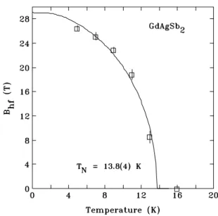
Figure 2.Temperature dependence of the155Gd M¨ossbauer
hyperfine field (Bhf) of GdAgSb2showing the N´eel temperature to be
13.8(4) K. The solid line is a fit using a J = 7
2Brillouin function,
appropriate for the Gd3+ion.
made in transmission mode. Temperatures down to 3.6 K were obtained using a closed-cycle refrigerator with the sample in a partial pressure of helium to ensure thermal uniformity. All full-pattern magnetic and structural refinements employed the FullProf/WinPlotr suite [11,12] with neutron scattering length coefficients for natural Gd taken from the tabulation by Lynn and Seeger [13], as detailed in our previous work on Gd3Ag4Sn4 [14]. No absorption correction was applied,
however the data were truncated at 2θ = 60◦to minimize the
impact of angle-dependent absorption effects.
3. Results
3.1.155Gd M¨ossbauer spectroscopy
The155Gd M¨ossbauer spectrum of GdAgSb2 at 16 K (above
TN) shown in figure 1 can be fitted as a paramagnetic
pattern with an axially symmetric electric field gradient and
e QVzz = 2.09(3) mm s−1. On cooling through TN, a clear
magnetic contribution develops and at 5 K the spectrum can be fitted with a hyperfine field (Bhf) of 26.5(5) T and an angle
(θ ) of 90◦between B
hfand Vzz. The 4mm point symmetry of
the Gd(2c) site in the ZrCuSi2-type structure requires that the
principal axis of the electric field gradient tensor lie along the crystallographic c-axis, so the observation that θ = 90◦means
that the Gd moments must lie in the ab-plane. This provides an independent and local confirmation of the ordering deduced from resonant x-ray scattering [7].
Tracking the temperature dependence of the hyperfine field allows us to determine the ordering temperature to be 13.8(4) K, as shown in figure 2. While this transition temperature is consistent with the 14.0 K originally reported by Solugub et al [2] on arc-melted samples, it is somewhat higher than the N´eel temperatures found by bulk measurements of single crystals (12.8(1) K [3]) and by xrms (13 K [7]). 2
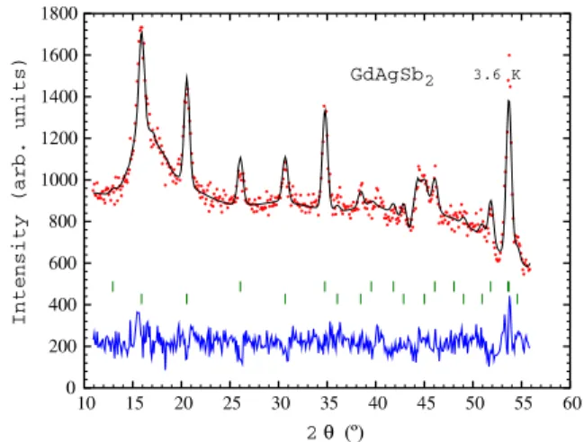
Figure 3.Neutron diffraction patterns taken at 16 K (above TN) and
3.6 K (well below TN) showing the additional magnetic reflections
that appear as the material orders. The calculated difference pattern shown at the bottom emphasizes the magnetic peaks, the most prominent of which have been indexed. The two measured patterns have been offset vertically for visualization purposes, however the dashed lines at the right show the locations of the actual zero values in each case.
Table 1. Crystallographic data for GdAgSb2in the tetragonal P4/nmm space group [3].
Atom Site Point symmetry x y z
Gd 2c 4mm 3 4 3 4 0.7641(1) Ag 2b ¯4m2 3 4 1 4 1 2 Sb(1) 2c 4mm 1 4 1 4 0.6811(1) Sb(2) 2a ¯4m2 34 14 0 3.2. Neutron diffraction
While the neutron diffraction signal from the GdAgSb2sample
is weak, many nuclear peaks are clearly seen in the 16 K pattern taken above the magnetic ordering temperature (figure3). The weak scattering signal means that background effects are far more visible, and three clear contributions are apparent in figure3. Below 2θ = 10◦spill-over from the straight-through
beam becomes significant, between 2θ ∼ 15◦ and 2θ ∼ 20◦
there is a broad feature due to secondary scattering from detector components, which also leads to a further contribution for 45◦<2θ < 50◦. The gradual fall-off in the background at
higher angles is in large part due to absorption by the increasing effective thickness of the flat plate seen at higher scattering angles.
The atomic position parameters used in our refinement of the neutron diffraction patterns were taken from the work of Myers et al [3] and are shown in table1.
Cooling below TN leads to the appearance of several
new peaks associated with the magnetic ordering. These are especially clear in the difference pattern shown at the bottom of figure3. These new peaks can be indexed to the original crystallographic cell by a simple doubling of the a-axis, and the first three are marked in figure 3 as (12 0 0), (12 0 1) and (12 0 2). This indexing immediately confirms the cell
Figure 4.Intensities of the (1
2 0 0) and ( 1
20 1) peaks for GdAgSb2as
functions of temperature showing an average N´eel temperature of 13.9 ± 1.4 K. The solid lines are fits to squared J =7
2 Brillouin
functions (the intensity is proportional to the moment squared).
doubling deduced from the resonant x-ray scattering work [7] and places the gadolinium moments in the ab-plane. We note here that the tetragonal symmetry of the P4/nmm space group places significant limits on what we can determine about the magnetic structure. As the a- and b-directions are completely equivalent they cannot be distinguished in a powder diffraction experiment. This means that we cannot tell the difference between the Gd moments being ordered parallel to the b-axis (as is implied by the indexing in figure 3) or parallel to the
a-axis (which would make the indices (0 12 0), (0 12 1) and (0 12 2)). Unfortunately the155Gd M¨ossbauer data cannot be used to distinguish between these two cases either as the axially symmetric electric field gradient tensor (itself a consequence of the tetragonal symmetry) also makes it impossible to determine the direction of the Gd moments within the ab-plane.
As with the M¨ossbauer hyperfine field, tracking the intensities of the two strongest magnetic reflections in figure4
yields an estimate of the ordering temperature as 13.9 ± 1.4 K, somewhat less precise than the M¨ossbauer value in figure 2
because the peaks are quite weak, however it is fully consistent with it. In particular, there is a clear magnetic signal present at 13 K in both the M¨ossbauer data (figure1) and the neutron diffraction pattern (figure4), strongly suggesting that the N´eel temperature is slightly above 13 K. The origin of the small (∼0.8 K) discrepancy between the current determinations and that previously made using x-ray resonant exchange scattering [7] is unknown at this time, but we emphasize that in all other respects, we are in full agreement with the previously reported magnetic structure.
The refinement of the diffraction pattern obtained at 3.6 K is shown in figure 5. The lattice parameters were fixed to the values obtained from the refinement of the nuclear pattern taken at 16 K. As shown in the difference plot (figure3), the antiferromagnetic order of the Gd sublattice leads to two clear peaks at 2θ = 15.9◦ and 2θ = 20.6◦, which correspond
J. Phys.: Condens. Matter 23 (2011) 106003 D H Ryan et al
Figure 5.Refined diffraction pattern for GdAgSb2obtained at 3.6 K.
The lattice parameters of a = 4.277(4) ˚A and c = 10.498(5) ˚A were taken from the nuclear-only pattern at 16 K. The solid line is a full-profile refinement of the nuclear and magnetic contributions. Two rows of Bragg markers are shown (top) nuclear contribution (bottom) magnetic contribution. The residuals are plotted at the bottom of the figure.
to the (1
2 0 0) and ( 1
2 0 1) reflections, respectively. A third,
weaker, magnetic reflection can be seen at 2θ = 30.7◦ which
corresponds to the (12 0 2) reflection. The observation of this cell doubling along a tetragonal axis has also been seen in TbAgSb2, DyAgSb2and ErAgSb2by Andr´e et al [6].
In full agreement with the resonant x-ray work of Song
et al[7], we obtained the best refinement to the 3.6 K pattern by using a [12 0 0] propagation vector with the Gd magnetic moments oriented in the tetragonal basal plane, transverse to the propagation vector. The refined Gd moment at 3.6 K is 6.2(3) µB. As noted above, the tetragonal crystal symmetry
makes it impossible to distinguish ordering along the a-axis with doubling of the b-axis, from ordering along the b-axis with doubling of the a-axis.
4. Conclusions
We have demonstrated that neutron powder diffraction can indeed be performed on Gd-based compounds at thermal wavelengths and that it readily yields data of sufficient quality to enable a magnetic structure determination.
Both155Gd M¨ossbauer spectroscopy and thermal neutron powder diffraction confirm that the N´eel temperature of GdAgSb2is 13.8(4) K and the Gd moments lie in the tetragonal
ab-plane. Analysis of the neutron diffraction pattern at 3.6 K shows that the magnetic cell is doubled along one of the basal axes and that the 6.2(3) µB Gd moments are ordered within
the basal plane but oriented perpendicular to the doubling direction. These results confirm and extend the description of the magnetic ordering derived by Song et al from x-ray resonant exchange scattering measurements [7].
Acknowledgments
JMC acknowledges support from the Canada Research Chairs programme. Financial support for various stages of this work was provided by the Natural Sciences and Engineering Research Council of Canada and Fonds Qu´eb´ecois de la Recherche sur la Nature et les Technologies. The research performed at the Ames Laboratory was supported by the US Department of Energy, Office of Basic Energy Science, Division of Materials Sciences and Engineering. Ames Laboratory is operated for the US Department of Energy by Iowa State University under Contract No. DE-AC02-07CH11358. Many useful discussions with A I Goldman (Ames Laboratory) are gratefully acknowledged. We gratefully acknowledge the assistance of Raghu Rao and Robert Speranzini in arranging for the activation of the 155Gd M¨ossbauer source in the National Research Universal (NRU) research reactor, which is operated by Atomic Energy of Canada Ltd at Chalk River, Ontario.
References
[1] Brylak M, M¨oller M H and Jeitschko W 1995 J. Solid State
Chem.115305
[2] Sologub O, No¨el H, Leithe-Jasper A, Rogl P and Bodak O I 1995 J. Solid State Chem.115441
[3] Myers K D, Bud’ko S L, Fisher I R, Islam Z, Kleinke H, Lacerda A H and Canfield P C 1999 J. Magn. Magn. Mater. 20527
[4] Myers K D, Bud’ko S L, Antropov V P, Harmon B N and Canfield P C 1999 Phys. Rev. B6013371
[5] Myers K D, Canfield P C, Kalatsky V A and Pokrovsky V L 1999 Phys. Rev. B591121
[6] Andr´e G, Bour´ee F, Kolenda M, Le´sniewska B, Ole´s A and Szytuła A 2000 Physica B292176
[7] Song C, Good W, Wermeille D, Goldman A I, Bud’ko S L and Canfield P C 2002 Phys. Rev. B65172415
[8] Ryan D H and Cranswick L M D 2008 J. Appl. Crystallogr. 41198–205
[9] Canfield P C and Fisk Z 1992 Phil. Mag. B651117 [10] Voyer C J and Ryan D H 2006 Hyperfine Interact.17091 [11] Rodr´ıguez-Carvajal J 1993 Physica B19255–69
[12] Roisnel T and Rodr´ıguez-Carvajal J 2001 Mater. Sci. Forum 378–81118–23
[13] Lynn J E and Seeger P A 1990 At. Data Nucl. Data Tables 44191
[14] Cadogan J M, Ryan D H, Napoletano M, Riani P and Cranswick L M D 2009 J. Phys.: Condens. Matter 21124201
![Table 1. Crystallographic data for GdAgSb 2 in the tetragonal P 4/nmm space group [3].](https://thumb-eu.123doks.com/thumbv2/123doknet/14136859.469791/4.892.94.419.97.356/table-crystallographic-data-gdagsb-tetragonal-nmm-space-group.webp)