Review
Modulation of contractility in human cardiac hypertrophy by myosin
essential light chain isoforms
Marcus C. Schaub
a,), Martin A. Hefti
a, Richard A. Zuellig
a, Ingo Morano
b aInstitute of Pharmacology, UniÕersity of Zurich, Winterthurerstrasse 190, CH-8057 Zurich, Switzerland
b
Max-Delbruck-Center for Molecular Medicine, D-13122 Berlin, Germany¨
Received 15 August 1997; accepted 14 October 1997
Abstract
Cardiac hypertrophy is an adaptive response that normalizes wall stress and compensates for increased workload. It is accompanied by distinct qualitative and quantitative changes in the expression of protein isoforms concerning contractility, intracellular Ca2q-homeostasis and metabolism. Changes in the myosin subunit isoform expression improves contractility by an increase in force generation at a given
2q Ž 2q .
Ca -concentration increased Ca -sensitivity and by improving the economy of the chemo-mechanical transduction process per
Ž .
amount of utilised ATP increased duty ratio . In the human atrium this is achieved by partial replacement of the endogenous fast myosin by the ventricular slow-type heavy and light chains. In the hypertrophic human ventricle the slow-type b-myosin heavy chains remain
Ž .
unchanged, but the ectopic expression of the atrial myosin essential light chain ALC1 partially replaces the endogenous ventricular
Ž .
isoform VLC1 . The ventricular contractile apparatus with myosin containing ALC1 is characterised by faster cross-bridge kinetics, a higher Ca2q-sensitivity of force generation and an increased duty ratio. The mechanism for cross-bridge modulation relies on the extended Ala–Pro-rich N-terminus of the essential light chains of which the first eleven residues interact with the C-terminus of actin. A change in charge in this region between ALC1 and VLC1 explains their functional difference. The intracellular Ca2q-handling may be impaired in heart failure, resulting in either higher or lower cytosolic Ca2q-levels. Thus the state of the cardiomyocyte determines whether this hypertrophic adaptation remains beneficial or becomes detrimental during failure. Also discussed are the effects on contractility of long-term changes in isoform expression of other sarcomeric proteins. Positive and negative modulation of contractility by short-term phosphorylation reactions at multiple sites in the myosin regulatory light chain, troponin-I, troponin-T, a-tropomyosin and myosin binding protein-C are considered in detail. q 1998 Elsevier Science B.V.
Keywords: Cardiac hypertrophy; Heart failure; Cross-bridge kinetics; Contractility; Myosin heavy chain; Myosin light chain; Ca2q-sensitivity–tension relation
1. Introduction
Heart muscle performs work with high contractile
effi-w x
ciency 1,2 . Under increased workload it reacts with hy-pertrophy. This represents an adaptive response in order to normalise wall stress and compensate for the increased
w x
hemodynamic load 3 . When the load is chronically ele-vated, compensated hypertrophy may progress to pump failure. Except in the case of acute global ischemia which is accompanied by a collapse of energy production, it is not clear which process, or processes, are responsible for
)
Ž . Ž .
Corresponding author. Tel.: q41-1 635 5919; fax: q41-1 635 5708; e-mail: [email protected]
turning compensated hypertrophy into heart failure. Nu-m erous physiological characteristics concerning metabolism, contractility and cellular structure are found to be impaired in heart failure. However, contractile function is often preserved or even improved in hypertrophy as well as in failure when studied at the subcellular level, i.e. on permeabilised muscle fibers or on isolated proteins in the in vitro motility assay and in solution studies.
Ž .
The changes in the contractile apparatus Figs. 1 and 2 result from long-lasting changes in the gene expression
Ž w x.
program reviewed in 4,5 . Because cardiomyocytes are
Time for primary review 26 days. 0008-6363r98r$19.00 q 1998 Elsevier Science B.V. All rights reserved.
Ž .
Fig. 1. Scheme of the cardiac sarcomere in relation to the membranous structures responsible for the intracellular Ca2q-handling. TT, transverse tubule;
Ž 2q .
TC, terminal cisternae; DHPR, dihydropyridine receptor voltage sensor and slow inward Ca -channel at the TT membrane; RyR, ryanodine receptor
ŽCa2q-release channel of the SR with foot structures at the TC membrane 183 . M-line consists of myomesin, 165 kDa M-line protein and creatine. w x w x
kinase; Z-line contains mainly a-actinin, desmin and CapZ protein 32 . MyPB-C localizes to the eleven transverse stripes on either side of the M-line and binds to myosin and titin. Titin is anchored with its N-terminus at the Z-line and, running along the myosin filament, reaches the M-line with its C-terminal head portion where it interacts with myomesin and the M-line protein. Two nebulette molecules are associated with each actin filament, which originate
w x
with their C-terminus from the Z-line, and Tropomodulin caps the actin filaments at their pointed end 5 .
no longer able to increase muscle mass by proliferation they resort to hypertrophy. Hypertrophy does not mean more of the same but involves specific qualitative
alter-w x
ations in the cell phenotype 5–7 . Reexpression of ‘fetal genes’ which are normally active in the fetal period coding
Ž . Ž
for proteins such as b-myosin heavy chain b-MHC see
.
Table 1 for abbreviations , a-skeletal and a-smooth mus-cle actin or atrial natriuretic factor, may occur. The devel-opment of hypertrophy in humans is a slow and chronic process extending over years. However, the long-term changes in expression of contractile proteins is not simply a reactivation of the ‘fetal gene program’, but follows a pattern that seems to be determined by the physiological demands. These physiological demands are transmitted by a panoply of different stimuli, besides mechanical loading, involving hormones, catecholamines, growth factors,
cy-Ž w x.
tokines and vasoactive peptides reviewed in 8 . Most of these hypertrophic stimuli use different intracellular signal-ing pathways which results in individual phenotypic re-sponses as characterised by gene expression pattern, cell morphology and function. The effects of various
hyper-trophic stimuli are likely to be balanced in vivo, even during compensatory hypertrophy. However, the distur-bance of any single factor may tip this balance for the worse. The precise knowledge of the function of the contractile apparatus including its regulatory mechanisms in the normal heart and during hypertrophy is indispens-able when considering potential therapeutic strategies aimed at avoiding development of heart failure.
The purpose of this review is to put together recently published results on the structure and function of the
Ž .
myosin light chain MLC isoforms whose expression changes during the development of cardiac hypertrophy. The discussion includes the position and relation of the MLC in the myosin head next to the motor domain, the variation in MHC and MLC isoforms and their possible hybrid molecular assemblies in atrial and ventricular mus-cles. The knowledge how calcium controls the myosin cross-bridge kinetics is instrumental in delineating the
Ž .
details of the mechanism by which the atrial MLC1 ALC1 positively affects contractility of the ventricular muscle. The pathophysiological consequences of the expression of
Fig. 2. Schematic detail of the sarcomere with the proteins affecting contractility and which are described in the text. AF, thin actin filament;
Ž . 2q Ž .
MF, thick myosin filament; inhibitory TnI , Ca -binding TnC and
Ž .
tropomyosin binding TnT troponin components; S1, myosin
subfrag-Ž .
ment-1 myosin head portion ; ELC and RLC, essential and regulatory myosin light chains; MyBP-C, hypothetical localization of the myosin binding protein-C spaced 43 nm apart along the myosin filament. The M-line would be to the left and the Z-line to the right.
the ALC1 are discussed in view of the functional viability of the cardiomyocyte with regard to intracellular Ca2q -handling and energy supply during hypertrophy and in heart failure. In order to recognize the relevance of modu-lation of contractility by myosin essential light chain, we give an overview of other sarcomeric proteins associated with the contractile machinery and point out the role of protein phosphorylation in Ca2q-sensitivity and
cross-bridge kinetics. Taken together, we provide evidence for the functional significance of the change in MLC isoform expression in the human heart ventricle where the MHC remain unaltered during hypertrophy. In view of space limitation, reviews which contain references to primary information are often cited.
2. The myosin motor
Ž .
The myosin muscle motor myosin type-II is a
hexam-Ž .
eric protein 520 kDa consisting of two heavy chains
ŽMHC. of around 220 kDa Ž1930–1940 amino acid
. Ž .
residues and of two pairs of light chains MLC of around 20 kDa each. The MHC subunits, which provide both the motor and the filament forming properties, can each be divided into two functional domains: the globular
N-termi-Ž
nal head domain corresponding to the proteolytic
subfrag-.
ment-1 or S1 of 120 kDa and the elongated a-helical domain that, together with the corresponding domain of the second MHC, forms the coiled coil C-terminal rod
ŽFig. 2 . The S1 has an approximate length of 16.5 nm and.
the rod domain one of 140 nm. The S1 domain can be
Ž .
further subdivided into two parts, i the motor domain, running from the N-terminus through to residue 770 and containing the nucleotide binding pocket and the actin
Ž .
binding cleft, and ii the 8 nm long a-helical light chain
Ž .
binding or regulatory domain neck region which extends from residue 771 to residue 843 and links the motor
w x Ž .
domain to the rod domain 9,10 Fig. 3 . The a-helix of this regulatory domain is stabilized by non-covalent associ-ation of one MLC of each type. Near its C-terminus the
a-helix bends sharply at the residues W829–P830–W831
and this last stretch of the a-helix points along the myosin filament axis towards the middle of the sarcomere, before it joins the second MHC in the rod. The precise interac-tions of the MLC with the MHC in this region have been
w x
revealed by crystal structures of the chicken S1 9 and of the regulatory domain of the molluscan scallop myosin
w11 . The two MLC are located in series and are wrappedx
around the MHC in an anti-parallel orientation. In the
Ž .
chicken S1, MLC type-1 essential light chain binds to
Ž
residues from L783 to M806 and MLC type-2 regulatory
. w x
light chain to residues from E808 to L842 of MHC 9 . It
Table 1
Abbreviations, names and explanations
2q Ž .
A1, atrial a a -MHC myosin pCa, negative decadic logarithm of the free Ca -concentration
Ž .
A2, atrial bb -MHC myosin PKA, protein kinase-A
ALC1, atrial myosin essential light chain PKC, protein kinase-C
ALC2, atrial myosin regulatory light chain RLC, myosin regulatory light chain
2q
Ž .
CAMK, Ca -calmodulin dependent protein kinase S1, myosin subfragment-1 myosin head portion
cAMP, cyclic adenosine monophosphate SDS-PAGE, sodium dodecylsulfate polyacrylamide gel electrophoresis
2q
DCM, dilated cardiomyopathy SERCA2, cardiac Ca -pump of the SR
DTNB, dithiobis-nitrobenzoic acid SR, sarcoplasmic reticulum
EF, ejection fraction Tm, tropomyosin
2q
EF-hand, Ca -binding helix–loop–helix motif Tn, troponin complex
2q
ELC, myosin essential light chain TnC, Ca -binding Tn component FHC, familial hypertrophic cardiomyopathy TnI, inhibitory Tn component
ICM, ischemic cardiomyopathy TnT, Tm-binding Tn component
Ž .
MHC, myosin heavy chain V1, ventricular a a -MHC myosin
Ž .
MLC, myosin light chain V2, ventricular ab -MHC myosin
Ž .
MLCK, myosin light chain kinase V3, ventricular bb -MHC myosin
MyBP-C, myosin-binding protein-C VLC1, ventricular myosin essential light chain NYHA, New York heart association classification VLC2, ventricular myosin regulatory light chain Single amino acid residues are given by the three-letter code and protein sequences in the one-letter code.
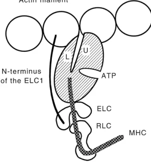
Fig. 3. Schematic drawing, approximately to scale, of the interactions of
Ž .
the myosin head subfragment-1 with the actin filament. The myosin
Ž .
motor domain hatched displays the ATP binding pocket and the actin
Ž . Ž .
binding cleft. The upper U and lower L portions of the 50 kDa segment are interacting with two adjacent actin monomers. MHC denotes the a-helix which constitutes the regulatory domain with the two bound
Ž .
light chains ELC and RLC . The extended N-terminus of the ELC1 reaches to the proximate actin. This state represents the rigor
conforma-w x
tion as modeled from the crystal structure 23 . The Z-line would be to the left and the M-line to the right. For further details see text.
has been agreed to give the position numbers for amino acids in the sequence of the chicken skeletal muscle MHC even when referring to mammalian skeletal or heart
mus-w x
cle myosins 12,13 .
Ž
Myosin cross-bridges myosin head portions protruding
.
from the myosin filament provoke movement during con-traction by repetitive interactions with the actin filament in the sarcomere. Thereby the cross-bridges are thought to swing through a number of power strokes, each time displacing the two filaments relative to one another by a
w x
distance of 5 to 10 nm 14,15 . Recent studies on
time-re-w x
solved X-ray diffraction of live frog sartorius muscles 16 and fluorescence polarisation coupled with rapid length
Ž
steps imposed on permeabilised rabbit psoas fibers
mech-. w x
anically or chemically skinned fibers 17 , show attach-ment of cross-bridges with actin and synchronized head movements in the elementary force-generating process. However, the rowing cross-bridge model as accepted in the
w x
textbooks since 1971 18 requires an update. The tip of the motor domain is now envisaged to bind actin with a more or less fixed geometry throughout the power stroke, while it is the distal C-terminal light chain binding region
w x
of the myosin molecule that actually moves 10,17,19 . The a-helix of this regulatory domain serves as a ‘lever arm’ to amplify relatively restricted rotational movements among different structural parts within the motor domain. It was shown that the length of the lever arm is directly
proportional to the sliding velocity in an in vitro motility
w x
assay system 20 . For this purpose, the lever arm of the S1 domain of myosin-II from the slime mould Dictyostelium discoideum, a myosin that is homologous to vertebrate striated muscle myosin, was altered in length by addition or subtraction of light chain binding regions by genetic engineering. The advent of nano-technology has recently allowed to determine discrete stepwise movement averag-ing 11 nm under low load and saverag-ingle force transients averaging 3 to 4 pN under isometric conditions of rabbit
w x
fast skeletal muscle myosin 21 . In this case, myosin molecules were fixed on silica beads and one molecule was let to interact with an actin filament held at both ends by a feedback enhanced laser trap system.
X-ray structural data of the individual proteins together with data obtained by fiber diffraction and electron mi-croscopy have been used to build detailed models of the
w x
rigor interaction of myosin with the actin filament 22–24 . In the contracting muscle this state is thought to occur transiently at the end of the power stroke after release of the products Pi and ADP before a new substrate ATP molecule gets bound to myosin. In the rigor state one myosin head is suggested to interact with two adjacent
Ž .
actin monomers Fig. 3 . A number of hydrophobic residues on the opposing faces of actin and myosin contribute to the main contacts. These contacts are flanked by charged myosin surface loops which form predominantly ionic interactions with adjacent regions of actin. Two such loops represent the connector regions between the segments of the MHC of S1 that can be obtained by proteolysis with trypsin, i.e. the 25-kDa, 50-kDa and 20-kDa segments
w x
starting from the N-terminus 25 . These loops exhibit considerable sequence variability and it appears that their composition influences the kinetic behavior of myosin from different sources. Replacement of the 50–20-loop in Dictyostelium myosin with the equivalent myosin se-quence from rabbit fast skeletal, chicken smooth or rat cardiac muscle leads to an actin activated ATPase activity that corresponds to the one of the respective parent myosin
w25 . The 25–50-loop has been suggested to affect thex
release of ADP from the nucleotide binding pocket and thus may be responsible for the observed differences in
w x
motility between different myosins 26 . In addition, and as discussed in the section on MLC below, the N-terminus of the large isoform of the essential MLC1 has also been shown to interact with the C-terminus of an actin monomer
ŽFig. 3 and to affect the actin activated ATPase activity..
3. Cardiac myosin heavy chains
At least eight different MHC types are expressed in mammalian striated muscles: two developmental isoforms
ŽMHC-embryonic and MHC-perinatal , four fast isoforms. ŽMHC-fast 2A, 2B, 2XrD and MHC-extraocular as well.
as MHC-slow which is identical to the cardiac b-MHC. In addition, the heart also expresses the cardiac a-MHC
Ž w x.
isoform for review see 27 . The human a-MHC contains
w x w x
1939 28 and the b-MHC 1935 29 amino acid residues. Among them they share 93.1% sequence identity. The rat
w x
a-MHC contains 1938 and the b-MHC 1935 residues 30 ,
and they share 93.2% sequence identity. A total of 131 residues differ between them and most of these differences are confined to regions of biological significance in the S1 subfragment such as the N-terminus, the ATP binding pocket, the actin binding cleft, the light chain binding domain and in the two hinge regions further down in the rod domain. In inter-species comparison between human and rat, a-MHC shows over 97% sequence identity. The same holds for b-MHC.
During development and under pathophysiological con-ditions, the expression of the two cardiac MHC isoforms is regulated in a tissue-specific manner. Their expression is under additional hormonal control, notably by thyroid
w x
hormones 5,31,32 . In all mammalian species a-MHC is expressed in the atria throughout life. In small mammals such as rat and mouse, a-MHC is also the predominant isoform in the ventricles post partum and during adult-hood. In addition, a-MHC is expressed in extraocular muscles as well as in some mandibular muscles of carni-vores. b-MHC is expressed in the embryonicrfetal ventri-cles of all mammals and disappears soon after birth in small animal species. In larger animals such as rabbit, dog, pig and human, b-MHC remains the predominant
ventricu-Ž .
lar isoform throughout adulthood Table 2 .
The fact that the b-MHC is also expressed in skeletal muscle fibers of the slow type-1 allows to perform func-tional studies on mutated b-MHC from patients with
famil-Ž .
ial hypertrophic cardiomyopathy FHC . In the in vitro motility assay of purified myosin from the soleus muscle of FHC patients with seven different point mutations in the
b-MHC gene, the velocity of actin translocation was on
average about half or less than that of myosin from healthy
Ž y1. w x
control persons average control value 0.48 mm s 33 .
In addition, myosin prepared from interventricular septal sites as well as from the soleus muscle of FHC patients
Ž .
with the R403Q position 405 in chicken mutation
exhib-Ž y1
ited the same low motility 0.11 and 0.13 mm s ,
respec-.
tively . Permeabilised slow fibers from the soleus muscle
Ž
of FHC patients with either the R403Q or G741R position
.
743 in chicken mutations also showed impaired contrac-tile functions, although the fraction of mutant MHC in
w x
these fibers was not known 34 .
4. Cardiac myosin light chains
Ž .
The myosin light chains MLC comprise two
subfami-Ž .
lies, the essential light chains ELC and the regulatory
Ž .
light chains RLC . Both MLC subfamilies belong, to-gether with the calmodulin and the troponin-C subfamilies, to the superfamily of intracellular Ca2q-binding proteins
that characteristically contain four EF-hand domains
Žhelix–loop–helix motif. w35 . However, during evolution,x all four EF-domains of vertebrate muscle myosin ELC have lost their ability to bind Ca2q, while in the case of the RLC only the first EF-domain can bind either Ca2q or Mg2q with high affinity in situ in the myosin molecule
Žbinding affinities in the range of 10 M7 y1. w36 . The ELCx
is called ‘essential’ because it was originally thought to be essential for the hydrolytic activity of myosin; but later,
w x
this proved not to be the case 37,38 . Alternative names
Ž
are alkali MLC since ELC can be removed from myosin
.
by elevated pH or MLC1. The RLC is termed ‘regulatory’ because in vertebrate smooth muscle and non-muscle cells, contractile activity is triggered by phosphorylation of this
w x
myosin light chain 39 . Alternative names are DTNB-MLC
Žsince it can be removed from myosin by
DTNB-treat-. Ž .
ment , PLC it can be reversibly phosphorylated or MLC2. The ATPase activity of isolated myosin does not seem to be affected by the ELC or RLC. It has however become increasingly evident, that within the complex sarcomeric structure, the MLC are involved in the fine-tuning of the contractile activity.
Table 2
Ž .
Myosin heavy chain MHC and MLC1 isoform composition in all four chambers of six explanted hearts from patients with end-stage heart failure compared to five hearts from patients with compensated hemodynamics. Muscle specimens were obtained from the free wall of all four heart chambers.
w x
Relative content of MHC and MLC1 was densitometrically evaluated after electrophoretic resolution 68,69 . Type of disease and functional state of the patients is given in the text. Average percentages"SEM are given
Heart condition Protein isoform Right atrium Left atrium Right ventricle Left ventricle
Compensated hearts a-MHC 90"2.1 93"3.4 - 3 - 3
b-MHC 10"2.1 7"3.4 100 100
ALC1 100 100 -1 -1
VLC1 -1 -1 100 100
Failing hearts a-MHC 52"9 56" - 3 - 3
b-MHC 48"9 44"9 100 100
ALC1 90.3"2.1 91.9"3.3 5.5"1.4 7.8"4.4
Two varieties of ELC and RLC are expressed in the heart, characteristic for the atrial and the ventricular tis-sues. They are thus designated ALC1 and VLC1 for atrial and ventricular ELC, respectively, and ALC2 and VLC2 for the corresponding RLC species. ALC1 and VLC1
Žhuman and rat contain 190–200 Table 3 and the VLC2. Ž . Ž
around 165 amino acid residues no data available for
. w x
ALC2 35 . The molecular mass for the MLC1 and MLC2 types is around 22 and 19 kDa, respectively. However, the MLC1 migrates with a higher apparent mass of around 27 kDa in SDS gel-electrophoresis because of its particular N-terminus. The most striking difference between the two types of MLC concerns an additional peptide stretch of around 30 residues at the N-terminus of the ELC. An unusual accumulation of around 10 Ala and 10 Pro is found in this extra stretch. In addition, several positively and negatively charged amino acid residues are clustered
Ž .
near the N-terminus Table 3 . Such sequences rich in Ala
w x
and Pro form rigid extended structures 40 . It has been shown by biochemical in solution studies that the
N-Ž .
terminus of fast skeletal muscle ELC ELC1fast binds to
w x
the C-terminus of actin 41,42 . This extended N-terminal structure of the ELC1fast cannot be seen in the crystal
w x
structure of the chicken S1 subfragment 9 . The last visible residue in the fast skeletal muscle ELC corresponds to positions 46–53 in the sequences of human and rat ALC1 and VLC1, and this residue lies roughly in the centre of the 3-dimensional structure of the ELC at a distance of 7–8 nm from the actin filament surface. Thus a stretch of around 50 residues is available for the N-terminus of ALC1 and VLC1 to bridge the gap of 7–8 nm and to contact the actin filament. In principle, only 24 residues are required to span a distance of 8 nm if present in an
extended b-sheet conformation. Taken together, the odd 50 N-terminal residues in ALC1 and VLC1 are ample to run along the myosin head moiety and to interact with a
Ž . w x
proximate actin monomer Fig. 3 24 . It has recently been shown that the first 11 N-terminal residues of human ALC1 are responsible for binding to the actin C-terminus
w43 . The interaction of the N-terminus of ELC has indeedx
been shown to affect contractility and actin activated
AT-Ž
Pase activity in skeletal muscle protein systems for
re-w x.
views see 44–47 . It is this structural feature that seems to be of relevance for the pathophysiological aspects of cardiac hypertrophy discussed below. Interestingly, all ELC isoforms expressed in vertebrate striated muscles have such an extended N-terminus that is found neither in invertebrate ELC nor in the vertebrate smooth and non-muscle myosin ELC.
The ALC1 is identical to the embryonic ELC1emb which is also transiently expressed in fetal skeletal muscles
w x
as well as in fetal ventricular heart tissue 48 . On the other hand, the VLC1 is identical to the skeletal muscle ELC1bslow which is expressed in adult slow skeletal
w x
muscles 49 . VLC2 is also expressed in adult slow skeletal muscles as RLC2slow. An additional ventricular RLC designated VLC2)
that has a different isoelectric point but the same mass as VLC2 and can be resolved by
2-dimen-Ž .
sional gel electrophoresis Fig. 4 , is found in ventricles of
w x
most mammals 50,51 . Both VLC2 varieties have differ-ent amino acid sequences and each contains one phospho-rylation site for a Ca2q-calmodulin dependent light chain
Ž . w x
kinase MLCK 52 . In contrast, the sole ALC2 variety in the human atrium can be phosphorylated at two sites. The significance of this feature for cardiac contractility is,
w x
however, not known 53 .
Table 3
Ž .
Amino acid sequence and charge distribution of the first 20 residues of the pre-domain of the myosin light chain type-1 MLC1 from human and rat
w x
cardiac and fast skeletal muscles. Numbering is based on the mature protein, neglecting the N-terminal Met 47 . Following the pre-domain, the rest of the
Ž
MLC1 comprises the four EF-domains with their respective interdomains and end with the last residue of the fourth EF-domain this part contains 143
.
amino acid residues in each case . For comparison the charge distribution is also given for the last 20 residues in the sequence of mammalian cardiac
w x
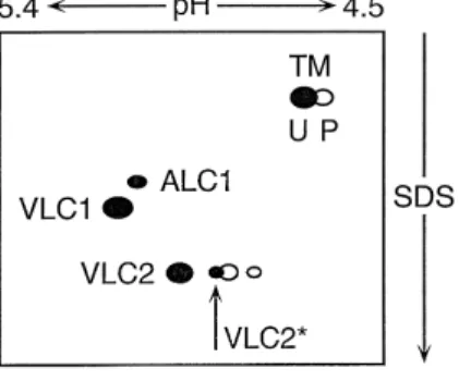
Fig. 4. Schematic drawing of the myosin light chains and a-tropomyosin
ŽTM resolved by two-dimensional gel electrophoresis from the ventricle.
of patients with hypertrophy andror end-stage heart failure. First dimen-sion, isoelectric focusing and second dimension in SDS with decreasing
Ž . Ž .
molecular mass. U unphosphorylated and P phosphorylated form of
)
Ž a-tropomyosin. Both VLC2 and VLC2 can be phosphorylated large
.
and small open symbols, respectively . Atrial ALC1 is present in addition
w x
to the endogenous VLC1 63,68,69 . For further details see text.
Several recently detected missense point mutations in either the ELC or RLC are associated with a rare variant of
w x
FHC and with skeletal muscle myopathy 54 . In contrast to most mutations in the MHC of FHC patients which are
w x
associated with impaired motility 33 , myosin from a patient with the M149V mutation in the VLC1 displayed an actin filament translocation velocity that was increased by 41% as compared to control myosin. The two mutations M149V and R154H found in the VLC1 are localized in the region where it contacts the motor domain of the MHC. This putative interaction may correspond to the base around
Ž .
which the regulatory domain lever arm moves during
Ž . w x
contraction Fig. 3 10 . Furthermore, three mutations in
Ž .
the VLC2 A13T, E22K and P94R all seem to be
local-w x
ized near the phosphorylation site at S15 54 .
5. Combinatorial associations of multiple myosin iso-form subunits
It is known that the different isoforms of MHC as well as of MLC are responsible for distinct contractile proper-ties. Therefore it is difficult to accept the idea that this isoform variety simply reflects an evolutionary relict of progredient accumulation of gene duplications, mutations and chromosomal rearrangements. The strict regulation during development and tissue-specific mode of expression seems more likely to favor the notion that the various isoforms present a store from which to choose in order to build molecular motors with functional isoform advan-tages. It might therefore be useful to consider the different heteromeric molecular assemblies that may possibly occur under normal and under pathological conditions.
Under normal conditions in adult human hearts the
a-MHC together with ALC1 and ALC2 are confined to the
atrium, while the b-MHC together with VLC1, VLC2 and
VLC2)
are to be found in the ventricles. Some b-MHC
Ž .
seems to accumulate in atria with increasing age Table 2 , which in the atrial appendages may reach values up to
w x
50% of total MHC 55–57 . However, pressure overload models in animals as well as hemodynamic overload in humans leads to characteristic changes in myosin composi-tion in both atria and ventricles. The ventricular myosin subunit isoforms VLC1 and VLC2 become ectopically expressed in atria in small mammals as well as in humans
w31,58 . b-MHC increases in human atria up to 50% orx
Ž . w x
more Table 2 56 and becomes reexpressed in ventricles of small animals which normally display predominantly
a-MHC. In normal human ventricles a small amount of a-MHC, estimated to vary between low levels and 10%,
w x
can be observed beside the abundant b-MHC 59–62 . This residual pool of a-MHC is absent in patients under hemodynamic overload. Electrophoretic resolution of na-tive myosin isolated from ventricles of hemodynamically overloaded small animals with varying proportions of a-MHC and b-a-MHC yields three species designated V1, V2 and V3 according to electrophoretic mobility. V1 and V3 consist of homodimeric a a and bb combinations, respec-tively, and V2 represents the heterodimer ab. Interest-ingly, myosin from overloaded atria resolves into two
Ž . Ž .
species only, namely A1 a a and A2 bb . A1 and A2 do not co-migrate exactly with the corresponding ventricu-lar species V1 and V3, because they contain different
w x
MLC complements 31 .
Ž
In normal human atria, two MHC homodimers a a and
.
bb together with ALC1 and ALC2 give rise to two myosin isoforms. In hemodynamically overloaded human atria, we are confronted with the co-existence of the two
Ž
MHC homodimers with two types of MLC ALC1 and
.
ALC2 as well as VLC1 and VLC2 . Both MHC homod-imers may combine with each MLC type in three ways forming two homodimers and one heterodimer with regard
Ž
to the MLC ALC1rALC1, VLC1rVLC1 and
ALC1rVLC1 as well as ALC2rALC2, VLC2rVLC2 and
.
ALC2rVLC2 . Combinatorially, this allows for 2 = 3 = 3
s18 different myosin species. Since both MLC1 and
MLC2 are present in two different isoforms, the sequential arrangement along one MHC may also play a role. Two sequential arrangements are possible in one myosin molecule: VLC1–VLC2rALC1–ALC2 as well as VLC1– ALC2rALC1–VLC2. Therefore, one additional species
Ž .
has to be added for each MHC homodimer a a and bb making a total of 20 instead of 18 isoforms. This number would increase even to 42 if the ventricular VLC2)
were also considered. This isoform has been shown to be ex-pressed in a proportion of VLC2)
to VLC2 of around 0.4
w x
in control as well as in overloaded human ventricles 63 . VLC2)
has been shown to occur also in significant
w x
amounts in overloaded human atria 64 . In any case, multiple possible myosin isoforms could account for the
w x
altered contractility of diseased atrium 65,66 when com-pared to normal atrium.
In normal human ventricle, mainly b-MHC and a small amount of a-MHC beside the VLC1 and two types of
Ž )
.
MLC2 VLC2 and VLC2 are present. Ignoring the rare
a-MHC, this would give rise to three myosin isoforms
co n tain in g V L C 2 r V L C 2 , V L C 2 r V L C 2)
o r VLC2)r
VLC2)
. As mentioned above, most of the a-MHC disappears under hemodynamic overload, leaving only the b-MHC species. However, we have reported that under such conditions the ALC1 becomes reexpressed in the ventricle varying in amounts up to 30% of total MLC1
ŽFig. 4 and Table 2. w67–69 . Therefore, in diseasedx human ventricle the bb-homodimer may combine with either VLC1 andror ALC1 to form two homodimers
ŽVLC1rVLC1 or ALC1rALC1. and the heterodimer VLC1rALC1. The same type of combinations are, of course, also possible with regard to the VLC2 and VLC2)
. Thus, in the diseased human ventricle as many as 1 = 3 = 3 s 9 myosin isoforms may co-exist. The combination with four different MLC in one molecule allows also in this case two possible sequential alignments on the MHC. This brings the total number of possible isoforms in the overloaded ventricle to 10 instead of 9.
As discussed below, shifts in expression of cardiac MHC as well as MLC are known to affect the contractile properties. In order to form heterodimeric molecules, the corresponding isoform subunits must be expressed in the same cell. This seems to be the case for ventricular myocytes as probed with monoclonal antibodies against
w x
a-MHC and b-MHC 62,70 . Atrial myocytes have also
w x
been shown to co-express a-MHC and b-MHC 55 ; nev-ertheless, MHC heterodimers are not observed in atrial tissue. Whether or not all possible subunit associations discussed above are actually realized in vivo is not known. Functional myosin requires a strict stoichiometric relation of its subunits. The parallel occurrence of b-MHC and VLC2 in the atria of hypertensive baboons was taken to
w x
indicate a coordinated expression of myosin subunits 71 . However, cardiac MLC exhibit a slower turnover rate than
w x
MHC 72,73 . Consequently there is a pool of unassembled MLC in the cytoplasm. In accordance, we found an excess of around 0.8 molar of VLC1 over MHC in human
ventri-w x
cle 69 . The additional expression of ALC1 in hyper-trophic ventricles added up to an excess of 3.5 molar of
Ž .
total MLC1 VLC1 plus ALC1 over MHC. Isolation and analysis of myosin from such tissue indicated that, com-pared to VLC1, the proportional contribution of ALC1 bound to bb-myosin was roughly the same as its content in total tissue samples. In contrast, in transgenic mice ectopically expressing the skeletal myosin ELC2fast, in-corporation into cardiac myosin was not proportional to
w x
the total amount actually expressed 74 . While in atria the foreign ELC2fast replaced the endogenous ALC2 almost entirely, this hardly occurred in the ventricles, despite the fact that in both tissues a a-myosin was the recipient protein. These cases illustrate that human ventricular bb-myosin readily combines with ALC1 and that murine atrial
a a-myosin accepts skeletal muscle ELC2fast while its
Ž .
ventricular counterpart also a a-myosin does not. It may thus be emphasised that correlated regulation of expression of MHC and MLC is not required for stoichiometric assembly. In general, synthesis of MLC may be in excess over that of MHC and a large portion of the unassembled MLC will probably be degraded. On the other hand, we observed a far larger amount of b-MHC in whole tissue samples of hemodynamically overloaded human atria than
Ž .
VLC1 Table 2 .
A further aspect may shed some light on the question of heteromeric myosin assemblies. During development the expression of MHC and MLC isoforms undergoes transi-tions in atria and ventricle which are temporally not closely
w x
linked 57 . This implies a lack of isotype-specific interac-tions between the MHC and MLC. Taken together, the available evidence suggests that different MLC can indeed combine with different MHC. Furthermore, the MHC and MLC may be regulated independently of one another and thus allow for a variety of mixed isotype assemblies to occur in vivo.
6. Myosin cross-bridge kinetics
Cardiac contractility is directly related to the type of predominant myosin species. The motor protein defines the frame within which contractility may vary in terms of
Ž .
force production unitary force per cross-bridge cycle and
Ž .
velocity of displacement kinetics . The structural and
Ž
mechanical changes in the cross-bridge a series of
succes-.
sive conformational changes in the myosin head domain are coupled to a concomitant series of chemical reactions involving the hydrolysis of ATP to ADP and inorganic
Ž w x.
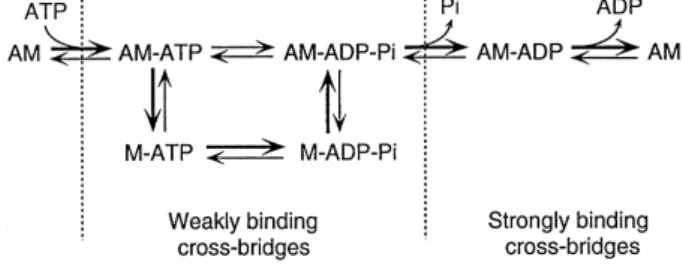
phosphate confer 75,76 . The simplified scheme in Fig. 5 combines the mechanical and kinetic cycles and delin-eates the main intermediate states in the actin–myosin
Ž . Ž .
cross-bridge cycle. The affinity of myosin M to actin A changes during the cross-bridge cycle. The species with
Ž
bound ATP or its products resulting from hydrolysis ADP
.
and Pi exhibit low affinity, do not produce force and
Ž .
oscillate rapidly on a time scale of ms or less between attached and detached states. Consequently, they do not present significant resistance to stretch of relaxed muscle.
Fig. 5. Simplified kinetic scheme of the cross-bridge cycle. M, myosin; A, actin. Main reaction pathways are indicated by heavy arrows. For explanations see text.
Force production is thought to be coupled to the release of Pi and comprises the transition from weakly to strongly
w x
binding states 77,78 . These force generating cross-bridges exhibit a slower rate of oscillation between attachment and detachment bind to actin with positive cooperativity. This results in an increased resistance to passive stretch. The complete cross-bridge cycle is terminated in the nu-cleotide-free state after dissociation of ADP which resem-bles the state of rigor mortis. In vivo, upon binding of a new substrate ATP, the cross-bridge dissociates from actin and the cycle is ready to start again.
Measurements of contractile parameters such as isomet-ric tension, shortening velocity and force redevelopment following a brief period of shortening at zero load can provide information about the distribution of cross-bridges among weakly and strongly binding states and the kinetics
w x
of transitions between them 79 . The first quantitative model which related experimental parameters to the kinet-ics of the cross-bridge cycle considered two states: one attached and force-generating, and one detached
non-w x
force-generating 80 . These two states are related by two rate constants: the forward rate constant f determining the rate of attachment, and the forward rate constant g deter-mining the rate of detachment. The same model is now applied to the reaction scheme with many intermediate
w x
cross-bridge states 76 . In analogy to the original model, the rate constant fapp now describes the transition from weakly binding, non-force generating states, to the strongly binding, force-generating states, and gapp describes that for the opposite transition via product release and
rebind-Ž .
ing of ATP Fig. 5 . Since weakly binding non-force-gen-erating cross-bridges are still in rapid equilibrium with
w x
actin 81 , the rate constants no longer describe attachment
Ž .
and detachment, respectively. The isometric force F of a muscle can thus be described as
fapp X F s F P n Ptot f qg app app X Ž .
where F is the force unitary force generated per cross-bridge and ntot is the total amount of cycling cross-bridges
Ž .
per half-sarcomere. The expression f r f qg rep-app app app
resents the fraction of cycling cross-bridges in the force-generating states, and hence, steady-state tension. Both an increase in fapp or a decrease in gapp will increase the fraction of force-generating cross-bridges and, therefore, increase tension generation of a muscle. The rate of transi-tion from weakly to strongly binding states may be esti-mated from the rate of tension development after a brief
w x
shortening at zero load 76,82 . The rate constant of
ten-Ž . Ž .
sion redevelopment ktd equals fappqgapp . The
un-Ž .
loaded maximal shortening velocity Vmax primarily de-pends on gapp which, in turn, is controlled by the release
w x
of ADP 83 . The ATPase activity during isometric
w x
steady-state tension can be described by the equation 82 ATPase s n P s P ftot appP gappr
Ž
fappqgapp.
where s is the number of half sarcomeres. Tension cost is then obtained by dividing the ATPase-equation by the force-equation yielding gappP srFX. Under isometric steady-state conditions s is constant, and if FX is equal between two myosin isoforms under consideration, then the relation of ATPase activity and tension may be directly proportional to gapp. An increase in gapp would predict a decrease in force, and vice versa.
To illustrate the complexity of the interrelation between the mechanical and kinetic properties of contractility, let us consider a muscle fiber that is stimulated to contract. The amount of force produced depends on how it is free to move. The velocity at which the fiber shortens depends on how great a load it must bear. If no load is applied, the
Ž .
fiber shortens at the maximal speed Vmax . If a force sufficient to prevent shortening is applied, the fiber devel-ops force under isometric conditions. Whatever parameter one measures, and regardless of how it is measured, the results are not only determined by the intrinsic properties of the motor protein alone, but also by the continuously varying mechanical conditions imposed on the system by the reaction partner actin as well as by the regulatory proteins in the sarcomere. This complex ensemble is
re-w x
flected in the force–velocity relation 32,79 . In perform-ing work against a load, muscle generates power, which is work per unit of time and is obtained by multiplying force with velocity. The maximal power output depends on Vmax,
Ž .
isometric tension P0 and on the curvature of the force–
w x
velocity curve 84 . The shape of the curve is dependent on the force coefficient a, normalised to isometric force s arP . The power output is zero during shortening without0 load at Vma x as well as under isometric conditions. Since force and velocity are inversely related, their product will be greatest at an intermediate load. It reaches a maximum at around 20–40% of Vma x while at the same time produc-ing around 30% of isometric tension.
P0 is determined by the number of attached cross-bridges per cross-sectional area at any given time, multi-plied by the force development of a single cross-bridge. The actual number of attached cross-bridges can in turn be estimated by multiplying the total number of cross-bridges
Ž .n by the fraction of time spent in the force-generating
Ž .
state duty ratio or duty cycle . Vmax under zero load cannot be measured directly and has to be extrapolated from the force–velocity curve, or better, may be
deter-w x
mined by Edman’s slack-test 85 . Determination of iso-metric tension of permeabilised muscle fibers yields incon-sistent results. This may be due, at least in part, to the
Ž w x .
experimental conditions see ref. 32 for discussion . In
Ž .
general, the specific tension force per cross-sectional area does not vary much among different muscles or animal
w x
species 86,87 . In vivo, movements are seldom performed
Ž .
at a speed close to Vmax. The range of speed VrVmax at which muscles mostly work, is close to the one where the
w x
maximal power output is generated 88 . Since Vmax of fast skeletal muscle fibers is about 2–4 times higher than in
slow fibers, the maximal power output is much lower in
w x
slow than in fast fibers 89 . It has been suggested that the maximal power output may be taken as a more reliable index of dynamic mechanical properties of a muscle than
w x
Vma x 86 . In view of the complex muscular mechanics, the diversity of myosin isoform compositions may be required to meet the physiological demands for a large number of optimal combinations between the power output and the corresponding shortening velocity in order to perform movement on an economic basis.
7. Ca2H-control of cross-bridge activity
The twitch properties of fast and slow skeletal muscles depend not only on the contractile apparatus but also on the Ca2q-transients in the cytosol. This is even more so in
the heart. The heart muscles do not relax fully during diastole, and neither do they contract maximally during
w x 2q
systole 90,91 . At submaximal Ca -concentrations, the rate of tension development shows pronounced Ca2q
-sensi-tivity. The sensitivity of the myofibrillar apparatus to the activating Ca2q is generally expressed by the pCa–tension relation as determined under isometric steady-state condi-tions on permeabilised fiber preparacondi-tions. Some studies have also been carried out on intact fibers using intra-cellular Ca2q-indicators. pCa denotes the negative decadic logarithm of the free Ca2q-concentration. The normalized
w x
tension–pCa data can be fitted to the Hill equation 92 H 2q
w
Cax
Y s H 2qw
x
pCa q Ca50where Y is the fractional force, pCa50 is the Ca2q -con-centration resulting in half-maximal activation, and H is an index for cooperativity. The value of pCa50 provides an index for the affinity of Ca2q to the contractile system.
The coefficient H gives the minimal number of
cooperat-Ž .
ing binding sites. Since cardiac troponin-C TnC contains only one functional Ca2q-binding regulatory site, any value
of H ) 1 indicates cooperativity involving multiple
com-Ž
plexes of the regulatory system on the actin filament for
w x.
references see 93 . Schematically, the cardiomyocyte may
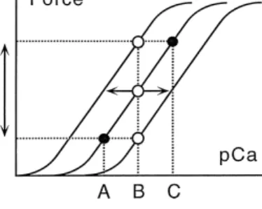
Ž .
modulate force of contraction in two ways Fig. 6 . First, force of contraction may be altered by changing the ampli-tude of the cytosolic Ca2q-transients. A rightward shift to higher Ca2q-concentration from A to C on the abscissa causes an increase in force by shifting the degree of activation from the lower to the upper filled circle on the
Ž .
same intermediate tension–pCa curve Fig. 6 . Second, changes in the sensitivity of the contractile apparatus to Ca2qmay equally allow alterations in the force of contrac-tion. A shift of the tension–pCa curve to the left or to the right as indicated by the horizontal arrows at a given Ca2q-concentration B, will increase or decrease the degree
Fig. 6. Schematic illustration how the cardiomyocyte may modulate contractile force by two main mechanisms. Force is plotted versus log of increasing Ca2q-concentrations. Increasing Ca2q from A to C increases
2q
Ž
force in proportion without affecting the Ca -sensitivity filled circles on
. 2q Ž .
the same curve . At a given Ca -concentration B open circle , force can be increased by increasing the Ca2q-sensitivity of the contractile system Žleftward shift , or lowered by decreasing Ca. 2q-sensitivity rightwardŽ
.
shift . The steepness of the force–pCa curve is a measure for
cooperativ-Ž .
ity of the activation process Hill coefficient and is unchanged in the given examples. For further discussion see text.
Ž .
of activation open circles , respectively. The contractile
Ž .
force will be altered accordingly. The Hill coefficient H determines the steepness of the tension–pCa curve.
In general, permeabilised fibers from slow muscles exhibit a higher Ca2q-sensitivity than fibers from fast
w x
muscles 94 . For activation levels up to around 20% of maximal tension, increasing Ca2q seems to increase n
tot Žrecruitment of cross-bridges . At higher levels of activa-.
tion, Ca2q seems to increase tension by increasing f , app
thus affecting the cross-bridge turnover kinetics directly. These two modes of regulation do overlap, however, when going from rest to activation. The effect of Ca2qon f is
app
consistent with the observation that Ca2qhas only a small,
if any, effect on Vmax, since this latter is mainly determined
2q w x
by gapp which is almost independent of Ca 76 . Thus, factors which may affect cross-bridge kinetics via fapp or gapp can become important parameters for modulation of muscle activity.
8. Effects of myosin subunit isoforms on contractility
In general, Vmax is roughly proportional to the ATPase activity and speed of contraction in heart as well as in
Ž w x.
skeletal muscles for reviews see 32,79 . The ATPase
Ž .
activity is highest in V1 a a , intermediate in the
het-Ž . Ž .
erodimer V2 ab and lowest in V3 bb cardiac isomyosin
w x 2q
of a given species 31 . In skeletal muscles the Ca -sensi-tivity is about 0.2–0.3 pCa units higher in slow than in fast fibers. On the other hand, fast fibers exhibit a much steeper tension–pCa curve with an H value of 4–6, while slow fibers have one of 2–3. Human cardiac fibers from atrium and ventricle have an equally low H value of 2–3
w51 . This may have to do with the fact that the cardiacx
TnC is also expressed in slow skeletal muscle fibers. This cardiac TnC isoform contains only one Ca2q-binding
regu-latory site instead of the two in the fast skeletal muscle
w x 2q
TnCf 95 . In addition, the Ca -sensitivity is 0.17 units higher in ventricular than in atrial fibers concomitant with
Ž .
an unloaded Vmax given as muscle length per second of 2.6 for atrial and a lower one of 1.7 for ventricular fibers
w51 . These characteristic differences seem to be mainlyx
due to the atrial myosin containing mainly a-MHC as opposed to the ventricular myosin which in man contains predominantly b-MHC. Measuring the ATPase activity during isometric tension generation at various Ca2q
-con-centrations allows to establish the ATPase-force relation
w x
for human atrial and ventricular muscle fibers 96 . Force at saturating Ca2q-concentrations was 14.0 for atrial and
21.1 kNrm2 for ventricular fibers. The amount of ATP
Ž .
used per force tension cost was independent of the Ca2q-concentration and just about three times higher for atrial than for ventricular fibers. The Ca2q-sensitivity was again 0.08 pCa units higher in ventricular fibers. While both Vmax and isometric tension generation differ only moderately between atrial and ventricular fibers, the differ-ence in the rate of tension development is quite
pro-Ž .
nounced. The rate constant of tension development ktd after photolytic release of ATP from ‘caged ATP’ is seven-fold higher in atrial than in ventricular porcine
w x
muscle fibers 97 .
As outlined above, the ATPase-force relation is propor-tional to gapp and the force of a muscle depends on its cross-bridge kinetics. Taken together, these results suggest that atrial myosin cross-bridges differ in their kinetics from the ventricular myosin by having a higher gapp, implying a faster transition rate from force-generating into non-force
Ž X.
generating states. If the unitary force value F were different, one fiber type would still generate different absolute tension at a given Ca2q-concentration, but it should not change the normalized tension–pCa curve. A lower gapp in the kinetic of V3 myosin is consistent with a longer fraction of the cross-bridge cycle being spent in the
Ž .
force-generating states increased duty ratio or duty cycle . This difference in kinetics between human atrial and ven-tricular myosin is thought to reside in the a-MHC and
b-MHC. It has to be born in mind, however, that these two
myosins differ in their MLC complement.
V1 and V3 myosin isoforms with the same ventricular
Ž .
MLC complement VLC1 and VLC2 can be generated in
Ž
rabbits by making them hypothyroid producing V3 myosin
. Ž
by feeding them propylthiouracil or hyperthyroid
produc-. w x
ing V1 myosin by administering levothyroxine 98 . V1 myosin has twice the actin activated ATPase activity and three times the actin filament sliding velocity in an in vitro motility assay system, when compared to V3 myosin. Yet, V1 myosin produces only half the average cross-bridge
Ž
force per cycle 0.15 pN for V1 and 0.30 pN for V3
.
myosin . These findings also point to a kinetic difference between the a-MHC and the b-MHC in V1 and V3 myosin, respectively. Such a difference in kinetics may provide the molecular basis for the observation made
w x
earlier by myothermal techniques 99 . The economy of
Ž
ATP utilisation for isometric force production i.e.
cross-.
bridge tension–time integral per ATP by V3 was found to be twice that for V1 myosin. The force–time integral reflects the cross-bridge attachment time and is positively related to economy of contraction and negatively related to ATPase activity, shortening velocity and cross-bridge cy-cling rate. It should be noted that slower actin translocation velocities are not always associated with higher average cross-bridge force. Both V1 and V3 myosin have slower actin filament velocities than fast skeletal muscle myosin,
w x
but neither generates greater average force 100,101 . Hyperthyroid rats supplied with triiodothyronine con-tain exclusively a-MHC in the atria as well as in the
w x
ventricles 102 . There is no evidence that the tissue-specific MLC isoforms of atrium and ventricles undergo changes in relation to the thyroid state. The actin filament proteins do not seem to differ between these two tissues
Žreviewed in w102 . Thus, atrial and ventricular my-x. ocardium of hyperthyroid rats have identical composition of myofibrillar proteins except for the tissue-specific MLC.
Ž
ATPase activities of myofibrils in solution fully
stimu-.
lated at pCa 4.4 and when inhibited at pCa ) 8 as well as of isolated myosin are the same in atrial and in ventricular preparations, yet the contractile properties differ markedly between permeabilised fibers from atrium and ventricle. Isometric tension is 9.7 for atrial and 22.6 kNrm2 for
Ž
ventricular fibers compare corresponding values given
.
above for human . Maximal shortening velocity and maxi-mal power output are almost twice as high in atrial fibers
w102 . These findings suggest that in addition to the MHCx
also the MLC contribute to the contractile properties, while the myofibrillar ATPase activity in solution is determined entirely by the MHC species. The possible involvement of the MLC in modulation of contractility and ATPase activ-ity has recently been suggested for skeletal muscle systems
w32,46,103 .x
9. Modulation of contractility in hypertrophy by the myosin essential light chain
The relative composition of MHC and MLC1 isoforms in tissue samples from the free wall of all four chambers is
Ž
summarised in Table 2 for six failing human hearts five cases with dilated cardiomyopathy, DCM, and one with
.
ischemic cardiomyopathy, ICM obtained during transplan-tation and is compared to samples from five hemodynami-cally compensated subjects. The functional state of the patients was characterised by the New York Heart
Associ-Ž .
ation NYHA classification IV and by an ejection fraction
ŽEF of 16 " 2% normal EF ) 60% . The mean pressure. Ž .
Ž .
in the right and left atrium was 14 " 1 normal - 5 and
Ž .
b-MHC can be quantified after resolution by SDS-PAGE
while two-dimensional gel electrophoresis is required for
w x
quantification of the MLC isoforms 68,69 . Both ALC1 and ALC2 display a higher apparent mass than the
corre-w x
sponding ventricular species 104 . For the ALC1 this
Ž .
applies only for human and pig Fig. 4 . In most other animals, including bovine, baboon and dogs, the ALC1 has a lower apparent mass than VLC1.
Ž .
The change in myosin isoforms from V1 a a to V3
Žbb. in the ventricle of small animals under pressure overload is associated with reduced ATPase activity and Vma x, concomitant with an increased economy of isometric
Ž w x.
force development reviewed in 99 . The hemodynami-cally overloaded human atrium seems to follow a similar pattern. As mentioned earlier, the a-MHC gets partially replaced by b-MHC in human atria under hemodynamic
Ž .
overload Table 2 . In accordance, permeabilised fibers from hypertrophic atria of patients with various valve diseases exhibit a Ca2q-sensitivity of tension 0.16 units
w x
higher and a Vmax 44% lower than in controls 65 . Be-cause of the close relation between MHC isoform and myocardial function, energetics and biochemistry, it is assumed that the MHC is the main determinant of the contractile characteristics.
Ž .
In human ventricles however, V3 bb myosin with low ATPase activity is the predominant species under normal
Ž
conditions as also in large animals such as dog, pig,
.
bovine and does not significantly change under hemody-namic overload. The a-MHC content of the right and left ventricles was below the biochemical detection limit both in the hemodynamically compensated and diseased hearts
w67,68x ŽTable 2 .. Nevertheless, isometric tension,
tension–time integral and maximal rate of tension rise are 2–3 times higher in muscle fibers from hypertrophic left ventricles of patients with severe mitral regurgitation than
w x 2q
in controls 99 . The Ca -sensitivity of tension was re-cently reported to be 0.28 units higher in left ventricular papillary muscle fibers of patients with DCM and heart
Ž .
failure NYHA IV than in samples of subjects without
w x 2q
heart affection 105 . This shift in Ca -sensitivity of
q0.28 units is even larger than those observed between
Ž .
normal and diseased atrium q0.16 or between normal
Ž .
atrium and normal ventricle q0.08 and q0.17 . Taken together, these findings indicate that ventricular muscle fibers from patients in end-stage heart failure behave me-chanically in a similar way as do fibers from overloaded atria where a-MHC is partially replaced by b-MHC. If a change in expression of a myosin subunit isoform were responsible for the altered contractility of hypertrophic human ventricle it could only concern the reexpression of
Ž .
ALC1 Table 2 . The ALC2 has never been detected in normal ventricle during development, neither at high age
w57 , nor in diseased ventricles 68,69 .x w x
At mid-gestation the ALC1 constitutes roughly half of
w x
the total MLC1 in the human ventricle 106,107 . After birth it decreases rapidly to almost zero by the age of one
year. The expression of the ALC1 persists, however, in patients with congenital heart diseases, causing an over-load for the right ventricle. The persistence of ALC1 in ventricles of patients with congenital heart diseases, or its reexpression in patients with various types of heart affec-tions, is consistently coupled with hemodynamic overload
w63,106,107 . The amount of ALC1 in the ventricle mayx
vary from a few up to over 30% of total MLC1. In 16% of over 100 patients with hemodynamic overload ALC1 was not detected in the ventricle.
The results of mechanical tests with permeabilised fibers from patients containing from zero up to 20% ALC1 are
Ž w x.
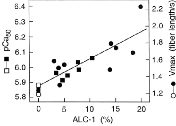
summarised in Fig. 7 data taken from Refs. 63,107 . Material without detectable ALC1 was included from sub-jects with normal hemodynamics. We showed for the first time, assuming other things being equal, a positive linear correlation between V and Ca2q-sensitivity of isometric
ma x
tension versus the ALC1 content. The correlation
coeffi-Ž . 2q Ž .
cient r for Vmax 12 cases and Ca -sensitivity 8 cases
Ž .
is 0.82 and 0.92, respectively p - 0.01 in both cases . Thus, both these two mechanical parameters are propor-tional to the ALC1 content. If taken together, the com-bined regression line in Fig. 7, would have a correlation coefficient of 0.86 with a significance level of p - 0.001. An increase of the Ca2q-sensitivity by q0.36 units and an increase of Vmax by a factor of 1.7 can be derived from the regression for an increase of 20% ALC1 in the ventricular tissue. In addition to Vmax, the curvature of the force– velocity relation changes as indicated by a decrease of the normalised tension coefficient arP0 from 0.21 in fibers
2q Ž 2q
Fig. 7. Correlation of Ca -sensitivity of tension pCa , Ca50 required
.
for half-maximal force generation and maximal shortening velocity
ŽVma x, expressed as muscle length per second MLrs. versus ALC1
Ž .
content expressed as % of total MLC1 isoforms assessed on perme-abilised ventricular muscle fibers. The regression line for pCa50 of left ventricular papillary muscle fibers from seven patients with end-stage
Ž . Ž .
heart failure filled squares and one control open square has a
correla-Ž .
tion coefficient r of 0.92 p- 0.01 . The regression line for Vma x of right ventricular infundibular muscle fibers from 11 patients with
congen-Ž . Ž .
ital heart diseases filled circles and one control open circle has a
Ž .
correlation coefficient r of 0.82 p- 0.01 . The combined r for all 20
Ž . w x
cases would be 0.86 p- 0.001 . Data taken from Refs. 63,107 . For further discussion see text.
lacking ALC1 to 0.15 in fibers containing 20% ALC1
w107 . The rate of tension development is almost twice asx
fast in fibers with high ALC1 when compared to fibers with low ALC1 content. Isometric tension at saturating Ca2q-concentrations was 352, 660 and 835 kNrm for
fibers with zero, 3–5% or with 20% ALC1, respectively. All muscle specimens were treated with cardioplegic solu-tion prior to chemical skinning and the MLC2 were thus
w x
fully dephosphorylated 63,107 .
Taken together, these findings indicate that ALC1 mod-ulates cross-bridge kinetics and thus contractility in human ventricular muscle. Increasing ALC1 increases Vmax which is proportional to gapp as well as the rate of tension
Ž .
development ktd which equals the sum of fappqgapp .
ALC1 thus accelerates the cycling kinetics. This may be brought about by increasing either fapp or gapp. Which one occurs with ALC1 cannot be decided from the
measure-w x
ments of Vmax and ktd 107 . Either change would affect the fraction of force-generating cross-bridges, n s
Ž . 2q
f r f qg . The Ca -sensitivity of isometric ten-app app app
sion would, however, be affected in opposite direction by
Ž . Ž
an increase in either fapp increasing it or gapp decreasing
. w x
it 76,82 . In view of the positive correlation with an
2q Ž .
increase in Ca -sensitivity Fig. 7 , one may conclude that ALC1 increases cross-bridge kinetics by accelerating selectively fapp over gapp. Even if both rate constants were increased in the presence of ALC1, fapp should be acceler-ated significantly more than gapp. This results in a bb-myosin cross-bridge with ALC1 having a longer duty
Ž
cycle fraction of time spent in force-generating states per
.
ATP-cycle in conjunction with faster kinetics than normal ventricular bb-myosin with VLC1. These kinetic charac-teristics are compatible with the mechanics described for ventricular muscle fibers from patients with severe mitral
Ž .
regurgitation mentioned above for which the ALC1
con-w x
tent was not known 99 . Assuming constant unitary force FXof bb-myosin with either MLC1, the species containing ALC1 with a longer duty cycle would represent an eco-nomic improvement over that with VLC1.
10. Molecular mechanism
The molecular mechanism for MLC1 to affect the cross-bridge kinetics seems to reside in its Ala–Pro-rich extended N-terminus which has been shown to interact
w x
with the C-terminus of actin 41,42,44 . The first hint that the MLC1 might affect the actin–myosin interaction and contractility came from experiments with skeletal muscles
Žreviewed in 45 . In vertebrate fast skeletal muscles, twow x.
Ž .
isoforms of the MLC1 ELC1fast and ELC2fast are ex-pressed which result from different transcription sites and
w x
differential splicing of the same gene 108 . The ELC2fast is missing the N-terminal 42 amino acid residues and does not bind to actin. Vmax is twice as high for rabbit fast psoas muscle fibers reconstituted with ELC2fast than with
w x
ELC1fast 45 . A similar difference in translocation veloc-ity between myosin with either ELC2fast or ELC1fast is
w x
also observed in the in vitro motility assay system 109 . A number of key experiments shed some light on the molecular mechanism how the MLC1 modulates contrac-tion in cardiac muscle. Human ALC1 can be reconstituted
Ž .
to rabbit fast skeletal muscle myosin subfragment-1 S1 for probing its effect on the actin activated ATPase activ-ity. Incorporated into S1, the ALC1 can also be chemically
w x
crosslinked to actin 43,47 . After removal of the first 11 residues by genetic engineering, the ALC1 can no longer be crosslinked to actin and the ATPase kinetics resemble those of the S1-actin complex with the short ELC2fast isoform. In conclusion, the first 11 residues at the N-terminus of the human ALC1 are sufficient to interact with actin and to modulate the ATPase kinetics. It is likely that this also applies to the human VLC1 as well as to the other MLC1 types. Indeed, removal of the first 13 residues from ELC1fast of chicken skeletal muscle myosin also converts the ATPase kinetics of the S1-actin complex with ELC1fast
w x
to those of S1 containing the short ELC2fast 110 . The 13-residue peptide 1-APKKDVKKPAAAA-13 was re-moved by proteolytic cleavage with papain and is identical in sequence to the corresponding N-terminus of the rat
Ž . a
ELC1fast Table 3 . In particular, it contains the N -tri-methylalanine at its N-terminus like all MLC1 beginning
w x
with Ala 111 . This 13-residue peptide can itself be crosslinked to actin and, when added to the S1-actin complex containing the endogenous intact ELC1fast, it
Ž .
increases the apparent Michaelis constant Km as well as the rate of ATPase activity. This was interpreted as indicat-ing that the bindindicat-ing of the 13 residue peptide to actin prevents the normal interaction of the endogenous ELC1fast of S1 with actin and thus converts its ATPase kinetics to those of S1 with the short ELC2fast. Interestingly, a synthetic decapeptide 1-APKKDVKKPA-10 missing the N-terminal Na-trimethyl group had hardly any effect on
w x
the ATPase kinetics 110 .
All MLC1 given in Table 3 comprise four positive charges within the first nine residues distributed over two
Ž .
couples of Lys with one negative charge Asp or Glu in between. Removal of the positive charges in Lys3 and Lys4 by exchanging them for Ala by site directed mutage-nesis in ALC1 also affects the ATPase kinetics approach-ing those of an S1-actin complex with the short ELC2fast
w43 . Changing the first two Lys3 and Lys4 to Ala or allx
four, including also Lys7 and Lys8, leads to a gradual increase in Vma x of rabbit fast psoas muscle fibers after incorporation of the mutated rat ELC1fast species in
com-w x
parison to fibers with its wild type ELC1fast 45 . This demonstrates that the four positive charges among the first nine amino acid residues of MLC1 are involved in
long-Ž .
range electrostatic interactions salt bridges with the C-terminus of actin. The C-C-terminus of actin does, indeed, contain three negative charges in the positions 12, 13, and
Ž .