Abstract Solitary fibrous tumors of the orbit (SFT) are
mesenchymal lesions that can develop either as malignant
or benign neoplasias. We describe the histological
fea-tures leading to the diagnosis in two females and review
the current literature. Diagnosis of SFT only can be
per-formed by histological examination, since clinical signs
and radiological features are not specific enough. Even a
malignant or benign course cannot be predicted, since
clinical and radiological features do not correlate with
his-tological signs of malignancy and vice versa. Radical
re-section is the treatment of choice, since no other treatment
option has been proven to be efficient.
Keywords Solitary fibrous tumor · Orbit ·
Immunohistochemistry · Diagnosis · Treatment
Introduction
Orbital tumors can be segregated into vascular,
hemato-poietic, neural, mesenchymal and inflammatory-induced
tumors. In addition, there are neoplasms and cysts from
the lacrimal gland, infiltrating tumors from adjacent
struc-tures as well as metastases (Table 1). Solitary fibrous
tu-mors (SFT) are rare mesenchymal spindel cell tutu-mors
usually occurring in the pleura, but also in many other
ex-tra-thoracal areas [26]. Since the first description of
or-bital SFT by Dorfmann et al. [13] in 1994, the segregation
of mesenchymal tumors of the orbit has been enlarged.
To our knowledge, there are up to 50 SFTs of the orbit
de-scribed [1, 4, 5, 6, 8, 13, 15, 16, 17, 18, 19, 20, 21, 26, 27,
28, 30, 31, 32, 33, 34, 35, 36, 37, 38, 39, 40, 41, 42, 43,
44, 45, 46]. The aim of the present study is to present two
cases and to review the current literature.
Subjects and methods
There were two patients with histologically confirmed SFT of the orbit seen in the University Hospital of Zurich between October 2002 and January 2003. Both wee diagnosed, operated on and fol-lowed up at the Department of Otorhinolaryngology Head and Neck Surgery, and clinical information was obtained from the files of the University Hospital, Zurich, Switzerland, and follow-up in-formation by contacting the attending physicians. Searching the current literature in the PubMed database, only English and Ger-man articles were considered using the keywords “solitary fibrous tumor” and “orbit.”
For histological examinations, the specimens were fixed in for-malin and embedded in paraffin according to standard procedures. Sections (1–2 mu) were cut for conventional histology and im-munohistochemistry. Immunohistochemistry was performed on deparaffinized, pretreated sections (either enzymatical prediges-tion or heating in citrate buffer for 3 min using a pressure cooker) using the Ventana NexES automated staining system with DAB as substrate. Reactions with antibodies against cytokeratin (Biomed-icals AG, Switzerland; dilution 1:250), S100 (DAKO, Glostrup, Denmark; 1:500), CD34 (Serotec, Oxford, UK; 1:20), bcl2 (Dako, Glostrup, Denmark; 1:200) and CD99 (Novocastra Laboratories, Newcastle upon Tyne, UK; 1:25) were performed. The quality of the reactions was controlled on tissue slides with known reaction patterns stained in parallel with the examined probes.
Case 1
Eighteen years prior to this presentation, this 77-year-old female had undergone an angiography and superselective embolization for an extraconal suspected hemangioma in the left orbit and remained blind as a consequence of this procedure. Seven years ago (1995), the tumor had to be excised surgically because of progressive growth using a superotemporal orbitotomy (i.e., Krönlein proce-dure). During the surgery, the tumor was well circumscribed and separated from adjacent tissue by a capsule-like structure. Except for the amaurosis on this side, she remained symptom free until 12 months prior to referral with a progressive swelling of the lat-eral aspect of the left lower eyelid and a mild painless proptosis.
M. Romer · B. Bode · B. Schuknecht · S. Schmid ·
D. Holzmann
Solitary fibrous tumor of the orbit – two cases and a review of the literature
DOI 10.1007/s00405-003-0731-7Received: 16 September 2003 / Accepted: 20 November 2003 / Published online: 13 August 2004
H E A D A N D N E C K O N C O L O G Y
M. Romer · S. Schmid · D. Holzmann (✉)
Department of Otorhinolaryngology and Head and Neck Surgery, University Hospital, 8091 Zurich, Switzerland
Tel.: +41-1-2551111, Fax: +41-1-2554556, e-mail: david.holzmann@usz.ch
B. Bode
Institute of Clinical Pathology, University Hospital, Zurich, Switzerland B. Schuknecht
Institute of Neuroradiology,
University Hospital, Zurich, Switzerland © Springer-Verlag 2004
The noncontrast and contrast-enhanced computed tomography exam-ination of the orbit showed an extraconal 3×1.5×2.5-cm tumor in the
lateral aspect of the left orbit. The lateral rectus muscle was not in-filtrated, but displaced medially. There were no calcifications within the lesion, showing a homogeneous contrast uptake (Fig. 1a–d). Magnetic resonance imaging (MRI) was not performed. Radical ex-cision by a coronal inex-cision, including releasing the temporal mus-hemangioma
Lymphangioma Langerhans’ Orbital meningioma Leiomyosarcoma Cellulitis/abscess Dermoid cell histiocytosis
Lipoma Infiltrating tumors from
adjacent structures Liposarcoma Metastasis Fibrous dysplasia Osteoma Osteosarcoma Fibroma Fibrous histiocytoma Fibrosarcoma Fibromatosis Nodular fasciitis Solitary fibrous tumor
Fig. 1 Solitary fibrous tumor of infero-lateral quadrant of left or-bit. The axial noncontrast CT depicts a well-defined ovoid-shaped lesion, isodense compared to muscle (a) with marked homoge-neous contrast enhancement (b). The globe is displaced anteriorly. The coronal images (c, d) show the lateral rectus muscle medially displaced corresponding to an “extraconal” origin of the tumor that lacks osseous changes on HR bone window images (d)
cle and temporarily removing the lateral wall of the orbit, was per-formed. The patient died of cardiac problems 6 months after the last operation.
Case 2
This 75-year-old female had noticed a slowly progressing swelling in the medial canthus on the left side for 2.5 years. This swelling was not painful and caused no double vision, but induced intermit-tent epiphora. On clinical examination, there was a marked dis-placement of the globe inferiorly and laterally, and in the medial canthus area, there was a well-divided tumor with a smooth sur-face. There was no hypaesthesia.
On contrast-enhanced MRI, there was a 2×2×3-cm extraconal
and well-defined tumor, which was homogeneous, situated adja-cent to the medial orbital wall and slightly flattened. On T1, the
tu-mor was isointense relative to gray matter and after contrast ho-mogeneously hyperintense with hypointense foci (Fig. 2a–d), while on CT the tumor showed mild pressure erosion of the lamina pa-pyracea. A fine-needle biopsy was performed. The tumor could be removed radically by a Linch incision. The tumor was of white color and was well divided from the adjacent structures (medial rectus muscle). The patient recovered without sequelae and has been symptom free up to 7 months.
Results
Pathology
Case 1
The 1995, the resected tumor measured 2.7×1.4×0.6 cm.
Histology showed focally strongly collagenized tissue
con-sisting of bland spindle cells with alternating cellularity
and patternless architecture. Many fine branching blood
vessels were seen, leading initially to the false
interpreta-tion of the tumor as a hemangioma. The mitotic activity
was low (less than one mitotic figure per ten high power
fields). The resection margins were involved. The
recur-rent tumor was resected in 2002 in two parts measuring
Fig. 2 Solitary fibrous tumor of left medial orbit. A markedlyen-hancing well-defined ovoid lesion is visible stretching the optic nerve because of displacement of the globe antero-laterally (a). The left orbit appears slightly enlarged because of flattening and thick-ening of the medial orbital wall. The extraconal tumor location is delineated to better advantage on the coronal T1-weighted noncon-trast MR image (b). Following connoncon-trast administration (c) the tumor shows marked signal intensity increase. On the T2-weighted coro-nal image (d), a hypointense (dark) sigcoro-nal is present reflecting the fibrous and cellular nature of the lesion
1.2 and 2.5 cm. The solid, firm cut surface was gray to
slightly tan. Compared to the tumor resected in 1995, the
cellularity, atypia and mitotic rate (four to five mitotic
fig-ures per ten high power fields) were increased. No
necro-sis or infiltrative growth was seen. Positivity for CD 34
and bcl2 was demonstrated in both of the tumors resected
in 1995 and 2002. During the autopsy 6 months after the
last operation, no recurrent tumor or metastases were
dem-onstrated.
Case 2
The fine-needle aspirate contained few cohesive groups
of monomorphic spindle cells with sparse cytoplasm
(Fig. 3). Immunocytochemically, a distinct positivity for
CD 34 could be demonstrated (Fig. 3, upper left). The
pre-ferred diagnosis was solitary fibrous tumor. The resected
tumor (Fig. 4) was firm, well circumscribed and measured
3.2×2.9×2.0 cm, with a solid, whitish cut section. A
mi-croscopically moderately cellular tumor with alternating
hypocellular and more cellular areas was found (Fig. 5).
Thick collagen fibers, perivascular fibrosis and a rich
net-work of branching blood vessels could be seen. The
mi-toses were rarely seen, not exceeding three normal mitotic
figures per ten high power fields. No necrosis or atypia was
demonstrated. Immunohistochemically strong diffuse
pos-itivity for CD 34 (Fig. 5, upper left) and a focally positive
reaction for bcl2 and CD99 were found, all other markers
being negative.
Review of the current literature
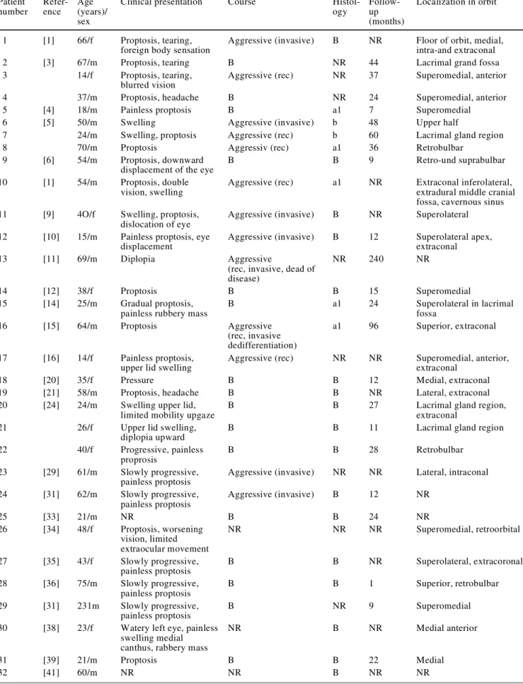
According to the present literature, 50 patients have been
described with orbital SFT (Table 2). In 9 out of 50, the
histological work-up is not described, and in 20, no
fol-low-up is indicated. For 25, it occurred on the left side and
for 14 on the right orbit, and for 11, the affected side was
not indicated. The most often described tumor locations
were extraconally in 11 cases (22%) and intraconally in
3 cases (6%), either in the superomedial quadrant (11 cases,
22%) or adjacent to the lacrimal gland (6 cases, 12%). From
the first clinical manifestation to diagnosis, there was a
mean course of 24.6 months. The mean age for all 50
pa-tients was 43.2 years (from 14 to 76 years), while the male
to female ratio was 28:22. The most frequent findings
were progressive painless proptosis (n=29, 58%), epiphora
(n=6, 12%) and a clinically detectable mass within the orbit.
Twenty-four out of the 50 (48%) described tumors were
described clinically as benign, while 15 (30%) showed an
aggressive course with local recurrences (n=6; 12%),
inva-sive growth (n=6; 12%) or both (n=2, 4%). Distant
metas-tasis after 7 years was described in one [26]. In 11 (22%)
Fig. 3 Fine-needle aspirate (Papanicolau staining) showingspin-dle mesenchymal cells without any significant pleomorphism. The immunocytochemical reaction with an antibody against CD34 is shown in the upper left with brown reaction product
Fig. 4 Cut surface of the solitary fibrous tumor of the second patient
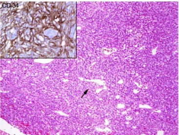
Fig. 5 Histology (standard hematoxilin and eosin staining) of the tumor of the second patient with an arrow pointing to a typical branching “hemangiopericytoma-like” blood vessel. Bland spindle cells in patternless arrangement with strong positive reaction for CD 34 (upper left)
Table 2 Review of the literature. Rec recurrence, B benign; a1 one histologic sign of malignancy, NR not reported Patient number Refer-ence Age (years)/ sex
Clinical presentation Course
Histol-ogy Follow-up (months) Localization in orbit 1 [1] 66/f Proptosis, tearing,
foreign body sensation
Aggressive (invasive) B NR Floor of orbit, medial,
intra-and extraconal
2 [3] 67/m Proptosis, tearing B NR 44 Lacrimal grand fossa
3 14/f Proptosis, tearing,
blurred vision
Aggressive (rec) NR 37 Superomedial, anterior
4 37/m Proptosis, headache B NR 24 Superomedial, anterior
5 [4] 18/m Painless proptosis B a1 7 Superomedial
6 [5] 50/m Swelling Aggressive (invasive) b 48 Upper half
7 24/m Swelling, proptosis Aggressive (rec) b 60 Lacrimal gland region
8 70/m Proptosis Aggressiv (rec) a1 36 Retrobulbar
9 [6] 54/m Proptosis, downward
displacement of the eye
B B 9 Retro-und suprabulbar
10 [1] 54/m Proptosis, double
vision, swelling
Aggressive (rec) a1 NR Extraconal inferolateral,
extradural middle cranial fossa, cavernous sinus
11 [9] 4O/f Swelling, proptosis,
dislocation of eye
Aggressive (invasive) B NR Superolateral
12 [10] 15/m Painless proptosis, eye
displacement
Aggressive (invasive) B 12 Superolateral apex,
extraconal
13 [11] 69/m Diplopia Aggressive
(rec, invasive, dead of disease)
NR 240 NR
14 [12] 38/f Proptosis B B 15 Superomedial
15 [14] 25/m Gradual proptosis,
painless rubbery mass
B a1 24 Superolateral in lacrimal fossa 16 [15] 64/m Proptosis Aggressive (rec, invasive dedifferentiation) a1 96 Superior, extraconal 17 [16] 14/f Painless proptosis,
upper lid swelling
Aggressive (rec) NR NR Superomedial, anterior,
extraconal
18 [20] 35/f Pressure B B 12 Medial, extraconal
19 [21] 58/m Proptosis, headache B B NR Lateral, extraconal
20 [24] 24/m Swelling upper lid,
limited mobility upgaze
B B 27 Lacrimal gland region,
extraconal
21 26/f Upper lid swelling,
diplopia upward
B B 11 Lacrimal gland region
22 40/f Progressive, painless
proprosis
B B 28 Retrobulbar
23 [29] 61/m Slowly progressive,
painless proptosis
Aggressive (invasive) NR NR Lateral, intraconal
24 [31] 62/m Slowly progressive, painless proptosis Aggressive (invasive) B 12 NR 25 [33] 21/m NR B B 24 NR 26 [34] 48/f Proptosis, worsening vision, limited extraocular movement NR NR NR Superomedial, retroorbital 27 [35] 43/f Slowly progressive, painless proptosis B B NR Superolateral, extracoronal 28 [36] 75/m Slowly progressive, painless proptosis B B 1 Superior, retrobulbar 29 [31] 231m Slowly progressive, painless proptosis B NR 9 Superomedial
30 [38] 23/f Watery left eye, painless
swelling medial canthus, rabbery mass
NR B NR Medial anterior
31 [39] 21/m Proptosis B B 22 Medial
cases, the clinical course was not indicated. However,
only 4 (27%) of the 15 clinically aggressive growths had
histological signs of malignancy, whereas 7 (47%) showed
no such signs, and in four no grading was indicated.
Among the 24 clinically benign lesions, histology was
be-nign in 19 (79%), malignant in 2 (8%), and in the
remain-ing three, no histological investigation was indicated. In
summary, in more than 30% of the orbital SFT, a
clini-cally aggressive course can be expected.
Discussion
In contrast to the first description in 1931 by Klemperer
und Rabin [26], ultrastructural, immunohistochemical as
well as electron microscopic investigations of SFT
re-vealed that this tumor has to be considered as having a
mesenchymal, submesothelial origin [10, 46]. Most SFTs
are seen in the pleura and less frequently in the Jung,
me-diastinum, pericard, dura, upper respiratory tract, salivary
gland, thyroid gland, peritoneum, liver, retroperitoneal
space, pelvis, adrenal gland, kidney, bladder, prostate gland,
cervix, spinal cord, periosteum and soft tissue [26].
Ac-cording to the ENT literature, there are case reports
involv-ing the upper respiratory tract, i.e., the nasal cavity, the
paranasal sinuses as well as in the epipharynx [47, 48].
These described tumors did not show an aggressive
clini-cal course and were not revealed to be malignant at
histo-logical examination [47, 48]. Twelve to 23% of pleural
SFT show an aggressive clinical course with infiltration
into adjacent structures, recurrences and/or metastases [3,
14]. Although worrisome histological features of
malig-nancy (i.e., hypercellularity, cytologic atypia, tumor
necro-sis and increased mitotic rate with more than four mitoses
per ten high power fields) were described, there is no strict
correlation between morphology and behavior.
Radical surgical resection seems to be the most
impor-tant prognostic factor according to a study on 223 pleural
SFT in which 82 (36.8%) were histologically malignant.
Forty-five percent of the latter could be healed
success-fully by resection alone. Most of them were pedicled or
circumscribed [3, 14]. However, there were lesions with
aggressive courses despite lacking signs of malignancy on
histology, as in our case 1 [3]. The high incidence of
ex-trapleural manifestations contradicts the hypothesis of a
mesothelial origin of SFT.
To our knowledge, 50 orbital SFT have been described,
apart from our two cases (Table 2). The clinical picture of
our two cases is very similar to most of the described
cases. Neither clinical presentation nor the localization of
SFT within the orbit is specific [32]. SFT can occur at any
age (mean age 43.2 years), even in children [1, 32, 36].
34 [42] 44/m Slowly progressive, painless proptosis B B 18 Medial 35 65/f Slowly progressive, painless proptosis B B 144 Medial 36 69/f Slowly progressive, painless proptosis Aggressive (rec) a1 NR NR
37 [46] 76/m Ptosis, pressure B B 12 Superolateral in lacrimal
fossa
38 [47] 24/f Blurred vision NR B NR Superomedial
39 69/m Proptosis NR B NR Superomedial
40 72/m None NR B NR Inferolateral
41 45/f Eyelid swelling NR B NR Superolateral
42 31/m Proptosis NR B NR Superolateral
43 18/f Ptosis, palpable mass NR B NR Inferior
44 [48] 40/f Tearing, disturbed
vision, swelling left upper lid, mass
B B 18 Superolateral, extraconal
45 20/f Watery eye, swelling B B 23 Anteromedial floor of orbit
46 19/f Swelling, mass B B 72 Lower anterior orbit into
lower eyelid, extraconal
47 32/f Slowly progressive, painless proptosis B B NR Lateral intraconal 48 48/m Slowly progressive, painless proptosis NR NR NR Superomedial
49 48/f Upper eyelid swelling Aggressive (metastasis) B 94 Upper eyelid
Our cases demonstrated histologically the typical
mor-phology of the solitary fibrous tumor: well-circumscribed,
firm, solid tumors consisting of rather bland spindle cells
haphazardly arranged in alternating hypo- and
hypercellu-lar areas with intervening thick collagen fibers.
Pattern-less architecture without a tendency to form bundles as
well as perivascular fibrosis are characterictic. A rich
net-work of typically branching, staghorn, so called
“heman-giopericytic” blood vessels are at least focally prominent.
Neither morphologic nor immunohistochemical features
of epithelial, neural or myogenic differentiation are
ob-served. Sometimes myxoid areas or a few adipocytes can
be found. The worrisome histological signs have been
dis-cussed above and must be sought after in representatively
sampled tumor tissue by the pathologist. In a typical case
of a solitary fibrous tumor, the histological diagnosis is
fairly easy if considered. Small biopsies may make the
di-agnosis difficult, especially if the clinical context is not
known to the pathologist. The immunohistochemical
pro-file (positivity for CD34, bcl2 and often CD99) is helpful,
although non-specific without the proper morphologic
cor-relation. CD 34 (human hematopoietic progenitor cell
anti-gen), which is consistently expressed in solitary fibrous
tumor, is also expressed in a broad range of various soft
tissue mesenchymal tumors (dermatofibrosarcoma
protu-berans, neural tumors, epithelioid sarcoma and Kaposi
sarcoma). Similarily, bcl2 and CD99 can be found in
var-ious other mesenchymal lesions. No typical genetic
alter-ations in SFTs have been identified to date [25]. Solitary
fibrous tumor is thought to be of a fibroblastic nature. The
diagnosis of a solitary fibrous tumors can be suspected
al-ready cytologically on fine-needle aspiration [6], as shown
in our case 2.
The histological differential diagnosis of the solitary
fi-brous tumor is primarily hemangiopericytoma, which is at
present histopathologically a very disputed entity. It seems
that this entity, first described in the 1940s by Stout [25],
is composed of many very different entities, which have
one thing in common: “hemangiopericytic”-like, branching
blood vessels. The new classifications separate these pooled
tumor types into synovial sarcomas, mesenchymal
chon-drosarcomas, solitary fibrous tumor and others. There is
still a subset of tumors, which morphologically can be
di-agnosed as hemangiopericytoma (and is strongly positive
for CD 34), but – interestingly – its prognostic features
seem to be very similar to SFTs. Giant cell angiofibroma,
first described in 1995 [9] and often occurring in the
or-bita, is nowadays considered as a morphologic variant of
the solitary fibrous tumors containing multinucleated
stro-mal giant cells and angectoid spaces [24]. Other tumors
that have to be considered are benign and malignant neural
tumors, meningeoma as well as benign deep fibrous
histio-cytoma.
On CT, this lesion shows a heterogeneous pattern in the
contrast-enhanced sequences and is sharply defined [20].
Hypodense areas within the tumor may represent myxoid
structures. MR imaging of orbital SFT are far more
char-acteristic. In the T1 sequences, they are isointense to gray
matter with moderate homogeneous or heterogeneous
gad-olinium enhancement, while on T2 they are hypointense.
SFTs frequently show centrally located, strongly
hypo-dense foci in T1 as well as in T2, representing
collagen-rich areas [7, 20]. Other collagen-containing tumors such
as fibromatosis, sclerotic pseudotumors or scirrhous
carci-noma metastases show a similar T1 and T2 pattern.
How-ever, the latter are not as sharply separated from the
adja-cent tissue. Contrary to SFT, cavernous hemangiomas
pre-dominantly occur within the muscle cone and exhibit a high
signal on T2-weighted sequences. The role of
fluorodeoxy-glucose positron emission tomography (PET) has not yet
been defined [28].
The aggressive SFT form of the orbit with its invasive
growth, local recurrence or metastasis seems to occur more
frequently (i.e., 30%) than in pleural SFT [3, 14, 26].
Sev-eral studies point to the fact that no conclusion of the
clin-ical course can be drawn from the histologclin-ical findings
[39]. There are histologically malignant tumors having both
an aggressive as well as benign outcome; on the other
hand, histologically benign features are no guarantee of a
good outcome. Hence, Iong-term fellow-up is mandatory.
Radical resection not only provides a good specimen for
diagnostic analysis, but also seems to be proven to be the
therapy of choice, since radiotherapy as well as
chemo-therapy are not proven to be effective [6,12, 40]. In
addi-tion, radical surgery is even more advantageous for a good
prognosis than histological signs of malignancy [3, 5, 20].
However, only in two out of the eight local recurrences
described in the literature were the primary resections
de-scribed as not having been performed radically [1, 28].
Conclusion
Whenever a mesenchymal tumor of the orbit is suggested,
SFT should be listed in the differential diagnosis. Thirty
percent show an aggressive course with local invasive
growth, local recurrence or metastasis. Histological signs
do not correlate with clinical behavior. Diagnosis only can
be made providing there is evidence of histological
fea-tures, immunohistological evidence of diffuse CD-34 and
vimentin positivity. There are no specific features of
ra-diological imaging showing moderate homogeneous or
heterogeneous contrast enhancement. Complete resection
is the therapy of choice, although the effect of
radiother-apy has not been evaluated. Radical resection seems to be
more important for prognosis than histological evidence
of malignancy.
References
1. Alexandrakis G, Johnson TE (2000) Recurrent orbital solitary fi-brous tumor in a 14-year-old girl. Am J Ophthalmol 130:373–376 2. Alford MA, Nerad JA (1998) Orbital tumors. In: Bailey BJ (ed) Head and neck surgery-otolaryngology, 2nd edn. Lippincott-Raven, Philadelphia New York, p 1471
3. Briselli M, Mark EJ, Dickersin GR (1981) Solitary fibrous tu-mors of the pleura: eight new cases and review of 360 cases in the litersture. Cancer 4:2678–2689
cases diagnosed by fine-needle aspiration. Diagn Cytopathol 25:172–176
7. Dalley RW (1999) Fibrous histiocytoma and fibrous tissue tu-mors of the orbit. Radiol Clin North Am 37:185–194
8. DeBacker CM, Bodker F, Putterman AM, Beckmann E (1996) Solitary fibrous tumor of the orbit. Am J Ophthalmol 121:447– 449
9. Dei Tos AP, Seregard S, Calonje E, Chan JK, Fletcher CD (1995) Giant cell angiofibroma. A distinct orbital tumor in adults. Am J Surg Pathol 19:1286–1293.
10. Dervan PA, Tobin B, O’Connor M (1986) Solitary (localized) fibrous mesothelioma: evidence against mesothelial cell origin. Histopathology 10:867–875
11. de Saint Aubain Somerhausen N, Rubin BP, Fletcher CD (1999) Myxoid solitary fibrous tumor: a study of seven cases with em-phasis on differential diagnosis. Mod Pathol 12:463–471 12. Dietrich CG, Roeb E, Breuer E, Matern S (2001) Solitary
fi-brous thoracic wall tumor. Progression with percutaneous ra-diotherapy. Dtsch Med Wochenschr 5;126:12–15
13. Dorfman DM, To K, Dickersin GR, Rosenberg AE, Pilch BZ (1994) Solitary fibrous tumor of the orbit. Am J Surg Pathol 18:281–287
14. England DM, Hochholzer L, McCarthy MJ (1989) Localized benign and malignant fibrous tumors of the pleura. A clinico-pathologic review of 223 cases. Am J Surg Pathol 13:640–658 15. Fan X, Semchyshyn TM, Mawn LA, Atkinson JB, Anderson JC, Toms SA, Johnson MD (2002) Sixty-six-year-old female with a 1-year history of progressive left proptosis. Brain Pathol 13:111–117
16. Fenton S, Moriarty P, Kennedy S (2001) Solitary fibrous tu-mour of the orbit. Eye 15:124–126
17. Festa S, Lee HJ, Langer P, Klein KM (1999) Solitary fibrous tumor of the orbit: CT and pathologic correlation. Neuroradiol-ogy 41:52–54
18. Fukunaga M, Naganuma H, Nikaido T, Harada T, Ushigome S (1997) Extrapleural solitary fibrous tumor: a report of seven cases. Mod Pathol 10:443–450
19. Fukunaga M, Ushigome S, Nomura K, Ishikawa E (1995) Soli-tary fibrous tumor of the nasal cavity and orbit. Pathol Int 45:952–957
20. Gigantelli JW, Kincaid MC, Soparkar CN, Lee AG, Carter SR, Yeatts RP, Holck DE, Hartstein ME, Kennerdell JS (2001) Or-bital solitary fibrous tumor: radiographic and histopathologic correlations. Ophthal Plast Reconstr Surg 48.17:207–214 21. Giufre I, Faiola A, Bonanno E, Liccardo G (2001) Solitary
fi-brous tumor of the orbit. Case report and review of the litera-ture. Surg Neurol 56:242–246
22. Guillou L, Gebhard S, Coindre JM (2000) Lipomatous heman-giopericytoma: a fat-containing variant of solitary fibrous tu-mor? Clinicopathologic, immunohistochemical, and ultrastruc-tural analysis of a series in favor of a unifying concept. Hum Pathol 31:1108–1115
23. Guillou L, Gebhard S, Coindre JM (2000) Orbital and extra-orbital giant cell angiofibroma: a giant cell-rich variant of soli-tary fibrous tumor? Clinicopathologic and immunohistochemi-cal analysis of a series in favor of a unifying concept. Am J Surg Pathol 24:971–979
24. Guillou L, Bridge JA (2002) Giant cell angiofibroma. In: Fletcher CDM, Unni KK, Mertens F (eds) WHO pathology and genetics. Tumours of soft tissue and bone. IARC Press, Lyon, pp 79–80 25. Guillou L, Fletcher JA, Fletcher CDM, Mandahl N (2002)
Ex-trapleural solitary fibrous tumour and hemangiopericytoma. In: Fletcher CDM, Unni KK, Mertens F (eds) WHO pathology and genetics. Tumours of soft tissue and bone. IARC Press, Lyon, pp 86–90
28. Hayashi S, Kurihara H, Hirato J, Sasaki T (2001) Solitary fi-brous tumor of the orbit with extraorbital extension: case re-port. Neurosurgery 49:1241–1245
29. Heathcote JG (1997) Pathology update: solitary fibrous tumour of the orbit. Can J Ophthalmol 32:432–435
30. Holbach LM, Colombo F, Schlotzer-Schrehardt U, Kirchner T (2002) Solitary fibrous tumor of the orbit presenting 20 years after Hodgkin’s disease. Orbit 21:49–54
31. Ing EB, Kennerdell JS, Olson PR, Ogino S, Rothfus WE (1998) Solitary fibrous tumor of the orbit. Ophthal Plast Re-constr Surg 14:57–61
32. Johnson TE, Onofrey CB, Ehlies FJ (2003) Echography as a useful adjunct in the diagnosis of orbital solitary fibrous tumor. Ophthal Plast Reconstr Surg 19:68–74
33. Kim HY, Lee SY, Kang SJ, Kim HJ (1999) Solitary fibrous tu-mor of the orbit: a poorly-recognized orbital lesion. Acta Oph-thalmol Scand 77:7048
34. Lanuza A, Lazaro R, Salvador M, Solanes C, Ramos F, Sorli E (1998) Solitary fibrous tumour of the orbit. Report of a new case. Int Ophthalmol 22:265–268
35. Lucas DR, Campbell RJ, Fletcher ChD, Garrity JA, Nascimento AG, McCartney AC (1995) Solitary fibrous tumor of the orbit. Int J Surg Pathol 2:193–198
36. Lucci LM, Anderson RL, Harrie RP, Mamalis N, Coffin C, Crandall DC (2001) Solitary fibrous tumor of the orbit in a child. Ophthal Plast Reconstr Surg 17:369–373
37. McElvanney AM, Noble JL, O’Donovan DG, Bonshek RE, Banerjee SS (1996) Solitary fibrous tumour: an atypical pre-sentation within the orbit. Eye 10:396–399
38. Miyagi N, Sugita Y, Terasaki M, Uchikado H, Takasaki K, Shigemori M, Nakashima A, Morimatsu M (1998) Solitary fi-brous tumor of the orbita: a case report. Jpn J Neurosurg 7: 243–247
39. O’Donovan DA, Bilbao JM, Fazl M, Antonyshyn OM (2002) Solitary fibrous tumor of the orbit. J Craniofac Surg 13:641– 644
40. Polito E, Tosi M, Toti P, Schurfeld K, Caporossi A (2002) Orbital solitary fibrous tumor with aggressive behavior. Three cases and review of the literature. Graefes Arch Clin Exp Oph-thalmol 240:570–574
41. Ramdial PK, Nadvi S (1996) An unusual cause of proptosis: orbital solitary fibrous tumor: case report. Neurosurgery 38: 1040–1043
42. Ruska KM, Westra WH (1996) Pathologic quiz case 1. Solitary fibrous tumor (SFT) of the orbit. Arch Otolaryngol Head Neck Surg 122:1130, 1132
43. Sciot R, Goffin J, Fossion E, Wilms G, Dom R (1996) Solitary fibrous tumour of the orbit. Histopathology 28:188–191 44. Scott IU, Tanenbaum M, Rubin D, Lores E 1996 Solitary
fi-brous tumor of the lacrimal gland fossa. Ophthalmology 103: 1613–1618
45. Takamura H, Kanno M, Yamashita H, Maeda K (2001) A case of orbital solitary fibrous tumor. Jpn J Ophthalmol 45:412–419 46. Westra WH, Gerald WL, Rosai J (1994) Solitary fibrous tumor. Consistent CD34 immunoreactivity and occurrence in the orbit. Am J Surg Pathol 18:992–998
47. Witkin GB, Rosai J (1991) Solitary fibrous tumor of the upper respiratory tract. A report of six cases. Am J Surg Pathol 15: 842–848
48. Zukerberg LR, Rosenberg AE, Randolph G, Pilch BZ, Good-man ML (1991) Solitary fibrous tumor of the nasal cavity and paranasal sinuses. Am J Surg Pathol 15:126–130