Supporting Information
Modified Biovectors for the Tuneable Activation
of Anti-platelet Carbon Monoxide Release
Lucas Prietoa, Jeremie Rossierc, Katarzyna Derszniakb, Jakub Dybasb, René M. Oetterlia, Emmanuel Kottelatc, Stefan Chlopickib,d *Felix Zeldera,* Fabio Zobic*
aUniversity of Zurich, Department of Chemistry, Winterthurerstrasse 190, CH-8057 Zurich,
Switzerland. Fax: +41 44 635 6803; E-mail: [email protected], www.felix-zelder.net
bJagiellonian Centre for Experimental Therapeutics (JCET), Jagiellonian University, Krakow,
Poland.
cDepartment of Chemistry, University of Fribourg, Chemin du Musée 9, 1700 Fribourg,
Switzerland. E-mail: [email protected]
dChair of Pharmacology, Jagiellonian University Medical College, Krakow, Poland. E-mail:
S2
Contents
Materials and Methods S4
Experimental procedures S6
Figures
- Figure S1 S12
1H-NMR spectrum and ESI-MS of 2
- Figure S2 S13
1H-NMR spectrum and ESI-MS of 3
- Figure S3 S14
- 1H-NMR spectrum and ESI-MS of 4
- Figure S4 S15
1H-NMR spectrum and ESI-MS of 5.
- Figure S5 S16
1H-NMR spectrum and ESI-MS of 6.
- Figure S6 S17 1H-NMR and 13C spectra of 7 Figure S7 S18 ESI-MS spectra of 7 - Figure S8 S19 CV of 2, 4 and 7 - Figure S9 S20 IR spectra of 9, 10, 11, 12, 13 and 14. Figure S10-S13 S21 UV-Vis spectra of 2, 3, 6, 8, 9, 10, 13. Figures S14-S18 S23 HR-MS spectra of 9, 10, 11, 13 and 14. Figure S19 S28
UV-Vis spectrum change of B12-ReCORM species in H2O
Figure S20 S29
Normalized shift of the 410 nm band of compounds 8-14 in H2O
Figure S21 S30
S3
Table S1 S31
Half-life (t½) of stability of species 8-14 in DMSO
Figure S22 S32
Deoxy-myoglobin assay
Figure S23 S33
Formations of MbCO.
S4
Materials and Methods
Materials: Chemicals were of reagent grade quality or better, obtained from commercial suppliers and used without further purification. Vitamin B12 was a generous gift from DSM
Nutritional products AG (Basel/Switzerland) and Prof. B. Jaun (retired ETH Zurich). All solvents were of reagent, analytical, HPLC or LC-MS grade respectively and obtained from commercial suppliers. Bidistilled water was used in all reactions.
Analytical HPLC: Spectra were recorded on an Acquity Waters system equipped with a PDA detector and an autosampler using a Nucleosil C18 250/3 column from Macherey-Nagel. A gradient (0-5 min 25% A; 5-30 min 25-100% A) of methanol (solvent A) versus bidistilled water containing 0.1% trifluoroacetic acid (solvent B) was applied using a flow rate of 0.3 mL/min.
Preparative HPLC: Separations were conducted on a VWR LaPrep system equipped with a PDA detector and a Nucleosil C18 250/40 column from Macherey-Nagel. A gradient (0-3 min 5% C; 3-3.20 min 25% C; 3.20-30 min 25-33% C) of acetonitrile (solvent C) versus bidistilled water containing 0.1% trifluoroacetic acid (solvent B) was applied using a flow rate of 40 mL/min.
ESI-MS: Spectra were recorded on a Bruker Daltonics HTC ESI-MS operated in the positive or negative mode. Injection rate 3 PL/min. Nebulizer P =10 psi, dry gas flow rate 5 L/min, gas T = 350 ºC. All solvents used were of LCMS grade.
ICP/OES: Inductively coupled plasma/optical emission spectrometry (ICP/OES)
measurements were performed on a Perkin Elmer Optima 7300 V HF ICP-OES Spectrometer.
HR-ESI-MS: Spectra were recorded on a Bruker maXis QTOF-MS instrument (Bruker Daltonics GmbH, Bremen, Germany). The samples were dissolved in MeOH and analyzed via continuous flow injection at 3 μL/min. The mass spectrometer was operated in positive ion mode with a capillary voltage of 4 kV, an endplate offset of –500 V, nebulizer pressure of 5.8 psig, and a drying gas flow rate of 4 L/min at 180°C. The instrument was calibrated with a sodium formate solution (500μl H2O: 500μl iPrOH: 20μl HCOOH: 20μl 0.1 M NaOHaq). The
S5
resolution was optimized at 30’000 FWHM in the active focus mode. The accuracy was better than 2 ppm in a mass range between m/z 118 and 1600.
Spectroscopy: UV-Vis spectra were recorded on a Varian Cary 50 using quartz cells with a path length of 1 cm. Citation of λmax (log ε) in nm. For platelet studies, spectra were recorded
on aPerkin Elmer double beam spectrophotometer Lambda 950 using quartz cells with a 1 cm path length. The kinetics of carbon monoxide release from CO donor compounds were studied by recording spectra at the Soret band region (Omax = 424 nm) and observing the
conversion from deoxyMb to MbCO at room temperature. Solutions of Mb (Myoglobin from equine heart, SIGMA) were prepared fresh by dissolving the protein in deionized water in such amount to obtain final concentrations 10 or 100 μM. Sodium dithionite (1 mg/ml, eq. 0.1%) was added just before measurements to convert myoglobin to deoxyMb. Before every experiment the reference spectra of deoxygenated protein were recorded. Solutions of CO donor compounds dissolved in DMSO:PBS (50:50) were added to the quartz cuvettes with deoxyMb in such amounts to give final concentrations of 10 or 100 μM. All solutions were always overlaid with mineral oil (0.5 cm3) to prevent CO escaping and myoglobin being oxygenated. 1H- and 13C-NMR as well as 2D-NMR spectra were recorded on a 500 MHz
Oxford NMR AS 500 using a QNP probe head and MestReNova 6.0.2 as evaluation tool. All
spectra were recorded in D2O at 300 K and TSP was used as a reference for all 13C-NMR
experiments.
Cyclic voltammetry: Cyclic voltammograms were obtained on a Metrohm 757 VA Computrace System. Measurements were performed using a glassy carbon electrode (working electrode) and an Ag/AgCl electrode (reference electrode). Samples were dissolved in 2 mL of 0.1 M TRIS buffered at pH 8. Hexacyanoferrate (0.5 mM) was used as an internal reference. Samples were purged with N2 (g) for 5 minutes prior to each measurement. The
measurement was accepted if EK3[Fe(CN)6] was found between +179 and +186 mV.
Solid Phase Extraction: Chromafix C18ec columns were applied for solid phase extraction (SPE). The compounds were dissolved in water, transferred to the adsorbent, washed with water and eluted with MeOH.
S6
Experimental procedures
Cyanocobalamin-c-lactone (2). The synthesis was performed according to a procedure reported by Bonnet et al.S1 Vitamin B12 (100 mg 74 Pmol, 1 equiv) was dissolved in 1 M HCl
(10 mL) and H2O (100 mL). Chloramin-T (25.2 mg, 110 Pmol 1.5 equiv) was dissolved in
H2O (50 mL) and was added drop wise over a period of 60 minutes. The pink solution was
desalted over Amberlite XAD-2. Purification by preparative HPLC and lyophilization afforded 2 in good yields (82 mg, 60.6 Pmol, 82%) UV-Vis (c = 1.66.10-5 M) O/nm (log ε) =
278 (4.0), 289 (3.9), 306 (4.1), 359 (4.5), 409 (3.4), 523 (3.7), 551 (3.7). HPLC tR = 14.9 min.
ESI-MS (H2O:MeOH 1:1) m/z = 677.9 [100%; M+2H]2+, 1354.5 [95%; M+H]+ (m/zcalc for
C63H86BrCoN13O15P: 1354.5). CV (0.1 M TRIS pH 8, K3[Fe(CN)6]) Ered = -929 mV. 1H
NMR (D2O; 500 MHz; 300K) G/ppm = 7.32 (s, 1H), 7.12 (s, 1H), 6.44 (s, 1H), 6.37 (d, J = 3.2 Hz, 1H), 6.09 (s, 1H), 4.29 (m, 2H), 4.20 (dd, J = 8.9, 1.9 Hz, 1H), 4.09 (m, 2H), 3.93 (dd, J = 12.9, 2.4 Hz, 1H), 3.75 (dd, J = 12.9, 4.0 Hz, 1H), 3.61 (d, J = 14.4 Hz, 1H), 3.38 (d, J = 9.8 Hz, 1H), 3.30 – 3.26 (m, 1H), 2.99 – 2.92 (m, 2H), 2.85-1.15 (m, 35H), 2.61 (s, 3H), 2.57 (s, 3H), 2.29 (s, 3H), 2.27 (s, 3H), 1.93 (s, 3H), 1.49 (s, 3H), 1.42 (s, 3H), 1.38 (s, 3H), 1.26 (d, J = 6.3 Hz, 3H), 1.17 (s, 3H), 0.50 (s, 3H).
10-bromo-cyanocobalamin (3). The synthesis was performed by a modified procedure of Wagner.S2 Vitamin B12 (100 mg, 74 μmol, 1 equiv) was dissolved in glacial AcOH (3 ml) and
NBS (13 mg, 74 μmol, 1 equiv) was added in small portions (~0.5 mg) over a period of 3 h. The resulting dark purple solution was desalted with solid phase extraction (SPE) and the solvents were removed under reduced pressure. Purification by preparative HPLC and lyophilization afforded 3 in quantitative yields (105 mg, 73 μmol, 99 %) as a purple powder. UV-vis (c = 6.05.10-5 M; H2O) O/nm (log ε) = 283 (3.9), 289 (3.9), 365 (4.3), 415 (3.3), 550
(3.7), 576 (3.7). HPLC tR = 16.5 min. ESI-MS (H2O:MeOH 1:1) m/z = 718.2 [100%;
M+2H]2+, 1435.5 [70%; M+H]+ (m/zcalc for C63H88BrCoN14O14P: 1435.48); HR-ESI-MS
(MeOH, NaI) m/z = 740.2279 [100%; M+2Na]2+ (m/zcalc for C63H87BrCoN14O14PNa2:
740.2281). CV (0.1 M TRIS pH 8, K3[Fe(CN)6]) Ered = -798 mV. 1H NMR (500 MHz, D2O)
δ/ppm = 7.31 (s, 1H), 7.13 (s, 1H), 6.52 (s, 1H), 6.37 (d, J = 3.1 Hz, 1H), 4.43 – 4.39 (m, 1H), 4.33 – 4.31 (m, 2H), 4.25 (d, J = 8.4 Hz, 1H), 4.07 (t, J = 8.6 Hz, 3H), 3.96 – 3.92 (m, 1H), 3.78 (dd, J = 12.9, 3.8 Hz, 2H), 3.69 – 3.56 (m, 3H), 3.40 (d, J = 8.8 Hz, 1H), 2.99 (dd, J = 14.4, 9.3 Hz, 1H), 2.80-1.10 ppm (m, 36H), 2.61 (s, 3H), 2.58 (s, 3H), 2.29 (s, 3H), 2.28 (s,
S7
3H), 1.93 (s, 3H), 1.83 (s, 3H), 1.41 (s, 3H), 1.39 (s, 3H), 1.34 (s, 3H), 1.28 (d, J = 6.2 Hz, 3H), 0.39 (s, 3H).
10-chloro-cyanocobalamin (4). To vitamin B12 (100 mg, 74 μmol, 1 equiv) dissolved in
glacial AcOH (3 ml) NCS (10 mg, 73 μmol, 1.0 equiv) was added over a period of 3 h. The resulting dark purple solution was desalted with solid phase extraction (SPE) and the solvents were removed under vacuum. Purification by preparative HPLC and lyophilization afforded 4 (75 mg, 55 μmol, 75%) as a purple powder. UV-vis (c = 4.75.10-5M; H2O) O/nm (log ε) = 282
(3.9), 289 (3.9), 364 (4.2), 408 (3.3), 551 (3.6), 574 (3.7); HPLC tR = 16.0 min; ESI-MS
(H2O:MeOH 1:1) m/z = 695.3 [100%; M+2H]2+, 1389.5 [70%; M+H]+ (m/zcalc for
C63H88ClCoN14O14P: 1389.53); HR-ESI-MS (MeOH, NaI) m/z = 717.2531 [100%;
M+2Na]2+ (m/zcalc for C63H87ClCoN14O14PNa2: 717.2534). ). CV (0.1 M TRIS pH 8,
K3[Fe(CN)6] Ered= -810 mV . 1H NMR (500 MHz, D2O) δ/ppm = 0.40 (s, 3H), 1.28 (d, J = 6.3 Hz, 3H), 1.36 (s, 3H), 1.39 (s, 3H), 1.42 (s, 3H), 1.79 (s, 3H), 1.93 (s, 3H), 2.27 (s, 3H), 2.29 (s, 3H), 2.58 (s, 3H), 2.61 (s, 3H), overlapped by 1.09-2.80 (m, 38H), 3.00 (dd, J = 14.4, 9.2 Hz, 1H), 3.39 (d, J = 9.1 Hz, 1H), 3.65 (d, J = 14.3 Hz, 1H), 3.79 (dd, J = 12.9, 3.7 Hz, 1H), 3.94 (d, J = 10.6 Hz, 1H), 4.06 – 4.11 (m, 2H), 4.24 (dd, J = 9.3, 5.9 Hz, 3H), 4.30 – 4.35 (m, 2H), 6.38 (d, J = 3.0 Hz, 1H), 6.51 (s, 1H), 7.11 (s, 1H), 7.32 (s, 1H).
10-chloro-cyanocobalamin-c-lactone (5). Vitamin B12 (200 mg, 148 Pmol, 1 equiv) was
dissolved in glacial acetic acid (3 mL) and NCS (20 mg, 150 μmol, 1.0 equiv) was added over a period of 3 h. The resulting dark purple solution was purified with solid phase extraction (SPE) and preparative HPLC. Solvents were removed under vacuum. 5 (14 mg, 10 μmol, 7%) was obtained as a purple powder.UV-vis (c = 1.80.10-5M; H2O) O/nm (log ε) = 281 (3.2), 289
(3.2), 363 (4.6), 421 (3.4), 551 (3.6), 577 (3.6); HPLC tR = 17.45 min; ESI-MS (H2O:MeOH
1:1) m/z = 694.7 [100%; M+2H]2+, 1388.2 [70%; M+H]+ (m/zcalc for C63H85CoN13O15PCl:
1388.50); HR-ESI-MS (MeOH NaI): m/z = 694.75596 [M+2H]2+ (m/zcalc for
C63H85CoN13O15PCl: 694.75568) CV (0.1 M TRIS buffer at pH 8, K3[Fe(CN)6] Ered= -784
mV; 1H NMR (500 MHz, CD3OD, TMS) δ/ppm = 0.40 (s, 3H), 1.25 (d, 3H), 1.29 (s, 3H),
1.79 (s, 3H), 2.00 (s, 3H), 2.29 (s, 3H), 2.31 (s, 3H), 2.62 (s, 3H), 2.63 (s, 3H), overlapped by 1.79-2.87 (m, ~46H), 3.39 (d, J = 9.4, 1H), 3.67 (d, J = 14.0 Hz, 1H), 3.76-3.92 (m, 1H), 4.08 (m, 1H), 4.20 (m, 1H), 4.54 (m, 1H), 4.62 (m, 1H), 6.32 (d, J = 2.9 Hz, 1H), 6.46 (s, 1H), 7.14 (s, 1H), 7.34 (s, 1H).
S8
c-(α,α-dibromo)-lactone-cyanocobalamin (6): The synthesis was performed according to recent literature procedures.S3
c-(α,α-dichloro)-lactone-cyanocobalamin (7): To vitamin B12 (100 mg, 74 μmol, 1 equiv)
dissolved in glacial AcOH (3 ml), NCS (99 mg, 744 μmol, 10 equiv) was added in one portion. The dark red solution was stirred at rt for 1 h. The resulting dark violet solution was desalted with solid phase extraction (SPE) and the solvents were removed under vacuum. Purification by preparative HPLC and lyophilization afforded 3 (58 mg, 41 μmol, 55%) as a violet powder. UV-vis (c = 4.21.10-5M; H2O) O/nm (log ε) = 279 (4.0), 288sh (3.9), 310 (3.8),
363 (4.2), 412 (3.3), 531 (3.7), 554 (3.7); HPLC (Method 1) tR = 19.0 min; ESI-MS
(H2O:MeOH 1:1) m/z = 712.7 [100%; M+2H]2+, 1424.4 [85%; M+H]+ (m/zcalc for
C63H84CoN13O15PCl2: 1424.4); HR-ESI-MS (MeOH, NaI): m/z = 711.73666 (m/zcalc for
C63H85CoN13O15PCl2: 711.73619) CV (0.1 M TRIS pH 8, K3[Fe(CN)6] Ered= -715 mV; 1
H-NMR (500 MHz, 300 K, D2O) δ/ppm = 0.54 (s, 3H), 1.16 (s, 3H), 1.26 (d, JH,H = 6.5 Hz, 3H), 1.41 (s, 3H), 1.47 (s, 3H), 1.54 (s, 3H), 2.10 (s, 3H), 2.29 (s, 3H), 2.33 (s, 3H), 2.63 (s, 3H), 2.74 (s, 3H), overlapped by 1.21-2.80 (m, ~36H), 2.96-3.00 (m, 1H), 3.39 (t, JH,H = 5.5 Hz, 1H), 3.61 (d, JH,H = 14.5 Hz, 1H), 3.76 (dd, JH,H = 4.0 Hz, 13.0 Hz, 1H), 3.94 (dd, JH,H = 2.5 Hz, 13.0 Hz, 1H), 4.07-4.09 (m, 1H), 4.18 (d, JH,H = 8.5 Hz, 1H), 4.23 (d, JH,H = 9.5 Hz, 1H), 4.29-4.35 (m, 2H), 6.12 (s, 1H), 6.39 (d, JH,H = 4.5 Hz, 1H), 7.10 (s, 1H), 7.34 (s, 1H). 13 C-NMR (126 MHz, 300 K, CD3OD) δ/ppm= 182.2,181.0,180.0, 177.6, 177.4, 175.5, 175.3, 175.3, 174.5, 167.5, 166.6, 166.3, 158.6, 143.4, 132.2, 134.3, 131.6, 127.1, 116.8, 113.1, 110.8, 107.7, 93.3., 92.2, 88.2, 86.7, 84.9, 76.6, 75.2, 73.6, 70.5, 65.2, 62.3, 60.6, 57.6, 55.0, 50.3, 46.7, 43.0, 40.1, 40.1, 36.1, 35.3, 32.2, 31.9, 31.8, 31.6, 30.1, 29.7, 29.5, 27.5, 25.00, 24.00, 21.1, 20.6, 20.3, 20.1, 19.2, 17.6, 17.5, 16.7, 16.3.
Cyanocobalamin-PP-CN-[Re(CO)2Br2(CH3OH)] (8). The synthesis was performed by modifications of a reported procedure.S4 Vitamin B12 (100 mg, 74 μmol, 1 equiv) and 1 (114
mg, 164 μmol, 2.2 equiv) were stirred in MeOH (60 mL) at 50 ºC for 90 minutes. The solvent was subsequently removed and the resulting red powder was washed several times with dichloromethane (20 mL) and acetone (20 mL). 8 (105.9 mg, 59 μmol, 80%) was obtained as a red microcrystalline powder. UV-vis (c = 1.81.10-4 M; H2O) O/nm (log ε) = 276 (3.9), 323
(3.9), 361 (4.3), 407 (3.3), 518 (3.7), 547 (3.7); ESI-MS (H2O:MeOH 1:1) m/z = 865.6
S9
IR (solid state, KBr, cm-1) v C≡N = 2184, v C≡O =1989, 1839. ICP/OES measurements of Re
content by relative weight: calcd 10.41; found 9.88 ± 0.05.
Cyanocobalamin-c-lactone-PP-CN-[Re(CO)2Br2(CH3OH)] (9). 2 (15.9 mg, 12 μmol, 1 equiv) and 1 (20 mg, 29 μmol, 2.4 equiv) were dissolved in methanol (10 mL) and stirred at 50ºC for 90 minutes. The solvent was subsequently removed and the resulting red powder was washed several times with dichloromethane (20 mL) and acetone (20 mL). 9 was obtained as a red microcrystalline powder (16.5 mg, 9 μmol, 75%). UV-vis (c = 1.81.10-4 M; H2O) O/nm
(log ε) = 276 (4.0), 323 (3.7), 361 (4.3), 407 (3.4), 518 (3.6), 547 (3.6); HR-ESI-MS (H2O:MeOH 1:1) m/z = 894.3 [100%; M+2H]2+, 1757.3235 [100%; M+H]+ (m/zcalc for
C63H86CoN13O15PReC2O2Br2: 1757.3236). IR (solid state, KBr, cm-1) v C≡N 2190, v C≡O 1989,
1841, 1789 (v C=O lactone). ICP/OES measurements of Re content by relative weight: calcd
10.41; found 9.89 ± 0.32.
10-bromo-cyanocobalamin-P-CN-[Re(CO)2Br2(CH3OH)] (10). 3 (8 mg, 5.5 μmol, 1 equiv) and 1 (10 mg, 14 μmol, 2.5 equiv) were dissolved in methanol (7 ml) and stirred for 30 minutes at 50 ºC. The solvent was subsequently removed and the resulting red powder was washed several times with dichloromethane (20 mL) and acetone (20 mL). 10 was obtained as a dark red microcrystalline powder (7.5 mg, 4 μmol, 66%). UV-vis (c = 1.89.10-4 M; H2O)
O/nm (log ε) = 279 (3.9), 366 (4.3), 411 (3.3), 550 (3.6), 574 (3.6); HR-ESI-MS (H2O:MeOH
1:1) m/z = 1836.2687 [100%; M+H]+ (m/zcalc for C63H88CoN14O14PReC2O2Br3: 1836.2647).
IR (solid state, KBr, cm-1) Q C≡N = 2176, Q C≡O = 1992, 1845. ICP/OES measurements of Re
content by relative weight: calcd 9.96; found 9.33 ± 0.12.
10-chloro-cyanocobalamin-P-CN-[Re(CO)2Br2(CH3OH)] (11). 4 (20 mg, 14 μmol, 1equiv) and 1 (34 mg, 48 μmol, 3.5 equiv) were dissolved in methanol (12 mL) and stirred for 30 minutes at 50 ºC. The solvent was subsequently removed and the resulting red powder was washed several times with dichloromethane (20 mL) and acetone (20 mL). 11 was obtained as a purple microcrystalline powder (10.5 mg, 6 μmol, yield 85%). UV-vis (c = 1.84.10-4 M; H2O) O/nm (log ε) = 279 (3.9), 287 (3.9), 363 (4.2), 415 (2.3), 545 (3.6), 570 (3.6);
HR-ESI-MS (H2O:MeOH 1:1) m/z = 1792.3110 [100%; M+H]+ (m/zcalc for
S10
=1973, 1841. ICP/OES measurements of Re content by relative weight: calcd 10.20; found 9.79 ± 0.23.
10-chloro-cyanocobalamin-c-lactone-PP-CN-[Re(CO)2Br2(CH3OH)] (12). 5 (6.5 mg, 5 μmol, 1 equiv) and 1 (8 mg, 11 μmol, 2.2 equiv) were dissolved in methanol (6 mL) and the mixture was stirred for 20 minutes at 50 ºC. The solvent was subsequently removed and the resulting red powder was washed several times with dichloromethane (15 mL) and acetone (15 mL). 12 was obtained as a dark purple microcrystalline powder (7.9 mg, 4 μmol, 80%). UV-vis (c = 1.84.10-4 M; H2O) O/nm (log ε) = 277 (3.2), 285 (3.2), 362 (4.2), 417 (3.4), 545
(3.6), 570 (3.6); ESI-MS (H2O:MeOH 1:1) m/z = 895.3 [100%; M+2H]2+, 1790.6 [70%;
M+H]+ (m/zcalc for C63H86ClCoN13O15PReC2O2Br2: 1790.29). IR (solid state, KBr, cm-1) v C≡N
= 2124, v C≡O = 1988, 1840, 1783. ICP/OES measurements of Re content by relative weight:
calcd 10.20; found 9.75 ± 0.36.
c-(α,α-dibromo)-lactone-cyanocobalamin-P-CN-[Re(CO)2Br2(CH3OH)] (13). 6 (10.5 mg, 7 μmol, 1equiv) and 1 (12 mg, 17 μmol, 2.4 equiv) were dissolved in methanol (5 ml) and stirred for 30 minutes at 50 ºC. The solvent was subsequently removed and the resulting red powder was washed several times with dichloromethane (20 mL) and acetone (20 mL).13 was obtained as a dark purple microcrystalline powder (10 mg, 5.2 μmol, 75%). UV-vis (c = 1.63.10-4 M; H2O) O/nm (log ε) = 278 (4.1), 311 (3.9), 363 (4.2), 413 (3.7), 529 (3.6), 558
(3.6); HR-ESI-MS (H2O:MeOH 1:1) m/z = 1915.1439 [100%; M+H]+ (m/zcalc for
C63H84Br2CoN13O15PReC2O2Br2: 1915.1426). IR (solid state, KBr, cm-1) v C≡N = 2181, v C≡O
= 1987, 1841, 1794. ICP/OES measurements of Re content by relative weight: calcd 9.56; found 9.14 ± 0.05.
c-(α,α-dichloro)-lactone-cyanocobalamin-P-CN-[Re(CO)2Br2(CH3OH)] (14). 7 (10 mg, 7 μmol, 1equiv) and 1 (12 mg, 17 μmol, 2.4 equiv) were dissolved in methanol (7 ml) and stirred for 25 minutes at 50 ºC. The solvent was subsequently removed and the resulting red powder was washed several times with dichloromethane (20 mL) and acetone (20 mL).14 was obtained as a dark purple microcrystalline powder (10 mg, 5.2 μmol, 77%). UV-vis (c = 1.81.10-4 M; H2O) O/nm (log ε) = 278 (3.6), 288 (3.5), 310 (3.4), 362 (4.2), 413 (3.1), 527
(3.6), 553 (3.6); HR-ESI-MS (H2O:MeOH 1:1) m/z = 1825.2319 [100%; M+H]+ (m/zcalc for
S11
1988, 1838, 1808. ICP/OES measurements of Re content by relative weight: calcd 10.02; found 9.67 ± 0.10.
S12
Figure S1. Top: 1H-NMR spectrum of cyanocobalamin-c-lactone (2) recorded in D2O [500
S13
Figure S2. Top: 1H-NMR spectrum of 10-bromo-cyanocobalamin (3) recorded in D2O [500
MHz]. Region of missing H10 signal is assigned. Bottom: measured (top row) and calculated (bottom row) ESI-MS spectra of 3.
S14
Figure S3. Top: 1H-NMR spectrum of 10-chloro-cyanocobalamin (4) recorded in D2O [500
MHz]. Region of missing H10 signal is assigned. Bottom: measured (top row) and calculated (bottom row) ESI-MS spectra of 4.
S15
Figure S4. Top: 1H-NMR spectrum of 10-chloro-lactone cyanocobalamin (5) recorded in CD3OD/TMS [500 MHz]. Region of missing H10 signal is assigned. Bottom: measured (top row) and calculated (bottom row) ESI-MS spectra of 5.
S16
Figure S5. Top: 1H-NMR spectrum of c-(α,α-dibromo)-lactone-cyanocobalamin (6) recorded in D2O//TMS [500 MHz]. H10 signal is assigned. Bottom: measured (top row) and calculated
S17
Figure S6. Top: 1H-NMR spectrum of c-(α,α-dichloro)-lactone-cyanocobalamin (7) recorded in D2O//TMS [500 MHz]. H10 signal is assigned. Bottom: 13C-NMR spectrum of 7 recorded
S18
S19 0.6 0.4 0.2 0.0 -0.2 -0.4 -0.6 -0.8 -1.0 -1.2 -2.5 -2.0 -1.5 -1.0 -0.5 0.0 0.5 1.0 1.5 Fe3+/Fe2+ Fe3+/Fe2+ I / A E / V 0.6 0.4 0.2 0.0 -0.2 -0.4 -0.6 -0.8 -1.0 -1.2 -2.5 -2.0 -1.5 -1.0 -0.5 0.0 0.5 1.0 1.5 Fe3+/Fe2+ Fe3+/Fe2+ I / A E / V 0.6 0.4 0.2 0.0 -0.2 -0.4 -0.6 -0.8 -1.0 -1.2 -5.0 -4.0 -3.0 -2.0 -1.0 0.0 1.0 2.0 Fe3+/Fe2+ Fe3+/Fe2+ I / A E / V
Figure S8. Cyclovoltammograms of (from top to bottom) 2, 4 and 7.Red/solid arrow: reduction potential of CoIII > CoI.
S20
S21
Figure S10. UV-visible spectra of an unbuffered aqueous solution of B12 (black line) and 8
(red line).
Figure S11. UV-visible spectra of an unbuffered aqueous solution of 2 (black line) and 9 (red line).
S22
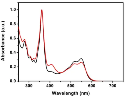
Figure S12. UV-visible spectra of an unbuffered aqueous solution of 3 (black line) and 10 (red line).
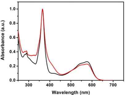
Figure S13. UV-visible spectra of an unbuffered aqueous solution of 6 (black line) and 13 (red line).
S23
S24
S25
S26
S27
S28
Figure S19. Changes in the UV-visible spectrum of an unbuffered aqueous solution of selected B12-ReCORM species. Spectra were recorded at fixed time intervals at 25 qC.
S29
Figure S20. Normalized exponential hypochromic shift of 410 nm band in the UV-Vis spectrum of compounds 8-14 in H2O.
S30
Figure S21. Normalized exponential hypochromic shift of 410 nm band in the UV-Vis spectrum of compounds 8-14 in DMSO.
S31
Table S1. Half-life (t½) of stability of species 8-14 in DMSO. Compound t½ hypochromic shift of 410 nm band in DMSO a
8 2.3 r 0.3 9 1.6 r 0.6 10 0.99 r 0.2 11 > 3 12 n. d. 13 1.9 r 0.4 14 > 3
S32
Figure S22. Spectrum changes (5 min intervals) of a solution of deoxy-myoglobin (Mb, 20 PM, phosphate buffer, pH = 7.4) solution after addition of 1 equivalent of species 8-14.
S33
Figure S23. The formation of MbCO in sixth (A-C) and fifteenth (D-F) minutes of B12
-ReCORMcomplexes 8-14 incubation with Mb (mean and standard deviation of N = 4-5) normalized to MbCO formation for B12-ReCORM.
S34
S1. Bonnett, R.; Cannon, J.; Johnson, A.; Todd, A., 226. Chemistry of the vitamin B 12 group. Part IV. The isolation of crystalline nucleotide-free degradation products. J. Chem.
Soc. 1957, 1148-1158.
S2. Wagner, F., Vitamin B and Related Compounds. Ann. Rev. Biochem. 1966, 35 (1), 405-434.
S3. Oetterli, R. M.; Prieto, L.; Spingler, B.; Zelder, F., Synthesis of a B Ring Opened 7,8-seco-Vitamin B12 Derivative with Grob Fragmentation. Org. Lett. 2013, 15 (18), 4630-4633. S4. Zobi, F.; Blacque, O.; Jacobs, R. A.; Schaub, M. C.; Bogdanova, A. Y., 17 e− rhenium dicarbonyl CO-releasing molecules on a cobalamin scaffold for biological application. Dalton