REVIEW
The endodermis
—development and differentiation
of the plant
’s inner skin
Julien Alassimone&Daniele Roppolo&Niko Geldner&
Joop E. M. Vermeer
Received: 18 June 2011 / Accepted: 22 June 2011 / Published online: 8 July 2011 # Springer-Verlag 2011
Abstract Controlling external compound entrance is essen-tial for plant survival. To set up an efficient and selective sorting of nutrients, free diffusion via the apoplast in vascular plants is blocked at the level of the endodermis. Although we have learned a lot about endodermal specification in the last years, information regarding its differentiation is still very limited. A differentiated endodermal cell can be defined by the presence of the“Casparian strip” (CS), a cell wall modifica-tion described first by Robert Caspary in 1865. While the anatomical description of CS in many vascular plants has been very detailed, we still lack molecular information about the establishment of the Casparian strips and their actual function in roots. The recent isolation of a novel protein family, the CASPs, that localizes precisely to a domain of the plasma membrane underneath the CS represents an excellent point of entry to explore CS function and formation. In addition, it has been shown that the endodermis contains transporters that are localized to either the central (stele-facing) or peripheral (soil-facing) plasma membranes. These features suggest that the endodermis functions as a polar plant epithelium.
Keywords Endodermis . Polarity . Casparian strip . Cell differentiation . Epithelium . Arabidopsis thaliana Abbreviations
AUX1 AUXIN RESISTANT1 AXR4 AUXIN RESISTANT 4
BOR1 BORON EFFLUX CARRIER 1 BOR4 BORON EFFLUX CARRIER 4 CS Casparian strip
CSD Casparian strip membrane domain GFP Green fluorescent protein
FM4-64 N-(3-triethylammoniumpropyl)-4-(6-(4-(diethyla-mino)phenyl)hexatrienyl)pyridinium dibromide NIP5; 1 NOD26-LIKE INTRINSIC PROTEIN 5; 1 NPSN12 Novel plant snare 12
PEN3 Penetration resistant 3 PI Propidium iodide PIN PINFORMED
PIS1 Polar auxin transport inhibitor sensitive 1 PM Plasma membrane
PP2A PROTEIN PHOSPHATASE 2A
SCR SCARECROW
SHR SHORT ROOT
Introduction
Every single cell in a higher plant is supplied with mineral compounds drawn from the soil by the roots and distributed to the aerial organs via the vascular tissues. Because of their immobility, terrestrial plants are fully dependent on their surrounding environment and must deal with the environ-mental conditions they live with. The roots have the
Handling Editor: David Robinson
J. Alassimone
:
D. Roppolo:
N. Geldner:
J. E. M. Vermeer (*)Department of Plant Molecular Biology, University of Lausanne, Quartier Sorge, Lausanne 1015, Switzerland e-mail: Joop.Vermeer@unil.ch J. Alassimone e-mail: Julien.Alassimone@unil.ch D. Roppolo e-mail: Daniele.Roppolo@unil.ch N. Geldner e-mail: Niko.Geldner@unil.ch
fundamental role of being the interface involved in nutrients intake. Yet, they must at the same time provide an efficient boundary against external biotic stresses, such as pathogenic microorganisms or abiotic stresses, such as excessive ion concentrations.
External compounds can reach the stele and be distrib-uted throughout the plant following two routes; they either move from one cell to the other via the symplastic pathway or progress between cells via the apoplast (Marschner
1995) (Fig. 1).
In the symplastic pathway, the plasma membrane (PM) acts as the first soil/plant interface by selecting ion uptake through specific transporters (Marschner 1995). Passive diffusion through the membrane or unspecific uptake may lead to the unintended entrance of compounds, but trans-porters regulating active export are known to be present in the epidermis and can counteract the presence of undesired molecules (Miwa et al. 2007). Controlling external com-pound entrance via the apoplastic space is more complicated since free diffusion occurs within cell walls. Thus, in order to allow an efficient and selective sorting of nutrients, an apoplastic diffusion barrier is present in the endodermis of all vascular plants, making passage through endodermal cells a mandatory step for molecules to reach the stele.
Endodermis specification
Endodermis specification has been intensively studied over the last years. Most of our knowledge regarding this process comes from research carried out in the model
plant Arabidopsis thaliana. In roots, cell types and developmental stages are easily recognizable: tissues are organized in concentric rings along the radial axis while developmental zones can be distinguished along the longitudinal axis. The cell specification of initials leading to endodermal cells is regulated by the interaction between two transcription factors, SHORTROOT (SHR) and SCARECROW (SCR), that together trigger the periclinal division of cortex–endodermal initials leading to the formation of the endodermis and the cortex (Helariutta et al. 2000). This process requires the movement of SHR from the stele, where it is expressed, into the endodermis; here, SHR interacts with SCR leading to the transcription of target genes and eventually determination of endoder-mal cell identity (Benfey and Scheres 2000; Cui et al.
2007; Di Laurenzio et al. 1996; Gallagher et al. 2004; Helariutta et al.2000; Nakajima et al.2001; Sozzani et al.
2010; Wysocka-Diller et al.2000).
Although specification of endodermal initials is a well-understood process, we are still completely in the dark about the molecular events that determine endodermis differentiation. This review will focus on cell differentiation by addressing the development, early differentiation, and CS formation of endodermal cells.
The endodermis: not just another cell layer
It has been a long time since the first description and suggestion of a diffusion barrier present in the“plant inner skin” by Robert Caspary, which is nowadays known as the
Fig. 1 The endodermis, and not the epidermis, is the barrier for the extracellular diffusion of molecules. a Schematic drawing of an Arabidopsis plant and b a detailed view of a cross-section of the root. Purple lines show the apoplastic route molecules can take until they are blocked by the CS. The inset shows an enlargement of a few endoder-mal cells with proposed central (green) and peripheral (red) lo-calized transporters that facili-tate the loading of nutrients in the inner space. Red arrows indicate symplastic route of up-take of compounds
Casparian strip (CS) (Caspary 1865). This structure refers to a modification that occurs by impregnation with ligno-suberic material of the anticlinal primary cell wall of endodermal cells, resulting in the formation an equatorial belt-like structure (Fig.1). The CS occupancy is normally about one third of anticlinal walls but can increase in size when exposed to environmental stresses, suggesting a certain plasticity of this structure (Karahara et al. 2004). The CS is the primary developmental stage of endodermal differentiation into a “border sheath”, and it is generally followed by a suberin deposition, forming suberin lamellae that eventually coat the entire cell (for a review, see Enstone et al. 2003). The coated cell is still connected to the surrounding cells by plasmodesmata (Clarkson et al.1987), although plasmodesmata are not observed in the region of the CS itself (Bonnett 1968). In some plant species, U-shaped tertiary cell walls have been described in the very late developmental stage of endodermal cells (for a review, see Enstone et al. 2003). Other examples of apoplastic barriers featuring CS-like structures exist in the root exodermis, in the needles of Pinaceae, and in specific cells involved in nectar secretion (Enstone et al. 2003; Liesche et al.2011).
The CS is an endodermal differentiation feature consid-ered to be crucial for plant development and survival. Undeniably, it provides a diffusion barrier between the apoplastic space facing the soil and the innermost apo-plastic space, in the stele. The efficiency of the CS to act as a diffusion barrier has been highlighted by several studies demonstrating that the transport of dyes (Moon et al.1986; Rufz de Lavison1910; Weerdenburg and Peterson1984) or ions (Peterson 1987; Robards and Robb 1974; Rufz de Lavison 1910; Singh and Jacobson 1977) but also heavy metals (Nagahashi et al.1974; Robards and Robb1972) are blocked and remain in the apoplast of cortical tissues. As a direct consequence of the establishment of this barrier, a directional and selective transport of nutrients should occur across the endodermis (Fig. 1b). Indeed, influx and efflux carriers have been localized to the central (stele-facing) and peripheral (soil-facing) sides of the endodermal plasma membrane (Alassimone et al.2010; Ma et al. 2006,2007; Takano et al. 2010). Thus, the CS isolates the stele’s
apoplastic space and distinct domains are organized in the endodermal plasma membrane. Beyond selective nutrient uptake, this is beneficial to prevent extensive ion backflow from the stele to the soil, and to maintain root pressure (Luttge and Laties1966; Peterson et al.1993). The latter is known to be important in sieve transport under low transpiration and in preventing xylem embolism in case of xylem cavitations (Tyree and Sperry1989). Currently, the role of the endodermis in defense against biotic stresses remains unclear despite evidence indicating that the CS is resistant to cell-wall-degrading enzymes that are generally
used by pathogens to degrade primary cell walls (Schreiber et al. 1999). An accumulation of pathogens at this apoplastic border has not been reported so far. Interestingly, juvenile root knot nematodes avoid the CS on their way to the vasculature: after penetration of the root epidermis, they move through the cortical apoplast towards the root tip and finally penetrate the vascular bundle at the meristematic or elongation zone, where CS are not yet established (Williamson and Gleason 2003).
Endodermis differentiation
In the last decades, several reports have studied CS structure and function of already differentiated endodermal cells, but only recently the developmental sequence of events that leads to this differentiation has been described in Arabidopsis thaliana (Alassimone et al. 2010). In this study, molecular and histochemical methods were used to highlight several differentiation events: propidium iodide was used as an apoplastic tracer and revealed the establishment of a functional apoplastic barrier; CS impreg-nations were directly observed by visualizing their auto-fluorescence; and physical attachment of the PM to cell wall was observed after plasmolysis, while diffusion in the PM was evaluated by using the fluorescent lipophilic styryl dye FM4-64. The combination of these techniques allowed the identification of a membrane domain where free diffusion from one side to the other of the PM was prevented. This PM domain is formed underneath the CS and is referred to as the“Casparian strip membrane domain (CSD)”. After plasmolysis, the CSD remained attached to the cell wall, which is in agreement with the hypothesis that a protein-like structure may scaffold the CSD to the CS (Alassimone et al. 2010; Behrisch 1926; Bonnett 1968; Karahara and Shibaoka1992).
As suggested, cell wall impregnations and PM attach-ment might be driven by protein scaffolding at the exact position of the CS, as previously suggested by ultrastruc-tural studies (Bonnett1968). Moreover, this protein scaffold may represent the barrier that prevents free diffusion of FM4-64 and plasma membrane proteins that are also excluded from the CSD (Alassimone et al. 2010). The establishment of a protein exclusion zone, the appearance of membrane–cell wall attachment sites, and the deposition of the CS all occur in a narrow time frame, while PI diffusion is blocked slightly later, suggesting that formation of a functional apoplastic barrier is a late differentiation step.
Therefore, several events can be observed during the development of mature endodermal cells: some of these events may be linked and occur concomitantly, while other seems to follow a step-by-step process (Alassimone et al.2010).
A novel protein family localizes to the Casparian strip domain
Recently, it has been reported that the plasma membrane at the CS is inaccessible to plasma membrane proteins, which allowed defining this“Casparian strip membrane domain” (CSD) as a zone of protein exclusion. Although this important feature has never been described before, another group recently reported that the auxin efflux carrier PIN3 is also excluded from the CSD (Alassimone et al.2010; Ding et al.2011).
It can be hypothesized that the spatial separation of the two lateral membrane domains is mediated by a tight protein scaffold that would also cause the general exclusion of membrane proteins from the CSD. This hypothesis is supported by ultrastructural analysis of the CS (Bonnett
1968). Until recently, we were completely in the dark regarding the proteins that might localize to this exclusion zone. However, a recent publication describes the identifi-cation of a novel protein family consisting of five members that localize precisely to this exclusion zone (Roppolo et al.
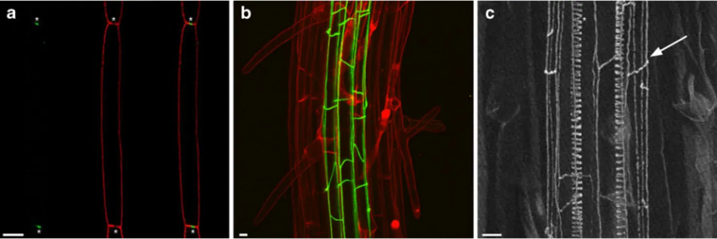
2011). These proteins were named CASP1-5 (CAsparian Strip membrane domain Protein). CASPs are 4-transmembrane spanning proteins that initially localize to the plasma membrane, but prior to the development of the CS localize to the exclusion zone (Fig. 2a). CASP transcription starts ~2 cells prior to the CSD formation, which matches the time it takes from its initial protein accumulation and final localization. This localization appears about five cells prior to the establishment of a functional diffusion barrier. This means that the CASPs are early markers for CSD and CS formation. In other words, the CASPs mark the differentiation of endodermal cells into
CS-containing endodermal cells (Roppolo et al. 2011). Their localization to the CSD domain prior to the establishment of the CSD implies an early role for the CASP proteins in CSD formation. Moreover, Roppolo et al. (2011) observed that in a casp1;casp3 double mutant, the CS appeared to be disorganized and more fluorescent compared to the CS in wild-type plants, although it still functions as a barrier. This observation does suggest a role for the CASP proteins in regulating cell-wall modification needed for the formation of the CS. It also shows that the other remaining CASP proteins can sufficiently compensate for the loss of CASP1 and CASP3 to maintain the diffusion barrier. It is likely that higher order casp mutants are needed to reveal the exact role of CASPs in CS formation.
When tagged with the green fluorescent protein (GFP), CASP proteins show a similar net-like localization as the autofluorescence of the CS reveals (Fig.2b, c). In addition, it has been shown that once the CASP proteins get localized into the CSD, they become completely immobile as compared to their initial plasma membrane localization (Roppolo et al.2011). Immuno-electron microscopy experi-ments on plants expressing CASP1–GFP under the control of its own promoter showed that CASP1 localizes to a spatially restricted domain in the plasma membrane that it is tightly aligned to the CASP1 domain in the adjacent plasma membrane of the next cell, separated by the CS cell wall. It is of interest to note that the CASP proteins seem only able to form the CSD domain in endodermal cells as ectopic expression results in only plasma membrane localization or accumulation in aggregates. This suggests that there is a specific signal in the endodermis that triggers the accumulation of CASP proteins in the CSD. How CASP proteins change their localization from an evenly
Fig. 2 CASP proteins localize to the CS domain and mediate CS formation in Arabidopsis. a Confocal image showing a medial longitudinal section of a differentiated endodermal cell co-expressing CASP1-mCherry (green) and YFP-NPSN12 (red). Note that CASP1 localizes to the middle of the apical and basal membrane, precisely labeling the depletion zone, highlighted by the asterisks. b Maximal
projection of an image stack taken from a plant expressing CASP1– GFP (green). Cell outlines are stained with PI (red). CASP1–GFP forms a net-like structure encompassing the endodermis. c Maximal image projection of a cleared Arabidopsis root, showing the CS
distributed to a highly localized one at the CSD and how they align themselves between cells is currently not known. Initially, CASP proteins are sensitive to treatment with the fungal toxin Brefeldin A (BFA) that affects endocytic recycling, but once localized to the CSD they are no longer sensitive to BFA and they also do not seem to turn over anymore (Roppolo et al.2011). In addition, it was shown that CASP proteins can interact with each other, and this might result in the formation of a large, tightly organized protein complex capable of preventing diffusion of mem-brane proteins through this domain and providing the scaffold for the formation of the CS. The identification of the CASP proteins and the use of the fluorescent CASP protein fusions now open up a new field of research and will allow us to get more fundamental insights into how the CS is made and what roles it plays during plant develop-ment. In addition, now that the first proteins localizing to the CSD have been identified, it will facilitate the identification and functional analysis of other proteins that make up the CSD. We hope that the identification of the CASPs will cause a similar boost in our understanding of CS formation as the identification of the first tight junction-localized protein ZONULA OCCLUDENS-1 (ZO-1) did for research in animal epithelia.
Since the CSD defines two distinct plasma membrane domains, proteins confined to one lateral domain should not be able to diffuse into the opposite domain. Under these conditions, several influx or efflux carriers might be localized either on the central (stele-facing) or peripheral (soil-facing) domains of endodermal cells. Hence, studying the endodermis should allow the investigation of this polarity in a relevant context.
Plasma membrane organization in plant roots: existence of distinct domains
Research on plant polarity has been intensively focused on the auxin-transport-related proteins, the PINFORMED (PIN) or AUXIN RESISTANT 1 (AUX1) proteins, that display an apical–basal (AB) polarity (for review, see Feraru and Friml2008; Grunewald and Friml2010).
In addition to AB polarity, some proteins have been shown to exhibit central–peripheral (CP) polarity as shown for silicon efflux and influx carriers in rice (Ma et al.2006,
2007). In Arabidopsis, the BORON EFFLUX CARRIER 4 (BOR4) and the boron influx carrier NOD26-LIKE IN-TRINSIC PROTEIN 5;1 (NIP5;1) localize to the peripheral PM domain (Miwa et al. 2007; Takano et al. 2006). In contrast, the BORON EFFLUX CARRIER 1 (BOR1) is observed at the central PM domain (Alassimone et al.2010; Takano et al.2010). The plant pathogen-defense-related and hormone precursor transporters PEN3/PDR8/ABCG36 and
PIS1/PDR9/ABCG37 are also exclusively localized to the peripheral PM domain (Langowski et al.2010; Strader and Bartel2009). So far, it has been shown that BOR1, NIP5;1, and PIS1 display polar localization in endodermal cells, although PIS1 was ectopically expressed (Alassimone et al.
2010; Langowski et al.2010; Takano et al.2010) (Fig.3b). It has also been shown that these markers, when ectopically expressed, exhibited polar localization in epidermal and cortical cells (Alassimone et al. 2010; Langowski et al.
2010; Takano et al. 2010).
Our hypothesis regarding CP-polarity function is that it is needed to direct nutrient flux from the soil to the stele, but when nutrients reach the stele, CP polarity is no longer required. The sequestration of nutrients in the vascular cylinder, by the virtue of the CS, might make their directional delivery obsolete once inside the stele. The fact that also non-differentiated cells exhibit CP polarity also fits with this hypothesis. In fact, nutrients may well not solely move through symplastic or apoplastic routes but can also use a combination of both (Fig. 4). Interestingly, our hypothesis is supported by the fact that ectopically expressed PIS1 seems to lose its CP polarity inside the stele (Langowski et al.2010).
As the CP polarity seems to be organized with respect to the stele position (Alassimone et al.2010; Langowski et al.
2010; Takano et al. 2010) (Fig. 3a), it is tempting to speculate that a mobile signal from the stele might regulate CP polarity.
Do AB polarity and CP polarity share common mechanisms?
Intuitively, two mechanisms could explain polar localiza-tion; either proteins are actively targeted to a defined domain or they are randomly secreted and specifically removed from the opposite domain (for a review, see Geldner2009). In the absence of lateral diffusion barriers, continuous endocytosis should be required for the mainte-nance of polarity, even if non-localized and constitutive, simply to counteract the inevitable dissipation of polarity by lateral diffusion of proteins (Valdez-Taubas and Pelham
2003). Up to now, the major insights underlying establish-ment and maintenance of AB polarity in plant roots have been based on research studying the PIN family of auxin-efflux carriers (Feraru and Friml 2008; Grunewald and Friml2010).
It is known that the basal localization of PIN1 is dependent on endocytic recycling mediated by the ADP-ribosylation factor guanine nucleotide exchange factor (ARF-GEF) GNOM as PIN1 polarity is strongly affected in gnom mutants or by inhibiting GNOM activity by the BFA (Geldner et al. 2003). In addition, it has been shown
that PIN1 is initially asymmetrically secreted and that subsequent polarization involves AtRabF2b/ARA7-mediat-ed endocytosis and/or recycling. However, apical localizAtRabF2b/ARA7-mediat-ed proteins like PIN2 and AUX1 seemed to be hardly affected, suggesting that there is at least another mechanism that regulates apical polarity (Dhonukshe et al.2008). Over the years, several additional mechanisms have been shown to act in the internalization and cycling of PIN proteins (Furutani et al.2011; Kitakura et al.2011; Naramoto et al.
2010). For example, sterol composition of the PM, the cytoskeleton, and cell-wall anchoring have been suggested to be important for PIN polarity (Feraru et al.2011; Geldner et al. 2001; Heisler et al. 2010; Kleine-Vehn et al. 2008; Men et al.2008).
Although only few reports have currently addressed this question, it seems that the mechanism underlying CP-polarity establishment and maintenance differs from that underlying AB polarity. It has been shown by Fluorescence Recovery After Photobleaching (FRAP) experiments that
PIS1 seems to exhibit a polar exocytosis to the peripheral plasma membrane domain, whereas FRAP studies on PIN1 revealed non-polar exocytosis (Dhonukshe et al. 2008; Langowski et al.2010). Treatments with BFA had no effect on the polar localization of BOR1, NIP5;1, or PIS1. However, BFA treatment did result in the re-localization of these proteins into endosomal aggregates, indicating that they are partially trafficked through BFA-sensitive endo-somes (Alassimone et al. 2010; Langowski et al. 2010; Takano et al.2010). In addition, it was shown that PIS1 was still correctly localized in the gnom mutant (Langowski et al. 2010). This does not mean that endocytic recycling is not as important for CP polarity as is the case for AB polarity, but just that GNOM seems not to be crucial for CP polarity establishment and maintenance. Additional studies are required to identify the molecular mechanisms mediat-ing CP polarity. One intrigumediat-ing observation pointmediat-ing to an important role for membrane trafficking in CP polarity is that treatment with the PI3-kinase inhibitor wortmannin
Fig. 3 Polarity establishment in the endodermis precedes CS develop-ment. Schematic drawing of an Arabidopsis root. Boxed regions are
enlarged in (a)–(c) to highlight endodermal polarity establishment. a
Root tip of Arabidopsis displaying already stele-facing (green) and
soil-facing (red) polarity. b Zoom-in of undifferentiated endodermal cells showing an intermixing of the two polar lateral domains (yellow). c Zoom-in of differentiated endodermal cells. The establishment of CSD (black) results in a strictly separated central and peripheral polarity
(WM) induces different effects on NIP5;1 compared to BOR1 in endodermal cells. High WM concentration does not affect BOR1 polarization, but does depolarize NIP5;1. Interestingly, this only occurred in endodermal cells where the CS was not yet established (Alassimone et al. 2010). This depolarization is thought to be a consequence of a block of PM internalization by WM, allowing lateral diffusion to distribute NIP5;1 to the whole PM domain in the absence of the CSD. This also suggests a role for the CS in lateral polarity maintenance by providing a PM diffusion barrier and resulting in the confinement of the proteins to one lateral PM domain.
In addition to endocytic recycling, it is known that phosphorylation/dephosphorylation processes involving the Ser/Thr protein kinase PINOID (PID) and the protein phosphatase 2A (PP2A) act in PIN polarity establishment (Friml et al.2004; Michniewicz et al.2007). However, PID and PP2A do not seem to be required for peripheral polarity (Langowski et al. 2010). Besides this, it has been shown that the ER-localized AUXIN RESISTANT 4 (AXR4), needed for the correct polar localization of AUX1, is also not required for BOR4, PIS1, or PEN3 polarity (Langowski
et al. 2010) and that the sterol composition of the plasma membrane has no effect on CP polarity (Langowski et al.
2010).
No obvious correlation between lateral polarity and the cytoskeleton has been reported so far. Chemical treatments or imaging of the cytoskeleton using GFP probes have not revealed any link to polarity establishment (Alassimone et al. 2010; Langowski et al. 2010; Takano et al. 2010). Disrupting the actin polymerization revealed that PIS1 and PEN3 have a tendency to accumulate in intracellular aggregations (Langowski et al. 2010). Very similar inter-cellular aggregations of a non-polar PM marker protein could also be observed after chemically induced actin depolymerization in endodermal cells (Alassimone et al.
2010). Thus, intracellular aggregations observed after actin depolymerization might not be specific to targeting of polar proteins, but rather a result of a general interference with membrane trafficking.
Although only a few proteins exhibiting CP polarity have been identified, there is already evidence indicating differences between some of them. For example, indepen-dent of polarity-related recycling, BOR1 endocytosis and degradation is regulated by boron availability whereas NIP5;1 and BOR4 are not (Miwa et al. 2007; Takano et al.2005,2010). Although both are partially BFA sensitive, BOR1 seems to be more sensitive to BFA than NIP5;1 (Takano et al.2010). Ubiquitination has been reported to be required for BOR1 sorting but seems not to be required for polar localization (Kasai et al. 2011). The lysine residue shown to be responsible for the ubiquitination-dependent vacuolar sorting in BOR1 seems to be absent from BOR4 (Kasai et al. 2011). Moreover, tyrosine motifs have been shown to be necessary for the proper polar localization of BOR1 (Takano et al.2010). It seems unlikely that tyrosine motifs are important for NIP5;1 localization since the two potential tyrosine-based signals are predicted in transmem-brane and extracellular regions (Takano et al. 2010). It is clear that we need to isolate additional proteins displaying CP polarity to be able to start addressing the mechanistic framework required for this cellular process.
A function for the CSD in endodermal polarity? Endodermal cells are able to organize lateral polarity even before the partitioning of their PM into two lateral domains (Alassimone et al. 2010). Furthermore, lateral polarity is observed in cells lacking the CSD such as epidermal or cortical cells (Alassimone et al. 2010; Langowski et al.
2010; Takano et al. 2010). The presence of a functional barrier seems not to be required for polarity establishment or maintenance, but is effective for a physical separation of the two lateral membrane domains. Indeed, polar proteins
Fig. 4 Different routes for nutrient uptake by plant roots. Schematic representation of an Arabidopsis root section showing different routes of compound uptake. From the epidermis to the endodermis there are stele- (green) and soil-faced (red) carriers which could provide a flux of molecules to the stele. Once passed the endodermis, this intrinsic lateral polarity seems lost (highlighted by a yellow-colored plasma membrane). Red arrows indicate the symplastic route and the purple arrows the apoplastic route. Of course, transport can also consist of a combination of the two pathways. Plasmodesmata are shown in orange, CSD in black, and the CS in blue. For clarity, plasmodesmata present in the anticlinal walls have been omitted. Pe pericycle, En endodermis, Co cortex, Ep epidermis
in differentiated endodermal cells are no longer intermixed, but confined to one lateral side of the cell (Alassimone et al.
2010) (Fig.3c). This suggests that the observed localization of polar proteins in the anticlinal PM of undifferentiated endodermal cells could be due to lateral diffusion. In addition, Langowski et al. proposed a dose-dependent effect on polarity as the localization of PIS1 became less pronounced when overexpressed (Langowski et al.2010), suggesting that without the physical restriction of lateral domains by the CS, a possible saturation of the machinery involved in polarity maintenance could occur resulting in inefficient confinement of polar proteins.
The endodermis as a polar epithelium of plants
The endodermis provides a barrier that separates the inner (stele) tissue from the outer (soil) interface and thus contains stele-facing and soil-facing plasma membrane domains that are separated by the CSD, to regulate uptake of nutrients (Figs.3and4). In addition, the barrier made up out of the CSD and CS bears functional similarity with mammalian tight junctions as it separates two membrane domains and forms a barrier that seals off the outer from the inner tissues. Although plants lack homologues of proteins known to be required for the establishment of polarized epithelia in vertebrates, such as PAR proteins, CLAUDINS, OCCLUDINS, ZO proteins, and others (Cereijido et al.
2004; Martin-Belmonte and Mostov 2008; Tsukita et al.
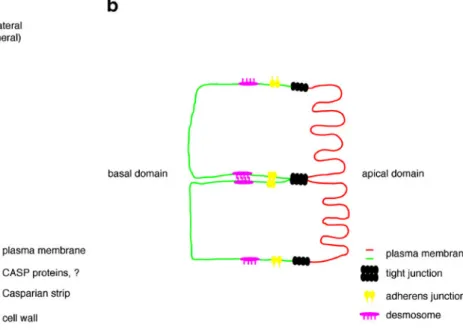
2009), the endodermis does have all the functional features of a polar epithelium. While the plant epithelium has only one barrier complex, the CS, the mammalian epithelial junctional complex consists of tight junctions, adherens junctions, and desmosomes. Since the tight junction is the most apical complex and it forms a tight seal between adjacent cells thereby separating the external and internal environment, it bears most similarity to the CSD and CS (Gupta and Ryan 2010; Steed et al. 2010; Bazzoni and Dejana 2004; Zeeb et al. 2010; Forster 2008). Indeed, although the CASP proteins do not show any obvious sequence similarity when compared with CLAUDINS, they do share similarity in overall structure (both 4-TM proteins) and size (Grebe2011). Moreover, it has been reported that similar to CASPs, the CLAUDINs show very limited diffusion in tight junctions, whereas other tight junction localized proteins such as OCCLUDINs and ZO-1 show much more mobility (Shen et al. 2008). CLAUDINs are viewed as the core constituents of tight junctions, a function that is also suggested for CASPs (Roppolo et al.2011; Shen et al. 2008). Figure5 shows a schematic representation of the endodermal and epithelial cells. The most obvious difference between the CS and tight junctions is the fact that in the CS there seems to be a “no-touch policy”; the two adjacent CSD are separated by the cell-wall modifica-tion known as the CS. Hence, the CASPs of one endodermal cell will never be in contact with the CASPs
Fig. 5 The endodermis as a polar epithelium of plants. The endodermis bears some overall similarity with polar epithelium of vertebrates. The Casparian strip forms a barrier that will separate the stele from the soil interface. The CASP proteins are suggested to be important for the formation of this barrier. In epithelia, the junctional complex is made up by tight junctions, adherens junctions, and
desmosomes. The tight junctions form the barrier that seals of the paracellular space from the lumen and the inside tissue, resembling the function of the Casparian strip. The big difference is that tight
junctions are mediated by protein–protein contacts, whereas the CS is
a cell-wall modification that does not depend on protein–protein
of the adjacent endodermal cell. This is in strong contrast to tight junctions, whose function is based on direct protein– protein interactions of tight junction complex-localized proteins between two adjacent cells (e.g., CLAUDINs) (Bazzoni and Dejana2004; Forster2008; Gupta and Ryan
2010; Shen et al.2008; Steed et al.2010; Zeeb et al.2010). Tight junctions are viewed as dynamic structures that function as selective barriers allowing certain molecules to pass through the paracellular space whereas others are blocked (Steed et al. 2010). At the moment, we do not know whether the CS can function in a similar way, i.e., as a kind of molecular sieve that allows selective passage of small molecules (e.g., water). Future research will be able to provide some answers to this. However, even if there is a functionally relevant selectivity of the CS toward certain small molecules, it is very probable that this selectivity is not subject to dynamic regulation as can be seen for the permeability and selectivity of animal tight junctions. This is because the more complex structure of the CS is not solely based on protein interactions but represents a localized modification/impregnation of the plant cell wall with hydrophobic polymers that probably cannot easily be altered in response to environmental stimuli.
Future prospects
Although described almost 150 years ago by Robert Caspary, until recently we have not gained much knowl-edge underlying CS formation in plants and in which biological process(es) it is involved. Now, with the discovery of the CASP proteins, we finally can start to unravel how this barrier gets established. For example, genetic screens on CASP1::CASP1–GFP plants might reveal how CASPs are regulated, but might also reveal how they get localized so precisely to the CSD. In parallel, expression-based analysis, as successfully used for the CASP proteins, together with proteomics could be used to identify additional proteins that localize to the CSD and CS. The generation of higher order CASP mutants, together with newly identified mutants that are required for barrier formation, will be instrumental to finally obtain genetic evidence for the biological roles of the CS. What will be the consequences of alterations of CS on drought, nutrient stress resistance, or the defense against soil borne pathogens?
Another interesting question is whether plants also are capable of locally modifying or altering their CS during lateral root formation. In order to emerge, lateral roots first need to pass the fully differentiated and CS-containing endodermis. Although our knowledge about lateral root formation has greatly advanced, surprisingly little is known about what happens to the endodermis during this process.
Some older reports on studies on other species speculate that this seems to be a regulated process (Bell and McCully
1970; Bonnett1969; Karas and McCully1973). Therefore, it might be that plants have devised a way to locally modify their CS to facilitate lateral root emergence. Besides deciphering the biological roles of the CS, the endodermis now also provides a beautiful system to study cell differentiation with powerful genetic and cell biological tools. We are still far away from a mechanistic understand-ing of CS formation, but the discovery of the CASP proteins and CS mutants provides us with the perfect tools to start investigating these processes.
Acknowledgments JA acknowledges Caroline Gutjahr for
stimulat-ing discussions. JEMV acknowledges financial support by a Marie-Curie IEF grant. Research in the group of NG is financed by grants from the European Research Council (ERC) and the Swiss National Science Foundation (SNSF).
Conflict of interest The authors declare that they have no conflict of
interest.
References
Alassimone J, Naseer S, Geldner N (2010) A developmental framework for endodermal differentiation and polarity. Proc Natl
Acad Sci USA 107(11):5214–5219
Bazzoni G, Dejana E (2004) Endothelial cell-to-cell junctions: molecular organization and role in vascular homeostasis. Physiol Rev 84(3):869–901
Behrisch R (1926) Zur Kenntnis der Endodermiszelle. Berichte der Deutschen Botanischen Gesellschaft 44:162–164
Bell JK, McCully ME (1970) A histological study of lateral root
initiation and development in Zea mays. Protoplasma 70:179–
205
Benfey PN, Scheres B (2000) Root development. Curr Biol 10(22):
R813–R815
Bonnett HT Jr (1968) The root endodermis: fine structure and
function. J Cell Biol 37(1):199–205
Bonnett HT Jr (1969) Cortical cell death during lateral root formation.
J Cell Biol 40(1):144–159
Caspary R (1865) Bemerkungen u¨ber die Schutzscheide und die Bildung des Stammes und der Wurzel. Jahrb wissensc Botanik 4:24
Cereijido M, Contreras RG, Shoshani L (2004) Cell adhesion, polarity,
and epithelia in the dawn of metazoans. Physiol Rev 84(4):1229–
1262
Clarkson DT, Robards AW, Stephens JE, Stark M (1987) Suberin lamellae in the hypodermis of maize (Zea mays) roots; development and factors affecting the permeability of hypoder-mal layers. Plant Cell Environ 10(1):83–93
Cui H, Levesque MP, Vernoux T, Jung JW, Paquette AJ, Gallagher KL, Wang JY, Blilou I, Scheres B, Benfey PN (2007) An evolution-arily conserved mechanism delimiting SHR movement defines a
single layer of endodermis in plants. Science 316(5823):421–425
Dhonukshe P, Tanaka H, Goh T, Ebine K, Mahonen AP, Prasad K, Blilou I, Geldner N, Xu J, Uemura T, Chory J, Ueda T, Nakano A, Scheres B, Friml J (2008) Generation of cell polarity in plants links endocytosis, auxin distribution and cell fate decisions.
Di Laurenzio L, Wysocka-Diller J, Malamy JE, Pysh L, Helariutta Y, Freshour G, Hahn MG, Feldmann KA, Benfey PN (1996) The SCARECROW gene regulates an asymmetric cell division that is essential for generating the radial organization of the Arabidopsis root. Cell 86(3):423–433
Ding Z, Galvan-Ampudia CS, Demarsy E, Langowski L, Kleine-Vehn J, Fan Y, Morita MT, Tasaka M, Fankhauser C, Offringa R, Friml J (2011) Light-mediated polarization of the PIN3 auxin trans-porter for the phototropic response in Arabidopsis. Nat Cell Biol
13(4):447–452
Enstone DE, Peterson CA, Ma F (2003) Root endodermis and exodermis: structure, function, and responses to the environment.
J Plant Growth Reg 21(4):335–351
Feraru E, Friml J (2008) PIN polar targeting. Plant Physiol 147
(4):1553–1559
Feraru E, Feraru MI, Kleine-Vehn J, Martiniere A, Mouille G, Vanneste S, Vernhettes S, Runions J, Friml J (2011) PIN polarity maintenance by the cell wall in Arabidopsis. Curr Biol 21
(4):338–343
Forster C (2008) Tight junctions and the modulation of barrier function in disease. Histochem Cell Biol 130(1):55–70 Friml J, Yang X, Michniewicz M, Weijers D, Quint A, Tietz O,
Benjamins R, Ouwerkerk PB, Ljung K, Sandberg G, Hooykaas PJ, Palme K, Offringa R (2004) A PINOID-dependent binary switch in apical–basal PIN polar targeting directs auxin efflux.
Science 306(5697):862–865
Furutani M, Sakamoto N, Yoshida S, Kajiwara T, Robert HS, Friml J, Tasaka M (2011) Polar-localized NPH3-like proteins regulate polarity and endocytosis of PIN-FORMED auxin efflux carriers.
Development 138(10):2069–2078
Gallagher KL, Paquette AJ, Nakajima K, Benfey PN (2004) Mechanisms regulating SHORT-ROOT intercellular movement.
Curr Biol 14(20):1847–1851
Geldner N (2009) Cell polarity in plants: a PARspective on PINs. Curr
Opin Plant Biol 12(1):42–48
Geldner N, Friml J, Stierhof YD, Jurgens G, Palme K (2001) Auxin transport inhibitors block PIN1 cycling and vesicle trafficking.
Nature 413(6854):425–428
Geldner N, Anders N, Wolters H, Keicher J, Kornberger W, Muller P, Delbarre A, Ueda T, Nakano A, Jurgens G (2003) The Arabidopsis GNOM ARF-GEF mediates endosomal recycling, auxin transport, and auxin-dependent plant growth. Cell 112
(2):219–230
Grebe M (2011) Plant biology: unveiling the Casparian strip. Nature
473(7347):294–295
Grunewald W, Friml J (2010) The march of the PINs: developmental plasticity by dynamic polar targeting in plant cells. EMBO J 29
(16):2700–2714
Gupta IR, Ryan AK (2010) Claudins: unlocking the code to tight junction function during embryogenesis and in disease. Clin
Genet 77(4):314–325
Heisler MG, Hamant O, Krupinski P, Uyttewaal M, Ohno C, Jonsson H, Traas J, Meyerowitz EM (2010) Alignment between PIN1 polarity and microtubule orientation in the shoot apical meristem reveals a tight coupling between morphogenesis and auxin transport. PLoS Biol 8(10):e1000516
Helariutta Y, Fukaki H, Wysocka-Diller J, Nakajima K, Jung J, Sena G, Hauser MT, Benfey PN (2000) The SHORT-ROOT gene controls radial patterning of the Arabidopsis root through radial signaling. Cell 101(5):555–567
Karahara I, Shibaoka H (1992) Isolation of Casparian strips from pea
roots. Plant Cell Physiol 33(5):555–561
Karahara I, Ikeda A, Kondo T, Uetake Y (2004) Development of the Casparian strip in primary roots of maize under salt stress. Planta
219(1):41–47
Karas I, McCully ME (1973) Further studies of the histology of lateral
root development in Zea mays. Protoplasma 77:243–269
Kasai K, Takano J, Miwa K, Toyoda A, Fujiwara T (2011) High boron-induced ubiquitination regulates vacuolar sorting of the BOR1 borate transporter in Arabidopsis thaliana. J Biol Chem 286(8):6175–6183
Kitakura S, Vanneste S, Robert S, Lofke C, Teichmann T, Tanaka H, Friml J (2011) Clathrin mediates endocytosis and polar distribu-tion of PIN auxin transporters in Arabidopsis. Plant Cell [Epub ahead of print]
Kleine-Vehn J, Langowski L, Wisniewska J, Dhonukshe P, Brewer PB, Friml J (2008) Cellular and molecular requirements for polar PIN
targeting and transcytosis in plants. Mol Plant 1(6):1056–1066
Langowski L, Ruzicka K, Naramoto S, Kleine-Vehn J, Friml J (2010)
Trafficking to the outer polar domain defines the root–soil
interface. Curr Biol 20(10):904–908
Liesche J, Martens HJ, Schulz A (2011) Symplasmic transport and
phloem loading in gymnosperm leaves. Protoplasma 248(1):181–
190
Luttge U, Laties GG (1966) Dual mechanisms of ion absorption in relation to long distance transport in plants. Plant Physiol 41 (9):1531–1539
Ma JF, Tamai K, Yamaji N, Mitani N, Konishi S, Katsuhara M, Ishiguro M, Murata Y, Yano M (2006) A silicon transporter in rice. Nature 440(7084):688–691
Ma JF, Yamaji N, Mitani N, Tamai K, Konishi S, Fujiwara T, Katsuhara M, Yano M (2007) An efflux transporter of silicon in
rice. Nature 448(7150):209–212
Marschner H (1995) Mineral nutrition of higher plants, 2nd edn. Academic, London
Martin-Belmonte F, Mostov K (2008) Regulation of cell polarity
during epithelial morphogenesis. Curr Opin Cell Biol 20(2):227–
234
Men S, Boutte Y, Ikeda Y, Li X, Palme K, Stierhof YD, Hartmann MA, Moritz T, Grebe M (2008) Sterol-dependent endocytosis mediates post-cytokinetic acquisition of PIN2 auxin efflux carrier polarity.
Nat Cell Biol 10(2):237–244
Michniewicz M, Zago MK, Abas L, Weijers D, Schweighofer A, Meskiene I, Heisler MG, Ohno C, Zhang J, Huang F, Schwab R, Weigel D, Meyerowitz EM, Luschnig C, Offringa R, Friml J (2007) Antagonistic regulation of PIN phosphorylation by PP2A and PINOID directs auxin flux. Cell 130(6):1044–1056 Miwa K, Takano J, Omori H, Seki M, Shinozaki K, Fujiwara T (2007)
Plants tolerant of high boron levels. Science 318(5855):1417 Moon GJ, Clough BF, Peterson CA, Allaway WG (1986) Apoplastic
and symplastic pathways in Avicennia marina (Forsk.) Vierh. roots revealed by fluorescent tracer dyes. Funct Plant Biol 13
(5):637–648
Nagahashi G, Thomson WW, Leonard RT (1974) The Casparian strip as a barrier to the movement of lanthanum in corn roots. Science
183(4125):670–671
Nakajima K, Sena G, Nawy T, Benfey PN (2001) Intercellular movement of the putative transcription factor SHR in root
patterning. Nature 413(6853):307–311
Naramoto S, Kleine-Vehn J, Robert S, Fujimoto M, Dainobu T, Paciorek T, Ueda T, Nakano A, Van Montagu MC, Fukuda H, Friml J (2010) ADP-ribosylation factor machinery mediates endocytosis in plant cells. Proc Natl Acad Sci USA 107 (50):21890–21895
Peterson CA (1987) The exodermal Casparian band of onion roots blocks the apoplastic movement of sulphate ions. J Exp Bot 38
(12):2068–2081
Peterson CA, Murrmann M, Steudle E (1993) Location of the major barriers to water and ion movement in young roots of Zea mays
Robards AW, Robb ME (1972) Uptake and binding of uranyl ions by
barley roots. Science 178(4064):980–982
Robards AW, Robb ME (1974) The entry of ions and molecules into roots: an investigation using electron-opaque tracers. Planta 120 (1):1–12
Roppolo D, De Rybel B, Tendon VD, Pfister A, Alassimone J, Vermeer JE, Yamazaki M, Stierhof YD, Beeckman T, Geldner N (2011) A novel protein family mediates Casparian strip formation
in the endodermis. Nature 473(7347):380–383
Rufz de Lavison JD (1910) Du mode de pénétration de quelques sels dans la plante vivante. Rev Gen Bot 22:16
Schreiber L, Hartmann K, Skrabs M, Zeier J (1999) Apoplastic barriers in roots: chemical composition of endodermal and
hypodermal cell walls. J Exp Bot 50(337):1267–1280
Shen L, Weber CR, Turner JR (2008) The tight junction protein complex undergoes rapid and continuous molecular remodeling
at steady state. J Cell Biol 181(4):683–695
Singh C, Jacobson L (1977) The radial and longitudinal path of ion
movement in roots. Physiol Plant 41(1):59–64
Sozzani R, Cui H, Moreno-Risueno MA, Busch W, Van Norman JM, Vernoux T, Brady SM, Dewitte W, Murray JA, Benfey PN (2010) Spatiotemporal regulation of cell-cycle genes by SHORTROOT links patterning and growth. Nature 466(7302):128–132 Steed E, Balda MS, Matter K (2010) Dynamics and functions of tight
junctions. Trends Cell Biol 20(3):142–149
Strader LC, Bartel B (2009) The Arabidopsis PLEIOTROPIC DRUG RESISTANCE8/ABCG36 ATP binding cassette transporter mod-ulates sensitivity to the auxin precursor indole-3-butyric acid.
Plant Cell 21(7):1992–2007
Takano J, Miwa K, Yuan L, von Wiren N, Fujiwara T (2005) Endocytosis and degradation of BOR1, a boron transporter of
Arabidopsis thaliana, regulated by boron availability. Proc Natl
Acad Sci USA 102(34):12276–12281
Takano J, Wada M, Ludewig U, Schaaf G, von Wiren N, Fujiwara T (2006) The Arabidopsis major intrinsic protein NIP5;1 is essential for efficient boron uptake and plant development under boron limitation. Plant Cell 18(6):1498–1509
Takano J, Tanaka M, Toyoda A, Miwa K, Kasai K, Fuji K, Onouchi H, Naito S, Fujiwara T (2010) Polar localization and degradation of Arabidopsis boron transporters through distinct trafficking
path-ways. Proc Natl Acad Sci USA 107(11):5220–5225
Tsukita S, Katsuno T, Yamazaki Y, Umeda K, Tamura A (2009) Roles of ZO-1 and ZO-2 in establishment of the belt-like adherens and tight junctions with paracellular permselective barrier function.
Ann N Y Acad Sci 1165:44–52
Tyree MT, Sperry JS (1989) Vulnerability of xylem to cavitation and
embolism. Annu Rev Plant Physiol Plant Mol Biol 40(1):19–36
Valdez-Taubas J, Pelham HR (2003) Slow diffusion of proteins in the yeast plasma membrane allows polarity to be maintained by
endocytic cycling. Curr Biol 13(18):1636–1640
Weerdenburg CA, Peterson CA (1984) Effect of secondary growth on the conformation and permeability of the endodermis of broad bean (Vicia faba), sunflower (Helianthus annuus), and garden balsam (Impatiens balsamina). Can J Bot 62(5):907–910 Williamson VM, Gleason CA (2003) Plant–nematode interactions.
Curr Opin Plant Biol 6(4):327–333
Wysocka-Diller JW, Helariutta Y, Fukaki H, Malamy JE, Benfey PN (2000) Molecular analysis of SCARECROW function reveals a radial patterning mechanism common to root and shoot.
Development 127(3):595–603
Zeeb M, Strilic B, Lammert E (2010) Resolving cell–cell junctions: lumen