TRANSPORT PHYSIOLOGY
Expression of renal and intestinal Na/Pi cotransporters
in the absence of GABARAP
Sonja C. Reining&Annette Liesegang&Heinrich Betz&
Jürg Biber&Heini Murer&Nati Hernando
Received: 11 December 2009 / Revised: 12 March 2010 / Accepted: 17 March 2010 / Published online: 31 March 2010 # Springer-Verlag 2010
Abstract We have recently shown that the abundance of the renal sodium (Na)/inorganic phosphate (Pi) cotrans-porter NaPi-IIa is increased in the absence of the GABAA receptor-associated protein (GABARAP). Accordingly, GABARAP-deficient mice have a reduced urinary excre-tion of Pi. However, their circulating levels of Pi do not differ from wild-type animals, suggesting the presence of a compensatory mechanism responsible for keeping serum Pi values constant. Here, we aimed first to identify the molecular basis of this compensation by analyzing the expression of Na/Pi cotransporters known to be expressed in the kidney and intestine. We found that, in the kidney, the upregulation of NaPi-IIa is not accompanied by changes on the expression of either NaPi-IIc or PiT2, the other cotransporters known to participate in renal Pi reabsorption.
In contrast, the intestinal expression of NaPi-IIb is down-regulated in mutant animals, suggesting that a reduced intestinal absorption of Pi could contribute to maintain a normophosphatemic status despite the increased renal retention. The second goal of this work was to study whether the alterations on the expression of NaPi-IIa induced by chronic dietary Pi are impaired in the absence of GABARAP. Our data indicate that, in response to high Pi diets, GABARAP-deficient mice downregulate the expression of NaPi-IIa to levels comparable to those seen in wild-type animals. However, in response to low Pi diets, the upregula-tion of NaPi-IIa is greater in the mutant mice. Thus, both the basal expression and the dietary-induced upregulation of NaPi-IIa are increased in the absence of GABARAP. Keywords Proximal tubule . Epithelial transport . Na+/Pi cotransporter . Phosphate . Homeostasis
Introduction
Homeostasis of inorganic phosphate (Pi) is an essential physiological function as manifested by syndromes that arise from hypo- or hyperphosphatemic states. Pi wasting is linked with rickets and osteomalacia, whereas hyperphos-phatemia is associated with an increased risk of cardiovas-cular diseases. The maintenance of constant circulating levels of Pi depends on the coordinated activity of two major organs, the intestine and the kidney. In both tissues, the uptake of Pi across the apical membrane of the epithelia is, at least partially, mediated by members of the SLC34 family of sodium (Na)-coupled solute carriers: NaPi-IIa/ SLC34a1 and NaPi-IIc/SLC34a3 are expressed in the brush border membrane (BBM) of renal tubules, whereas NaPi-IIb/SLC34a2 is located in the intestinal microvilli (for
S. C. Reining
:
J. Biber:
H. Murer:
N. Hernando (*) Institute of Physiology, University of Zurich, Winterthurerstr. 190,8057 Zurich, Switzerland e-mail: hernando@physiol.uzh.ch
S. C. Reining
:
J. Biber:
H. Murer:
N. Hernando Zurich Center for Integrative Human Physiology (ZIHP), University of Zurich,Winterthurerstr. 190, 8057 Zurich, Switzerland A. Liesegang
Institute of Animal Nutrition, Vetsuisse Faculty, University of Zurich,
Winterthurerstr. 260, 8057 Zurich, Switzerland H. Betz
Department of Neurochemistry, Max-Planck Institute for Brain Research, Deutschordenstr 46,
60528 Frankfurt am Main, Germany DOI 10.1007/s00424-010-0832-2
review, see [7]). The SLC20 family, in particular PiT2/ SLC20a2, also contributes to the renal handling of Pi [33]. The intestine absorbs up to 70% of the ingested Pi. Although the relative contributions of transcellular and paracellular pathways to the overall absorptive process have long been under discussion, recent data show that NaPi-IIb is primarily involved in this process [26]. In mice, NaPi-IIb is expressed in the small intestine [12], preferentially in the ileum [23] and its abundance in the BBM is regulated by dietary Pi and 1,25-dihydroxyvitamin D3 (vitamin D3; [10]). The effect of vitamin D3 involves transcriptional regulation in young animals [34] but seems to work posttranscriptionally in adult mice [10, 34]. Recently, two groups generated NaPi-IIb-deficient mice. Shibasaki et al. reported that full ablation of NaPi-IIb results in embryonic lethality [31], whereas Sabbagh et al. described that an inducible conditional knockout is characterized by fecal Pi wasting, hypophosphaturia due to upregulation of NaPi-IIa, and normophosphatemia [26]. Apparently, in the absence of NaPi-IIb, Pi homeostasis is achieved through a compensatory increase in renal reabsorption resulting from overexpression of NaPi-IIa. Sabbagh et al. proposed that NaPi-IIb mediates up to 90% of the active reabsorption of Pi in the ileum [26].
The kidney, and in particular the proximal tubule, is the major organ involved in Pi homeostasis. It does so by modulating the reabsorption of Pi accordingly to the body’s needs, primarily via changes on the expression of NaPi-IIa. The phenotype of NaPi-IIa-deficient mice includes hyper-phosphaturia, hypophosphatemia, and undeveloped trabec-ular bone [2]. The Na/Pi cotransport into renal BBM of homozygous mutant mice was reduced by about 70%, despite a heavy upregulation of NaPi-IIc [27]. In contrast, NaPi-IIc knockouts do not exhibit either hypophosphatemia or skeletal abnormalities [29]. Furthermore, the Na/Pi cotransport into renal BBM as well as the expression of NaPi-IIa remains unaffected in the absence of NaPi-IIc. Ablation of NaPi-IIa [32] and NaPi-IIc [29] results in hypercalcemia and hypercalciuria, probably due to a vitamin D3-induced upregulation of intestinal Ca++ absorp-tion. Together, these findings suggest a minor contribution of NaPi-IIc to the renal handling of Pi in the murine kidney that is probably restricted to young animals [27]. However, this concept has been recently challenged by the detection of NaPi-IIc mutations in patients with Pi wasting pheno-types [3,13,16]. In addition to NaPi-IIa and NaPi-IIc, PiT2 also contributes to renal Pi reabsorption. PiT2 is a member of the SLC20 family of solute carriers first identified as retroviral receptors (for review, see [5]) that has a wide tissue distribution. Recent data show that PiT2 is expressed in the proximal BBM in rat kidneys and that its abundance is regulated by dietary Pi [33]. Exploiting the different sensitivities of SLC34 and SLC20 cotransporters to inhibition by phosphonophormic acid, this study suggests
that the relative contribution of SLC34-independent cotrans-porters to overall Na/Pi uptake in rat kidney varies between 25% and 50%, depending on the dietary conditions.
As the major Pi storage site, bones also contribute to Pi homeostasis. Osteoblast and osteoblast-like cells express several members of the SLC34 and SLC20 families of Na/ Pi cotransporters [18,35], whereas osteoclasts are known to express NaPi-IIa and PiT1 [9]. As indicated above, NaPi-IIa-deficient mice are hypophosphatemic, and in young animals, this associates with undeveloped trabecular bone [2, 9]. No skeletal abnormalities result upon ablation of NaPi-IIb [26] or NaPi-IIc [29] consistent with the normo-phosphatemic status of both animal models.
Unlike other transport systems whose activity can be modulated by molecular modifications, the activity of Na/Pi cotransporters seems to be mostly controlled by changes in their epithelial expression. Different hormones (parathyroid hormone, PTH), vitamins (vitamin D3), and metabolic factors (dietary Pi or acidosis) regulate the apical abundance of SLC34 and/or SLC20 transporters (for review, see [7]). The expression of membrane proteins is often controlled by interaction with partners that directly or indirectly connect them to the cytoskeleton. Such mechanism has been reported for NaPi-IIa: its C-terminal PDZ domain is engaged in interactions with, among other binding partners, the four members of the Na/H-exchanger regulatory factor (NHERF) family [8]. In particular, the interaction with NHERF1 appears to be required for stabilizing the cotransporter in the proximal BBM [11,30]. In contrast, we reported recently that the expression of NaPi-IIa is substantially upregulated in renal BBM from GABAA receptor-associated protein (GABARAP)-deficient mice [24]. Although this increase is mirrored by a reduction in the urinary excretion of Pi, GABARAP-deficient mice are normophosphatemic. This suggests the existence of a compensatory mechanism/s that allows maintaining Pi homeostasis despite a higher renal retention of Pi. Therefore, the first goal of this paper was to investigate the nature of this compensatory mechanism. For that purpose, we compared the expression of known Na/Pi cotransporters in kidney and intestine as well as the overall mineral density and Pi/Ca++ contents of bones in samples from wild-type and GABARAP-deficient mice. In addition, we examined whether the absence of GABARAP interferes with the regulation of NaPi-IIa by chronic dietary Pi.
Materials and methods Animal handling
Animal experiments comply with the Swiss Animal Welfare law and were performed with the approval of the local veterinary authority (Kantonales Veterinäramt Zürich).
Experiments were done with male wild-type (WT) and GABARAP knockout (KO) mice [22] aged 12 to 14 weeks. GABARAP knockout mice were generated by Lexicon Pharmaceuticals injecting C57BL/6J albino blastocytes with 129SvEvBrd stem cells [22]. Prior to this study, the mice were backcrossed with the C57BL/6J strain for seven generations.
For the studies under normal dietary conditions, animals were fed normal chow and had free access to water. Mice were sacrificed and kidneys (from five WT and five KO) collected, decapsulated, and frozen immediately in liquid nitrogen. From each mouse, one kidney was later on processed for the isolation of BBM and the second one for the preparation of total RNA. Ileum and bones (femurs and L3/L4 lumbar vertebras) were extracted from a new set of mice (four WT and four KO). Ileum (last two thirds of the small intestine [23]) was rinsed with cold 0.9% NaCl and inverted with the help of a thin metal rod. Then, the intestinal mucosa was scraped off and frozen in liquid nitrogen. Bones were immediately cleaned from attached tissue. All samples were kept at−20°C until further use.
For the chronic dietary adaptation, WT and KO animals were randomly divided into two groups of eight animals each, placed in metabolic cages, and fed with either a high (1.2%) or low (0.1%) Pi diet (Kliba, NAFAG) for 5 days. Overnight urine was collected under mineral oil during the last 12 h. In the morning, blood samples were taken from the vena cava and kidneys collected as indicated above. From six animals, one kidney was later on processed to isolate BBM, whereas total RNA was prepared from the second one. Two mice per group were independently handled for immunostainings. Ileum and bone samples were collected from a second set of mice (six to seven animals per dietary condition) and processed as indicate above.
Urinary and plasma concentrations of creatinine and phosphate were determined according to the Jaffe (Wako Chemicals) and Fiske Subarov (Sigma Diagnostics) methods, respectively.
Isolation of renal and intestinal BBM vesicles
BBMV were prepared using the Mg2+ precipitation tech-nique [4]. Briefly, frozen kidneys were cut into small pieces and transferred into 800μl homogenization buffer contain-ing (in mM) 300 mannitol, 5 EGTA and 12 Tris–HCl, pH 7.1. Samples were homogenized (2 min on ice) with a polytron (Kinematica). Upon the addition of 1,120 μl of water and 22 μl of 1 M MgCl2, membranes were precipitated on ice for 15 min. Frozen mucosa of the ileum was mixed with 200μl homogenization buffer and 1,000 μl of water and then homogenized (1 min on ice) using an Omnimixer (Sorval). Upon the addition of 11 μl of 1 M MgCl2, membranes were precipitated on ice for 30 min.
After the precipitation step, samples (from kidney/ intestine) were centrifuged in a Sorvall centrifuge for 15 min at 4,500 rpm. Small aliquots of the supernatants, representing total homogenates, were immediately frozen, whereas the rest was further centrifuged at 18,500 rpm for 30 min. The pellets were resuspended in 1 ml of membrane buffer (300 mM mannitol, 20 mM Hepes–Tris, pH 7.4) and centrifuged again at 18,500 rpm for 30 min. Pellets containing the BBMV were resuspended in 150 μl of membrane buffer. Protein concentration was measured using a BioRad determination kit. Small aliquots of the kidney BBMV and the whole ileum preparation were frozen for further analysis by Western blot. The rest of the renal samples were immediately processed for32P uptakes. 32
P uptake into isolated BBMV
Na+-dependent uptake of 32Pi into renal BBMV was determined by a rapid filtration technique as described previously [4]. Freshly prepared BBMV were incubated for 30 s at 25°C in a solution containing either (in mM) 100 NaCl, 0.1 K2HPO4, 80 mannitol, and 16 Hepes–Tris, pH 7.4 and 1-2 μCi32P (as orthophosphoric acid) or with the NaCl replaced by KCl. The reactions were terminated by incubation in 1 ml ice-cold stop solution consisting of (in mM) 100 mannitol, 150 NaCl, 5 K2HPO4, and 5 Tris-HCl, pH 7.4. The incorporation of32Pi was measured in a beta counter. The Na-dependent uptake of Pi was calculated as the difference between the incorporation of 32Pi in the presence and absence of NaCl.
Immunoblots
Renal and intestinal BBM were prepared as described above. Samples (15 µg of renal BBM or 50 µg of intestinal BBM) were separated on 10% SDS/PAGE gels, transferred to PVDF membranes and incubated overnight with anti-bodies against IIa (1:3,000), IIc (1:3,000), NaPi-IIb (1: 3,000), PiT-2 (1:1,000), and β-actin (1:10,000). After several washes, membranes were incubated for 2 h at room temperature with HRP-linked secondary antibodies (GE Healthcare) followed by incubation with a chemilumi-nescence reagent (ECL, Amersham Pharmacia Biotech). Immunoreactive signals were detected using the DIANA III-chemiluminescence detection system (Raytest) and quantification was performed with AIDA software (Advanced Image Data Analyser AIDA, Raytest). Data are shown as ratios between the protein of interest toβ-actin.
Immunostainings
Mice were perfused-fixed as indicated previously by perfusion through the left ventricle with a prewarmed
fixative solution containing 3% paraformaldehyde, 3 mM MgCl2, and 4.5 mM picric acid, prepared in a 6:4 mixture of 0.1 M cacodylate buffer (pH 7.4; adjusted to 300 mOsm with sucrose) and 10% hydroxyethyl starch [6]. Kidneys were then cut in slices, mounted onto thin cork plates, and frozen in liquid propane, cooled with liquid nitrogen. Renal cryosections (about 6μm thick) were prepared in a Leica CM 1850 microtome and preincubated with 0.1% SDS for 5 min. Upon blocking unspecific binding with 2% BSA and 0.02% Na-azide in PBS, sections were incubated overnight at 4°C with primary antibodies (NaPi-IIa 1:600, NaPi-IIc 1:400) diluted in blocking solution. Secondary antibody (Alexa 488-conjugated anti-rabbit antibodies; Invitrogen) was then added together with Texas Red-coupled phalloidin (Invitrogen). Fluorescences were analyzed with an epifluor-escence microscope (Nikon).
Determination of bone mineral content and bone mineral density
The lengths of femurs were measured with a digital calliper. The total bone mineral contents (BMC) and total bone mineral densities (BMD) of 1-mm thick scans were determined by quantitative computer tomography in the distal femurs (10% of length, 7 mm of distal length) as well as in L4 lumbar vertebras (1.4 mm ventral to the foramen) using a Stratec XCT-960A bone scanner (Stratec Medi-zinaltechnik GmbH, Pforzheim, Germany; contour mode 1, voxel size 0.1 mm). The following parameters were calculated by automated computation: cortical BMC (cortical mode 2; thresholds for cortical bone: femurs, 930 mg/cm3; L4 vertebras, 640 mg/cm3), trabecular BMD (peel mode 2; thresholds for trabecular bone: femurs, 930 mg/cm3; L4 vertebras, 710 mg/cm3).
Determination of mineral concentrations in bone ashes The concentration of Pi and Ca2+ in L3+L4 lumbar vertebras and femurs was determined as follows. Bones were first dried for 96 h at 105°C and then put in a muffle furnace at 600°C for 96 h to obtain bone ash. The ashes were resuspended in 8% HCl and then filtered. The minerals were determined by colorimetry with an autoan-alyzer (COBAS MIRA, Roche) using commercial kits. Analyses of Pi and Ca2+ contents were based on the phosphomolybdate and methylthymol blue methods, respec-tively [15].
Real-time polymerase chain reaction
Real-time polymerase chain reaction (PCR) on total kidney RNA was performed using the ABI PRISM 7700 Sequence Detection System as previously reported [24]. RNA was
first transcribed with the TaqMan reverse transcription kit (Applied Biosystems) followed by PCR amplification using TaqMan universal PCR master mix using commercial primers and probes for 1αOHase and 24OHase (Taqman gene expression assays) as well as primers and probes for NaPi-IIa and NaPi-IIc previously designed in our group [19]. Hypoxanthine-guanine phosphoribosyltransferase (HPRT) was used as control for normalization. Relative expression ratios were calculated as R=2{Ct[HPRT]−Ct[test gene]}, where Ct represents the cycle number at the threshold 0.2.
Results and discussion
Expression of Na/Pi cotransporters in renal BBM from WT and GABARAP-deficient mice Until very recently, it was assumed that reabsorption of Pi in the murine kidney is a process mediated exclusively by NaPi-IIa/SLC34a1 and NaPi-IIc/SLC34a3, with the first transporter playing a major quantitative role [2,27]. More recently, we showed that also PiT2/SLC20a2 contributes to this process [33]. Here, we expanded our previous study [24] and compared the expression of these three cotransporters in renal BBM of WT and GABARAP-deficient mice by Western blot. As previously reported, NaPi-IIa was upregulated in GABARAP-deficient animals without changes in the expression of NaPi-IIc (Fig.1). The antibody raised against rat PiT2 [33] also detected this cotransporter in mouse BBM (Fig.1). However, the abundance of PiT2 was similar in kidneys from WT and mutant mice. Therefore, the overexpression of NaPi-IIa observed in the absence of GABARAP is not compensated by a reduced expression of other renal Na/Pi cotransporters. This finding was not unexpected in view of the reduced urinary excretion of Pi in the mutant mice and further supports the notion that in mice NaPi-IIa represents the major regulator of renal Pi reab-sorption. Compensatory changes in the abundance of renal cotransporters have been reported in some but not all available animal models. Thus, although NaPi-IIc is upregulated in NaPi-IIa-deficient mice [27], the expression of NaPi-IIa remains normal in the absence of NaPi-IIc [29]. Expression of Na/Pi cotransporters in ileum BBM from WT and GABARAP-deficient mice Sabbagh et al. have shown recently that upon administration of a low Pi diet (to induce maximal expression of NaPi-IIb) the overall intestinal absorption of Pi is reduced by 50% in NaPi-IIb/SLC34a2-deficient mice as compared to WT animals [26]. This finding shows that NaPi-IIb plays a major role in the intestinal absorption of Pi. Furthermore, this report also demonstrates that Na/Pi cotransport takes place preferentially in the ileum, with duodenum and jejunum contributing less than 25% to the process. Ablation of NaPi-IIb results in the
loss of up to 90% of the active transport of Pi in the ileum and completely abolishes the transport in the duodenum and jejunum [26]. This is consistent with the segmental localiza-tion of the transporter along the murine intestinal tract [23]. As shown in Fig.2, the expression of NaPi-IIb was reduced in BBM isolated from ileum samples of GABARAP-deficient animals as compared to WT. Thus, a reduced intestinal absorption, via NaPi-IIb, might compensate for the increased renal reabsorption, via NaPi-IIa, in the mutant mice. This compensatory change is just the opposite to that seen in NaPi-IIb-deficient mice, where the reduced intestinal absorption of Pi seems to be balanced by an increased renal reabsorption due to upregulation of NaPi-IIa [26]. In addition
to NaPi-IIb, also PiT2 messenger RNA (mRNA) has been reported in small intestine [1]. Although especially enriched in jejunum, PiT2 transcripts are also expressed in ileum. As shown in Fig.2, we could also detect PiT2 protein in ileum BBM; however, its abundance did not differ significantly between WT and mutant animals. It should be noticed that the segmental distribution of PiT2 protein along the intestinal track is not known. Further studies are therefore required to address whether the pattern of PiT2 protein expression overlaps with the mRNA data [1].
Vitamin D3is a major regulator of NaPi-IIb expression [10]. The renal production of vitamin D3 is controlled by the antagonistic effects of 25-hydroxyvitamin D3
-1α-Fig. 1 Expression of Na/Pi cotransporters in renal BBM from WT and GABARAP-deficient mice. PVDF membranes containing BBM (15μg) isolated from kidneys of wild-type (WT) and
GABARAP-deficient mice (KO) were incubated with antibodies against NaPi-IIa, NaPi-IIc, PiT2, andβ-actin. The abundance of each cotransporter was normalized to theβ-actin content; quantifications are shown as bar graphs. Data represent means ± SE (n=5). **P≤0.01, unpaired Student’s t test
Fig. 2 Expression of Na/Pi cotransporters in BBM from ileum from WT and GABARAP-deficient mice. PVDF membranes containing BBM (50μg) isolated from ileum of WT and GABARAP-deficient mice were incubated with antibodies against NaPi-IIb, PiT2, andβ-actin. As in Fig.1, the abundance of each cotransporter was normalized to theβ-actin content, and the quantifications displayed as bar graphs. Data represent means ± SE (n=4). *P≤0.05, unpaired Student’s t test
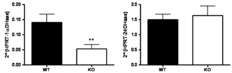
hydroxylase (1αOHase) and 25-hydroxyvitamin D3 -24-hydroxylase (24OHase) which generate active or inactive vitamin D3metabolites, respectively (for review, see [20]). Real-time PCR on renal RNA samples revealed lower levels of 1αOHase mRNA in kidneys from GABARAP-deficient mice, whereas the expression of the 24OHase mRNA was similar in both groups (Fig. 3). These results suggest that the reduced expression of NaPi-IIb in mutant mice could be due to low circulating levels of vitamin D3. Mineral density and Pi/Ca++content in bones from WT and GABARAP-deficient mice Up to 85% of the body’s Pi is stored in bones in the form of hydroxyapatite. Therefore, bone’s physiology is tightly connected to Pi homeostasis. An increased deposit of Pi in bones could compensate for the increase in renal Pi reabsorption observed in GABARAP-deficient mice. To address this possibility, we first analyzed the mineral density (as amount of matter per cubic centimeter) of the trabecular and cortical components
of bones (femurs and L3+L4 lumbar vertebras). As shown in Fig. 4a, the mineral density was similar in femurs from WT and GABARAP-deficient mice. Although the trabecular density was higher in vertebras from mutant mice as compared to WT (Fig.4b), no differences were observed between both groups of animals in the cortical and the total densities of lumbar bones. Moreover, the Pi and Ca++contents of femurs and vertebras were also similar in both groups of mice (Fig. 4c, d). These data suggest that the bones do not contribute to the compensation of Pi balance observed in the absence of GABARAP.
Adaptation of urinary and circulating Pi values to chronic changes on dietary Pi In response to changes in dietary intake, the kidney modulates the amount of Pi reabsorbed along the proximal tubule and thus adjusts the urinary Pi output to the intestinal input. As expected, the urinary excretion of Pi was lower in WT animals fed a low Pi diet as compared to mice fed a high Pi chow (Fig. 5a). This
Fig. 3 Quantification of 1αOHase and 24OHase transcripts in kidneys of WT and GABARAP-deficient mice. Real-time PCR was performed on RNA samples extracted from kidneys of WT and GABARAP-deficient mice. Transcript levels of 1αOHase (left) and
24OHase (right) are shown as relative ratio to hypoxanthine-guanine phosphoribosyltransferase (HPRT) mRNA. Data are given as means ± SE (n=5). **P≤0.005, unpaired Student’s t test
Fig. 4 a, b Mineral density and c, d Pi/Ca++content in bones from WT and GABARAP-deficient mice. Mineral density (milligram per cubic centimeter) was measured in a femurs and b L4 vertebras from four WT and four GABARAP-deficient mice as indicated under“Materials and methods”. The contents of c
Pi and d Ca++are given as percentage of dry matter in femurs and L3+L4 vertebras from two WT and two GABARAP-deficient mice. Data represent means ± SE. **P≤0.05, unpaired Student’s t test
Fig. 5 Effect of chronic dietary Pi in urinary and serum concen-trations of Pi as well as in the 32P uptake into BBM. a Urinary concentrations of Pi, given as ratio to urinary creatinine; b32P uptake into renal BBM; and c serum concentration of Pi in samples from WT
and GABARAP-deficient mice fed with either a high (H) or low (L) Pi diets. Parameters were measured as indicated under “Materials and methods”. Data are given as means ± SE (n=6). ***P≤0.005,
unpaired Student’s t test
Fig. 6 Adaptation of renal Na/Pi cotransporters to chronic dietary intake of Pi. a PVDF membranes containing renal BBM (15μg) from wild-type (WT) and GABARAP-deficient mice (KO) fed either high (H) or low (L) Pi diets were incubated with antibodies against NaPi-IIa, NaPi-IIc, PiT2, andβ-actin. NaPi-IIa and NaPi-IIc immunoreactive bands were visualized sequentially on the same PVDF membrane upon stripping, whereas a second membrane was used to analyze PiT2. Each
group consisted of six animals and the pictures show representative immunoblots. b β-Actin normalization of the abundance of each cotransporter. c Real-time PCR to quantify the expression of NaPi-IIa (top) and NaPi-IIc (bottom) mRNAs. Transcript levels are given as relative ratio to HPRT mRNA. Data are given as means ± SE (n=6). *P≤0.05, **P≤0.01, ***P≤0.005, unpaired Student’s t test
change correlated with an increased Na-dependent uptake of32Pi into renal BBM from low Pi-fed mice (Fig.5b). A proper adaptative response of urinary Pi and Na/Pi cotransport activity was also observed in GABARAP-deficient mice (Fig. 5a, b). This suggests that similar to the acute (hours) response [24], the overall adaptation of the kidney to chronic changes in dietary Pi is not impaired in the absence of GABARAP. As in the acute studies, the urinary excretion of Pi was similar in WT and GABARAP-deficient mice when fed a high Pi diet. However, under chronic Pi restriction, GABARAP-deficient animals re-duced the urinary excretion of Pi to levels even lower than those of WT mice (Fig. 5a), a difference that was not observed upon acute feeding [24]. This reduced urinary excretion in the mutant mice was paralleled by an increased uptake of 32P that was just below statistical significance (Fig.5c; P=0.054). Regarding the circulating levels of Pi, no differences were detected either between both genotypes or between dietary conditions (Fig. 5c). This finding contrasts the results obtained upon acute dietary adaptation, where both WT and mutant mice had lower serum levels of Pi when fed diets with a low as compared to a high Pi content [24]. This transitory difference in serum values most probably reflects the time lag required for the kidney to readjust basal circulatory Pi levels.
Adaptation of renal Na/Pi cotransporters to chronic changes on dietary Pi Readjustment of circulatory levels of Pi upon dietary changes is mediated by adaptive
alterations in the expression of NaPi-IIa [14, 17, 19, 25] and NaPi-IIc [21, 27, 28]. Both cotransporters are down-regulated in response to a high dietary intake of Pi, whereas their expression is increased upon administration of low Pi diets. More recently, it was shown that also the abundance of PiT2 in the renal BBM of rats is regulated by the dietary Pi [33]. Based on dietary switches, NaPi-IIa was suggested to be the fastest responding cotransporter with changes in expression levels being detected already a few hours after the dietary switch; NaPi-IIc levels change only slowly, whereas the time course of the adaptive response of PiT2 is somewhere between both SLC34 cotransporters. We have already shown that the adaptation of NaPi-IIa to acute changes on dietary Pi is not impaired in the absence of GABARAP [24]. Here, we investigated whether the absence of GABARAP interferes with the adaptation of the three renal Na/Pi cotransporters to chronic changes of dietary Pi. Western blot analysis (Fig.6) revealed that NaPi-IIa and NaPi-IIc were upregulated by a low Pi diet both in WT and GABARAP-deficient mice. Adaptation of NaPi-IIc was quantitatively similar in both groups of animals (Fig. 6a, b). However, the increase of NaPi-IIa in the low Pi groups was more pronounced in the absence of GABARAP. Thus, while no differences were detected between WT and GABARAP-deficient mice fed a high Pi diet, the expression of the cotransporter was higher in the mutant mice than in WT upon ingestion of a low Pi chow (Fig. 6a, b). This finding, which was not observed in the acute study [24], most probably explains the urinary
Fig. 7 Adaptation of renal Na/Pi cotransporters to chronic dietary intake of Pi. Immunos-tainings of kidney sections from wild-type (WT) and
GABARAP-deficient mice (KO) fed either high (H) or low (L) Pi diets using antibodies against a NaPi-IIa and b NaPi-IIc
excretion data shown in Fig. 5a, i.e., that GABARAP-deficient mice fed a low Pi diet excrete less Pi than their WT counterparts. In contrast to the two SLC34 cotransporters, PiT2 was not regulated by chronic dietary Pi, neither in WT nor in GABARAP-deficient mice (Fig. 6a, b). This is in contrast to the observations made in rats discussed above [33]. Thus, interspecies variations in the regulation of this transporter may exist.
The adaptation of NaPi-IIa and NaPi-IIc was also analyzed by immunofluorescence (Fig.7). As reported, NaPi-IIa was detected in proximal tubules from cortical and juxtamedular nephrons in kidneys from WT animals fed a low Pi diet, whereas its expression was confined to juxtamedular nephrons in those animals that received a high Pi chow (Fig. 7a) [19]. In agreement with the Western blot data, a similar adaptation of NaPi-IIa was also observed in GABARAP-deficient mice. As expected, NaPi-IIc-related signal was weaker than that of NaPi-IIa, but it was nevertheless also upregulated in WT and mutant mice fed low Pi (Fig.7b).
The cellular mechanism responsible for the dietary regulation of NaPi-IIa probably differs among species. In the rat kidney, this regulation seems to involve changes in mRNA levels, as demonstrated by Northern blot [14] and
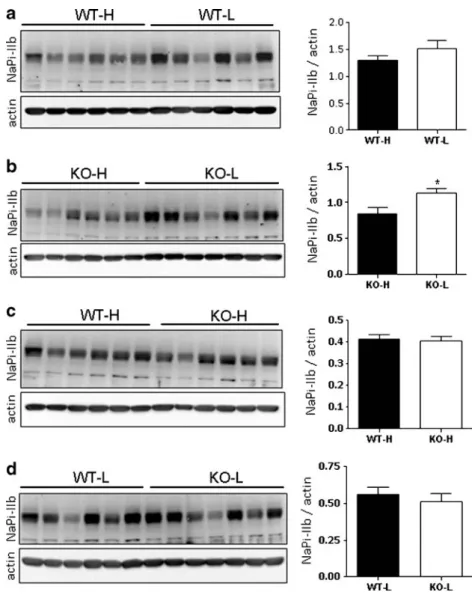
Fig. 8 Adaptation of NaPi-IIb to chronic dietary intake of Pi. PVDF membranes containing ileum BBM (50μg) from wild-type (WT) and
GABARAP-deficient mice (KO) fed either high (H) or low (L) Pi diets were incubated with antibodies against NaPi-IIb and β-actin. a BBM from WT mice fed with high (H) and low (L) Pi diets. b BBM from KO animals fed with high (H) and low (L) Pi diets. c BBM from WT and KO animals fed with high (H) Pi diets. d BBM from WT and KO animals fed with low (L) Pi diets. The abundance of the cotransporter was normalized to theβ-actin content, and the quantifications are shown in the bar graphs. Data are given as means ± SE (n=6, except for the KO-L group that consists of seven mice). *P≤0.05, unpaired Student’s t test
Fig. 9 Bone mineral concentrations. Content of Pi and Ca++ (as percentage of dry matter) in femurs from wild-type (WT) and GABARAP-deficient mice (KO) fed either high (H) or low (L) Pi diets. Data represent means ± SE (n=6)
in situ hybridization [25] data. In contrast, the adaptation in mouse appears to involve posttranscriptional mechanisms, as the amount of NaPi-IIa mRNA in dissected nephron segments was similar in samples isolated from mice fed high or low Pi diets [19]. This result was also reproduced in our study: as shown in Fig.6c, we found that the Pi content of the diet does not affect the amount of NaPi-IIa transcripts neither in WT nor in GABARAP-deficient mice. Further-more, no differences were detected between genotypes under all dietary conditions tested. The dietary regulation of NaPi-IIc in the mouse kidney has been previously shown to involve changes in mRNA expression [19], a finding that was also reproduced here: Fig.6c shows that the levels of NaPi-IIc mRNA were increased in kidneys from both WT and GABARAP-deficient fed a low Pi diet. No differences between genotypes were observed regardless whether high or low Pi diets were given.
Adaptation of NaPi-IIb to chronic changes in dietary Pi In addition to the kidney, the intestine also responds to changes in dietary Pi by regulating the amount of NaPi-IIb in the apical membrane of the small intestine. In mice fed with normal Pi diets, NaPi-IIb is highly expressed in the ileum, whereas its expression is very low in the duodenum and jejunum [23]. The expression of this transporter is upregulated in ileum as well as jejunum under low Pi dietary conditions [10, 23]. Whether such upregulation depends on transcriptional [23] or posttranscriptional [10] mechanisms is still controversial. Here, we analyzed whether the dietary regulation of NaPi-IIb proceeds normally in the absence of GABARAP. As shown in Fig. 8, the expression of NaPi-IIb tended to be higher in ileum BBM from WT animals fed a low Pi diet as compared to high Pi diet (Fig. 8a). Also in GABARAP-deficient mice, the amount of NaPi-IIb increased upon dietary restriction of Pi (Fig.8b). Importantly, no differences were detected between WT and mutant mice either when fed a high (Fig.8c) or a low (Fig.8d) Pi diet. Taken together, these data suggest that the absence of GABARAP does not interfere with the dietary regulation of NaPi-IIb.
Pi/Ca++content in bones from WT and GABARAP-deficient mice Finally, we analyzed whether the different dietary conditions described above lead to changes in the content of Pi and Ca++in bones. Again, the concentrations of both elements in femurs (Fig.9) and vertebras (data not shown) were not affected by changes in dietary Pi. Furthermore, there were no differences between the values measured in femurs from WT and GABARAP-deficient mice.
In conclusion, this study shows that: (a) In GABARAP-deficient mice, the expression of the intestinal Na/Pi cotransporter NaPi-IIb is reduced. Thus, the resulting decrease in the intestinal absorption of Pi may compensate
for its increased renal reabsorption caused by the upregu-lation of NaPi-IIa, thereby allowing normophosphatemic values to be maintained in the mutant mice. (b) In response to chronic deprivation of Pi, a signal for Pi saving, the upregulation of NaPi-IIa is potentiated in the absence of GABARAP. Together with our previously reported findings, this further supports the concept that GABARAP destabilizes NaPi-IIa in the renal BBM by enhancing its internalization and/or degradation.
Acknowledgments We thank Dr. V. Sorribas (Zaragoza, Spain) for kindly providing us with the anti-PiT2 antibody. This work was supported by the Swiss National Science Foundation Grant 44342003 (to HM) and the Sixth European Frame Work EuReGene Project Grant 005085 (to HM). SC Reining was supported by a Ph.D. student fellowship from the University Research Priority Program“Integrative Human Physiology” from the University of Zurich.
References
1. Bai L, Collins JF, Ghishan FK (2000) Cloning and characterization of a type III Na-dependent phosphate cotransporter from mouse intestine. Am J Physiol Cell Physiol 279(4):C1135–C1143 2. Beck L, Karaplis AC, Amizuka N, Hewson AS, Ozawa H,
Tenenhouse HS (1998) Targeted inactivation of Npt2 in mice leads to severe renal phosphate wasting, hypercalciuria, and skeletal abnor-malities. Proc Natl Acad Sci U S A 95(9):5372–5377
3. Bergwitz C, Roslin NM, Tieder M, Loredo-Osti JC, Bastepe M, Abu-Zahra H, Frappier D, Burkett K, Carpenter TO, Anderson D, Garabedian M, Sermet I, Fujiwara TM, Morgan K, Tenenhouse HS, Juppner H (2006) SLC34A3 mutations in patients with hereditary hypophosphatemic rickets with hypercalciuria predict a key role for the sodium–phosphate cotransporter NaPi-IIc in maintaining phosphate homeostasis. Am J Hum Genet 78(2):179– 192
4. Biber J, Stieger B, Stange G, Murer H (2007) Isolation of renal proximal tubular brush-border membranes. Nat Protoc 2(6):1356– 1359
5. Collins JF, Bai L, Ghishan FK (2004) The SLC20 family of proteins: dual functions as sodium–phosphate cotransporters and viral receptors. Pflugers Arch 447(5):647–652
6. Dawson TP, Gandhi R, Le Hir M, Kaissling B (1989) Ecto-5 ′-nucleotidase: localization in rat kidney by light microscopic histochemical and immunohistochemical methods. J Histochem Cytochem 37:39–47
7. Forster IC, Hernando N, Biber J, Murer H (2006) Proximal tubular handling of phosphate: a molecular perspective. Kidney Int 70(9):1548–1559
8. Gisler SM, Stagljar I, Traebert M, Bacic D, Biber J, Murer H (2001) Interaction of the type IIa Na/Pi cotransporter with PDZ proteins. J Biol Chem 276(12):9206–9213
9. Gupta A, Tenenhouse HS, Hoag HM, Wang D, Khadeer MA, Namba N, Feng X, Hruska KA (2001) Identification of the type II Na(+)–Pi cotransporter (Npt2) in the osteoclast and the skeletal phenotype of Npt2−/− mice. Bone 29(5):467–476
10. Hattenhauer O, Traebert M, Murer H, Biber J (1999) Regulation of small intestinal Na–P(i) type IIb cotransporter by dietary phosphate intake. Am J Physiol 277(4 Pt 1):G756–G762 11. Hernando N, Déliot N, Gisler SM, Lederer E, Weinman EJ, Biber
of type IIa Na/P(i) cotransporters. Proc Natl Acad Sci U S A 99 (18):11957–11962
12. Hilfiker H, Hattenhauer O, Traebert M, Forster I, Murer H, Biber J (1998) Characterization of a murine type II sodium–phosphate cotransporter expressed in mammalian small intestine. Proc Natl Acad Sci U S A 95(24):14564–14569
13. Ichikawa S, Sorenson AH, Imel EA, Friedman NE, Gertner JM, Econs MJ (2006) Intronic deletions in the SLC34A3 gene cause hereditary hypophosphatemic rickets with hypercalciuria. J Clin Endocrinol Metab 91(10):4022–4027
14. Levi M, Lötscher M, Sorribas V, Custer M, Arar M, Kaissling B, Murer H, Biber J (1994) Cellular mechanisms of acute and chronic adaptation of rat renal P(i) transporter to alterations in dietary P(i). Am J Physiol 267(5 Pt 2):F900–F908
15. Liesegang A, Loch L, Bürgi E, Risteli J (2005) Influence of phytase added to a vegetarian diet on bone metabolism in pregnant and lactating sows. J Anim Physiol and Anim Nutr 89 (3-6):120–128
16. Lorenz-Depiereux B, Benet-Pages A, Eckstein G, Tenenbaum-Rakover Y, Wagenstaller J, Tiosano D, Gershoni-Baruch R, Albers N, Lichtner P, Schnabel D, Hochberg Z, Strom TM (2006) Hereditary hypophosphatemic rickets with hypercalciuria is caused by mutations in the sodium–phosphate cotransporter gene SLC34A3. Am J Hum Genet 78(2):193–201
17. Lötscher M, Wilson P, Nguyen S, Kaissling B, Biber J, Murer H, Levi M (1996) New aspects of adaptation of rat renal Na–Pi cotransporter to alterations in dietary phosphate. Kidney Int 49 (4):1012–1018
18. Lundquist P, Murer H, Biber J (2007) Type II Na+–Pi cotransporters in osteoblast mineral formation: regulation by inorganic phosphate. Cell Physiol Biochem 19(1–4):43–56
19. Madjdpour C, Bacic D, Kaissling B, Murer H, Biber J (2004) Segment-specific expression of sodium–phosphate cotransporters NaPi-IIa and -IIc and interacting proteins in mouse renal proximal tubules. Pflugers Arch 448(4):402–410
20. Miller WL, Portale AA (2000) Vitamin D 1α-hydroxylase. Trends Endocrinol Metab 11(8):315–319
21. Ohkido I, Segawa H, Yanagida R, Nakamura M, Miyamoto K (2003) Cloning, gene structure and dietary regulation of the type-IIc Na/Pi cotransporter in the mouse kidney. Pflugers Arch 446(1):106–115 22. O’Sullivan GA, Kneussel M, Elazar Z, Betz H (2005) GABARAP
is not essential for GABA receptor targeting to the synapse. Eur J NeuroSci 22(10):2644–2648
23. Radanovic T, Wagner CA, Murer H, Biber J (2005) Regulation of intestinal phosphate transport. I. Segmental expression and adaptation to low-P(i) diet of the type IIb Na(+)–P(i) cotransporter in mouse small intestine. Am J Physiol Gastrointest Liver Physiol 288(3):G496–G500
24. Reining SC, Gisler SM, Fuster D, Moe OW, O’Sullivan GA, Betz H, Biber J, Murer H, Hernando N (2009) GABARAP deficiency
modulates expression of NaPi-IIa in renal brush-border membranes. Am J Physiol Renal Physiol 296(5):F1118–F1128
25. Ritthaler T, Traebert M, Lötscher M, Biber J, Murer H, Kaissling B (1999) Effects of phosphate intake on distribution of type II Na/Pi cotransporter mRNA in rat kidney. Kidney Int 55(3):976– 983
26. Sabbagh Y, O’Brien SP, Song W, Boulanger JH, Stockmann A, Arbeeny C, Schiavi SC (2009) Intestinal Npt2b plays a major role in phosphate absorption and homeostasis. J Am Soc Nephrol 20 (11):2348–2358
27. Segawa H, Kaneko I, Takahashi A, Kuwahata M, Ito M, Ohkido I, Tatsumi S, Miyamoto K (2002) Growth-related renal type II Na/Pi cotransporter. J Biol Chem 277(22):19665–19672
28. Segawa H, Yamanaka S, Ito M, Kuwahata M, Shono M, Yamamoto T, Miyamoto K (2005) Internalization of renal type IIc Na–Pi cotransporter in response to a high-phosphate diet. Am J Physiol Renal Physiol 288(3):F587–F596
29. Segawa H, Onitsuka A, Kuwahata M, Hanabusa E, Furutani J, Kaneko I, Tomoe Y, Aranami F, Matsumoto N, Ito M, Matsumoto M, Li M, Amizuka N, Miyamoto K (2009) Type IIc sodium-dependent phosphate transporter regulates calcium metabolism. J Am Soc Nephrol 20(1):104–113
30. Shenolikar S, Voltz JW, Minkoff CM, Wade JB, Weinman EJ (2002) Targeted disruption of the mouse NHERF-1 gene promotes internalization of proximal tubule sodium–phosphate cotransporter type IIa and renal phosphate wasting. Proc Natl Acad Sci U S A 99 (17):11470–11475
31. Shibasaki Y, Etoh N, Hayasaka M, Takahashi MO, Kakitani M, Yamashita T, Tomizuka K, Hanaoka K (2009) Targeted deletion of the type IIb Na(+)-dependent Pi-co-transporter, NaPi-IIb, results in early embryonic lethality. Biochem Biophys Res Commun 381 (4):482–486
32. Tenenhouse HS, Gauthier C, Martel J, Hoenderop JG, Hartog A, Meyer MH, Meyer RA Jr, Bindels RJ (2002) Na/P(i) cotransporter (Npt2) gene disruption increases duodenal calcium absorption and expression of epithelial calcium channels 1 and 2. Pflugers Arch 444(5):670–676
33. Villa-Bellosta R, Ravera S, Sorribas V, Stange G, Levi M, Murer H, Biber J, Forster IC (2009) The Na+–Pi cotransporter PiT-2 (SLC20A2) is expressed in the apical membrane of rat renal proximal tubules and regulated by dietary Pi. Am J Physiol Renal Physiol 296 (4):F691–F699
34. Xu H, Bai L, Collins JF, Ghishan FK (2002) Age-dependent regulation of rat intestinal type IIb sodium–phosphate cotransporter by 1, 25-(OH)(2) vitamin D(3). Am J Physiol Cell Physiol 282(3): C487–C493
35. Zoidis E, Ghirlanda-Keller C, Gosteli-Peter M, Zapf J, Schmid C (2004) Regulation of phosphate (Pi) transport and NaPi-III transporter (Pit-1) mRNA in rat osteoblasts. J Endocrinol 181 (3):531–540