Arch Orthop Trauma Surg (1998) 117 : 39-42 © Springer-Verlag 1998
A . K o h l e r • R . H o f f m a n n " A . P l a t z - M . B i n o
Diagnostic value of duplex ultrasound
and liquid crystal contact thermography
in preclinical detection of deep vein thrombosis
after proximal femur fractures
Received: 29 August 1996
A b s t r a c t During a prospective clinical study the diag- nostic value of the two non-invasive examinations colour- coded duplex ultrasound (Duplex) and fluid crystal con- tact thermography (LCCT) was investigated in relation to phlebography, the standard examination for the diagnosis of deep vein thrombosis (DVT), in 112 patients with prox- imal femur fractures. In 19% of the patients, DVT was di- agnosed by phlebography, with the main localisation in the lower leg in 19 of 21 (90%) thromboses. With a nega- tive prediction value of 83%, Duplex is less suitable than LCCT under such difficult examination conditions as the early postoperative period. The specificity of Duplex is 95%, but the sensitivity only 18%. The specificity of LCCT is 85% and the sensitivity 75%. Considering the frequency of postoperative DVT after surgery on the legs, especially hip surgery, a postoperative screening for DVT should become mandatory. LCCT has proved to be a suit- able, cheap, non-invasive examination with a negative prediction value of 94%.
Introduction
Deep leg vein thrombosis (DVT) is, in spite of prophy- laxis with heparin, still a frequent postoperative complica- tion, especially after operations involving the lower ex- tremities. Operations around the hip carry a very high risk, with published rates of thrombosis from 20% up to 40% [1, 6-10, 13, 17]. Therefore, these patients should be checked routinely. To prevent dangerous lung embolism and chronic vein insufficiency, an early diagnosis of DVT at the level of the lower leg is needed before appositional growth of the clot has occluded the popliteal vein and
A. Kohler ( ~ ) - R. Hoffmann • A. Platz
Departement Chirurgie, Universit/itsspital Ztirich, Rfimistrasse 100, CH-8091 Ztirich, Switzerland
M. Bino
Institute for Diagnostic Radiology, University of Ziirich Hospital, Ztirich, Switzerland
starts to give rise to typical clinical symptoms. This re- quires very precise diagnostic methods at the calf and the feasibility of monitoring in the first postoperative days.
The golden standard for the diagnosis of DVT is still anterograde phlebography [1, 4, 10]. Despite the high in- cidence of DVT with possibly severe consequences [11], testing with phlebography has not become standard prac- tice for high-risk operations such as hip surgery. This con- trasts with the many obligatory preoperative examina- tions, which give indications of risks much lower than those associated with DVT. Arguments against using phlebography are the application of intravenous contrast, high dosage of X-radiation, pain and costs.
In addition to phlebography there are various techni- cal examinations for the diagnosis of DVT. Colour- coded duplex ultrasound (Duplex) and liquid crystal con- tact thermography (LCCT) are two non-invasive ones which can be done a few days after the operation [1-6, 8, 10]. Duplex has a high sensitivity and specificity for the diagnosis of DVT, but the collective of patients is in most studies highly selected and the interpretation espe- cially at the lower leg is controversial [1, 5, 6, 10, 17]. L C C T also has a good sensitivity but a slightly lower specificity [2, 8, 16]. L C C T is easy to use, with the pa- tient lying in bed on the ward. The technique and inter- pretation of the results of L C C T are easy to learn. A non- invasive, reliable screening method for DVT diagnosis would be very helpful in postoperative management. Therefore, these two methods were evaluated during a prospective study under difficult clinical examination conditions during the early postoperative period with mostly very elderly patients.
Patients and methods
In a prospective, randomised, monocentric clinical study, the effi- cacy of low molecular weight heparin (LMWH; Sandoparin) given as one subcutaneous (s.c.) application of 3000 international units (IU) per 24 h was compared with the standard prophylaxis for high-risk operations, low-dose heparin (not fractionated heparin; LDH; Liquemin) given 3 times 5000 IU s.c. per 24 h. As a stan- dard control to prove or exclude thrombosis, a phlebography was
40
Table 1 Contraindications for prophylaxis with heparin bleeding shock, polytrauma
- severe bum injury
- haemorrhagic diathesis (anamnestic tendency for suffusions, increased bleeding tendency)
- severe hypertension (blood pressure over 200 m m H g systolic or 115 mmHg diastolic)
- florid gastrointestinal ulcer head and brain injury
- oral anticoagulation with derivatives of coumarin
or medication with non-steroidal anti-rheumatics
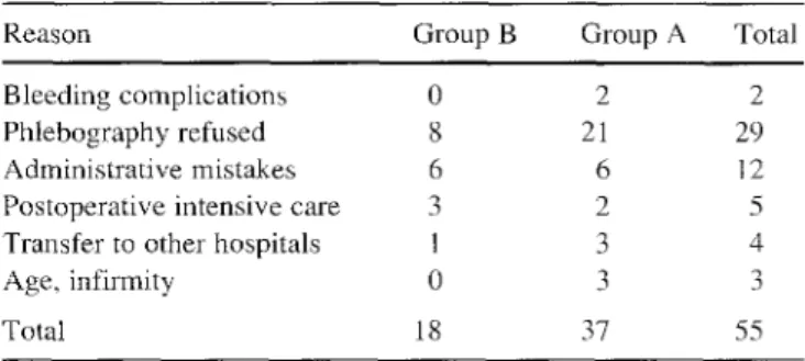
Table 2 Reasons for prematurely ending study
Reason Group B Group A Total
Bleeding complications 0 2 2
Phlebography refused 8 21 29
Administrative mistakes 6 6 12
Postoperative intensive care 3 2 5
Transfer to other hospitals 1 3 4
Age, infirmity 0 3 3
Total 18 37 55
performed in the first week after the operation. Before the phle- bography L C C T and Duplex were done to investigate these two methods of screening for the preclinical detection of deep DVT. Sensitivity, specificity, and positive and negative prediction values were calculated in relation to the results of the phlebography. All patients with proximal femur fractures (femur neck and in- tertrochanteric fractures) were included who did not have con- traindications according to Table 1. Admission to the hospital oc- curred for all patients on the day of trauma. The randomisation to group A (Liquemin) or group B (Sandoparin) was done in the emergency room after confirming the diagnosis by X-ray. The first dose was given preoperatively in the emergency room afier pri- mary diagnostics and randomisation. For postoperative care on the operated leg, an elastic bandage was applied from the foot to the pelvis (hip-spika bandage). The bandage was removed on the sec- ond postoperative day. Mobilisation beside the bed was started on the 2nd postoperative day, and walking training was instituted on the 2nd or 3rd day after the vacuum drains (Redon) had been re- moved. Prophylaxis with heparin was always given for a minimum of 10 days. Before the control examinations the patient was in- formed a second time about the study (the first information given in the emergency room is commonly not satisfactory either for the patient or the doctor) and gave written consent. Between the 4th and 6th postoperative days clinical examination and LCCT were performed by one of three surgeons experienced in this technique, then the Duplex control and lastly phlebography were done at the Institute for Diagnostic Radiology. The examiners were not in- formed of each other's results.
LCCT was performed on the patient in his bed on the ward. In contrast to infrared thermography or indirect thermography, air conditioning or darkening of the examination room is not neces- sary. Fifteen minutes before the examination, the leg end of the bed was elevated to 15 ° . To achieve symmetric conditions both legs were freed from bandages, bed splints and coverlet, up to the hip. From 1 h beforehand neither physiotherapy nor ointment ap- plication was allowed, Outside rotation of the operated leg and then symmetric positioning of the opposite leg in supine position gave the best contact zone for the LCCT detector. In every in- stance all three projections, lower legs, knees and thigh, were checked.
Duplex was performed in the supine position. Changes of posi- tion to prone or standing up were not feasible due to the short time after operation and the old age of most of the patients.
Anterograde phlebography was done bilaterally on a tilt table. After injection of contrast in a dorsal vein of the foot, standard ex- posures at the level of the lower leg, knee, thigh and pelvis were taken. If the phlebography was not performed for any reason, the patient was excluded from the study. Oral anticoagulation with acenocoumarol (Sintrom) was started for 3 months if DVT was di- agnosed in the lower leg with phlebography, or if full mobilisation after 10 days was not anticipated. Thrombosis of the popliteal, femoral or iliac veins was treated with full dosage continuous in- travenous heparin, bed rest for 5-6 days and overlapping oral anti- coagulation with acenocoumarol for 6 months.
The study was approved by the Ethics Committee of the Uni- versity Hospital of Z(irich.
Statistical analysis
The mean values were compared with the z-test. The contingency tables were analysed with the chi-square test. The limit for signifi- cance was always alpha = 0.05 (5%). Sensitivity and specificity were calculated as usual. The positive/negative prediction value
(p) is the possibility that the diagnosis is true if the result of the ex-
amination is positive/negative. Positive predictive value = p(K/T+);
K =ill patients and T+ = positive result of the examination. Nega- tive predictive value = p(G/T-); G = healthy patients and T - = neg- ative result of the examination,
Patients
From June 1993 until June 1995 at the University Hospital of Zurich, 167 patients with a proximal femur fracture (femur neck and intertrochanteric fractures) were randomised for the study, giving 125 women and 42 men with a mean age of 75.9 (+/- 13.5) years (range 28-96 years). The study was completed by 112 pa- tients, 55 patients interrupted the study for different reasons (see Table 2), the most frequent one being the absence of phlebogra- phy, which some patients refused.
Results
In all, 112 p a t i e n t s c o m p l e t e d t h e study, 45 in g r o u p A a n d 67 in B. T h e t w o g r o u p s w e r e c o m p a r e d f o r t h e f o l l o w i n g criteria: age, sex, w e i g h t , h e i g h t , p r e o p e r a t i v e b e d rest, v a r i c o s i s , a n a m n e s t i c state a f t e r t h r o m b o s i s o r p u l m o n a r y e m b o l i s m , atrial f i b r i l l a t i o n , n e o p l a s m , s y s t e m i c a n g i o p a - thy, d i a g n o s i s o f o p e r a t i o n , t y p e o f o p e r a t i o n , s u r g e o n a n d t y p e o f a n a e s t h e s i a . T h e r e w a s n o s i g n i f i c a n t d i f f e r e n c e e i t h e r f o r a n y o f t h e s e c r i t e r i a b e t w e e n t h e t w o g r o u p s o r in r e l a t i o n to p a t i e n t s w i t h t h r o m b o s i s . G r o u p A h a d sig- n i f i c a n t l y (P = 0 . 0 0 1 ) m o r e d r o p - o u t s (n = 37) t h a n g r o u p B (n = 18) ( T a b l e 2). O f t h e 112 p a t i e n t s w i t h p h l e b o g r a - p h y results, w e f o u n d 21 c a s e s o f D V T ( 1 9 % ) , 12 o f 45 p a t i e n t s in g r o u p A ( 2 7 % ) a n d 9 o f 67 p a t i e n t s in g r o u p B ( 1 3 % ) . T h e d i f f e r e n c e b e t w e e n t h e g r o u p s is n o t s i g n i f i - c a n t (P = 0.08). W i t h 19 p a t i e n t s ( 9 0 % ) t h e t h r o m b o s i s w a s l i m i t e d to t h e l o w e r leg, a n d t h u s o n l y 2 h a d t h r o m - b o s i s in t h e f e m o r a l a n d i l i a c v e i n . L C C T w a s d o n e o n 107 p a t i e n t s (Figs. 1, 2); f o r 5 pa- t i e n t s t h e i n s t r u m e n t w a s o u t o f o r d e r b e c a u s e o f a t e c h n i - c a l d e f e c t . M a n y p a t i e n t s c o m p l a i n e d o f p a i n d u r i n g D u - p l e x b e c a u s e o f t h e a t t e m p t e d p o s i t i o n i n g p r o c e d u r e . In c o n s i d e r a t i o n o f t h e u n e q u i v o c a l s t a t i s t i c a l results, w e s t o p p e d p e r f o r m i n g D u p l e x a f t e r 79 p a t i e n t s b e c a u s e o f p o o r a c c e p t a n c e . T h e r e s u l t s o f p h l e b o g r a p h y , L C C T , D u -
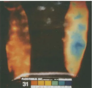
Fig. 1 LCCT finding in the ventral lower leg showing regional hy- perthermia on the left side laterally (blue area)
41 Table 3 Results of the diagnostic methods in relation to phlebo- graphy (DVT deep vein thrombosis, LCCT liquid crystal contact thermography, Duplex colour-coded duplex ultrasound)
a) clinical examination vs phlebography Clinical exam Phlebography
DVT yes DVT no Total DVT yes 2 (1.8%) 19 (17.0%) 21 (18.8%) DVT no 3 (2.7%) 88 (78.5%) 91 (81.2%) Total 5 (4.5%) 107 (95.5%) 112 (100%) b) LCCT vs phlebography LCCT Phlebography DVT yes DVT no Total DVT yes 15 (14.0%) 5 (4.7%) 20 (18.7%) DVT no 13 (12.1%) 74 (69.2%) 87 (81.3%) Total 28 (26.1%) 79 (73.9%) 107 (100%) c) Duplex vs phlebography Duplex Phlebography DVT yes DVT no Total DVT yes 3 (3.8%) 14 (17.7%) 17 (21.5%) DVT no 3 (3.8%) 59 (74.7%) 62 (78.5%) Total 6 (7.6%) 73 (92.4%) 79 (100%) Table 4 Statistical characteristics of LCCT and Duplex LCCT
Sensitivity 75%
Specificity 85%
Positive predictive value 54% Negative predictive value 94% Duplex
Sensitivity 18 %
Specificity 95%
Positive predictive value 46% Negative predictive value 83 %
p l e x and c l i n i c a l e x a m i n a t i o n are s u m m a r i s e d in Table 3. In r e l a t i o n to the results o f the p h l e b o g r a p h y , the statisti- cal v a l u e s w e r e c a l c u l a t e d (Table 4).
Fig.2 Corresponding phlebography of the same patient with a fresh thrombosis in the fibular veins
Discussion
T h e rate o f t h r o m b o s i s in our s t u d y is 19%, w h i c h is a b o u t the a v e r a g e o f m a n y other p u b l i c a t i o n s [1, 4 - 7 , 9, 13, 17]. P r o p h y l a x i s w i t h L M W H o n c e a d a y has p r o v e n to b e at least as e f f i c a c i o u s as L D H three t i m e s a day. T h e P v a l u e o f 0.08 e v e n s h o w s a t e n d e n c y t o w a r d s s i g n i f i c a n t differ- e n c e for the b e n e f i t o f L M W H .
T h e c l i n c i a l d i a g n o s i s o f D V T p o s t o p e r a t i v e l y is unre- liable. The d i f f e r e n t i a t i o n b e t w e e n p o s t - t r a u m a t i c or post-
42
operative oedema and deep haematomas leads to false clinical judgements. In our study, with most diagnosed D V T in the lower leg, clinical evaluation is not sufficient. Duplex has been assessed as having a high specificity of 95% but in contrast to the results from other studies, a low sensitivity of only 18% [1, 5, 18]. Reasons for this low sensitivity include the main localisation of D V T in the lower leg in 19 of 21 patients, the early postoperative time period of the examination and the old age of our pa- tients. The low diagnostic precision of Duplex in the lower leg has also been confirmed by Ginsberg et al. and Magnusson et al. [4, 10]. Elderly patients often cannot co- operate in changing position to get more information with the Duplex because of postoperative pain and the m a n y handicaps of old age.
L C C T was also limited by the early postoperative time period and the patient's inability to cooperate; in particu- lar the prone position for dorsal exposure was not feasible. L C C T had a lower specificity of 85% but a better sen- sitivity of 75%. The positive predictive value of 54% (Du- plex 46%) and the negative predictive value of 94% (Du- plex 83%) shows that L C C T is suitable for screening pa- tients early in the postoperative period, to exclude D V T with a probability of 94%, even under difficult examina- tion conditions. Cooke and Pilcher [2, 3] described ther- m o g r a p h y already in the 1970s as a suitable technique for the preclinical diagnosis of DVT. But the system was too complicated to be routinely applied. The technique of L C C T described by Pochaczevsky and collaborators [14] makes the examination much easier and allows one to check patients on the ward without difficult preparations. In spite of our aggressive antithromboctic prophylaxis, thrombosis remains a severe postoperative problem with a rate of 19%. Considering that over 60% of thromboses originate intraoperatively [12], early postoperative diag- nosis is decisive to identify patients with D V T as soon as possible to institute adequate therapy to prevent apposi- tional growth of the clot. The remaining high incidence of D V T with different types of prophylaxis involving he- parin also seems to indicate that the rate of intraoperative origin is not influenced much by the type of prophylaxis. The localisation pattern of D V T with increased incidence in the lower leg in more recent publications suggests that the prophylaxis m a y slow down the postoperative growth of the clot in the bigger veins above the lower leg. This should give enough time for a postoperative diagnosis.
In our study L C C T has proven to be a suitable exami- nation for the early postoperative detection of D V T before the manifestation of clinical symptoms. For screening for D V T of the lower leg, L C C T is superior to Duplex. Con- sidering all the preoperative diagnostics used to reduce the perioperative risk for risks much lower than that of DVT, a postoperative control to exclude D V T should be- come mandatory. L C C T is a suitable non-invasive screen- ing examination after high-risk operations like hip surg- ery. It allows a carefully directed early further diagnosis with phlebography and therapy with oral anticoagulation
for patients with DVT. This management limits most D V T at the level of the lower leg before irreparable changes [11] in the big veins above the popliteal vein or embolism can occur.
References
1. Barrellier MT, Bosson JL, Vignon C, Rousseau JF, Bernard M, Boissel M, Fauchon G, Pegoix M, Thomassin C, Trahay A (1994) Echo-doppler for early diagnosis of deep venous throm- bosis in orthopedic surgery and traumatology. J Mal Vasc 19 : 298-307
2. Cooke ED, Pilcher MF (1973) Thermography in the diagnosis of deep venous thrombosis. Br Med J 2 : 523-526
3. Cooke ED, Pilcher MF (1974) Deep vein thrombosis: preclini- cal diagnosis by thermography. Br J Surg 61:971-978 4. Ginsberg JS, Caco CC, Brill-Edwards PA, Panju AA, Bona R,
Demers CM, Tuters LM, Nugent P, McGinnis J, Grant BM (199 I) Venous thrombosis in patients who have undergone ma- jor hip or knee surgery: detection with compression US in im- pedance plethysmography. Radiology 181 : 651-654
5.Girasole GJ, Cuomo F, Denton JR, O'Conner D, Ernst A (1994) Diagnosis of deep vein thrombosis in elderly hip-frac- ture patients by using the duplex scanning technique. Orthop Rev 23:411-416
6.Grady-Benson JC, Oishi CS, Hanson PB, Cowell CW, Otis SM, Walker RH (1994) Routine postoperative duplex ultra- sonography screening and monitoring for the detection of deep vein thrombosis. A survey of 110 total hip arthroplasties. Clin Orthop 307 : 130-141
7.Hoffmann R (1991) The thrombo-embolic risk in surgery. He- patogastroenterology 38:272-278
8.Hoffmann R, Brtitsch HP, Schwarz R (1990) Die Fltissig- kristall-Kontaktthermographie in der Diagnostik der tiefen Ve- nenthrombose. Angiologie 60 : 12-17
9. Knudson MM, Lewis FR, Clinton A, Atkinson K, Mergerman J (1994) Prevention of venous thromboembolism in trauma pa- tients. J Trauma 37(3):480-487
10. Magnusson M, Eriksson BI, KNebo P, Sievertsson R (1996) Is colour doppler ultrasound a sensitive screening method in di- agnosing deep vein thrombosis after hip surgery? Thromb Haemost 75 : 242-245
11. Milne AA, Stonebridge PA, Bradbury AW, Ruckley CV (1994) Venous function and clinical outcome following deep vein thrombosis. Br J Surg 81 : 847-849
12. Nicolaides AN (1975) Thromboembolism. Medical and techni- cal publishing, Lancaster p 205
i3.Platz A, Hoffmann R, Kohler A, Bischoff T, Trentz O (1993) Thromboembolieprophylaxe bei Htiftfraktur: Unfraktioniertes Heparin versus niedermolekulares Heparin. Z Unfallchir Ver- sicherungsmed 86:184-188
14.Pochaczevsky R, Pillari G, Feldman F (1982) Liquid crystal contact thermography of deep venous thrombosis. Am J Radiol 138 : 717-723
15. Ritchie WG, Soulen RL, Lapayowker MS (1979) Thermo- graphic diagnosis of venous thrombosis. Radiology 131:341- 344
16. Sandler DA, Martin JF (1985) Liquid crystal thermography as a screening test for deep-vein thrombosis. Lancet 1 (8430): 665-667
17.Walker RH (1994) Secondary prevention of venous throm- boembolism in joint replacement using duplex ultrasonogra- phy. Orthopedics 17 [Suppl] : 14-17
18.Wenda K, Jaeger U, Das Gupta K, Degreif J, Runkel M, Ritter G (1993) Zur Entstehung der Thrombosen in der Htiftgelenk- endoprothetik. Eine Studie mit der farbcodierten Duplexsono- graphie. Unfallchirurg 96 : 373-381