Endogenous retroviral elements, but not exogenous retroviruses,
are detected in CD30-positive lymphoproliferative disorders of the
skin
Werner Kempf
1,2,4, Marshall E.Kadin
2, Ann M.Dvorak
2,
Carol C.Lord
1, Gu¨nter Burg
4, Norman L.Letvin
1and
Igor J.Koralnik
1,31Beth Israel Deaconess Medical Center, Harvard Medical School, Department of Medicine, Division of Viral Pathogenesis, Boston, MA, 2Beth Israel Deaconess Medical Center, Harvard Medical School, Department of Pathology, Boston, MA,3Beth Israel Deaconess Medical Center, Harvard Medical School, Department of Neurology, Boston, MA, USA and4University Zurich, Department of Dermatology, Zurich, Switzerland
To whom correspondence should be addressed. 4Email: [email protected]
Introduction
Primary cutaneous CD30-positive lymphoproliferative
dis-orders (LPD) comprise a spectrum of disdis-orders including
lymphomatoid papulosis (LyP) and CD30⫹ pleomorphic or
anaplastic large T-cell lymphomas (1–3). LyP is a chronic,
recurrent lymphoproliferative disorder which manifests
clinic-ally as papulonodular, spontaneously regressing skin lesions
(4). Histologically, large pleomorphic and anaplastic tumor
cells are found which are activated T-lymphocytes expressing
the CD30 antigen (5,6).
Viruses are known etiologic factors in the pathogenesis of
non-Hodgkin’s lymphomas (NHL) (7). In particular,
retro-viruses such as human T-lymphotropic virus 1 and 2
(HTLV-1 and 2) have been linked to adult T-cell leukemia/lymphoma
(ATLL) and other forms of NHL (for review see refs 8 and
9). Primary cutaneous lymphomas, especially cutaneous T-cell
lymphomas, have been considered to be caused by viruses.
Several reports of the detection of HTLV-1 or 2 sequences in
cutaneous T-cell lymphomas (CTCL) (10–13) have not been
confirmed in other studies (8,9,14,15).
Ultrastructural and clinical features suggest that a virus may
be involved in the etiology of LyP (Kadin, unpublished data).
Viral-like particles (VLP) have been detected in LyP lesions
by electron microscopy (16). In addition, the immunophenotype
of LyP tumor cells is identical to the lymphoid tumor cells in
ATLL associated with HTLV-1. Moreover, CD30 antigen
expression is known to be triggered by viruses such as
HTLV-1 and Epstein–Barr virus (HTLV-17). HTLV-HTLV-1 proviral sequences
have been found in CD30⫹ large-cell cutaneous T-cell
lymphoma (18). Nevertheless, neither retroviruses nor other
oncogenic viruses have been detected in LyP (5,19–21). In up
to 20% of cases, LyP is preceded or followed by other
lymphoproliferative disorders such as Hodgkin’s lymphoma,
mycosis fungoides or systemic CD30⫹ anaplastic large-cell
lymphoma (22–24). These tumors have been shown to be
Abbreviations: ATLL, adult T-cell leukemia/lymphoma; CTCL, cutaneous
T-cell lymphomas; GAPDH, glyceraldehyde 3-phosphate dehydrogenase; HTLV-1 and 2, human T-lymphotropic virus 1 and 2; IUDR, 5⬘-iodo-2⬘ deoxy-uridine; LPD, lymphoproliferative disorders; LyP, lymphomatoid papulosis; NHL, non-Hodgkin’s lymphomas; RT–PCR, reverse transcription–polymerase chain reaction; VLP, viral-like particles.
clonally related to LyP occurring in the same individual
(25,26). Therefore, it is of importance to identify a putative
viral etiology underlying these lymphomas, if one exists.
The aim of the present study was to investigate whether
retro-viruses are involved in the pathogenesis of LyP. To accomplish
this goal, various methods were chosen to detect cytopathic
effects in cocultivation experiments, reverse transcriptase
activ-ity in supernatants of cultured tumor cells, nucleic acid sequences
of retroviruses in lesional tissue and cell lines established from
CD30⫹ LPD and electron microscopy of tissue biopsies and
cell lines to detect viral particles. To identify possible unknown
retroviruses putatively associated with CD30⫹ LPD, consensus
PCR for retroviral polymerase genes was used since these genes
are generally the most conserved sequences among retroviruses
(27). Here, we report the presence of viral-like particles,
tran-scripts of endogenous retroviral elements (ERV), and reverse
transcriptase (RT) activity, as well as the absence of exogenous
retroviruses, in LyP.
Material and methods
LyP biopsies and cell linesTwenty-seven glutaraldehyde-fixed biopsies from 13 patients with LyP were investigated by EM for the presence of viral-like particles. Eleven snap-frozen skin biopsies of LyP lesions from a total of 10 patients were available for extraction of nucleic acids. The tumor cell lines JK, Mac 1 and Mac 2A, had been established from progressive lesions of two LyP patients (‘JK’ and ‘Mac’) who showed development of CD30-positive anaplastic large-cell lymphoma during the course of LyP. Studies of T-cell receptor gene rearrange-ment showed that JK and Mac cells were each clonally related to all prior LyP skin lesions (for details see refs 25 and 28).
Cell cultures and cocultivation
All cells were cultured in RPMI 1640 medium supplemented with 10% fetal calf serum at 37°C. Peripheral blood mononuclear cells (PBMC) from a healthy individual were separated by Ficoll gradient and maintained in culture without stimulation. JK and Mac 2a were cocultivated with lymphoid cell lines SupT1, CemX, MT2 and H9 as well as with peripheral blood mononuclear cells of a healthy subject according to the protocols described by Miyoshi et al. (29).
Induction
In addition, JK cells were treated for 48 h with 5⬘-iodo-2⬘ deoxyuridine (IUDR) to enhance transcription of putative retroviruses. Alternatively, cells were treated with sodium butyrate (1µM) added to the medium containing RPMI 1640 and interleukin-2 (IL-2).
Electron microscopy
Twenty-seven skin biopsies of LyP lesions from a total of 13 patients, and cells from one tumor cell line were prepared and studied by electron microscopy (30). The controls included various inflammatory and neoplastic disorders such as eczema (n⫽ 3), lichen planus (n ⫽ 3), psoriasis (n ⫽ 3), and other cell T-cell lines (n⫽ 2).
RT assay
The supernatants of tumor cell lines were tested for reverse transcriptase (RT) activity with colorimetric highly sensitive manganese-based and magnesium-based RT assays (Cavidi HS-kit Mn2⫹RT, Cavidi Tech, Uppsala, Sweden), respectively. Supernatants were filtered through a 0.45 micrometer Nalgene filter, added to a Beckman centrifuge tube and centrifuged at 50 000 r.p.m. for 1 h in a 90 Ti Beckman rotor under vacuum at 4°C. HIV-1 infected cells (HIV B3 cells) and HTLV-1 infected cells (LCL-Mok and TCL-Kan cells) served as positive controls for the RT assays. Negative controls consisted of supernatants of cultured PBMC of three healthy, HIV-seronegative individuals.
by guest on November 24, 2016
http://carcin.oxfordjournals.org/
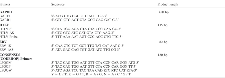
Table I. Oligonucleotides and length of amplification products used in the described PCR protocols
Primers Sequence Product length
GAPDH 480 bp
GAP.F1 5⬘-AGG CTG GGG CTC ATT TGC-3⬘
GAP.R1 5⬘-GTG CTC AGT GTA GCC CAG GAT G-3⬘
HTLV 135 bp
HTLV S 5⬘-CTA TGG AGA GTA CTA CCC CAA GG-3⬘ HTLV AS 5⬘-CTC GTC ATC CAT GTA CTG AAG-3⬘ HTLV Probe 5⬘ TTT AAA AAT AGT CCC ACC CTG TTC-3⬘
ERV 82 bp
ERV 1S 5⬘-CAA CTC TCT GCT TTG TAT CAT AAT C-3⬘ ERV 1AS 5⬘-ATA GAC CAG TGT GAT ATC TTG CG-3⬘
CONSENSUS 120 bp
(CODEHOP)-Primers
LPQGM 5⬘-TAC CAG TGG AAT GTT CTA CCN CAR GGN ATG-3⬘ LPQGF 5⬘-TAC CAG TGG AAT GTT CTA CCN CAR GGN TT-3⬘ LPQGW 5⬘-ATC AGA TCC TAC TAA CAD RTC RTC CAT RTA-3⬘
Y⫽ C / T; K ⫽ G / T; R ⫽ A / G; N ⫽ A / C / G / T
The Mn-based assay has been shown to detect RT from ERV with higher sensitivity than the Mg-based assay or conventional RT assays using radioactive substrates (31).
RNA extraction
RNA from tissue samples was extracted using the Atlas Pure RNS Isolation kit (Clontech, Bo. K1038-1, Palo Alto CA, USA). RNA was extracted from cells using the Qiagen RNAEasy Kit (Qiagen, Valencia, CA). RNA was reverse transcribed using a cDNA synthesis kit for RT–PCR (Reverse Transcription system, No. A 3500, Promega Corp., Madison, WI, USA) using oligo-dT primers and degenerate retroviral primers, respectively. RNA was extracted from ultracentrifuged supernatants by QIAamp viral RNA mini kit (Qiagen, Valencia, CA).
PCR and Southern blot hybridization
Successful amplification of a sequence of the human glyceraldehyde 3-phosphate dehydrogenase (GAPDH) (for sequences see Table I) mRNA indicated that the samples were adequate for RT–PCR analysis and that no PCR inhibitors were present. To avoid contamination and product carryover, DNA extraction, PCR and gel electrophoresis were done in separate laboratories.
Specific sequences of HTLV-1 and 2 were detected by means of a PCR protocol with virus-specific primers followed by Southern blot hybridization with specific radioactive labelled probes for HTLV-1 and 2 (for sequences of oligonucleotides and probe see Table I). HTLV-1 infected cells (LCL-Mok and TCL-Kan cells) served as positive controls for the amplification of corresponding retroviral sequences.
Amplification of ERV sequences
For amplification of ERV 52–123-related sequences, specific primers were designed which amplify a 82 bp long region (for sequences of oligonucleotides see Table I). The PCR conditions were as follows: 35 cycles consisting of 30 s denaturation at 96°C, 30 s annealing of oligonucleotides at 55°C and 30 s extension at 72°C.
Degenerate PCR
Degenerate primers with a 5⬘ end clamp and variable nucleotide positions at the 3⬘ end, recently described as Consensus-Degenerate Hybrid Oligonucleotide Primers (CODEHOP)-PCR (32), were used to amplify a highly conserved region of the reverse transcriptase genes of various retroviruses by a PCR protocol. The primers LPQGF, LPQGM, LPQGW and YMDD have been described in detail by Rose and coworkers (32). LPQGF have been designed to amplify preferentially oncogenic retroviruses, whereas LPQGW amplify predominantly lentiviruses. The cDNA templates were first amplified using 35 PCR cycles (denaturation at 94°C for 1 min, annealing at 55°C for 1 min, and extension at 72°C for 1 min). The amplification products were electrophoresed in a 2% agarose gel and visualized by UV transillumination after staining with ethidium bromide. The primers were shown to amplify HTLV-1 and HIV-1 sequences from infected cells. Carry over of trace amounts of genomic DNA was prevented by digestion of nucleic acid extracts from tissues by DNase. For each sample, replicate RT–PCR was performed (data not shown). The same amount of RNA was used for all samples analyzed by RT–PCR.
Cloning and sequencing
The RT–PCR products of expected size were cloned into a pCR2.1 vector (TOPO TA Cloning kit, No. K4500-01, Invitrogen, Carlsbad, CA), and the resulting recombinant plasmids were sequenced using the BigDye Terminator Ready Reaction kit (Perkin Elmer Cat. No. 4303151, Foster City, CA) on a ABI Prism 377 automated DNA sequencer (Perkin Elmer, Foster City, CA). Sequence analysis
Homology searches against various databases were performed using BLASTN and BLASTX programs (33,34).
Results
Electron microscopy
Nine of 27 biopsies from 13 patients with LyP showed multiple
large cytoplasmic membrane-bound vacuoles of variable sizes
up to 10
µm, either filled with or lined on their inner surfaces
with small viral-like particles (VLP) (Figure 1). Many of these
structures with poorly visible or non-electron dense centers
were morphologically similar to immature retrovirus particles
(10,35,36). A few particles contained irregularly shaped and
located electron dense core material. Budding of these particles
from the plasma membrane was not seen in biopsies. Nor were
visible free particles present in the nucleus or cytoplasm. The
diameter of the particles in cytoplasmic vacuoles was 60–
80 nm. No clinical and/or histologic differences between
virus-positive and negative lesions could be identified.
The tumor cell line JK, derived from a patient with LyP,
and cultured in the presence of IL-2 and sodium butyrate was
examined by electron microscopy. VLP analogous to those
present in biopsies were present in similar cytoplasmic vacuoles
and as clusters adjacent to tumor cell surfaces (Figure 2).
Classical images of budding were not evident in the EM sample.
Cocultivation
No cytopathic effect could be observed after cocultivation of
tumor cell lines JK and Mac 1 or Mac 2A with lymphoid cell
lines SupT1, CemX, MT2 and H9 as well as with peripheral
blood mononuclear cells of a healthy subject after 48 h, 7
days or 14 days. Furthermore, no syncytia formation of JK
cells was observed after induction with IUDR for 48 h.
RT assays
Conventional radioactive RT assays performed on supernatants
of JK and Mac cell lines and cocultures did not reveal
by guest on November 24, 2016
http://carcin.oxfordjournals.org/
Fig. 1. Viral-like particles in a tumor cell in a skin biopsy of a patient with
lymphomatoid papulosis. In (A), a giant-sized vacuole (V) in the cytoplasm contains numerous small structures resembling immature retroviral particles, most of which are attached to the inner vacuolar membrane. Two small vacuoles (arrowhead) close to the cell surface also contain viral-like structures. A portion of the inner vacuole membrane is enlarged in (B). This allows visualization of dense core-like structures (arrows) in some of the particles (C, indicated by arrow). (A)⫻43 000; (B) ⫻80 000; (C) ⫻200 000.
Fig. 2. Viral-like particles in the extracellular space adjacent to a tumor cell
in the JK tumor cell line. Note that some of the particles contain dense cores (arrows).⫻63 500.
detectable RT activity. Even after induction of cells with
IUDR, which is a known inducer of retroviral transcription,
no RT activity was detected by conventional RT assays. In
contrast, the colorimetric manganese-based RT assay allowed
detection of RT activity in the ultracentrifuged supernatant of
JK cells after induction with IUDR for 48 h, with a median
value of RT activity of 903
µU/µl compared with ⬍200 µU/
µl for uninduced JK cells. No RT was activity detected in the
control cell lines.
PCR studies
HTLV 1 and 2 sequences could not be detected by RT–PCR
with virus-specific oligonucleotides in lesional tissue of LyP
and cell lines (data not shown).
Degenerate PCR with CODEHOP-primers LPQGF and
YMDD revealed amplification products of expected size in
two of six LyP cases, JK cells and the ultracentrifuged
supernatant of JK cells induced for 48 h with IUDR (data not
shown). The PCR products of 87 bp length were subsequently
cloned and automatically sequenced. Homology searches
against various databases were performed using BLASTN and
BLASTX programs and demonstrated that nucleotides 1–72
of our 87 bp long amplified products showed similarity of
98% with nucleotides 16–87 of the pol region of ERV 52–123
(Genbank accession no. S71609) with one nucleotide change
at position 20 (a
→ t) of our amplified product. The sequence
of our fragment is: 5⬘-gct ttg tat cat aat ctt att cgg aga gaa ctt
gat agc ttt tca cat ccg caa gat atc aca ctg gtc tat-3
⬘ which does
not contain any stop codons. One of the putative corresponding
amino acid sequences is: A L Y H N L I R R E L D S F S
H P Q D I T L V Y. In this open reading frame, this amino
acid sequence has similarity of 66% to the pro-pol-dUTPase
polyprotein (aa 83–106) of murine endogenous retrovirus
ERV-L (Genbank accession no. T29097) and to the reverse
transcriptases. No sequence similarity to known gag or env
genes protein sequences was found by Genbank analysis.
Thus, oligonucleotides specific for ERV 52–123 were
designed to assess expression levels of this ERV in LyP and
cell lines. As shown in Figure 3, higher levels of mRNA
transcripts were detected in all three tumor cell lines JK and
Mac1 and 2A (lanes 1, 2, 5 and 6), supernatants of tumor cell
lines (lanes 3 and 4) and the majority of LyP biopsies (lanes
7–10) compared with PBMC of healthy individuals (lanes
11–13) and T-cell lines CEM-X, HTLV-1 infected H9 and
HTLV-1 negative Sup T1 (lanes 14–16).
by guest on November 24, 2016
http://carcin.oxfordjournals.org/
Fig. 3. RT–PCR for ERV 52–123: Higher levels of amplified mRNA
sequences in CD30⫹ tumor cell lines (lanes 1, 2, 5 and 6), supernatants of tumor cell lines (lanes 3 and 4) and LyP lesional tissue (lanes 7–10) compared with PBMC of healthy individuals (lanes 11–13) and T-cell lines CEM-X, HTLV-1 infected H9 and HTLV-1 negative Sup T1 (lanes 14–16). The upper images represent amplified ERV transcripts, whereas the lower images the GAPDH levels from corresponding samples. N: negative control; P: positive control (cloned PCR product); M: molecular weight marker.
Discussion
In this study, we demonstrate the presence of intra- and
extracellular viral-like particles in lesional tissue and tumor
cell lines from primary cutaneous CD30-positive LPD. By
electron microscopy, these particles exhibited the morphology
of immature retroviral particles (10,35,36). Similar particles
have been described in cultured peripheral blood cells and
T-cell lines from patients with Se´zary’s syndrome, a disorder
belonging to the group of CTCL (37,38), and we have
seen similar particles in cutaneous lesions in a patient with
erythrodermic CTCL (see Figs 279 and 284 in ref. 39). Similar
VLP have not been present in a wide variety of diagnostic
human skin biopsies including eczema, lichen ruber, and
psoriasis (Dvorak, unpublished data).
To characterize these particles, PCR-based approaches were
chosen to search for known, and for closely related, but yet
unknown exogenous and endogenous retroviral sequences. By
virus-specific PCR, no evidence of HTLV-1 and HTLV-2
presence could be found in lesional tissue and cell lines of
CD30⫹ LPD of the skin. These results are in accordance with
the data reported in another study (20). In addition, we
did not identify sequences of closely related, yet unknown
retroviruses by applying a consensus PCR method aimed at
detecting a highly conserved retroviral transcriptase region.
These data indicate that exogenous retroviruses closely related
to HTLV-1 or -2 are probably not present in CD30⫹ LPD of
the skin. Despite various features which may indicate the
involvement of oncogenic exogenous retroviruses in the
patho-genesis of CD30⫹ LPD, there is so far no evidence for a role
of these viruses in the pathogenesis of CD30
⫹ LPD based on
our findings and the data reported in the literature. Furthermore,
the absence of exogenous retroviruses is in accordance with
the lack of well-documented cases suggesting horizontal or
vertical transmission of CD30⫹ LPD.
However, using the same consensus PCR approach, we
detected transcripts of human endogenous retroviral sequences
(HERVs) in lesional tissue and cell lines of CD30
⫹ LPD of
the skin. ERV are inherited genomic elements with structural
features of integrated retroviruses (40,41). Whereas ERV
are mostly silent genomic elements, some groups can form
retrovirus-like particles and be enzymatically active (42,43).
Sequencing of PCR products found in LyP tissue and cell
lines revealed that the amplification products belong to ERV
52–123. These ERV transcripts were originally isolated from
PBMC of patients with systemic lupus erythematosus, a
systemic autoimmune disorder (44).
These retroviral elements are closely related to human
endogenous retroviral ERV 9 sequences which have been
isolated from the T-lymphoma Peer cell line (45). So far, these
ERV 52–123 have not been linked to human tumors. The
pol fragment has a 65% similarity to the pol fragment of
HTLV-1 and 2. Except for this fragment, other parts of the
genomic structure of ERV 52–123 remain unknown. Thus it
remains to be proven whether this sequence is part of a
complete provirus. Similarly, further work needs to be done
to identify the whole ERV sequence found in our samples.
The detection of mRNA shows that ERV 52–123 is
transcriptionally
active.
Moreover,
reverse
transcriptase
activity was detected in the supernatant of tumor cells by a
highly sensitive Mn-based RT assay developed to detect RT
activity of ERVs. Although it remains unclear whether this RT
activity originates from ERV particles, RT activity and high
levels of ERV transcripts were both found in LyP lesional
tissue and cell lines.
In addition, the same ERV sequences could be found in the
supernatant of tumor cell lines harboring ERV 52–123 which
might result from release of VLP in the extracellular space as
we observed by electron microscopy. Interestingly,
cocultiva-tion of tumor cells with other potentially susceptible T-cells
did not reveal detectable transmission. This is not surprising
since most ERV have lost their infectivity due to deletions
and mutations during evolution.
Since ERV are inherited, it is difficult to establish an
association between these viral elements and a particular
disease. To establish causality, one cannot only rely on the
presence or absence of these elements as in the case for
exogenous viruses. In this study, higher levels of transcription
of ERV 52–123 sequences could be found in lesional tissue
and cell lines of LyP, compared with controls. Similarly, a
high level of ERV expression has been detected in mouse
lymphoma and in human tumors such as germ cell tumors
(GCT) (46). Furthermore, viral ERV gag protein is produced
in large quantities by GCT and serum antibodies directed
against ERV env protein can be found in 85% of patients
with GCT, whereas healthy individuals and patients with
inflammatory diseases or tumors other than GCT very rarely
develop such antibodies (46). Recently, Boese and coworkers
reported that the HERV protein cORF supports cell
transforma-tion as shown by tumor inductransforma-tion in nude mice (47).
Our data suggest that exogenous retroviruses related to
HTLV-1 and HTLV-2 are not involved in the pathogenesis of
primary cutaneous CD30⫹ LPD. Because of the high
preferen-tial expression of HERV 52–123 in lesional tissue and cell
lines of LyP, and the detection of VLP by electron microscopy,
we postulate that the activation of endogenous retroviral
elements may be associated with the pathogenesis of primary
cutaneous CD30
⫹ lymphomas. Further studies are necessary
to demonstrate that the particles found by EM represent an
endogenous retrovirus as suggested by the data obtained by
RT–PCR and RT assays.
Acknowledgements
We thank Bijan Etemad-Moghadam, Dana Farber, Cancer Institute, Boston, MA, USA for performing Mg-based RT assays, and Patricia Fox, Karen Bryan, and Tracey Sciuto for electron microscopic assistance.
Dr Werner Kempf was a recipient of a Research Fellowship of the Schweizerische Stiftung fu¨r Medizinisch-Biologische Stipendien, Basel, Switzerland, and of a research grant of the MeritCare Foundation, Fargo, ND, USA when the studies were performed.
This work was supported in part by USPHS grant AI 33372.
References
1. Kadin,M.E. (1990) The spectrum of Ki-1⫹ cutaneous lymphomas. Curr. Probl. Dermatol., 19, 132–143.
by guest on November 24, 2016
http://carcin.oxfordjournals.org/
2. Kaudewitz,P. and Burg,G. (1991) Lymphomatoid papulosis and Ki-1 (CD30)-positive cutaneous large cell lymphomas. Semin. Diagn. Pathol.,
8, 117–124.
3. Willemze,R., Kerl,H., Sterry,W., et al. (1997) EORTC classification for primary cutaneous lymphomas: a proposal from the cutaneous lymphoma study group of the European Organization for Research and Treatment of Cancer. Blood, 90, 354–371.
4. Macaulay,W.L. (1968) Lymphomatoid papulosis. A continuing self-healing eruption, clinically benign–histologically malignant. Arch. Dermatol., 97, 23–30.
5. Kadin,M., Nasu,K., Sako,D., Said,J. and Vonderheid, E. (1985) Lymphomatoid papulosis. A cutaneous proliferation of activated helper T cells expressing Hodgkin’s disease-associated antigens. Am. J. Pathol.,
119, 315–325.
6. Kaudewitz,P., Stein,H., Burg,G., Mason,D.Y. and Braun-Falco,O. (1986) Atypical cells in lymphomatoid papulosis express the Hodgkin cell-associated antigen Ki-1. J. Invest. Dermatol., 86, 350–354.
7. Cesarman, E. and Mesri,E.A. (1999) Virus-associated lymphomas. Curr. Opin. Oncol., 11, 322–332.
8. Lisby,G., Konstatinov,K. and Lange Vejlsgaard,G. (1990) Retroviruses and cutaneous T-cell lymphoma. In Van Vloten,W.A., Willemze,R., Lange Vejlsgaard,G. and Thomsen,K. (eds) Cutaneous Lymphoma. Curr. Probl. Dermatol. Karger Basel, vol. 19, pp. 50–68.
9. Lessin,S.R., Vowels,B.R. and Rook,A.H. (1994) Retroviruses and cutaneous T-cell lymphoma. Dermatol. Clin., 12, 243–253.
10. Poiesz,B.J., Ruscetti,F.W. and Gazder,A.F. (1980) Detection and isolation of type C retrovirus particles from fresh and cultured lymphocytes of patients with cutaneous T-cell lymphoma. Proc. Natl Acad. Sci. USA, 77, 7415–7419.
11. Zucker-Franklin,D., Hooper,W.C. and Evatt,B.L. (1992) Human lymphotropic retroviruses associated with mycosis fungoides: evidence that human T-cell lymphotropic virus type OO (HTLV-II) as well as HTLV-I may play a role in the disease. Blood, 80, 1537–1545.
12. Pancake,B.A., Zucker-Franklin,D. and Coutavas,E.E. (1995) The cutaneous T cell lymphoma, mycosis fungoides, is a human T-cell lymphotropic virus-associated disease. A study of 50 patients. J. Clin. Invest., 95, 547–554.
13. Poiesz,B., Dube,D., Dube,S., Love,J., Papsidero,L., Uner,A. and Hutchinson,R. (2000) HTLV-II-associated cutaneous T-cell lymphoma in a patient with HIV-1 infection. N. Engl. J. Med., 342, 930–936. 14. Bazarbachi,A., Saal,F. and Laroche,L. (1997) Mycosis fungoides and
Se´zary syndrome are not associated with HTLV-I infection. An international study. Br. J. Haematol., 98, 927–933.
15. Wood,G., Schaffer,J.M., Boni,R., Dummer,R., Burg,G., Takeshita,M. and Kikuchi,M. (1997) No evidence of HTLV-1 proviral integration in lymphoproliferative disorders associated with cutaneous T-cell lymphoma. Am. J. Pathol., 150, 667–673.
16. Shamsuddin,A.K., Nedwich,A. and Toker,C. (1980) Lymphomatoid papulosis. Ultrastructural study with demonstration of intranuclear and intracytoplasmic viruslike particles. Dermatologica, 161, 238–242. 17. Stein,H., Mason,D.Y., Gerdes,J., et al. (1985) The expression of the
Hodgkin’s disease associated antigen Ki-1 in reactive and neoplastic lymphoid tissue: evidence that Reed-Sternberg cells and histiocytic malignancies are derived from activated lymphoid cells. Blood, 66, 848–858.
18. Agnastopoulos,I., Hummel,M., Kaudewitz,P., Braun-Falco,O. and Stein,H. (1990) Detection of HTLV-I proviral sequences in CD30 positive large cell cutaneous T-cell lymphoma. Am. J. Pathol., 137, 1317–1322. 19. Sangueza,O.P., Galloway,G., Eagan,P.A., Braziel,R.M. and Gulley,M.L.
(1996) Absence of Epstein-Barr virus in lymphomatoid papulosis: an immunohistochemical and in situ hybridization study. Arch. Dermatol.,
132, 279–282.
20. Ortiz-Romero,P.L., Vaqllejo,A., Lopez Estebaranz,J.L., Garcia Saiz,A., Fernandez,V. and Iglesias Diez,L. (1997) Absence of HTLV-1 proviral sequences in patients with lymphomatoid papulosis (letter). J. Invest. Dermatol., 109, 817–818.
21. Kempf,W., Kadin,M.E., Kutzner,H., Lord,C.L., Burg,G., Letvin,N.L. and Koralnik,I.J. (2001) Lymphomatoid papulosis and human herpesviruses – A PCR-based evaluation for the presence of human herpesvirus 6, 7 and 8 and related herpesviruses. J. Cut. Pathol., 28, 29–33.
22. Kaudewitz,P., Stein,H., Plewig,G., Schwarting,R., Gerdes,J., Burg,G., Kind,P., Eckert,F. and Braun-Falco,O. (1990) Hodgkin’s disease followed by lymphomatoid papulosis. J. Am. Acad. Dermatol., 22, 999–1006. 23. Cabanillas,F., Armitage,J., Pugh,W.C., Weisenburger,D. and Duvic,M.
(1995) Lymphomatoid papulosis: a T-cell dyscrasia with a propensity to transform into malignant lymphoma. Ann. Intern. Med., 122, 210–217. 24. Wang,H.H., Myers,T., Lach,L.J., Hsieh,C.C. and Kadin,M.E. (1999)
Increased risk of lymphoid and nonlymphoid malignancies in patients with lymphomatoid papulosis. Cancer, 86, 1240–1245.
25. Davis,T.H., Morton,C.C., Miller,C.R., Balk,S.P. and Kadin,M.E. (1992) Hodgkin’s disease, lymphomatoid papulosis, and cutaneous T-cell lymphoma derived from a common T-cell clone. N. Engl. J. Med., 326, 1115–1122.
26. Chott,A., Vonderheid,E.C., Olbricht,S., Miao,N.N., Balk,S.P., Kadin,M.E. (1996) The dominant T cell clone is present in multiple regressing skin lesions and associated T cell lymphomas of patients with lymphomatoid papulosis. J. Invest. Dermatol., 106, 696–700.
27. McClure,M.A., Johnson,M.S., Feng,D.F. and Doolittle,R.F. (1988) Sequence comparisons of retroviral proteins: relative rates of change and general phylogeny. Proc. Natl Acad. Sci. USA, 85, 2469–2473.
28. Schiemann,W.P., Pfeifer,W.M., Levi,E., Kadin,M.E. and Lodish,H.F. (1999) A deletion in the gene for transforming growth factor beta type I receptor abolishes growth regulation by transforming growth factor beta in a cutaneous T-cell lymphoma. Blood, 8, 2854–2861.
29. Miyoshi,I., Kubonishi,I., Yoshimoto,S., Akagi,T., Ohtsuki,Y., Shiraishi,Y., Nagata,K. and Hinuma,Y. (1981) Type C virus particles in a cord blood T-cell line derived from cocultivating normal human cord leukocytes and human leukaemic T-cells. Nature, 294, 770–771.
30. Dvorak,A.M. (1987) Monograph: Procedural guide to specimen handling for the diagnostic ultrastructural pathology service laboratory. J. Electron. Microsc. Tech., 6, 255–301.
31. Malmsten,A., Ekstrand,D.H., Akerblom,L., Gronowitz,J.S., Kallander,C.F., Bendinelli,M. and Matteucci D. (1998) A colorimetric reverse transcriptase assay optimized for Moloney murine leukemia virus, and its use for characterization of reverse transcriptases of unknown identity. J. Virol. Methods, 75, 9–20.
32. Rose,T.M., Schultz,E.R., Henikoff,J.G., Pietrokovski,S., McCallum,C.M. and Henikoff,S. (1998) Consensus-degenerate hybrid oligonucleotide primers for amplification of distantly related sequences. Nucleic Acids Res., 26, 1628–1635.
33. Altschul,S.F., Gish,W., Miller,W., Myers,E.W. and Lipman,D.J. (1990) Basic local alignment search tool. J. Mol. Biol., 215, 403–410.
34. Altschul,S.F., Madden,T.L., Scha¨ffer,A.A., Zhang,J., Zhang,Z., Miller,W. and Lipman,D.J. (1997) Gapped BLAST and PSI-BLAST: a new generation of protein database search programs. Nucleic Acids Res., 25, 3389–3402. 35. Biswas,P., Poli,G., Kinter,A.L., Justement,J.S., Stanley,S.K., Maury,W.J., Bressler,P., Orenstein,J.M. and Fauci,A.S. (1992) Interferon gamma induces the expression of human immunodeficiency virus in persistently infected promonocytic cells (U1) and redirects the production of virions to intracytoplasmic vacuoles in phorbol myristate acetate-differentiated U1 cells. J. Exp. Med., 176, 739–750.
36. Bugelski,P.J., Kirsh,R. and Hart,T.K. (1994) HIV protease inhibitors: effects on viral maturation and physiologic function in macrophages. J HIV protease inhibitors. J. Leukoc. Biol., 56, 374–380.
37. Kaltoft,K., Bisballe,S., Rasmussen,H.F., Thestrup-Pedersen,K., Boehncke,W.H., Volker,H. and Sterry,W. (1988) C-type particles are inducible in Se-Ax, a continuous T-cell line from a patient with Se´zary’s syndrome. Arch. Dermatol. Res., 280, 264–267.
38. Saal,F., Gessain,A., Lasneret,J., et al. (1989) Detection of retrovirus particles and reverse transcriptase activity in mid-term cultured peripheral blood and lymph node cells from a French woman with Se´zary syndrome. Nouv. Rev. Fr. Hematol., 31, 333–337.
39. Dvorak,A.M. and Monaham-Earley,R.A. (1992) Case 39. Soft tissue mass and lytic lesion of the scapula in a 55-year-old red man with a 16 year history of eczema. In Diagnostic Ultrastructural Pathology I. CRC Press, pp. 327–346.
40. Leib-Mo¨sch,C., Brack-Werner,R., Werner,T., Bachmann,M., Faff,O., Erfle,V. and Hehlman,R. (1990) Endogenous retroviral elements in human DNA. Cancer Res., 50 (Suppl. 17), 6536–5642.
41. Wilkinson,D., Mager,D. and Leong,J.C. (1994) Endogenous human retroviruses. In Levy,J.A. (ed.) The Retroviridae. Plenum Press, New York, vol. 3, pp. 465–535.
42. Lower,R., Boller,K., Hasenmaier,B., Korbmacher,C., Muller-Lantzsch,N., Lower,J. and Kurth,R. (1993) Identification of human endogenous retroviruses with complex mRNA expression and particle formation. Proc. Natl Acad. Sci. USA, 90, 4480–4484.
43. Lower,R. (1999) The pathogenic potential of endogenous retroviruses: facts and fantasies. Trends Microbiol., 7, 350–356.
44. Herrmann,M. and Kalden,J.R. (1994) PCR and reverse dot hybridization for the detection of endogenous retroviral transcripts. J. Virol. Methods,
46, 333–348.
45. Strazzullo,M., Parisi,T., Di Cristofano,A., Rocchi,M. and La Mantia,G. (1998) Characterization and genomic mapping of chimeric ERV9 endogenous retroviruses-host gene transcripts. Gene, 206, 77–83.
by guest on November 24, 2016
http://carcin.oxfordjournals.org/
46. Sauter,M., Schommer,S., Kremmer,E., et al. (1995) Human endogenous retrovirus K10: expression of Gag protein and detection of antibodies in patients with seminomas. J. Virol., 69, 414–421.
47. Boese,A., Sauter,M., Galli,U., Best,B., Herbst,H., Mayer,J., Kremmer,E., Roemer,K. and Mueller-Lantzsch,N. (2000) Human endogenous
retro-virus protein cORF supports cell transformation and associates with the promyelocytic leukemia zinc finger protein. Oncogene, 19, 4328– 4336.
Received December 5, 2001; revised July 9, 2002; accepted September 26, 2002
by guest on November 24, 2016
http://carcin.oxfordjournals.org/