Prevalence and predictors of fatigue in glioblastoma: a prospective
study
Philipp O. Valko, Asim Siddique, Claudia Linsenmeier, Kathrin Zaugg, Ulrike Held, and Silvia Hofer
Department of Neurology, University Hospital Zurich, Zurich, Switzerland (P.O.V., S.H.); Department of Oncology, University Hospital Zurich, Zurich, Switzerland (A.S., S.H.); Department of Radiation Oncology, University Hospital Zurich, Zurich, Switzerland (C.L., K.Z.); Department of Radiation Oncology, Inselspital, Bern University Hospital, University of Bern, Bern, Switzerland (K.Z.); Horten Center for Patient Oriented Research and Knowledge Transfer, University of Zurich, Zurich, Switzerland (U.H.)
Corresponding Author: Philipp O. Valko, MD, Department of Neurology, University Hospital Zurich, Frauenklinikstrasse 26, 8091 Zurich, Switzerland ([email protected]).
Background. The main goal of this study was to assess frequency, clinical correlates, and independent predictors of fatigue in a homogeneous cohort of well-defined glioblastoma patients at baseline prior to combined radio-chemotherapy.
Methods. We prospectively included 65 glioblastoma patients at postsurgical baseline and assessed fatigue, sleepiness, mean bedtimes, mood disturbances, and clinical characteristics such as clinical performance status, presenting symptomatology, de-tails on neurosurgical procedure, and tumor location and diameter as well as pharmacological treatment including antiepileptic drugs, antidepressants, and use of corticosteroids. Data on fatigue and sleepiness were measured with the Fatigue Severity Scale and the Epworth Sleepiness Scale, respectively, and compared with 130 age- and sex-matched healthy controls.
Results. We observed a significant correlation between fatigue and sleepiness scores in both patients (r ¼ 0.26; P ¼ .04) and con-trols (r ¼ 0.36; P , .001). Only fatigue appeared to be more common in glioblastoma patients than in healthy concon-trols (48% vs 11%; P , .001) but not the frequency of sleepiness (22% vs 19%; P ¼ .43). Female sex was associated with increased fatigue fre-quency among glioblastoma patients but not among control participants. Multiple linear regression analyses identified depression, left-sided tumor location, and female sex as strongest associates of baseline fatigue severity.
Conclusions. Our findings indicate that glioblastoma patients are frequently affected by fatigue at baseline, suggesting that fac-tors other than those related to radio- or chemotherapy have significant impact, particularly depression and tumor localization. Keywords: depression, fatigue, glioblastoma, sleepiness.
Glioblastoma is the most common primary brain tumor in adults, with an estimated incidence of about 3 per 100 000 in-habitants per year in Europe and North America.1The standard
of care for newly diagnosed glioblastoma, subsequent to sur-gery, comprises radiotherapy with concomitant temozolomide followed by adjuvant temozolomide. In the study defining this treatment regimen, median survival was limited to 15 months,2and median survival was reported to be only 12
months in a population-based analysis of more than 10 000 glioblastoma patients.3
Independent of any treatment, fatigue is a common symp-tom in cancer patients in general as well as in primary brain tumor patients, with an estimated prevalence of 50% – 90% and 40% – 70%, respectively.4,5Cancer-related fatigue is de-fined by the National Comprehensive Cancer Network (NCCN)
as a distressing, persistent, subjective sense of physical, emo-tional, and/or cognitive tiredness or exhaustion related to can-cer or cancan-cer treatment that is not proportional to recent activity and interferes with usual functioning.6The patients themselves indicate fatigue as one of the most distressing symptom related to cancer and its treatment.7It is a strong predictor of decreased patient satisfaction and health-related quality of life (QoL) and may represent one of the key reasons for discontinuing treatment.8–10Nevertheless, fatigue is be-lieved to be underdiagnosed and underestimated in cancer pa-tients despite its possible impact on treatment compliance.4,11 As a consequence, some groups have questioned whether the standard treatment for glioblastoma is justified in view of the limited benefit on survival and the severity of associated symptoms.12
Received 8 October 2013; accepted 3 June 2014
#The Author(s) 2014. Published by Oxford University Press on behalf of the Society for Neuro-Oncology. All rights reserved. For permissions, please e-mail: [email protected].
Neuro-Oncology
Neuro-Oncology 17(2), 274–281, 2015 doi:10.1093/neuonc/nou127
Compared with other tumor types, namely breast and lung cancer, few studies have addressed the problem of fatigue in glioblastoma patients in depth, and several limitations have to be mentioned. First, many groups have included patients with all sorts of primary brain tumors despite large differences in underlying neurobiology, treatment procedures, and progno-sis.5,13Second, baseline data are often missing, particularly in
studies using a cross-sectional design.5As a consequence,
fa-tigue was mainly assessed as a treatment complication, there-by failing to acknowledge the primary impact of the tumor itself and other treatment-independent factors.14Third, the re-sults of many older series cannot be directly compared with the current situation because of advances in radiation techniques. Finally, while many validated fatigue questionnaires are avail-able,9,15the large majority of neuro-oncological studies identi-fied and quantiidenti-fied fatigue in a very rudimental way, using the Visual Analogue Scale (VAS) or one single fatigue item appear-ing in tools such as the European Organisation for Research and Treatment of Cancer (EORTC) QoL questionnaire, the M D Ander-son Symptom Inventory –Brain Tumor Module, or the Symptom Distress Scale.9,12,16–18
Thus, in this prospective study, we aimed at examining fre-quency and predictors of fatigue severity in a homogeneous cohort of glioblastoma patients at baseline prior to combined radio-chemotherapy. For this goal, we used the Fatigue Severity Scale (FSS), which has been identified as the most widely adopted fatigue questionnaire in clinical practice and has been validated for a variety of neurological diseases.15,19,20
In addition, we explored the evolution of fatigue, sleepiness, and mood disorders during and after combined radio-chemotherapy.
Patients and Methods
This prospective, longitudinal study was conducted as a collab-oration of the Departments of Neurology, Oncology, and Radi-ation Oncology of the University Hospital Zurich between October 2008 and October 2012. The study protocol was ap-proved by the Ethics Committee of the Canton of Zurich, Swit-zerland, specialized subcommittee for Psychiatry, Neurology, Neurosurgery (Project E-43/2007), and informed consent was obtained from all patients prior to enrollment.
Participants and Controls
We prospectively included 65 patients with newly diagnosed and histologically proven glioblastoma, corresponding to an es-timated 60%– 65% of all eligible glioblastoma patients of the acquisition period. Patients aged≥18 years undergoing postop-erative standard radio-chemotherapy were eligible.2They also had to be fluent in the German language. The participants were examined clinically and by questionnaires at 3 different time points: (i) 28+7 days after the initial neurosurgical procedure (1 day prior to the first radiation), (ii) at the time of the last ra-diation, and (iii) prior to the initiation of adjuvant chemothera-py. Overall, the study period captured the first 10 weeks of postoperative standard treatment.
As control group, we included 130 healthy and age- and sex-matched individuals using a 1:2 case-control design. The con-trols were selected from a previously published cohort of 454
healthy subjects that we used in our original validation study of the German version of the FSS.19
Clinical Assessment and Questionnaires
Demographic variables included age, sex, and educational sta-tus (defined as highest degree attained). We ascertained sever-al tumor characteristics including type of neurosurgicsever-al procedure (biopsy, partial, complete or unclear resection), tumor topography, and brain magnetic resonance imaging-based tumor diameter. To elucidate whether the presenting symptomatology affected fatigue severity, we divided the medical history into seizure, motor weakness, cognitive deficit, headache/nausea/vomiting, apathy/asthenia, visual deficit, and accidental finding. Seizure type was further classified as partial, generalized, complex-focal, and unclassified. To esti-mate the influence of pharmacological treatment, we included at each time point details on anticonvulsive drugs, antidepres-sants, anxiolytics, CNS stimulants, hypnotics, and whether or not participants received corticosteroids (including the dose of the steroids). Clinical performance status was assessed by the Karnofsky performance score (KPS), with 100% indicating perfect physical health and 0% death. As mentioned earlier, we measured fatigue by means of the FSS. This self-administered questionnaire comprises 9 items exploring fa-tigue severity in different situations during the previous week, and the final score ranges from 1 to 7 with the latter value in-dicating maximal fatigue. The presence of clinically significant fatigue was defined as an FSS score≥4.0. The FSS has robust psychometric properties and has been validated for various neurological disorders but has not yet been validated for glio-blastoma patients. We therefore performed a reliability statistic in our cohort, which revealed excellent internal consistency as reflected by a Cronbach’s a of 0.94.21We used the German ver-sion of the Epworth Sleepiness Scale (ESS) for assessment of sleepiness; a score of≥10 indicates excessive daytime sleepi-ness (EDS).22We also determined the prevalence of overlap
be-tween fatigue and EDS, when participants presented both a FSS score≥4.0 and an ESS score ≥10. Sleep need was estimated using information on mean bedtimes. We arbitrarily defined mean bedtimes≥10 hours as “long bedtimes”, probably indi-cating increased sleep need per 24 hours (ie, hypersomnia). Fi-nally, for evaluation of anxiety and depression, we used the German version of the Hospital Anxiety and Depression Scale (HADS). It is a well-validated questionnaire, which is suitable for cancer populations because it contains only nonphysical symptoms for both anxiety and depression. Subjects indicate their agreement with each item on a scale ranging from 0 to 3. The questionnaire has 2 subscales for anxiety and depres-sion, each consisting of 7 items. A score of .10 is considered to indicate overt anxiety or depression.23,24
Data Analysis and Statistics
We used SPSS (version 19.0) for statistical analysis. Group data are described by means, standard deviations (SD), and confi-dence intervals (95% CI). To compare the mean values of FSS and ESS scores between glioblastoma participants and con-trols, we used the Student’ t test; the x2test was used to com-pare the frequency of fatigue and EDS between the 2 groups.
Longitudinal differences of scores were assessed using the Stu-dent’ paired t test. We applied Cronbach a statistics to calculate the internal consistency of the FSS in glioblastoma patients. To identify predictors of fatigue severity at baseline, we performed a multiple linear regression analysis with the FSS score as a de-pendent variable. Among the set of potential predictor vari-ables (age, sex, education, KPS, ESS, bedtimes, anxiety and depression scores, tumor localization, and use of steroids or an-tidepressants), we evaluated each variable for an estimated ef-fect of≥0.2 or ≤20.2 on the outcome score for fatigue severity and a P value ,.05 in a univariate comparison. Those predictor variables fulfilling the 2 criteria were included in the multiple linear regression model. Significance was accepted at P ,.05. 95% confidence intervals (CI) for mean differences between the groups were additionally presented when group compari-sons revealed significant differences.
Results
Characterization of Glioblastoma Patients
We included 65 glioblastoma patients, of whom 44 (68%) were male. Mean age was 57.3+10.1 years. The tumor was localized in the left brain hemisphere in 28 patients (43%), in the right brain hemisphere in 31 patients (48%), and bilateral in 6
patients (9%). A majority of tumors affected the frontotempo-ral lobes (57%) as compared with the parieto-occipital lobes (31%), basal ganglia (5%), or multiple sites (8%).
Comparison between Glioblastoma Patients and Controls
Glioblastoma patients had significantly higher FSS scores (3.9+ 1.7 vs 2.8+1.0; 95% CI 0,74–1.49; P , .001), fatigue frequency (48% vs 11%; P , .001), and longer bedtimes (8.8+1.2 h vs 7.7+0.9 h; 95% CI, 0.76–1.39; P , .001) than controls, where-as ESS scores and the prevalence of EDS were similar (Table1). FSS and ESS scores were significantly correlated in both glio-blastoma patients (r ¼ 0.26; P ¼ .04) and controls (r ¼ 0.36; P , .001). In glioblastoma patients, overlap of both fatigue and EDS was observed in 15%, while “isolated fatigue” was much more common (32%) than “isolated EDS” (6%) (Fig.1). Conversely, we observed more controls with isolated EDS (14%) than isolated fatigue (5%) (P , .001).
Comparison of Glioblastoma Patients With and Without
Fatigue
Almost half of all glioblastoma patients suffered from fatigue prior to radiotherapy. When compared with those not having fatigue, glioblastoma patients with fatigue revealed a higher prevalence of EDS (32% vs 12%; P ¼ .04), spent more time in bed (95% CI, 0.13 – 1.32; P ¼ .02), and were more depressed (95% CI, 0.78 –4.04; P ¼.005) (Table2). In addition, sex distribu-tion differed significantly: of the 21 female glioblastoma pa-tients, 14 had fatigue (67%), while only 39% of all male patients had fatigue (P ¼ .03). Of note, fatigue prevalence was similar in female and male controls (12% vs 10%; P ¼ .49). Finally, glioblastoma patients affected by a tumor in the left brain hemisphere appeared to suffer more frequently from fatigue than those with right-sided tumors (Table3). Pa-tients with left-sided tumor localization were also more prone to anxiety and depression. On the other hand, we did not ob-serve any group differences concerning educational status, pre-senting symptomatology, seizure type, use of antiepileptic drugs or corticosteroids, or extent of tumor resection.
Predictors of Fatigue Severity in Glioblastoma Patients
Using a multiple linear regression model, we identified higher HADS depression score (estimated effect ¼ 0.16 per 1-unit
Fig. 1. Frequency and overlap of fatigue and excessive daytime sleepiness in glioblastoma patients and controls. In glioblastoma patients, fatigue is often associated with excessive daytime sleepiness (EDS), but isolated EDS seldom occurs.
Table 1. Comparison of frequency and severity of fatigue, sleepiness, and mean bedtimes between glioblastoma patients and controls. Values are mean+standard deviation.
Glioblastoma Patients (n ¼ 65) Controls (n ¼ 130) P Value Age (y) 57.3+10.1 57.4+9.8 .93 Sex, male 44 (68%) 88 (68%) .57 FSS 3.9+1.7 2.8+1.0 <.001 Fatigue (FSS .4.0) 31 (48%) 14 (11%) <.001 ESS 5.9+4.3 6.2+3.6 .67 EDS (ESS . 10) 14 (22%) 25 (19%) .43 Mean bedtime (h) 8.8+1.2 7.7+0.9 <.001 Long bedtime (.10 h) 10 (16%) 4 (3%) .003 Abbreviations: EDS, excessive daytime sleepiness; ESS, Epworth Sleepiness Scale; FSS, Fatigue Severity Scale; h, hours; y, years.
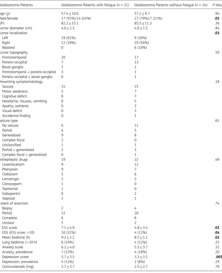
Table 2. Comparison of glioblastoma patients with and without fatigue at baseline. Data are described as mean+standard deviation.
Glioblastoma Patients Glioblastoma Patients with Fatigue (n ¼ 31) Glioblastoma Patients without Fatigue (n ¼ 34) P Value
Age (y) 57.4+10.6 57.2+9.7 .94 Male:female 17 (55%):14 (45%) 27 (79%):7 (21%) .03 KPS 82.2+15.1 85.5+11.3 .36 Tumor diameter (cm) 4.0+1.5 4.0+1.5 .84 Tumor localization .03 Left 19 (61%) 9 (26%) Right 12 (39%) 19 (56%) Bilateral 0 6 (18%) Tumor topography .59 Frontotemporal 20 17 Parieto-occipital 7 13 Basal ganglia 1 2 Frontotemporal+ parieto-occipital 3 1
Parieto-occipital+ basal ganglia 0 1
Presenting symptomatology .18
Seizure 15 15
Motor weakness 5 7
Cognitive deficit 8 7
Headache, nausea, vomiting 8 5
Apathy, asthenia 0 2 Visual deficit 0 3 Accidental finding 0 1 Seizure type .63 No seizure 6 12 Partial 4 5 Generalized 9 8 Complex-focal 2 0 Unclassified 1 1 Partial+ generalized 2 3 Complex-focal+ generalized 0 1 Antiepileptic drugs 19 22 .49 Levetiracetam 9 12 Phenytoin 9 7 Clobazam 5 6 Lamotrigin 2 3 Clonazepam 1 0 Topiramat 1 0 Gabapentin 0 2 Valproat 1 1 Extent of resection .74 Biopsy 2 4 Partial 22 20 Complete 6 8 Unclear 1 2 ESS score 7.1+4.9 4.8+3.4 .03
EDS (ESS score .10) 10 (32%) 4 (12%) .04
Mean bedtime (h) 9.2+1.2 8.5+1.1 .02 Long bedtime (.10 h) 6 (19%) 4 (12%) .25 Anxiety score 6.2+4.0 5.3+3.7 .32 Anxiety, prevalence 7 (23%) 4 (18%) .20 Depression score 5.7+3.5 3.3+3.1 .005 Depression, prevalence 4 (13%) 2 (6%) .29 Corticosteroids [mg] 2.7+3.7 2.5+2.7 .79 Continued
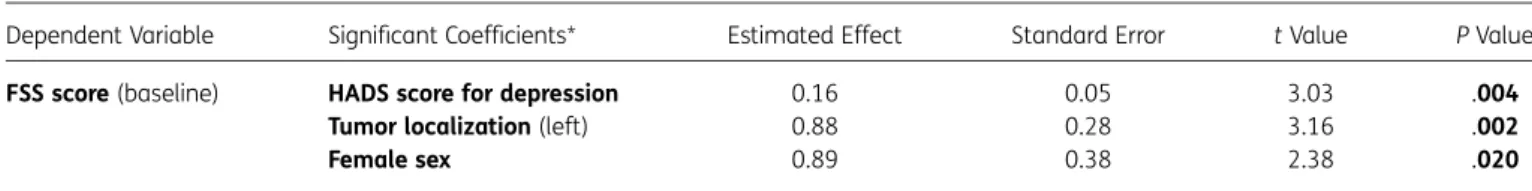
increase in depression score: P ¼ .004), left-sided tumor loca-tion (estimated effect ¼ 20.88; P ¼ .002), and female sex (esti-mated effect ¼ 0.89; P ¼ .02) as significant associates of fatigue severity at baseline prior to radio-chemotherapy (Table 4). The adjusted r2 and the multiple r2 of our final model were 0.29 and 0.32, respectively.
Evolution of Fatigue, Excessive Daytime Sleepiness, and
Mood Disorders
Unfortunately, the dropout rate was rather high: only 46 and 38 of the included 65 glioblastoma patients filled out all question-naires during and after radio-chemotherapy, respectively. Mean values for FSS and ESS scores, mean bedtimes, mean values for HADS anxiety and depression scores, and KPS did not show sig-nificant changes at subsequent time points (Fig.2).
Discussion
Our prospective study demonstrated that fatigue is a promi-nent pretreatment symptom in patients with newly diagnosed and operated glioblastoma, reaching a prevalence of 48% com-pared with only 11% among healthy controls. Surprisingly, our data represent the first controlled assessment of fatigue fre-quency in a selected and homogeneous cohort of glioblastoma patients that was measured with a specific and validated fa-tigue questionnaire. Although direct comparison is obviously hampered by major methodological differences, our finding roughly matches the reported 40% – 70% fatigue prevalence among patients with primary brain tumors.5,9,14On the other hand, we have found higher fatigue prevalence in patients with other neurological disorders such as multiple sclerosis (69%), idiopathic Parkinson’s disease (59%), episodic migraine (54%, unpublished data) or previous ischemic stroke (49%), al-ways using the FSS.19,20
Fatigue in patients with primary brain tumors has repeatedly been reported in relation to radiotherapy.13,25,26In contrast,
our study challenges the view that fatigue in glioblastoma pa-tients mainly represents a complication of radiotherapy and/or chemotherapy because the prevalence of fatigue was already high prior to radio-chemotherapy. Therefore, the contribution of toxicity from radio-chemotherapy to fatigue is probably only one factor among many. In addition, the longitudinal as-sessment of fatigue, sleepiness, and mood disturbances during and after radio-chemotherapy did not show significant chang-es, which might however reflect a bias due to the high dropout rate. It is conceivable, however, that the toxic effect of radio-chemotherapy on fatigue severity was obscured by the addi-tional presence of many other fatigue-inducing factors in our cohort. Of related interest is our observation that pharmacolog-ical treatment, including antiepileptic drugs, antidepressants or corticosteroids, was not associated with fatigue. This is in con-trast to a recent work by Struik et al, who reported an increase in fatigue severity among patients with low-grade glioma tak-ing antiepileptic drugs and corticosteroids.27
While fatigue was more than 4 times more prevalent in glio-blastoma patients than in controls, the frequency of excessive daytime sleepiness was similar (22% vs 19%). Fatigue and sleepiness are commonly regarded as 2 distinct symptoms, but they also present substantial overlap and presumably, at least to some extent, similar pathophysiology.28Our finding is surprising because increased frequency of sleepiness is indeed common in many neurological disorders with prominent fa-tigue. For instance, we found excessive daytime sleepiness in
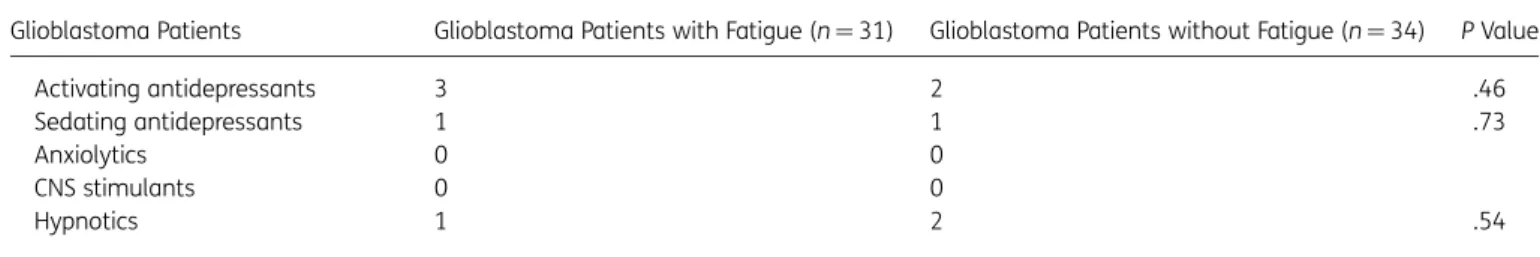
Table 2. Continued
Glioblastoma Patients Glioblastoma Patients with Fatigue (n ¼ 31) Glioblastoma Patients without Fatigue (n ¼ 34) P Value
Activating antidepressants 3 2 .46
Sedating antidepressants 1 1 .73
Anxiolytics 0 0
CNS stimulants 0 0
Hypnotics 1 2 .54
EDS, excessive daytime sleepiness; ESS, Epworth Sleepiness Scale; FSS, Fatigue Severity Scale; h, hours; KPS, Karnofsky performance status; y, years.
Table 3. Comparison of fatigue, sleepiness, and mood disorders between patients with left-sided and right-sided glioblastoma at baseline prior to radio-chemotherapy. Data are described as mean+ standard deviation. Left-sided Tumor Right-sided Tumor P Value (n ¼ 28) (n ¼ 31) Age (y) 55.8+9.5 57.9+10.5 .43 Male sex 20 (71%) 21 (68%) .60 Karnofsky performance status 86+14 83+12 .40 Tumor diameter (cm) 4.0+1.7 4.0+1.3 .92 FSS 4.6+1.3 3.5+1.8 .008 Fatigue (FSS .4.0) 19 (68%) 12 (39%) .004 ESS 5.8+4.2 6.0+4.7 .83 EDS (ESS .10) 5 (18%) 8 (26%) .73 Mean bedtime (h) 9.1+1.4 8.6+1.0 .13 Long bedtime (.10 h) 7 (25%) 3 (11) .18
HADS anxiety score 6.6+3.8 5.1+4.0 .16
Anxiety (prevalence) 8 (30%) 3 (10%) .03 HADS depression score 5.3+2.9 3.6+3.8 .05 Depression, prevalence 2 (9%) 4 (13%) .49 EDS, excessive daytime sleepiness; ESS, Epworth Sleepiness Scale; FSS, Fatigue Severity Scale; h, hours; HADS, Hospital Anxiety and Depression Scale; y, years.
48% of patients with idiopathic Parkinson’s disease and 38% of traumatic brain injury survivors, while other groups reported an even higher prevalence.20,29Degenerative or trauma-induced
disruption of arousal-promoting structures in the rostral brain-stem and hypothalamus may cause both fatigue and sleepi-ness, and this assumption is increasingly supported by neuropathological evidence.30–32On the other hand, fatigue
is a complex symptom influenced by a large variety of factors, and the composition of these contributing factors most likely differs between neurological disorders commonly associated with high fatigue burden. Thus, it is tempting to speculate whether the selective increase of fatigue with normal preva-lence of sleepiness may shed some light on the underlying eti-ology of glioblastoma-related fatigue. Of interest in this context, the discrepant prevalence of fatigue and sleepiness is reminiscent of patients with mood disorders, who often suf-fer from fatigue and insomnia, while sleepiness is not a consis-tent complaint.33 Along the same line, we could identify
depression as an independent predictor of fatigue severity at
baseline by using multiple regression analyses. To the best of our knowledge, this is the first study to highlight this important association between fatigue severity and depression in glio-blastoma patients; similar correlations have been reported in breast cancer patients but never in patients with primary brain tumors.34,35
Anxiety and depression represent normal emotional reac-tions to the diagnosis of glioblastoma, and they are significant and independent contributors to fatigue severity. However, as emphasized by the overview of Litofsky and Resnick and further corroborated by our observations, other factors have to be con-sidered.36Of note, fatigue, anxiety and depression all appeared
to be more common in glioblastoma patients with left-sided tumors compared with those having right-sided tumors; left-sided tumor location was consistently identified as an indepen-dent predictor of fatigue severity. Indeed, there is some evi-dence indicating that patients with left hemispheric lesions are prone to depressive reactions, whereas patients with right hemisphere lesions often show indifferent emotional
Table 4. Multiple linear regression model for coefficients of fatigue severity at baseline in glioblastoma patients
Dependent Variable Significant Coefficients* Estimated Effect Standard Error t Value P Value
FSS score (baseline) HADS score for depression 0.16 0.05 3.03 .004
Tumor localization (left) 0.88 0.28 3.16 .002
Female sex 0.89 0.38 2.38 .020
*Additional coefficients included in the model were age, education, Karnofsky performance status, Epworth Sleepiness Scale, bedtimes, Hospital Anxiety and Depression Scale score for anxiety, use of steroids, and use of antidepressants.
Abbreviations: FSS, Fatigue Severity Scale; HADS, Hospital Anxiety and Depression Scale.
Fig. 2. Compared with postsurgical baseline, the scores of the Fatigue Severity Scale (FSS), Epworth Sleepiness Scale (ESS), mean bedtimes, HADS depression and anxiety, and Karnofsky performance status did not show any significant changes during and immediately after combined radio-chemotherapy (RCT). Error bars indicate standard deviations.
reactions.37,38,39Likewise, Klein et al found significant impair-ment of attentional and executive functioning in patients with left-sided high-grade glioma compared with right-sided.40
However, several studies failed to observe any association be-tween depression and hemispheric laterality of gliomas.41Data on depression in ischemic stroke provide a similarly inconsistent picture. Few studies suggested a higher prevalence of depres-sion in left-sided ischemic stroke, but other studies could not confirm a significant impact of stroke location on depressive symptoms.42Whether tumor laterality plays such a significant role in the severity of fatigue, as suggested by our study, re-mains to be confirmed by future works.
Similar to previous work, we found that female sex was as-sociated with higher fatigue severity at baseline. Recently, Arm-strong et al assessed fatigue in 201 patients with primary brain tumors and demonstrated that moderate-to-severe fatigue was more common in females, while low fatigue was more common in males.5
Several limitations of our study have to be acknowledged. First, the number of participants was relatively small. Second, dropout rates at subsequent time points were high and might have introduced significant bias. Our observations on evolution of fatigue, sleepiness, and mood disorders during and after radio-chemotherapy must therefore be considered with cau-tion and require confirmacau-tion by larger studies with minimal dropout rates. However, elimination of a significant dropout will be challenging, as it is a well-known, notorious problem in longitudinal studies of primary brain tumor patients. Our dropout rate was similar, for instance, to that reported in a re-cent large randomized EORTC trial.43Third, a certain selection bias is likely, as we included only 60%–65% of all eligible glio-blastoma patients during the study period, which prevents di-rect generalizability of our findings. Finally, we did not measure the impact of fatigue and associated variables on health-related QoL.
In summary, roughly half of all glioblastoma patients are af-fected by fatigue at postsurgical baseline. Depression is among the strongest predictors of fatigue severity at baseline, which might also explain the unexpected absence of increased sleepi-ness in glioblastoma patients. Hence, treating physicians should be more vigilant for fatigue and depression in glioblas-toma patients because they both represent frequent comorbid-ities with mutually negative repercussions and are known to negatively impact QoL, treatment compliance, and overall survival.12,16,18,44
Funding
There was no funding for this study.
Conflict of interest statement. None declared.
References
1. Louis DN, Ohgaki H, Wiestler OD, et al. WHO Classification of Tumours of the Central Nervous System. Lyon: IARC: 2007.
2. Stupp R, Mason WP, van den Bent MJ, et al. Radiotherapy plus concomitant and adjuvant temozolomide for glioblastoma. N Engl J Med. 2005;352(10):987– 996.
3. Johnson DR, Ma DJ, Buckner JC, et al. Conditional probability of long-term survival in glioblastoma: a population-based analysis. Cancer. 2012;118(22):5608– 5613.
4. Campos MP, Hassan BJ, Riechelmann R, et al. Cancer-related fatigue: a practical review. Ann Oncol. 2011;22(6):1273–1279. 5. Armstrong TS, Cron SG, Bolanos EV, et al. Risk factors for fatigue
severity in primary brain tumor patients. Cancer. 2010;116(11): 2707–2715.
6. Berger AM, Abernethy AP, Atkinson A, et al. Cancer-related fatigue. J Natl Compr Canc Netw. 2010;8(8):904–931.
7. Stone P, Richardson A, Ream E, et al. Cancer-related fatigue: inevitable, unimportant and untreatable? Results of a multi-centre patient survey. Cancer Fatigue Forum. Ann Oncol. 2000;11(8):971–975.
8. Lis CG, Rodeghier M, Grutsch JF, et al. Distribution and determinants of patient satisfaction in oncology with a focus on health related quality of life. BMC Health Serv Res. 2009;9:190. 9. Armstrong TS, Gilbert MR. Practical strategies for management of
fatigue and sleep disorders in people with brain tumors. Neuro Oncol. 2012;4(suppl. 4):iv65–iv72.
10. Winningham ML, Nail LM, Burke MB, et al. Fatigue and the cancer experience: the state of the knowledge. Oncol Nurs Forum. 1994; 21(1):23– 36.
11. Pachman DR, Barton DL, Swetz KM, et al. Troublesome symptoms in cancer survivors: fatigue, insomnia, neuropathy, and pain. J Clin Oncol. 2012;30(30):3687–3696.
12. Taphoorn MJ, Stupp R, Coens C, et al. Health-related quality of life in patients with glioblastoma: A randomized controlled trial. Lancet Oncol. 2005;6(12):937– 944.
13. Faithfull S, Brada M. Somnolence syndrome in adults following cranial irradiation for primary brain tumours. Clin Oncol. 1998; 10(4):250– 254.
14. Lovely MP, Miaskowski C, Dodd M. Relationship between fatigue and quality of life in patients with glioblastoma multiformae. Oncol Nurs Forum. 1999;26(5):921–925.
15. Hjollund NH, Andersen JH, Bech P. Assessment of fatigue in chronic disease: a bibliographic study of fatigue measurement scales. Health Qual Life Outcomes. 2007;5:12.
16. Brown PD, Ballman KV, Rummans TA, et al. Prospective study of quality of life in adults with newly diagnosed high-grade gliomas. J Neurooncol. 2006;76(3):283 –291.
17. Flechl B, Ackerl M, Sax C, et al. Neurocognitive and sociodemographic functioning of glioblastoma long-term survivors. J Neurooncol. 2012;109(2):331–339.
18. Flechl B, Ackerl M, Sax C, et al. The caregivers’ perspective on the end-of-life phase of glioblastoma patients. J Neurooncol. 2013; 112(3):403 –411.
19. Valko PO, Bassetti CL, Bloch KE, et al. Validation of the fatigue severity scale in a Swiss cohort. Sleep. 2008;31(11):1601– 1607. 20. Valko PO, Waldvogel D, Weller M, et al. Fatigue and excessive
daytime sleepiness in idiopathic Parkinson’s disease differently correlate with motor symptoms, depression and dopaminergic treatment. Eur J Neurol. 2010;17(12):1428–1436.
21. Cronbach LJ. Coefficient alpha and the internal structure of test. Psychometrika. 1951;16(3):97 –102.
22. Bloch KE, Schoch OD, Zhang JN, et al. German version of the Epworth Sleepiness Scale. Respiration. 1999;66(5): 440 – 447.
23. Zigmond AS, Snaith RP. The hospital anxiety and depression scale. Acta Psychiatr Scand. 1983;67(6):361–370.
24. Herrmann C. International experiences with the Hospital Anxiety and Depression Scale - a review of validation data and clinical results. J Psychosom Res. 1997;42(1):17 –41.
25. Drappatz J, Schiff D, Kesari S, et al. Medical management of brain tumor patients. Neurol Clin. 2007;25(4):1035– 1071.
26. Powell C, Guerrero D, Sardell S, et al. Somnolence syndrome in patients receiving radical radiotherapy for primary brain tumours: a prospective study. Radiother Oncol. 2011;100(1): 131– 136.
27. Struik K, Klein M, Heimans JJ, et al. Fatigue in low-grade glioma. J Neurooncol. 2009;92(1):73 –78.
28. Hossain JL, Ahmad P, Reinish LW, et al. Subjective fatigue and subjective sleepiness: two independent consequences of sleep disorders?. J Sleep Res. 2005;14(3):235– 253.
29. Baumann CR, Werth E, Stocker R, et al. Sleep-wake disturbances 6 months after traumatic brain injury: a prospective study. Brain. 2007;130(7):1873– 1883.
30. Fronczek R, Overeem S, Lee SY, et al. Hypocretin (orexin) loss in Parkinson’s disease. Brain. 2007;130(6):1577– 1585.
31. Baumann CR, Bassetti CL, Valko PO, et al. Loss of hypocretin (orexin) neurons with traumatic brain injury. Ann Neurol. 2009; 66(4):555–559.
32. Harrington ME. Neurobiological studies of fatigue. Prog Neurobiol. 2012;99(2):93– 105.
33. Billiard M, Dolenc L, Aldaz C, et al. Hypersomnia associated with mood disorders: A new perspective. J Psychosom Res. 1994; 38(suppl. 1):41–47.
34. Gaston-Johansson F, Fall-Dickson JM, Bakos AB, et al. Fatigue, pain, and depression in pre-autotransplant breast cancer patients. Cancer Pract. 1999;7(5):240– 247.
35. Bower JE, Ganz PA, Desmond KA, et al. Fatigue in breast cancer survivors: occurrence, correlates, and impact on quality of life. J Clin Oncol. 2000;18(4):743–753.
36. Litofsky NS, Resnick AG. The relationships between depression and brain tumors. J Neurooncol. 2009;94(2):153– 161.
37. Gainotti G. Emotional behavior and hemispheric side of lesion. Cortex. 1972;8(1):41– 55.
38. Irle E, Peper M, Wowra B, et al. Mood changes after surgery for tumors of the cerebral cortex. Arch Neurol. 1994;51(2):164–174. 39. Pringle AM, Taylor R, Whittle IR. Anxiety and depression in patients with an intracranial neoplasm before and after tumour surgery. Br J Neurosurg. 1999;13(1):46– 51.
40. Klein M, Taphoorn MJB, Heimans JJ, et al. Neurobehavioral status and health-related quality of life in newly diagnosed high-grade glioma patients. J Clin Oncol. 2001;19(20):4037 – 4047.
41. Rooney AG, Carson A, Grant R. Depression in cerebral glioma patients: a systematic review of observational studies. J Natl Cancer Inst. 2011;103(1):61– 76.
42. Carson AJ, MacHale S, Allen K, et al. Depression after stroke and lesion location: a systematic review. Lancet. 2000;356(9224):122–126. 43. Soffietti R, Kocher M, Abacioglu UM, et al. A European Organisation
for Research and Treatment of Cancer phase III trial of adjuvant whole-brain radiotherapy versus observation in patients with one to three brain metastases from solid tumors after surgical resection or radiosurgery: quality-of-life results. J Clin Oncol. 2013;31(1):65– 72.
44. Hinz A, Krauss O, Hauss JP, et al. Anxiety and depression in cancer patients compared with the general population. Eur J Cancer Care. 2010;19(4):522–529.