Interplay between the Hsp90 Chaperone and the HslVU
Protease To Regulate the Level of an Essential Protein in
Shewanella oneidensis
Flora Ambre Honoré,aNathanael Jean Maillot,aVincent Méjean,aOlivier Genesta
aAix Marseille Université, CNRS, BIP UMR 7281, IMM, Marseille, France
ABSTRACT Protein synthesis, folding, and degradation are an accurately regulated process occurring in every organism and called proteostasis. This process is essential to maintain a healthy proteome since proteostasis dysregulation is responsible for devastating cellular issues. Proteostasis is controlled by a complex network of molec-ular chaperones and proteases. Among them, eukaryotic Hsp90, assisted by many cochaperones and the Hsp70 chaperone system, plays a major role in activating hundreds of client proteins, and Hsp90 inhibition usually leads to proteasomal deg-radation of these clients. In bacteria, however, the precise function of Hsp90 remains quite unclear, and only a few clients are known. Recently, we have shown that Hsp90 is essential at elevated temperature in the aquatic model bacterium
She-wanella oneidensis, and we have identified a client of Hsp90, TilS, involved in tRNA
modification. Here we found that two members of the proteostasis network with an-tagonist activities, the Hsp90 chaperone and the HslVU protease, which is consid-ered the proteasome ancestor, together regulate the level of TilS. In particular, we show that deletion of the genes coding for the HslVU protease suppresses the growth defect of an S. oneidensis strain with hsp90 deleted, by increasing the cellular level of the essential TilS protein. These results open up new avenues for under-standing how proteostasis is controlled in bacteria, and new Hsp90 clients are much needed now to confirm the interplay between Hsp90 and proteases.
IMPORTANCE Maintaining a healthy proteome is essential in every living cell from bacteria to humans. For example, proteostasis (protein homeostasis) imbalance in humans leads to devastating diseases, including neurodegenerative diseases and cancers. Therefore, proteins need to be assisted from their synthesis to their native folding and ultimately to their degradation. To ensure efficient protein turnover, cells possess an intricate network of molecular chaperones and proteases for protein folding and degradation. However, these networks need to be better defined and understood. Here, using the aquatic bacterium Shewanella oneidensis as a model or-ganism, we demonstrate interplay between two proteins with antagonist activities, the Hsp90 chaperone and the HslVU protease, to finely regulate the level of an es-sential client of Hsp90. Therefore, this work provides a new bacterial model to better study protein regulation and turnover, and it sheds light on how proteostasis by Hsp90 and proteases could be controlled in bacteria.
KEYWORDS heat shock, proteases, protein chaperone, protein folding, proteostasis, stress adaptation
P
roteostasis is controlled in every organism by a complex network of chaperonesand proteases (1, 2). Among them, the eukaryotic 90-kDa heat shock protein (Hsp90) chaperone, assisted by many cochaperones and the Hsp70 chaperone system, remodels and activates hundreds of client proteins, including kinases and receptors
Citation Honoré FA, Maillot NJ, Méjean V,
Genest O. 2019. Interplay between the Hsp90 chaperone and the HslVU protease to regulate the level of an essential protein in Shewanella
oneidensis. mBio 10:e00269-19.https://doi.org/ 10.1128/mBio.00269-19.
Editor Susan Gottesman, National Cancer
Institute
Copyright © 2019 Honoré et al. This is an
open-access article distributed under the terms of theCreative Commons Attribution 4.0 International license.
Address correspondence to Olivier Genest, ogenest@imm.cnrs.fr. Received 31 January 2019 Accepted 8 April 2019 Published 14 May 2019
crossm
®on July 11, 2019 by guest
http://mbio.asm.org/
Downloaded from
(3–7). In bacteria, Hsp90 also collaborates with the DnaK chaperone system, but cochaperones are absent, and its function needs to be clarified (8–13). In addition, only a few bacterial Hsp90 client proteins are known (8, 14–19). Using the aquatic proteo-bacteria Shewanella oneidensis, we have recently found that Hsp90 is necessary under heat stress conditions to protect and activate the TilS protein, leading to bacterial growth (17). TilS is an essential enzyme that modifies the specificity of the only tRNA that translates the AUG initiator codon in methionine into a tRNA that translates the AUA rare codon in isoleucine (20, 21). Given the major importance for whole proteome synthesis to correctly translate the AUG initiator codon, the level of TilS has to be finely regulated in the cell, since an excess of TilS could lead to depletion of the tRNA-AUG in favor of tRNA-AUA. On the other side, the absence of TilS prevents translation of proteins containing the AUA codon. Here we show that there is interplay between two components of the proteostasis network to regulate the level of the TilS protein: (i) the Hsp90 chaperone for protection and activation and (ii) the HslVU protease for degra-dation. Therefore, the level of TilS is precisely adjusted in the cell to allow correct protein translation and bacterial growth.
The absence of HslVU suppresses the phenotype of the⌬hsp90Sostrain. To get
insight into how proteostasis is controlled in S. oneidensis during heat stress, we wanted to identify new components of the proteostasis network that could work with S.
oneidensis Hsp90 (Hsp90So). As previously observed, we found that a strain with hsp90So
deleted did not grow under heat stress conditions in liquid cultures and on solid media compare to the wild type (WT), whereas it grew well at the permissive temperature of 28°C (Fig. 1A and B) (17). We hypothesized that in the absence of Hsp90 at high
temperature, one or several essential Hsp90Soclients are not correctly folded and are
therefore degraded. Consequently, inactivation of the protease responsible for this degradation could restore a sufficient level of the client protein and could allow growth
of the Δhsp90Sostrain. We thus deleted the genes coding for the HslVU protease and
for the ClpP subunit of the ClpAP and ClpXP proteases. These major proteolytic machines are composed of an ATP-dependent chaperone subunit belonging to the
AAA⫹ family (i.e., HslU, ClpA and ClpX) that unfolds substrates and directs them into
the catalytic chamber of the peptidase subunit (i.e., HslV and ClpP) (22). Strikingly, we
observed that the absence of the HslVU protease did rescue the growth of the Δhsp90So
strain at high temperature in liquid and solid media (Fig. 1A and B, compare the
Δhsp90SoΔhslVU strain with the Δhsp90Sostrain). In contrast, the ClpP protease did not
seem to be involved in this pathway since no growth improvement was observed in the
Δhsp90SoΔclpP strain compared to the Δhsp90Sostrain. At 28°C, all strains grew as wild
type (Fig. 1A and B).
We then confirmed the growth phenotypes by producing HslVU or Hsp90Sofrom
plasmids (Fig. 1C). We found that at high temperature, production of HslVU strongly
reduced the growth of the Δhsp90SoΔhslVU strain as expected, whereas production of
Hsp90Socomplemented the phenotype of the Δhsp90Sostrain.
Altogether, these results support the idea that some essential Hsp90Soclients that
are degraded in the Δhsp90So strain are stabilized in the absence of HslVU. They
therefore strongly suggest that these clients are degraded by the HslVU protease.
The essential Hsp90Soclient TilS is degraded by HslVU. Since we have previously
shown that the Hsp90Soclient TilS is responsible for the growth defect of the Δhsp90So
strain at high temperature (17), we looked at its level under stress conditions with or
without Hsp90Soand/or HslVU. To do that, a plasmid coding for TilS with a 6⫻ His tag
was introduced in the different genetic backgrounds of S. oneidensis. The strains were grown at sublethal high temperature, TilS expression was induced, and its amount was determined by Western blot analysis.
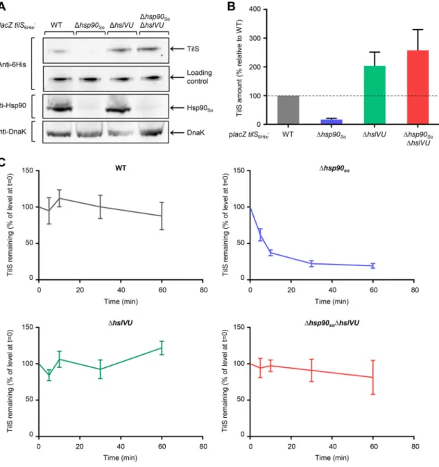
As already seen, we found that about 85% of TilS was degraded in the Δhsp90So
strain compared to the wild type (Fig. 2A and B) (17). Interestingly, the TilS level was
dramatically increased in the Δhsp90SoΔhslVU strain, reaching more than 15 times the
level observed in the absence of Hsp90So. These results strongly support the idea that
on July 11, 2019 by guest
http://mbio.asm.org/
FIG 1 hslVU deletion suppresses the growth phenotype of the Δhsp90Sostrain at high temperature. (A)
Strains grown at 28°C to late exponential phase were diluted to an optical density at 600 nm (OD600) of 1, and
2l of 10-time serial dilutions was spotted onto LB agar plates. The plates were incubated at 28 or 35°C. (B)
Strains grown at 28°C to late exponential phase were diluted to an OD600of 0.0005 and incubated with
shaking in a microplate reader at 28 or 37°C. (C) Strains were treated as in panel B, except that LB rich medium was supplemented with 0.015% arabinose to induce protein production from the plasmids. In panels B and
C, data from at least three replicates are shown as mean⫾ standard error of the mean (SEM).
on July 11, 2019 by guest
http://mbio.asm.org/
TilS is degraded by the HslVU protease. In the ΔhslVU strain, we observed that the TilS
level is higher than in the wild-type strain, suggesting that in the presence of Hsp90So,
part of the pool of TilS is degraded by the HslVU protease. In addition, we found that most of TilS protein was present in the soluble fraction of the different strains (see Fig. S1A and B in the supplemental material).
As a control, we showed that transcription from this plasmid did not vary in the different strains grown at sublethal temperature (see Fig. S2 in the supplemental
FIG 2 The HslVU protease degrades the Hsp90Soclient TilS. (A) Strains containing the placZ tilS6Hisplasmid, in which lacZ and tilS6Hisare
two independent genes under the control of the PBADpromoter, were grown at 35°C, a sublethal temperature. At an OD600of 0.6, 0.02%
arabinose was added. After 2 h at 35°C, the same amounts of total protein extract from each strain were loaded for SDS-PAGE, transferred
by Western blotting, and revealed with anti-6⫻ His antibody to detect the TilS protein, anti-Hsp90 antibody, or anti-DnaK antibody. The
loading control corresponds to a contaminating band revealed with the anti-6⫻ His antibody, indicating that the same amount of cellular
extracts was loaded. (B) Quantification of the amount of TilS was performed from 3 independent Western blots described in panel A,
revealed with the anti-6⫻ His antibody using ImageJ software. The amount of TilS measured in the wild-type strain was set to 100%. Data
are shown as mean⫾ SEM. (C) Chase experiments. Strains containing the placZ tilS6Hisplasmid were grown as in panel A, except that 0.2%
arabinose was added to increase the level of the TilS protein, in particular in the Δhsp90Sostrain. After 2 h of induction, 200g/ml
chloramphenicol was added to block protein translation (t⫽ 0). Samples were taken at several times after chloramphenicol addition, and
proteins were precipitated with trichloroacetic acid (TCA), loaded for SDS-PAGE, and quantified on a Western blot, revealed with anti-6⫻
His antibody using the ImageJ software. The amount of TilS measured in each strain at t⫽ 0 (chloramphenicol addition) was set to 100%.
Data are shown as mean⫾ SEM.
on July 11, 2019 by guest
http://mbio.asm.org/
material). We also checked that deletion of hsp90Soand/or hslVU did not modify the
heat shock response by quantifying the level of DnaK, whose gene is under the control of the RpoH sigma factor. Indeed, no significant variation was observed in the four strains (Fig. 2A).
Finally, chase experiments were performed to measure kinetic of TilS degradation in the different genetic backgrounds. Strains containing the plasmid coding for TilS with
a 6⫻ His tag were grown as described previously, and after induction, a high
concen-tration of chloramphenicol was added to block translation. The amount of TilS was quantified at several time points after chloramphenicol addition and was expressed relative to the level observed at time zero (Fig. 2C; see Fig. S3 in the supplemental
material). In the absence of Hsp90So, TilS was degraded with time, and its level reached
a plateau after about 30 min, whereas low or no degradation was found in the
wild-type, ΔhslVU, and Δhsp90So ΔhslVU strains (Fig. 2C; Fig. S3). These experiments
demonstrate that TilS is degraded by the HslVU protease in the absence of Hsp90. In this article, we found that the level of TilS is highly regulated at a posttranslational
level by the Hsp90Sochaperone and the HslVU protease, and we show that growth of
the Δhsp90Sostrain at elevated temperature strongly depends on the amount of the
TilS protein. Interestingly, this result is reminiscent of our previous work, in which
overproduction of TilS led to growth of the Δhsp90Sostrain at high temperature (17).
Therefore, increasing the level of TilS by two opposite mechanisms, overproduction or inactivation of the degradation, did result in the rescue of the phenotype of the
Δhsp90So strain. This finding reinforces the notion that the level of TilS needs to be
tightly controlled.
Interestingly, interplay between Hsp90 and HslVU has already been proposed for the posttranslational regulation of an unknown protein involved in the synthesis of toxins in extraintestinal pathogenic Escherichia coli (23). In addition, the level of the Cas3 protein, a client of E. coli Hsp90, is reduced in the absence of Hsp90 in E. coli; however, the protease involved in the degradation has not yet been identified (15). This thus suggests that the antagonist activities of Hsp90 and HslVU could serve as a general mechanism to control the level of some proteins. In eukaryotes, connections between folding by the Hsp90 chaperone and degradation by the proteasome have been well established (24, 25). It therefore becomes essential to identify new clients of bacterial Hsp90 to confirm this model.
SUPPLEMENTAL MATERIAL
Supplemental material for this article may be found athttps://doi.org/10.1128/mBio
.00269-19.
TEXT S1, DOCX file, 0.1 MB. FIG S1, PDF file, 0.1 MB. FIG S2, PDF file, 0.1 MB. FIG S3, PDF file, 0.1 MB.
ACKNOWLEDGMENTS
We thank Marianne Ilbert, Eric Durand, Aurélia Battesti, and members of our group for help and fruitful discussions.
This work was supported by the Centre National de la Recherche Scientifique and Aix Marseille Université (AMU). O.G. was supported by a grant from the Agence Nationale de la Recherche (ANR-16-CE11-0002-01).
REFERENCES
1. Balchin D, Hayer-Hartl M, Hartl FU. 2016. In vivo aspects of protein
folding and quality control. Science 353:aac4354. https://doi.org/10
.1126/science.aac4354.
2. Finka A, Mattoo RUH, Goloubinoff P. 2016. Experimental milestones in the discovery of molecular chaperones as polypeptide unfolding
en-zymes. Annu Rev Biochem 85:715–742.https://doi.org/10.1146/annurev
-biochem-060815-014124.
3. Schopf FH, Biebl MM, Buchner J. 2017. The HSP90 chaperone machinery.
Nat Rev Mol Cell Biol 18:345–360.https://doi.org/10.1038/nrm.2017.20.
4. Radli M, Rüdiger SGD. 2018. Dancing with the diva: Hsp90-client
inter-actions. J Mol Biol 430:3029 –3040.https://doi.org/10.1016/j.jmb.2018.05
.026.
5. Mayer MP, Le Breton L. 2015. Hsp90: breaking the symmetry. Mol Cell
58:8 –20.https://doi.org/10.1016/j.molcel.2015.02.022.
on July 11, 2019 by guest
http://mbio.asm.org/
6. Taipale M, Jarosz DF, Lindquist S. 2010. HSP90 at the hub of protein homeostasis: emerging mechanistic insights. Nat Rev Mol Cell Biol 11:
515–528.https://doi.org/10.1038/nrm2918.
7. Johnson JL. 2012. Evolution and function of diverse Hsp90 homologs
and cochaperone proteins. Biochim Biophys Acta 1823:607– 613.https://
doi.org/10.1016/j.bbamcr.2011.09.020.
8. Genest O, Wickner S, Doyle SM. 2019. Hsp90 and Hsp70 chaperones: collaborators in protein remodeling. J Biol Chem 294:2109 –2120.
https://doi.org/10.1074/jbc.REV118.002806.
9. Genest O, Hoskins JR, Camberg JL, Doyle SM, Wickner S. 2011. Heat shock protein 90 from Escherichia coli collaborates with the DnaK chap-erone system in client protein remodeling. Proc Natl Acad Sci U S A
108:8206 – 8211.https://doi.org/10.1073/pnas.1104703108.
10. Nakamoto H, Fujita K, Ohtaki A, Watanabe S, Narumi S, Maruyama T, Suenaga E, Misono TS, Kumar PKR, Goloubinoff P, Yoshikawa H. 2014. Physical interaction between bacterial heat shock protein (Hsp) 90 and Hsp70 chaperones mediates their cooperative action to refold
dena-tured proteins. J Biol Chem 289:6110 – 6119.https://doi.org/10.1074/jbc
.M113.524801.
11. Morán Luengo T, Kityk R, Mayer MP, Rüdiger S. 2018. Hsp90 breaks the deadlock of the Hsp70 chaperone system. Mol Cell 70:545–552.e9.
https://doi.org/10.1016/j.molcel.2018.03.028.
12. Bardwell JC, Craig EA. 1988. Ancient heat shock gene is dispensable. J
Bacteriol 170:2977–2983.https://doi.org/10.1128/jb.170.7.2977-2983
.1988.
13. Thomas JG, Baneyx F. 2000. ClpB and HtpG facilitate de novo protein folding in stressed Escherichia coli cells. Mol Microbiol 36:1360 –1370. 14. Motojima-Miyazaki Y, Yoshida M, Motojima F. 2010. Ribosomal protein
L2 associates with E. coli HtpG and activates its ATPase activity. Biochem
Biophys Res Commun 400:241–245.https://doi.org/10.1016/j.bbrc.2010
.08.047.
15. Yosef I, Goren MG, Kiro R, Edgar R, Qimron U. 2011. High-temperature protein G is essential for activity of the Escherichia coli clustered regularly interspaced short palindromic repeats (CRISPR)/Cas system.
Proc Natl Acad Sci U S A 108:20136 –20141.https://doi.org/10.1073/
pnas.1113519108.
16. Press MO, Li H, Creanza N, Kramer G, Queitsch C, Sourjik V, Borenstein E.
2013. Genome-scale co-evolutionary inference identifies functions and
clients of bacterial Hsp90. PLoS Genet 9:e1003631.https://doi.org/10
.1371/journal.pgen.1003631.
17. Honoré FA, Méjean V, Genest O. 2017. Hsp90 is essential under heat stress in the bacterium Shewanella oneidensis. Cell Rep 19:680 – 687.
https://doi.org/10.1016/j.celrep.2017.03.082.
18. Sato T, Minagawa S, Kojima E, Okamoto N, Nakamoto H. 2010. HtpG, the prokaryotic homologue of Hsp90, stabilizes a phycobilisome protein in the cyanobacterium Synechococcus elongatus PCC 7942. Mol Microbiol
76:576 –589.https://doi.org/10.1111/j.1365-2958.2010.07139.x.
19. Grudniak AM, Markowska K, Wolska KI. 2015. Interactions of Escherichia coli molecular chaperone HtpG with DnaA replication initiator DNA. Cell
Stress Chaperones 20:951–957. https://doi.org/10.1007/s12192-015
-0623-y.
20. Suzuki T, Miyauchi K. 2010. Discovery and characterization of tRNAIle
lysidine synthetase (TilS). FEBS Lett 584:272–277. https://doi.org/10
.1016/j.febslet.2009.11.085.
21. Nakanishi K, Bonnefond L, Kimura S, Suzuki T, Ishitani R, Nureki O. 2009. Structural basis for translational fidelity ensured by transfer RNA
lysidine synthetase. Nature 461:1144 –1148.https://doi.org/10.1038/
nature08474.
22. Sauer RT, Baker TA. 2011. AAA⫹ proteases: ATP-fueled machines of
protein destruction. Annu Rev Biochem 80:587– 612.https://doi.org/10
.1146/annurev-biochem-060408-172623.
23. Garcie C, Tronnet S, Garénaux A, McCarthy AJ, Brachmann AO, Pénary M, Houle S, Nougayrède J-P, Piel J, Taylor PW, Dozois CM, Genevaux P, Oswald E, Martin P. 2016. The bacterial stress-responsive Hsp90 chap-erone (HtpG) is required for the production of the genotoxin colibactin and the siderophore yersiniabactin in Escherichia coli. J Infect Dis 214:
916 –924.https://doi.org/10.1093/infdis/jiw294.
24. Theodoraki MA, Caplan AJ. 2012. Quality control and fate determination
of Hsp90 client proteins. Biochim Biophys Acta 1823:683– 688.https://
doi.org/10.1016/j.bbamcr.2011.08.006.
25. Edkins AL. 2015. CHIP: a co-chaperone for degradation by the
protea-some. Subcell Biochem 78:219 –242.https://doi.org/10.1007/978-3-319
-11731-7_11.