HAL Id: inserm-01143772
https://www.hal.inserm.fr/inserm-01143772
Submitted on 20 Apr 2015HAL is a multi-disciplinary open access archive for the deposit and dissemination of sci-entific research documents, whether they are pub-lished or not. The documents may come from teaching and research institutions in France or abroad, or from public or private research centers.
L’archive ouverte pluridisciplinaire HAL, est destinée au dépôt et à la diffusion de documents scientifiques de niveau recherche, publiés ou non, émanant des établissements d’enseignement et de recherche français ou étrangers, des laboratoires publics ou privés.
Distributed under a Creative Commons Attribution| 4.0 International License
Usefulness of Tc-99m Sestamibi studies for monitoring
response to therapy in patients with high grade gliomas:
a preliminary study
Jean-Marc Tacchella, Nathanaelle Yeni, Gregorio Petrirena, Mike-Ely Cohen,
Muriel Lefort, Rémy Guillevin, Marie-Odile Habert, Elodie Roullot, Aurélie
Kas, Frédérique Frouin
To cite this version:
Jean-Marc Tacchella, Nathanaelle Yeni, Gregorio Petrirena, Mike-Ely Cohen, Muriel Lefort, et al.. Usefulness of Tc-99m Sestamibi studies for monitoring response to therapy in patients with high grade gliomas: a preliminary study. Journées RITS 2015, Mar 2015, Dourdan, France. pp.48-49. �inserm-01143772�
Actes des Journées Recherche en Imagerie et Technologies pour la Santé - RITS 2015 48
Usefulness of Tc-99m Sestamibi studies for monitoring response to therapy in
patients with high grade gliomas: a preliminary study
Jean-Marc Tacchella
1,2*, N Yeni
3, G Petrirena
4, ME Cohen
1, M Lefort
1, R Guillevin
5, MO Habert
1,3,
E Roullot
2, A Kas
1,3, F Frouin
1,61
Sorbonne Universités, UPMC Univ Paris 06, Inserm, CNRS, Laboratoire d’Imagerie Biomédicale, Paris, France. 2
ESME-Sudria, Laboratoire PRIAM, Ivry-sur Seine, France. 3
AP-HP, CHU Pitié-Salpêtrière, Service de Médecine Nucléaire, Paris, France 4
AP-HP, CHU Pitié-Salpêtrière, Service de Neurologie, Paris, France 5
Université de Poitiers, CHU Poitiers, Service de Radiologie, Poitiers, France 6
Inserm/CEA/Université Paris Sud/CNRS, CEA/I2BM/SHFJ, Laboratoire IMIV, Orsay, France *
Corresponding author, jm.tacchella@gmail.com.
Abstract
–
Early and late Sestamibi studies were acquired in addition to conventional MRI protocol in 14 patients with high-grade gliomas to monitor an antiangiogenic treatment. Global and local indices were deduced from these SPECT studies and were compared with progression free survival (PFS) and overall survival (OS). Variations of intensity in late studies were not correlated with PFS, but were related to OS. This suggests the possible role of Sestamibi for monitoring response to treatment.Index terms - Image Processing, Therapy monitoring. I. INTRODUCTION
The RANO criteria [1] are currently proposed for monitoring patients with high-grade gliomas. They are based on 1) multi-modal MR imaging including T1 weighted images after injection of Gadolinium chelate and FLAIR T2 weighted images and 2) general status of the patient evaluated through the use of corticoids. These RANO criteria have been proposed as surrogate markers of Progression free survival (PFS), thus early end-points of overall survival (OS). The purpose of the pilot study that was conducted in our institution was to test the potential interest of Tc-99m Sestamibi exams in order to predict the response to therapy, as suggested by previous studies [2]. In this study, two SPECT volumes were acquired: one volume 15 minutes after the injection of the radiotracer (early study) and one volume 3 hours after the injection (late study). Different biomarkers resulting from SPECT volumes have been tested as possible surrogate markers of PFS and OS.
II. MATERIALS AND METHODS II.1. Patients and acquisition protocol
Fourteen patients with high-grade glioblastoma underwent two series of multimodal session imaging, each including conventional MRI session with (FLAIR and Gadolinium enhanced studies) and Tc-99m Sestamibi
SPECT imaging. All patients were selected to receive an antiangiogenic treatment based on Bevacizumab in combination with other treatments. The first imaging session was acquired just before the beginning of the treatment, and the second session one month later. For each session, delays between MRI and SPECT exams within 5 days. Progression free survival (PFS) and overall survival (OS) data were available for each patient.
II.2. Image processing
First volumes registration was performed to realign each modality (MRI, early and late SPECT) correctly with the MRI before treatment. For this purpose, SPECT-MR registration was performed according to the strategy described in [3]. For MRI-MRI registration, a conventional approach was used as described in [4]. The tumor was segmented on MRI data by an interactive tool, using level set and manual inputs of the operator. For SPECT volumes, data were first normalized according to the mean intensity inside a frontal region, delineated outside the tumor. Tumor volumes were segmented on SPECT data using a threshold approach. For each patient, the threshold was set equal to 40% of the maximal normalized intensity measured on the SPECT acquisition performed one month after treatment.
II.3. Image biomarkers extraction
Global indices (G1-G7) were defined including the relative variation of volumes using MRI (G1), early SPECT (G2) and late SPECT (G3) tumor segmentation, and relative variations of maximal (G4, G5) and mean intensities (G6, G7) in normalized early and late SPECT volumes were systematically computed. To further investigate the relationship between the different modalities, some local indices were defined using the method presented in [4] to compare the position of tumors. They were based on a voxel by voxel analysis of tumor volumes, defining the superimposition of these volumes (Dice index) and the part of non-overlapping volumes. These indices considered a margin to take into account for small errors of segmentation and registration.
49 Actes des Journées Recherche en Imagerie et Technologies pour la Santé - RITS 2015
III. RESULTS
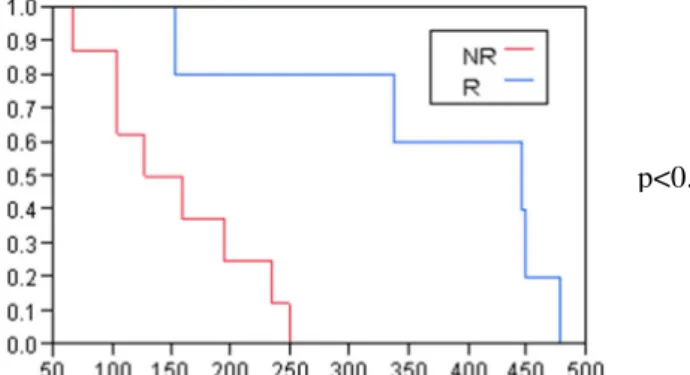
No significant correlation was found between the global indices and the PFS. However in three cases, the indices related to SPECT intensities were significantly correlated with OS (Table 1). This trend was larger for late SPECT studies than for early studies. A Kaplan–Meier analysis based on G5 index dividing the population in 7 responders and 7 non responders) versus OS provides significant discrimination (Figure 1), thus suggesting this biomarker could be a surrogate marker of OS.
Global indices Correlation (R2) with PFS Correlation (R²) with OS G4 NS 0.30 (p=0.04) G5 NS 0.47 (p<0.01) G6 NS 0.27 (p=0.06) G7 NS 0.56 (p<0.01)
Table 1
:
Comparison of global indices with PFS and OS (NS states for not significant correlation, with p>0.05).Figure 1: Kaplan-Meier plot giving G7 versus OS (days) The comparison of early and late SPECT studies showed differences between the two exams. If a high correlation (R²=0.92) was found between their relative variations of volumes, it was moderate (R²=0.44) between relative variations of intensities. The local indices (Table 2) also revealed some discrepancies. Figure 2 illustrates these criteria for one patient who was stable according to RANO criteria, but presented a short OS.
Dice index C1 C5
Mean SD 0.5 0.2 0.17 0.15 0.25 0.47 Table 2
:
Dice index, % of tumor voxels inside early and outside late SPECT (C1), % of tumor voxels inside late and outside early SPECT (C5), see [4] for precisionsIV. DISCUSSION – CONCLUSION
The proposed process is strongly related to the quality of tumor segmentation (for global and local indices) and of image registration (for local indices). The quality of registrations was judged high by two operators
independently, and the differences that were observed in the localization of maximal uptake in the tumor were clearly not due to registration errors. Despite the difficulty in segmenting the tumor zone on SPECT volumes, all results were concordant to indicate a variation of intensity uptake inside the tumor, thus reinforcing the hypothesis of the relationship between the variations of intensity inside the tumor with the overall surviving.
Figure 2: Voxel by voxel comparison of early (red) and late (blue) SPECT tumor volumes for one patient,
showing quite different patterns on both exams Of course this pilot study was not a random study and, as such, cannot be used to state the interest of the antiangiogenic treatment. The difficulties of MRI gadolinium enhanced study and SPECT Sestamibi study to predict PFS should be due to the restoration of the blood brain barrier induced by the antiangiogenic treatment. Thus a clear reduction of perfusion activity is observed in most of cases, but this reduction is not directly related to the tumor activity.
However, our study shows a clear difference between early and late Sestamibi acquisitions. Both intensity indices and localization of highest spots are different when comparing the two examinations. To explain it, our hypothesis is that early acquisition is more related to perfusion mechanism, while late acquisition could be related to mitochondrial process modified in case of tumor activity. Results on this small series were unexpected since the late uptake criteria are well correlated with OS, but not with PFS. However it is important to note that on this small series, PFS was not correlated with OS.
The methodology that we proposed enables a global and local comparison of different imaging modalities and was used to underline the differences between early and late Sestamibi studies. Furthermore late Sestamibi studies could define surrogate markers for overall survival.
REFERENCES
[1] PY Wen, DR Macdonald, DA Reardon et al. J Clin
Oncol 2010 Vol. 28, pp. 1963-72.
[2] F Prigent-Le Jeune, F Dubois, S Perez et al. Eur J
Nucl Med Mol Imag 2004 Vol. 31, pp. 714–9.
[3] J.M. Tachella, E. Roullot, M. Lefort et al., Phys Med
Biol 2014, Vol. 59, pp. 6997-7011.
[4] J.M. Tachella, N. Yeni, E. Roullot, et al, Conf Proc
IEEE EMBS 2014, pp. 1905-08. p<0.008