HAL Id: hal-02353212
https://hal.archives-ouvertes.fr/hal-02353212
Submitted on 14 Nov 2019
HAL is a multi-disciplinary open access
archive for the deposit and dissemination of
sci-entific research documents, whether they are
pub-lished or not. The documents may come from
teaching and research institutions in France or
abroad, or from public or private research centers.
L’archive ouverte pluridisciplinaire HAL, est
destinée au dépôt et à la diffusion de documents
scientifiques de niveau recherche, publiés ou non,
émanant des établissements d’enseignement et de
recherche français ou étrangers, des laboratoires
publics ou privés.
X inactivation in a mammal species with three sex
chromosomes
Frederic Veyrunes, Julie Perez
To cite this version:
Frederic Veyrunes, Julie Perez. X inactivation in a mammal species with three sex chromosomes.
Chromosoma, Springer Verlag, 2018, 127 (2), pp.261-267. �10.1007/s00412-017-0657-2�. �hal-02353212�
ORIGINAL ARTICLE
X inactivation in a mammal species with three sex chromosomes
Frédéric Veyrunes1 &Julie Perez1
Received: 27 July 2017 / Revised: 8 December 2017 / Accepted: 12 December 2017 # Springer-Verlag GmbH Germany, part of Springer Nature 2017
Abstract
X inactivation is a fundamental mechanism in eutherian mammals to restore a balance of X-linked gene products between XY males and XX females. However, it has never been extensively studied in a eutherian species with a sex determination system that deviates from the ubiquitous XX/XY. In this study, we explore the X inactivation process in the African pygmy mouse Mus minutoides, that harbours a polygenic sex determination with three sex chromosomes: Y, X, and a feminizing mutant X, named X*; females can thus be XX, XX*, or X*Y, and all males are XY. Using immunofluorescence, we investigated histone modi-fication patterns between the two X chromosome types. We found that the X and X* chromosomes are randomly inactivated in XX* females, while no histone modifications were detected in X*Y females. Furthermore, in M. minutoides, X and X* chromosomes are fused to different autosomes, and we were able to show that the X inactivation never spreads into the autosomal segments. Evaluation of X inactivation by immunofluorescence is an excellent quantitative procedure, but it is only applicable when there is a structural difference between the two chromosomes that allows them to be distinguished.
Keywords Histone modification . X inactivation . African pygmy mouse . Mus minutoides . Polygenic sex determination . Sex-autosome translocation
Introduction
In eutherian mammals, sex determination involves a genetic system with heterogametic male (XY) and homogametic fe-male (XX), that results in an unequal copy number of X-linked genes between the two sexes. This dosage imbalance of gene expression is resolved by the silencing of one of the two X chromosomes during early embryonic female development through a process called X inactivation (reviews in Heard and Disteche (2006), Veitia et al. (2015), and Marshall Graves (2016)). The silencing of the inactivated X is established through epigenetic factors initiated by the X inac-tivation centre (Xic) involving in particular the Xist gene that codes for a long non-coding RNA which is transcribed exclu-sively from the future inactivated X. The Xist transcript then spreads in cis from the Xic to coat the whole X, recruiting along the way a series of chromatin changes through histone
modifications and DNA methylation that trigger the inactiva-tion (review in Heard (2004)). The silenced X is chosen at random, and once established, a specific signature of epigenet-ic modifepigenet-ications maintains the same inactivated chromosome in all progeny cells. Therefore, females are mosaics for two populations of cells, one with the active maternal X and the other with paternal X (e.g. Heard (2004), Heard and Disteche (2006), Veitia et al. (2015), and Marshall Graves (2016)). In eutherians, there are a few natural exceptions to the standard XX/XY sex determination system (reviews in Fredga (1994) and Parma et al. (2016)), but for now, they have received little attention, in particular regarding X inactivation process. For example, constitutive X0 females known in Microtus oregoni, Tokudaia osimensis, and Ellobius lutescens can be seen as the ultimate evolutionary step of X inactivation with the physically suppression of one X. Nevertheless, the recent analysis of the E. lutescens Xic has shown that all genes known to control X inactivation are still present and well conserved (Mulugeta et al. 2016). In Ellobius tancrei, E. talpinus, and E. alaicus, both females and males are XX, and the presence of two X in males raises the question about the need for X inactivation: has the X inactivation been lost in females or been recruited in males as well? Finally, in Myopus schisticolor, three sex chro-mosome complements occur in females, i.e. XX, XX*, and
* Frédéric Veyrunes
frederic.veyrunes@umontpellier.fr
1
Institut des Sciences de l’Evolution, ISEM UMR 5554 (CNRS / Université Montpellier / IRD / EPHE), Montpellier, France
X*Y, due to the presence of a third sex chromosome, named X*, that carries a feminizing mutation that prevents the initia-tion of the male sex-determining program in X*Y individuals. Although limited investigation seemed to suggest random X inactivation of either the X or X* in XX* females (H. Winking, personal communication, in Schempp et al. (1985)), other re-sults showed more striking patterns, with complete non-random X or X* inactivation (100%) in specimens with nu-merically aberrant X*XY sex chromosome constitution (Schempp et al.1985). Recently, a novel case of unusual mam-malian sex determination system was described in the African pygmy mouse, Mus minutoides, a close relative of the house mouse (Veyrunes et al.2010; Rahmoun et al.2014; Zhao et al.
2017). In wild populations, up to 75% of females carry a Y chromosome and a peculiar feminizing X* chromosome and are fully fertile. The sex-determining system is in fact very similar to the one of the wood lemming Myopus schisticolor and can be characterized as polygenic, with three sex chromo-somes, X, Y, and X*, and two sex-determining genes, the regular mammalian male determiner Sry on the Y, and a still unknown dominant female determiner on the X*. Like in the wood lemming, females are XX, XX*, or X*Y, while all males are XY (Veyrunes et al.2010). Some life history and behav-ioural traits vary along with sex chromosome complement in M. minutoides, and interestingly, XX* females are much more similar to XX females than X*Y ones (Saunders et al.2014,
2016; Ginot et al.2017). This pattern is at least partly under the direct influence of genes on the sex chromosomes, but consid-ering the paucity of genes on the Y chromosome and that most of them are specialized in male reproduction (Marshall Graves
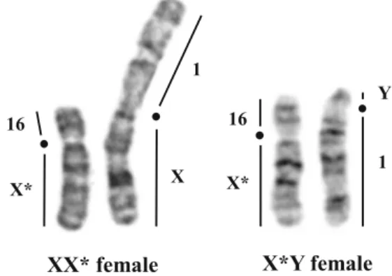
2006), other mechanisms might be involved, such as a prefer-ential X* inactivation in XX* females (Saunders et al.2014). The wild-type X and the mutant X* can be cytogenically dis-tinguished owing to important structural rearrangements (Veyrunes et al.2010). In addition, they are fused to different autosomes, the X to chromosome 1, and the X* to chromo-some 16 (while the Y is also fused to chromochromo-some 1; Fig.1).
The presence of two different X chromosomes in XX* females gives the opportunity to extensively test if both Xs are still randomly inactivated, or if one is preferentially inactivated. We thus investigated the pattern of histone modification char-acteristic of active and inactive chromatin on metaphase chro-mosomes from M. minutoides XX* female embryo fibroblasts. This work also provided insights into the X histone modifica-tions in rare genomic contexts of importance in human medi-cine, such as sex-reversed females and X-autosome transloca-tions (e.g. White et al.1998; Popova et al.2006).
Materials and methods
Animals and cell culture
The karyotype of M. minutoides was published previously (Veyrunes et al.2004). The karyotype is composed of 18 bi-armed chromosomes, all resulting from Robertsonian fusion events, even the sex chromosomes are fused, but with differ-ent autosomes: Rb(Y.1), Rb(X.1), and Rb(X*.16) (Veyrunes et al. 2007). Mice were bred in our own laboratory colony (CECEMA facilities of Montpellier University) established from wild-caught animals (for further details, see Saunders et al. (2014)). Mid-term embryos (between 12 and 16 days post coitum) of pregnant X*Y females (that give birth to XX* and X*Y daughters, and XY sons) were collected. Females were euthanized by CO2inhalation. Embryos were
first sexed by gonad examination. Female embryos were then genotyped using PCR amplification using the Y-specific Sry gene following Veyrunes et al. (2010), to distinguish between XX* and X*Y. Thereby, 11 XX* and two X*Y female em-bryos were selected for fibroblast cell cultures using half of the body and following standard procedures.
Chromosome preparations
Before harvest, fibroblast cultures (70–90% confluent in a 75-cm2flask) were incubated for 2 h with 1% to final volume of KaryoMAX Colcemid (GIBCO). Cells were harvested by trypsinisation and washed with culture medium then with PBS. Cell pellets were resuspended in 1 ml of PBS superna-tant before hypotonic treatment by slowly adding of 10 ml of 0.075 M KCl with 0.1% (vol/vol) glycerol and incubated for 20 min in a 37 °C water bath. Swollen cells were kept on ice up to 1 h before spreading. Three hundred to five hundred microlitres of swollen cell suspensions was spun onto slide using Cytospin (300 rpm, 5 min, low acceleration). Metaphase spread quality was highly dependent on the con-centration of the cell suspension; slides containing metaphase spreads (area of 6 mm in diameter) were air dried.
X X* 1 16 X* 16 1 Y
XX* female X*Y female
Fig. 1 G-banded sex chromosomes of the XX* and X*Y females. X and Y chromosomes are translocated to autosome pair 1, and X* to autosome 16. Black dots indicate centromere position
Antibodies and immunofluorescence
We studied the distribution of two active chromatin markers, namely, histone H4 acetylated at lysine 8 (H4K8ac—Abcam Ab15823) and histone H3 dimethylated at lysine 4 (H3K4me2—Millipore 07-030), and one inactive chromatin marker, histone H3 trimethylated at lysine 27 (H3K27me3— Millipore 07-449), on metaphase spreads (Heard et al.2001; Plath et al.2003; Rens et al. 2010). Before use, antibodies specific for modified histones were diluted 1:600 in TKCM buffer (120 mM KCl, 20 mM NaCl, 10 mM Tris HCl pH 7.5, 0.5 mM EDTA, 0.1% (vol/vol) Triton X-100) with 1.5% BSA (SIGMA). The immunofluorescence protocol was adapted from Rens et al. (2010). Briefly, slides were permeabilised at room temperature with TKCM buffer for 20 min. After re-moving the TKCM buffer, slides containing metaphases spreads were blocked for 20 min with 5% BSA in TKCM buffer (30μl onto cell area under plastic film) in humid cham-ber at room temperature then labelled with 30μl histone an-tibodies overnight at 4 °C. After three washes for 4 min each with TKCM buffer, slides were incubated with FITC-conjugated secondary antibody (anti-rabbit IgG from SIGMA) diluted 1:100 in TKCM buffer with 1% BSA for 30 min in humid chamber at room temperature. Slides were washed as previously and fixed during 10 min at room tem-perature with 5% paraformaldehyde (vol/vol) in TKCM buff-er. Slides were washed for 5 min with TKCM buffer and mounted in Vectashield mounting medium containing DAPI (Vector). Slides were examined with a Zeiss Axioplan 2 Imaging epifluorescence microscope equipped with Cytovision capturing software and CCD camera.
Results
Histone modification characteristic of active
chromatin
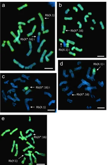
The distribution of the two active chromatin markers (H4K8ac and H3K4me2) on metaphase spreads of 11 M. minutoides XX* was consistent with the one found in the house mouse (Boggs et al.2002; Chaumeil et al.2002; Rens et al.2010), strong signals on the autosomes and one X chromosome, while the other X, supposedly the inactive one, was depleted (Fig.2a, b). In both sex-autosome trans-locations, Rb(X.1) and Rb(X*.16), the histone modifica-tions were asymmetrically distributed around the centro-mere, with a strong signal on the autosomal arm and no staining on the inactive X arm. The X*Y females showed a different hybridization pattern: H4K8ac and H3K4me2 were evenly distributed on all chromosomes including the X* and Y chromosomes (Fig.2e).
Histone modification characteristic of inactive
chromatin
On the same specimens but on different metaphase spreads, we also analysed the repressive modification of the histone H3K27me3 that is a specific mark of the inactivated X in the house mouse (Plath et al.2003; Rens et al.2010). On the M. minutoides XX* metaphases, there was a strong enrich-ment on only one single X arm with a clear cut-off limit at the centromere of the sex-autosome translocation (Fig.2c, d). On the X*Y females, H3K27me3 was not detected (data not shown).
The number of analysed cells and the percentage of inac-tive X and X* chromosomes for each XX* embryo are sum-marized in Table1. The number of metaphases varied between 16 and 126, depending of the cell suspension and the cytospin spreading quality. Arbitrarily, skewed (i.e. non-random) X
a
b
e
c
Rb(X.1) i Rb(X*.16) Rb(X*.16) i Rb(X.1) Rb(X*.16) i Rb(X.1) Rb(X*.16) Rb(Y.1) Rb(X.1) id
Rb(X*.16)Fig. 2 Immunofluorescence staining for a H4K8ac and b H3K4me2 histones, both characteristic of the active chromatin, on XX* female metaphase spreads; c, d H3K27me3 histone, an inactive chromatin marker, on XX* female metaphase spreads; e H4K8ac histone on an X*Y female metaphase spread. Bi^ indicates the inactive X chromosome type. Scale bar = 10μm
inactivation is often defined when the ratio is≥ 80:20% (e.g. Belmont (1996), Plenge et al. (2002), and Talebizadeh et al. (2005)). Interestingly, four of the 11 samples reached the threshold ratio of 80:20, three and one with a highly biased X* and X inactivation, respectively. In total, 781 metaphase spreads have been analysed, 439 (56%) with an inactive X* chromosome and 342 (44%) with an inactive X.
Discussion
X inactivation is a fundamental process in eutherian mammals to restore a balance of X-linked gene products between males and females. However, it has never been extensively studied in a mammal species with an atypical sex determination sys-tem which deviates from the ubiquitous XX/XY. In this study, we explore the X inactivation process in the African pygmy mouse, M. minutoides, that harbours a polygenic sex determi-nation with three sex chromosomes: Y, X, and X* (Veyrunes et al.2010). Such a system raises several questions. First, in XX* females, is the X inactivation mechanism still random, or is one type of X preferentially inactivated? Second, in X*Y females, the single copy of X chromosome is not expected to be silenced, could we thus provide direct evidence of the ab-sence of X inactivation? And finally, does the X inactivation spread onto the autosomal arms in the sex-autosome translo-cations, Rb(X.1) and Rb(X*.16)? Hence, to investigate the X inactivation process in this unusual sex determination system, we conducted immunofluorescence analyses to evaluate his-tone modification patterns between the two X chromosome
types. This appears to be an excellent quantitative procedure to detect any deviation from randomness.
In eutherian mammals, random X inactivation (50:50%) is the norm, but skewed X inactivation patterns have been re-ported and are most of the time detected in women who man-ifest severe X-linked genetic diseases (e.g. Van den Veyver (2001), Plenge et al. (2002), and Talebizadeh et al. (2005)). The causes of X inactivation skewing have been extensively discussed (Belmont 1996; Migeon 1998; Brown and Robinson2000). Particularly, it has been proposed that the main cause for it is somatic selection occurring after random X inactivation. This process may lead to a skewed X inacti-vation when one of the X chromosomes carries extensive chromosomal rearrangements or a single gene mutation that affects cell survival or growth. Consequently, a majority of cells bearing the inactive mutated X would remain. Other potential causes for X inactivation skewing are mutations on genes that could influence the choice of the inactivated X, such as Xic, Xist, or Xce loci (e.g. Plenge et al. 1997; Migeon 1998). In M. minutoides, recent studies comparing the three female genotypes have shown that XX* females are much more similar to XX females than X*Y ones in terms of fertility, behaviour, and morphology (Saunders et al.2014,
2016; Ginot et al.2017), and interestingly, it has been hypoth-esized that these findings could be partly explained by a pref-erential inactivation of the X* in XX* females (Saunders et al.
2014).
Our results on histone modification patterns support that the M. minutoides X* and X chromosomes remain randomly inactivated; they are found to be inactive in 56 and 44% of the 781 analysed metaphase spreads, respectively (Table 1). Hence, there is no evidence of a constitutional X inactivation skewing, as could have been suggested by the observations of behaviour and life history traits. However, the variance among the 11 samples was important. While some showed a perfect random inactivation pattern, e.g. one specimen, with respec-tively 56 and 55 cells with an inactive X* and X chromosome, four others showed important skews (≥ 80:20%), three in fa-vor of an inactive X* and one for the X (Table1). Skewed X inactivation can occur by chance as a result of the fact that the X inactivation is a stochastic process and at the time it is established, the progenitor cell population is relatively small. Consequently, it has been estimated that in a control popula-tion of healthy women, approximatively 10% present skewed X inactivation (e.g. Plenge et al. (2002) and Talebizadeh et al.
2005). However, in the case of M. minutoides, this proportion is much higher (four out of 11 specimens, 36%). One possible explanation is that cell selection took place in the cell cultures. For example, we could have accidentally sampled only one cell population that all derived from the same progenitor cell. To avoid such unbalanced sampling, we began the cell cul-tures from material representing half of the organism (lower part of the embryo, the other half was used for genotyping).
Table 1: Number of analysed cells with an inactive X* or X chromosome, and percentage of X* inactivation for each of the 11 XX* embryos individual No. metaphases inactive X* inactive X % of X* inactivation 1 73 15 58 20% 2 121 40 81 33% 3 111 56 55 50% 4 16 9 7 56% 5 115 65 50 57% 6 126 77 49 61% 7 74 50 24 68% 8 26 19 7 73% 9 32 27 5 84% 10 36 32 4 89% 11 53 49 4 92% Total 781 439 342 56%
Bold typeface indicates samples that reached skewed X inactivation with a treeshold ratio≥ 80:20
However, we cannot rule out the possibility that the cells grew out from only one or a few small tissue pieces before the first culture passage and harvest. Overall, our results suggest that the X* chromosome, although highly rearranged (Veyrunes et al.2010), does not affect X inactivation, and thus that the genes involved in X inactivation have not been affected by the rearrangements.
As expected, no histone modifications were detected on metaphase spreads of X*Y females (Fig.2), since in euthe-rian mammals, a counting mechanism exists to maintain only one active chromosome (Monkhorst et al. 2008; Augui et al.2011). This was first revealed in humans with abnormal numbers of X chromosomes: females with Turner syndrome (XO) show no inactivation of their unique X, while XXX females and even Klinefelter males (XXY) dis-play respectively two and one inactive X chromosomes. Another remarkable feature of the M. minutoides karyotype is the presence of two X-autosome translocations, Rb(X.1) and Rb(X*.16) (Veyrunes et al.2004,2007). X-autosome translocation is considered as one of most deleterious rear-rangements in mammals, since it can generate important perturbations of gametogenesis and gene expression, nota-bly due to the spreading of X inactivation into the adjacent autosome, that causes the silencing of autosomal genes (re-view in Dobigny et al. (2004)). This phenomenon has been demonstrated in humans and mice (e.g. Rastan (1983), Schanz and Steinbach (1989), White et al. (1998), Hall et al. (2002), and Popova et al.2006) and has been shown to generally lead to severe phenotypic effects (Waters et al.
2001). Fixation of X-autosome translocations is neverthe-less not that uncommon in mammalian karyotypes, and it has been suggested that the cost of such rearrangement can be overcome by the addition of a large block of heterochro-matin between the X and the autosomal segments that would serve as a boundary to functionally isolate the two components. Support for this hypothesis is found in almost all mammal species known to possess X-autosome translo-cations, in which a large block of heterochromatin is sys-tematically identified between the two chromosomal seg-ments (review in Dobigny et al. (2004)). However, very few studies have actually demonstrated its role as insulation buffer. In the common shrew Sorex araneus and in rodents of the genus Taterillus, replication banding pattern after BrdU treatment showed that a late-replicating segment, supposedly inactive, was limited to the ancestral X com-partment (Pack et al.1993; Dobigny et al.2004), and in the marsupial potoroo Potorous tridactylus, hypomethyla-tion was restricted to one of the X chromosome arms of the X-autosome translocations (Rens et al. 2010). Here, with modern technique of immunofluorescence to detect histone modifications, we confirmed that the X inactivation never extents to the translocated autosome in M. minutoides (Fig.
2). These results are even more interesting in M. minutoides since no blocks of C-positive heterochromatin were found near the centromeres of Rb(X.1) and Rb(X*.16) (Veyrunes et al.2004), and there is no pseudoautosomal region that could create a natural barrier preventing the spreading (the sex chromosomes being asynaptic; Britton-Davidian et al.
2012). These observations support our assumption that in this species, the isolation of the X and autosomal compart-ments may involve other types of repetitive sequences to act as a buffer (Veyrunes et al.2004; Colomina et al.2017), or some genomic characteristics, such as a low density of LINE-1 elements, as they are suggested to act as booster elements of the X inactivation propagation (Lyon 1998; Bala Tannan et al.2014; Cotton et al.2014).
In summary, we explored the X inactivation process in a mammalian model that presents a unique sex determination system with two types of X chromosomes. We provided direct evidence that in the XX* females, the mutant feminizing X* and the ancestral X are randomly inactivated, and that in X*Y females, inactivation does not occur. Evaluation of X inacti-vation by immunofluorescence is an excellent quantitative procedure, but it is only applicable when there is a structural difference between the two chromosomes that allows them to be distinguished. However, we warned about cell selection that could occur in cultures and bias the results. We also con-cluded that in the sex-autosome translocations found in this species, the X inactivation does not spread into the autosomal arm. More extensive molecular studies (e.g. RT-qPCR, methylation-specific PCR) on autosomal genes at the junction with the X arm would be required to confirm the chromosome-wide histone modification pattern and the ab-sence of X inactivation spreading.
Acknowledgements We are especially indebted to the animal breeding facility of Montpellier University (CECEMA). We thank M. Rahmoun for her technical help and PA Saunders for comments on the manuscript. Experiments were performed at the CytoEvol facilities of ISEM and CBGP (labex CeMEB). This article is dedicated to the memory of our friend, colleague, and mentor Janice Britton-Davidian. She was a great scientist and a remarkable woman, and it was through her intellectual leadership that the cytogenetic work on the African pygmy mouse was initiated.
Funding This study was supported by the ANR grantBSEXYMUS^ (no. 10-JCJC-1605).
Compliance with ethical standards
Conflict of interest The authors declare that they have no conflict of interest.
Ethical approval The experimental protocol was performed in accor-dance with European guidelines and with the approval of the Ethical Committee on Animal Care and Use of France (no. CEEA-LR-12170).
References
Augui S, Nora EP, Heard E (2011) Regulation of X-chromosome inacti-vation by the X-inactiinacti-vation centre. Nat Rev Genet 12(6):429–442.
https://doi.org/10.1038/nrg2987
Bala Tannan N, Brahmachary M, Garg P, Borel C, Alnefaie R, Watson CT, Thomas NS, Sharp AJ (2014) DNA methylation profiling in X;autosome translocations supports a role for L1 repeats in the spread of X chromosome inactivation. Hum Mol Genet 23(5): 1224–1236.https://doi.org/10.1093/hmg/ddt553
Belmont JW (1996) Genetic control of X inactivation and processes lead-ing to X-inactivation skewlead-ing. Am J Hum Genet 58(6):1101–1108 Boggs BA, Cheung P, Heard E, Spector DL, Chinault AC, Allis CD (2002) Differentially methylated forms of histone H3 show unique association patterns with inactive human X chromosomes. Nat Genet 30(1):73–76.https://doi.org/10.1038/ng787
Britton-Davidian J, Robinson TJ, Veyrunes F (2012) Systematics and evo-lution of the African pygmy mice, subgenus Nannomys: a review. Acta Oecol 42:41–49.https://doi.org/10.1016/j.actao.2012.01.001
Brown CJ, Robinson WP (2000) The causes and consequences of random and non-random X chromosome inactivation in humans. Clin Genet 58(5):353–363
Chaumeil J, Okamoto I, Guggiari M, Heard E (2002) Integrated kinetics of X chromosome inactivation in differentiating embryonic stem cells. Cytogenet Genome Res 99(1-4):75–84.https://doi.org/10. 1159/000071577
Colomina V, Catalan J, Britton-Davidian J, Veyrunes F (2017) Extensive amplification of telomeric repeats in the karyotypically highly di-verse African pygmy mice. Cytogenet Genome Res 152(2):55–64.
https://doi.org/10.1159/000478297
Cotton AM, Chen CY, Lam LL, Wasserman WW, Kobor MS, Brown CJ (2014) Spread of X-chromosome inactivation into autosomal se-quences: role for DNA elements, chromatin features and chromo-somal domains. Hum Mol Genet 23(5):1211–1223.https://doi.org/ 10.1093/hmg/ddt513
Dobigny G, Ozouf-Costaz C, Bonillo C, Volobouev V (2004) Viability of X-autosome translocations in mammals: an epigenomic hypothesis from a rodent case-study. Chromosoma 113(1):34–41.https://doi. org/10.1007/s00412-004-0292-6
Fredga K (1994) Bizarre mammalian sex-determining mechanisms. In: Short RV, Balaban E (eds) The differences between the sexes. Cambridge University Press, Cambridge, pp 419–431
Ginot S, Claude J, Perez J, Veyrunes F (2017) Sex reversal induces size and performance differences among females of the African pygmy mouse, Mus minutoides. J Exp Biol 220(11):1947–1951.https://doi. org/10.1242/jeb.157552
Hall LL, Clemson CM, Byron M, Wydner K, Lawrence JB (2002) Unbalanced X;autosome translocations provide evidence for sequence specificity in the association of XIST RNA with chromatin. Hum Mol Genet 11(25):3157–3165.https://doi.org/10.1093/hmg/11.25.3157
Heard E (2004) Recent advances in X-chromosome inactivation. Curr Opin Cell Biol 16(3):247–255.https://doi.org/10.1016/j.ceb.2004.03.005
Heard E, Disteche CM (2006) Dosage compensation in mammals: fine-tuning the expression of the X chromosome. Genes Dev 20(14): 1848–1867.https://doi.org/10.1101/gad.1422906
Heard E, Rougeulle C, Arnaud D, Avner P, Allis CD, Spector DL (2001) Methylation of histone H3 at Lys-9 is an early mark on the X chro-mosome during X inactivation. Cell 107(6):727–738.https://doi. org/10.1016/S0092-8674(01)00598-0
Lyon MF (1998) X-chromosome inactivation, a repeat hypothesis. Cytogenet Cell Genet 80(1-4):133–137.https://doi.org/10.1159/ 000014969
Marshall Graves JA (2006) Sex chromosome specialization and degen-eration in mammals. Cell 124(5):901–914.https://doi.org/10.1016/j. cell.2006.02.024
Marshall Graves JA (2016) Evolution of vertebrate sex chromosomes and dosage compensation. Nat Rev Genet 17:33–46
Migeon BR (1998) Non-random X chromosome inactivation in mamma-lian cells. Cytogenet Cell Genet 80(1–4):142–148.https://doi.org/ 10.1159/000014971
Monkhorst K, Jonkers I, Rentmeester E, Grosveld F, Gribnau J (2008) X inactivation counting and choice is a stochastic process: evidence for involvement of an X-linked activator. Cell 132(3):410–421.https:// doi.org/10.1016/j.cell.2007.12.036
Mulugeta E, Wassenaar E, Sleddens-Linkels E, van IJcken WJF, Heard E, Grootegoed JA, Just W, Gribnau J, Baarends WM (2016) Genomes of Ellobius species provide insight into the evolutionary dynamics of mammalian sex chromosomes. Genome Res 26(9):1202–1210.
https://doi.org/10.1101/gr.201665.115
Pack SD, Borodin PM, Serov OL, Searle JB (1993) The X-autosome translocation in the common shrew (Sorex araneus L.): late replica-tion in female somatic cells and pairing in male meiosis. Chromosoma 102(5):355–360. https://doi.org/10.1007/ BF00661279
Parma P, Veyrunes F, Pailhoux E (2016) Sex reversal in non-human placental mammals. Sex Dev 10(5-6):326–344.https://doi.org/10. 1159/000448361
Plath K, Fang J, Mlynarczyk-Evans SK, Cao R, Worringer KA, Wang H, de la Cruz CC, Otte AP, Panning B, Zhang Y (2003) Role of histone H3 lysine 27 methylation in X inactivation. Science 300(5616):131– 135.https://doi.org/10.1126/science.1084274
Plenge RM, Hendrich BD, Schwartz C, Arena JF, Naumova A, Sapienza C, Winter RM, Willard HF (1997) A promoter mutation in the XIST gene in two unrelated families with skewed X-chromosome inacti-vation. Nat Genet 17(3):353–356
Plenge RM, Stevenson RA, Lubs HA, Schwartz CE, Willard HF (2002) Skewed X chromosome inactivation is a common feature of X-linked mental retardation disorders. Am J Hum Genet 71(1):168– 173.https://doi.org/10.1086/341123
Popova BC, Tada T, Takagi N, Brockdorff N, Nesterova TB (2006) Attenuated spread of X-inactivation in an X;autosome translocation. Proc Natl Acad Sci U S A 103(20):7706–7711.https://doi.org/10. 1073/pnas.0602021103
Rahmoun M, Perez J, Saunders PA, Boizet-Bonhoure B, Wilhelm D, Poulat F, Veyrunes F (2014) Anatomical and molecular analyses of XY ovaries from the African pygmy mouse Mus minutoides. Sex Dev 8(6):356–363.https://doi.org/10.1159/000368664
Rastan S (1983) Non-random chromosome inactivation in mouse X-autosome translocation embryos-location of the inactivation centre. J Embryol Exp Morphol 78:1–22
Rens W, Wallduck MS, Lovell FL, Ferguson-Smith MA, Ferguson-Smith AC (2010) Epigenetic modifications on X chromosomes in marsu-pial and monotreme mammals and implications for evolution of dosage compensation. Proc Natl Acad Sci U S A 107(41):17657– 17662.https://doi.org/10.1073/pnas.0910322107
Saunders PA, Perez J, Rahmoun M, Ronce O, Crochet PA, Veyrunes F (2014) XY females do better than the XX in the African pygmy mouse, Mus minutoides. Evolution 68(7):2119–2127.https://doi. org/10.1111/evo.12387
Saunders PA, Franco T, Sottas C, Maurice T, Ganem G, Veyrunes F (2016) Masculinised behaviour of XY females in a mammal with naturally occurring sex reversal. Sci Rep 6(1):22881.https://doi.org/ 10.1038/srep22881
Schanz S, Steinbach P (1989) Investigation of theBvariable spreading^ of X inactivation into a translocated autosome. Hum Genet 82(3):244– 248.https://doi.org/10.1007/BF00291163
Schempp W, Wiberg U, Fredga K (1985) Correlation between sexual phenotype and x chromosome inactivation pattern in the X*XY wood lemming. Cytogenet Cell Genet 39(1):30–34.https://doi.org/ 10.1159/000132099
Talebizadeh Z, Bittel DC, Veatch OJ, Kibiryeva N, Butler MG (2005) Brief report: non-random X chromosome inactivation in females with autism. J Autism Dev Disord 35(5):675–681.https://doi.org/ 10.1007/s10803-005-0011-z
Van den Veyver IB (2001) Skewed X inactivation in X-linked disorders. Semin Reprod Med 19(02):183–191. https://doi.org/10.1055/s-2001-15398
Veitia RA, Veyrunes F, Bottani S, Birchler JA (2015) X chromosome inactivation and active X upregulation in therian mammals: facts, questions, and hypotheses. J Mol Cell Biol 7(1):2–11.https://doi. org/10.1093/jmcb/mjv001
Veyrunes F, Catalan J, Sicard B, Robinson TJ, Duplantier JM, Granjon L, Dobigny G, Britton-Davidian J (2004) Autosome and sex chromo-some diversity among the African pygmy mice, subgenus Nannomys (Muridae; Mus). Chromosom Res 12(4):369–382.
https://doi.org/10.1023/B:CHRO.0000034098.09885.e6
Veyrunes F, Watson J, Robinson TJ, Britton-Davidian J (2007) Accumulation of rare sex chromosome rearrangements in the African pygmy mouse, Mus (Nannomys) minutoides: a whole-arm reciprocal translocation (WART) involving an X-autosome fusion.
Chromosom Res 15(2):223–230. https://doi.org/10.1007/s10577-006-1116-8
Veyrunes F, Chevret P, Catalan J, Castiglia R, Watson J, Dobigny G, Robinson TJ, Britton-Davidian J (2010) A novel sex determination system in a close relative of the house mouse. Proc R Soc B 277(1684):1049–1056.https://doi.org/10.1098/rspb.2009.1925
Waters JJ, Campbell PL, Crocker AJ, Campbell CM (2001) Phenotypic effects of balanced X-autosome translocations in females: a retro-spective survey of 104 cases reported from UK laboratories. Hum Genet 108(4):318–327.https://doi.org/10.1007/s004390100465
White WM, Willard HF, Van Dyke DL, Wolff DJ (1998) The spreading of X inactivation into autosomal material of an X-autosome transloca-tion: evidence for a difference between autosomal and X-chromosomal DNA. Am J Hum Genet 63(1):20–28.https://doi. org/10.1086/301922
Zhao L, Quinn A, Ting Ng E, Veyrunes F, Koopman P (2017) Reduced activity of SRY and its target enhancer Sox9-TESCO in a mouse species with X*Y sex reversal. Sci Rep 7:41378.https://doi.org/ 10.1038/srep41378