HAL Id: hal-01772539
https://hal-amu.archives-ouvertes.fr/hal-01772539
Submitted on 20 Apr 2018
HAL is a multi-disciplinary open access
archive for the deposit and dissemination of
sci-entific research documents, whether they are
pub-lished or not. The documents may come from
teaching and research institutions in France or
abroad, or from public or private research centers.
L’archive ouverte pluridisciplinaire HAL, est
destinée au dépôt et à la diffusion de documents
scientifiques de niveau recherche, publiés ou non,
émanant des établissements d’enseignement et de
recherche français ou étrangers, des laboratoires
publics ou privés.
Distributed under a Creative Commons Attribution| 4.0 International License
Dysregulation in Q Fever Endocarditis: The Role of
IL-10 in PD-1 Modulation
Mignane Ka, Françoise Gondois-Rey, Christian Capo, Julien Textoris,
Mathieu Million, Didier Raoult, Daniel Olive, Jean-Louis Mege
To cite this version:
Mignane Ka, Françoise Gondois-Rey, Christian Capo, Julien Textoris, Mathieu Million, et al..
Im-balance of Circulating Monocyte Subsets and PD-1 Dysregulation in Q Fever Endocarditis: The Role
of IL-10 in PD-1 Modulation. PLoS ONE, Public Library of Science, 2014, 9 (9),
�10.1371/jour-nal.pone.0107533�. �hal-01772539�
Dysregulation in Q Fever Endocarditis: The Role of IL-10
in PD-1 Modulation
Mignane B. Ka1,2, Franc¸oise Gondois-Rey2, Christian Capo1, Julien Textoris1, Mathieu Million1, Didier Raoult1, Daniel Olive2, Jean-Louis Mege1*
1 Aix Marseille Universite´, Unite´ de Recherche sur les Maladies Infectieuses Tropicales et Emergentes, UMR 63, CNRS 7278, IRD 198, INSERM U1095, Marseille, France, 2 Inserm UMR 1068, Centre de Recherche en Cance´rologie de Marseille, Marseille, France
Abstract
Q fever endocarditis, a severe complication of Q fever, is associated with a defective immune response, the mechanisms of which are poorly understood. We hypothesized that Q fever immune deficiency is related to altered distribution and activation of circulating monocyte subsets. Monocyte subsets were analyzed by flow cytometry in peripheral blood mononuclear cells from patients with Q fever endocarditis and controls. The proportion of classical monocytes (CD14+CD162monocytes) was similar in patients and controls. In contrast, the patients with Q fever endocarditis exhibited a decrease in the non-classical and intermediate subsets of monocytes (CD16+monocytes). The altered distribution of monocyte subsets in Q fever endocarditis was associated with changes in their activation profile. Indeed, the expression of HLA-DR, a canonical activation molecule, and PD-1, a co-inhibitory molecule, was increased in intermediate monocytes. This profile was not restricted to CD16+monocytes because CD4+T cells also overexpressed PD-1. The mechanism leading to the overexpression of PD-1 did not require the LPS from C. burnetii but involved interleukin-10, an immunosuppressive cytokine. Indeed, the incubation of control monocytes with interleukin-10 led to a higher expression of PD-1 and neutralizing interleukin-10 prevented C. burnetii-stimulated PD-1 expression. Taken together, these results show that the immune suppression of Q fever endocarditis involves a cross-talk between monocytes and CD4+T cells expressing PD-1. The expression of PD-1 may be useful to assess chronic immune alterations in Q fever endocarditis.
Citation: Ka MB, Gondois-Rey F, Capo C, Textoris J, Million M, et al. (2014) Imbalance of Circulating Monocyte Subsets and PD-1 Dysregulation in Q Fever Endocarditis: The Role of IL-10 in PD-1 Modulation. PLoS ONE 9(9): e107533. doi:10.1371/journal.pone.0107533
Editor: Yolande Richard, COCHIN INSTITUTE, Institut National de la Sante´ et de la Recherche Me´dicale, France Received February 20, 2014; Accepted August 16, 2014; Published September 11, 2014
Copyright: ß 2014 Ka et al. This is an open-access article distributed under the terms of the Creative Commons Attribution License, which permits unrestricted use, distribution, and reproduction in any medium, provided the original author and source are credited.
Funding: These authors have no support or funding to report.
Competing Interests: The authors have declared that no competing interests exist. * Email: jean-louis.mege@univ-amu.fr.
Introduction
Q fever is a widespread zoonosis caused byCoxiella burnetii, a small gram-negative bacterium. Humans usually contract Q fever via aerosols and develop a primary infection that is symptomatic in a minority of exposed individuals. When symptomatic, primary Q fever consists of isolated fever, hepatitis or pneumonia [1]. Q fever may become chronic in patients with valvular damage, immuno-compromised patients and pregnant women [2]. The major clinical manifestation of chronic Q fever is an endocarditis, which accounts for 48% of blood culture-negative cases of infective endocarditis [3]. Patients with Q fever endocarditis exhibit spontaneous evolution to death in the absence of antibiotic treatment, and long-lasting treatment is not sufficient to prevent relapses [1]. However, antibiotic prophylaxis of patients with acute Q fever and valvulopathy prevents the evolution from acute Q fever to endocarditis [4].
The innate and adaptive immune response toC. burnetii has an impact on the evolution of Q fever. Monocytes and tissue macrophages are infected by C. burnetii [5,6], but bacterial clearance in vitro requires interferon (IFN)-c-mediated microbi-cidal signals [7,8]. The production of such signals is observed in primary Q fever and is associated with the formation of tissue
granulomas [9]. In contrast, Q fever endocarditis is characterized by a defective cell-mediated immune response, and tissue granulomas are replaced with lymphocyte infiltration [5]. The mechanisms governing the defective immune response in Q fever endocarditis are diverse involving alterations in cytokine amounts and distribution of immune cells. The overproduction of the immunoregulatory cytokine, interleukin (IL)-10, in Q fever endocarditis has been largely documented and plays a role in the inhibition of macrophage microbicidal activity and T cell proliferation [10,11]. On the other hand, an anomalous distribu-tion of circulating immune cell subsets may account for the inefficiency of the immune response in chronic Q fever. We recently demonstrated that a subset of regulatory T cells, namely naturally occurring CD4+CD25+Foxp3+T cells (Treg cells), was increased in Q fever endocarditis independent of changes in the CD4+T cell population, suggesting that the disease is associated with an expansion of this subset of CD4+T cells [12].
As observed in some chronic infectious diseases, the inefficient immune response observed in Q fever endocarditis may also involve co-inhibitory molecules, such as programmed cell death (PD)-1. The expression of the glycoprotein PD-1 (CD279), a member of CD28/B7 Ig superfamily, is increased after the activation of immune cells including CD4+T cells, CD8+T cells,
natural killer cells (NK cells), B cells and monocytes [13,14]. The PD-1 molecule delivers an inhibitory signal for T cells [15,16]. This is illustrated by the immune deficiency of patients with immunodeficiency virus (HIV) infection, in which PD-1 is significantly up-regulated in CD8+ T cells; such pattern of expression is related to impaired function of HIV-specific CD8+ T cells, plasma viral load and IL-10 production [17,18]. The role of PD-1 in bacterial infections such as Q fever is largely unknown but we reasoned that the chronic evolution of Q fever may be reminiscent of the chronic evolution of HIV infection.
The aim of this study was to assess the distribution of circulating monocyte subsets in patients with Q fever endocarditis using flow cytometry, a tool of choice for probing human immune phenotypes and for characterizing subsets of cells, including rare circulating subsets [19]. We demonstrated that the major subset of monocytes (classical monocytes) was not affected in Q fever endocarditis. In contrast, Q fever endocarditis is characterized by a decrease in non-classical and intermediate monocytes (CD16+ monocytes). We found that intermediate monocytes, but also CD4+ T lymphocytes and Treg cells, overexpressed PD-1 in patients with Q fever endocarditis. The overexpression of PD-1 is likely related to the overproduction of IL-10 we observed in Q fever endocarditis. Indeed, in vitro experiments revealed thatC. burnetii up-regulates the expression of PD-1 by monocytes via IL-10 production. These findings show that Q fever endocarditis is associated with changes in monocyte subsets and dysregulated expression of PD-1.
Materials and Methods Ethics statement
The study was conducted with the approval of the Ethics Committee of Aix-Marseille University, France. Blood samples were collected after written informed consent was obtained from each participant.
Patients
Suspected Q fever endocarditis was based on standardized questionnaire that included pathological evidence of endocarditis, positive echocardiograms, positive blood cultures, high titers of IgG specific for phase IC. burnetii. The diagnostic is considered certain after data scoring as previously described in our laboratory [20]. Patients with Q fever endocarditis consisted of 12 men (ranging from 36 to 75 years-old) and 5 women (ranging from 42 to 64 years-old) (Table 1). Eleven healthy individuals (6 men ranging from 26 to 65 years-old and 4 women ranging from 30 to 70 years-old) and 9 patients with acute Q fever (6 men ranging from 42 to 71 old and 3 women ranging from 40 to 68 years-old) were included as controls; the diagnostic of acute Q fever was based on serological determination of anti-phase IIC. burnetii antibodies (Abs) [20].
PBMCs and the preparation of monocytes
Total blood was drawn in 5-ml EDTA-anticoagulated tubes, and peripheral blood mononuclear cells (PBMCs) were separated using Ficoll cushions. PBMCs were recovered in RPMI 1640 containing 20 mM HEPES, 10% fetal calf serum (FCS), 2 mM L-glutamine, 100 U penicillin/ml and 50mg/ml streptomycin (Life Technologies, Saint Aubin, France). The cells were cryopreserved using 10% DMSO and frozen in liquid nitrogen.
Monocytes were obtained from PBMCs using magnetic beads coated with monoclonal Abs (mAbs) directed against CD14, according to the manufacturer’s instructions (Miltenyi Biotech, Paris, France). This procedure resulted in more than 95% monocyte purity, as assessed by flow cytometry. Monocyte viability was greater than 98% as determined by trypan blue exclusion.
Flow cytometry
PBMC samples were incubated with mAbs or isotypic controls and Aqua-Fluorescent Reactive dye (vivid), a viability dye, for Table 1. Patients with Q fever endocarditis.
Patients Sex Age Score IgG Valvulopathy Treatment 1 M 75 A1B2C3 1600 yes valvular prosthesis 2 M 36 A1B2C1 12800 yes mitral valvuloplasty 3 M 73 ND 6400 yes abdominal aortic endograft 4 F 56 A1B2C2 800 yes native heart valve disease 5 M 47 A0B2C2 6400 yes valvular prosthesis 6 M 54 A0B1C3 6400 yes aortic regurgitation 7 M 67 A1B1C3 25600 yes interventicular communication 8 M 66 A0B0C3 1600 yes aortic bicuspidy
9 F 54 A2B0C2 800 yes aortic stenosis 10 F 47 A0B0C3 6400 yes aortic bicuspidy 11 M 41 A0B2C0 25600 yes aortic homograft 12 F 64 ND 400 yes valvular prosthesis 13 M 48 A0B0C3 400 yes mitral valve prolapse 14 M 52 A0B0C3 400 yes valvular calcification 15 F 42 ND 3200 yes aortic stenosis
16 M 56 A0B1C1 800 yes valvulopathy without precision 17 M 65 A0B0C3 400 yes mitral regurgitation
The titer of IgG directed against C. burnetii in phase I is indicated. doi:10.1371/journal.pone.0107533.t001
20 min (the list of fluorescent reagents is presented inTable S1. PBMCs were then washed, fixed in 4% paraformaldehyde and data were acquired within 3 days. Flow cytometry was performed using a LSRII-SORP cytometer (Becton Dickinson, Le Pont de Claix, France) equipped with four lasers and 17 photomultipliers. The cytometer was routinely calibrated with cytometer Set-up and Tracking Beads (Becton Dickinson). The voltage of photomulti-pliers was determined using unstained and stained PBMCs. Compensation particle-set beads (Becton Dickinson) were used for each fluorescent reagent, except Aqua-Fluorescent Reactive dye, to calculate the compensation matrix with FlowJo tools. Approximately 106 events were acquired. After the exclusion of
debris on the forward/side scatter dot plot and dead cells with Aqua-Fluorescent Reactive dye, data were exported and analyzed with FlowJo Software (version 9.2, Tree Star Ashland, OR, USA). The gating strategy to explore circulating monocytes and T cells is presented in Figure 1A and Figure 2A, respectively, as previ-ously described [21]. The proportion of monocyte and T cell subsets is expressed as the ratio of living cells expressing the fluorescent marker to the total number of analyzed PBMCs. The phenotypic expression of HLA-DR is given as the mean
fluorescence intensity (MFI). The expression of PD-1 is presented as the ratio of MFIassayto MFIisotypic controlfor monocytes and the
percentage of PD-1 positive cells for T cell subsets. Flow cytometry was also used to analyze the level of apoptosis of monocyte subsets. In brief, monocyte subsets were gated according the expression of CD14 and CD16, and the expression of fluorescent annexin V by monocyte subsets was assessed.
In vitro stimulation of monocytes
C. burnetii (Nine Mile strain, RSA496) was cultured as previously described [22]. The L929 cells were infected, sonicated and centrifuged at 300 6g for 10 min. Supernatants were collected and centrifuged at 10,000 6g for 10 min. Bacteria were then washed and stored at -80uC. The concentration of organisms was determined by Gimenez staining, and the bacterial viability was assessed using the LIVE/DEAD BacLight bacterial viability kit (Molecular Probes, Life Technologies).
PBMCs or isolated monocytes were incubated with living or heat-denaturated (100uC for 30 min) phase I C. burnetii organisms (bacterium-to-cell ratio of 50:1), 10mg/ml LPS fromEscherichia coli (Sigma-Aldrich, Saint-Quentin Fallavier) or from phase I C.
Figure 1. Analysis of monocyte subsets. A, Gating strategy for studying monocyte subsets. PBMCs were gated according to their size and after the exclusion of dead cells. PBMCs that expressed HLA-DR and did not express CD3, CD19 or CD56 (exclusion of CD3+T cells, B cells and NK cells,
respectively) were selected. Selected cells were analyzed according the level of CD14 and CD16 expression (monocytes).B, Analysis of monocyte subsets in Q fever endocarditis. Monocytes from 10 healthy controls and 17 patients with Q fever endocarditis were analyzed with flow cytometry. The proportion of monocytes with respect of the total number of live PBMCs is presented for each individual. The nonparametric Mann-Whitney U test was used to compare the patient and control groups. *p,0.05. Horizontal bar, median value.
Figure 2. Analysis of T lymphocyte subsets. A, Gating strategy for studying T cell subsets. PBMCs were gated according to their size and after the exclusion of dead cells. PBMCs that expressed CD3 were selected. Selected cells were analyzed according the level of CD4 and CD8 expression. Treg cells were analyzed according the expression of CD127 and CD25.B, Analysis of T cell subsets in Q fever endocarditis. T cell subsets from 11 healthy controls and 14 patients with Q fever endocarditis were analyzed with flow cytometry. The proportion of T cells subsets with respect of the total number of living PBMCs is presented for each individual. The nonparametric Mann-Whitney U test was used to compare the patient and control groups. **p,0.005. Horizontal bar, median value.
burnetii (kindly provided by Pr R. Toman, Slovakia), 10mg/ml recombinant human IL-10 or neutralizing anti-IL-10 Abs (R&D Systems, Lille, France) for 24 hours. Cells were recovered, and the expression of PD-1 by monocytes was assessed by flow cytometry.
Cell supernatants were stored at 220uC before IL-10 measure-ments by immunoassay (R&D Systems). The intra- and interspe-cific coefficients of variation ranged from 5% to 10%.
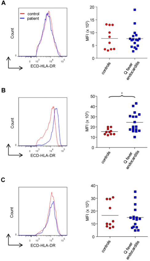
Figure 3. HLA-DR expression on monocyte subsets. The expression of HLA-DR on classical (A), intermediate (B) and non-classical (C) monocytes from 10 healthy controls and 17 patients with Q fever endocarditis was analyzed with flow cytometry. The level of HLA-DR expression was expressed as MFI. The nonparametric Mann-Whitney U test was used to compare the patient and control groups. *p,0.05. Horizontal bar, median value. doi:10.1371/journal.pone.0107533.g003
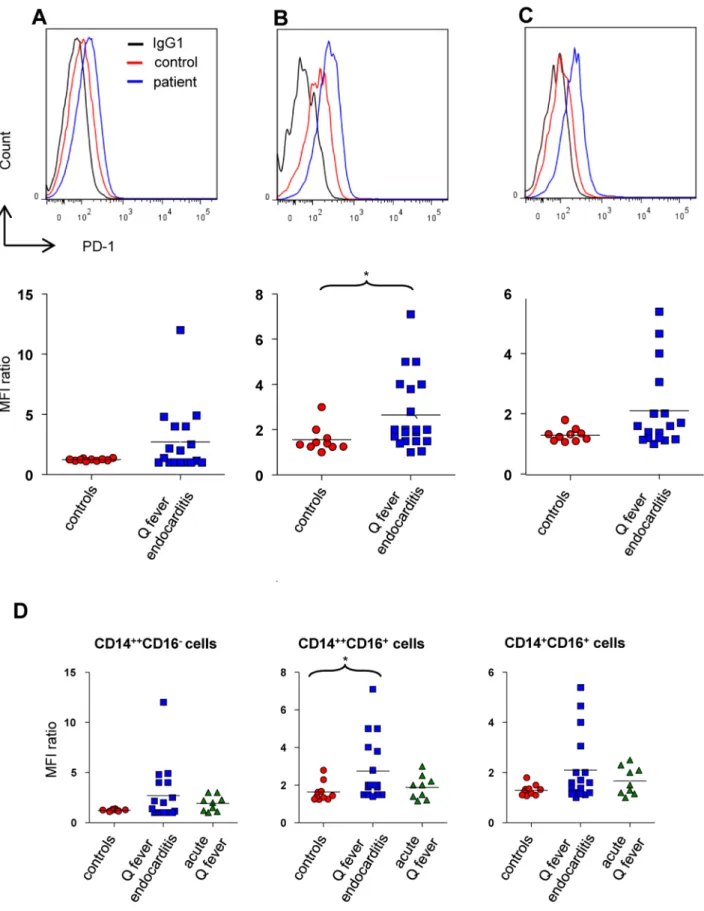
Figure 4. PD-1 expression on monocyte subsets. The expression of PD-1 on classical (A, D), intermediate (B, D), non-classical (C, D) from 10 healthy control, 17 patients with Q fever endocarditis and 9 patients with acute Q fever was analyzed with flow cytometry. The expression of PD-1 in the monocyte subsets is presented for each individual as the ratio of MFIPD-1to MFIisotypic control. The nonparametric Mann-Whitney U test was used to
compare the patient and control groups. *p,0.05. Horizontal bar, median value. doi:10.1371/journal.pone.0107533.g004
Statistical analysis
Data were analyzed using the Mann-WhitneyU test to compare healthy donors and patients with Q fever endocarditis or the Student’st-test to analyze in vitro data. The results are presented as the median or the mean 6 SD, and a p value ,0.05 was considered statistically significant. The relationship between specific Ab levels and the distribution of immune cells expressing or not PD-1 was analyzed with the Spearman correlation coefficient.
Results
CD16+monocytes and Treg cells are affected in Q fever endocarditis
The myeloid compartment was defined as the population of PBMCs that expressed HLA-DR but not the markers of T cells (CD3), B cells (CD19) or NK cells (CD56) (Figure 1A). Using CD14 and CD16 mAbs, we analyzed the distribution of monocyte subsets in controls and patients with Q fever endocarditis after the exclusion of debris, doublets and dead cells. The blood monocytes (5–15% of the total number of PBMCs) were divided into three
different subsets (Figure 1A), as previously described [23]. Approximately 90% of control monocytes expressed high levels of CD14 but not CD16 (CD14++CD162monocytes, also called classical monocytes); approximately 5% of monocytes expressed lower levels of CD14 but high levels of CD16 (CD14+CD16+ monocytes, also called non-classical monocytes). The remaining monocytes expressed high levels of CD14 and CD16 (CD14++CD16+ monocytes, also known as intermediate mono-cytes). We observed that the total number of monocytes was similar in healthy controls and patients with Q fever endocarditis (approximately 12% of the total number of PBMCs). However, while classical monocytes were similarly represented in controls and patients with Q fever endocarditis, the percentage (Fig-ure 1B) and the total count (Fig(Fig-ure S1A) of intermediate and non-classical monocytes was significantly (p,0.05) lower in patients with Q fever endocarditis than in controls. In parallel, we measured the proportion of CD3+, CD4+, CD8+T cells and Treg cells in patients with Q fever endocarditis. With the exception of Treg cells that were increased in Q fever endocarditis, the proportion of CD3+, CD4+and CD8+T cells was similar in patients and controls (Figure 2). Taken together, these results show that the minor monocyte subsets and the Treg population were affected in Q fever endocarditis.
HLA-DR expression on monocyte subsets in Q fever endocarditis
As intermediate and non-classical monocytes were found to be modulated in Q fever endocarditis, we wondered if this modulation is related to their activation, as determined by the expression of HLA-DR. Although the expression of HLA-DR on classical (Figure 3A) and non-classical (Figure 3C) monocytes was similar in patients and healthy donors, HLA-DR expression was significantly increased on intermediate monocytes in Q fever endocarditis (Figure 3B). Hence, the increased expression of HLA-DR on intermediate monocytes cannot account for the decreased number of non-classical monocytes. Taken together, these results suggest that the expression of HLA-DR is modestly affected in Q fever endocarditis and is not related to the modulation of the monocyte subsets.
PD-1 molecules are modulated on intermediate monocytes and CD4+T cells
PD-1 is associated with the chronic evolution of infectious diseases [17] and with IL-10 overproduction in HIV infection [18]. Because the overproduction of IL-10 is observed in Q fever endocarditis [10], we hypothesized that the expression of PD-1 by monocyte subsets was affected in Q fever endocarditis. The expression of PD-1 was not modulated in classical monocytes (Figure 4A), but it was significantly (p,0.05) increased in intermediate monocytes from patients with Q fever endocarditis (Figure 4B). It is noteworthy that the modulation of PD-1 expression by non-classical monocytes was close to that of intermediate monocytes although non-significant (Figure 4C). To assess the role ofC. burnetii infection in PD-1 expression, we measured the expression of PD-1 by monocytes from patients with acute Q fever. It was similar in the three monocyte subsets and was not different from that of controls (Figure 4D). These results suggest that the overexpression of PD-1 on CD16+monocytes is not a direct consequence of C. burnetii infection. We then determined the specificity of PD-1 overexpression by studying the expression of PD-1 on T cell subsets from controls and patients with Q fever endocarditis. The expression of PD-1 was similar on CD8+T cells from patients with Q fever endocarditis and controls,
Figure 5. PD-1 expression on T lymphocyte subsets. The expression of PD-1 on T lymphocytes (A), CD4+(B), CD8+(C) T cells
and Treg cells (D) from 11 healthy controls and 14 patients with Q fever endocarditis was analyzed with flow cytometry. The expression of PD-1 is presented for each individual as the percentage of PD-1-positive cells. The nonparametric Mann-Whitney U test was used to compare the patient and control groups. **p,0.005, ***p,0.0005. Horizontal bar, median value.
but was significantly higher on CD3+, CD4+T cells and Treg cells from patients than on control T cells (Figure 5). Hence, the modulation of PD-1 expression is not restricted to CD16+ monocytes but also involves CD4+ T cells, suggesting a global alteration of immune response in Q fever endocarditis.
Up-regulated expression of PD-1 depends on C. burnetii and IL-10
Because clinical features suggest that the modulation of PD-1 expression is not a direct consequence ofC. burnetii infection, we used anin vitro approach to test this hypothesis. To explore that further, PBMCs from healthy donors were incubated with living or heat-inactivated C. burnetii for 24 hours, and the expression of PD-1 by gated monocytes was assessed by flow cytometry. The expression of PD-1 by monocytes was dramatically increased after C. burnetii stimulation (Figure 6A). The responses of monocytes stimulated with heat-inactivated or living (Figure S1B)C. burnetii were not significantly different. We then isolated circulating monocytes by positive selection and stimulated them with
heat-inactivatedC. burnetii. The expression of PD-1 by monocytes was clearly up-regulated in response to C. burnetii (Figure 6B). Hence, in vitro C. burnetii infection may account for PD-1 modulation. As LPS is a major virulence factor ofC. burnetii [24], we investigated the putative role of LPS in PD-1 modulation. We observed that the E. coli LPS as positive control increased the expression of PD-1 on monocytes but that LPS from phase IC. burnetii did not modulate the expression of PD-1 by monocytes (Figure 6, A and B). This result demonstrates thatC. burnetii modulated the expression of PD-1 independently of its LPS, suggesting thatC. burnetii may affect PD-1 expression through an indirect mechanism.
As Q fever endocarditis is associated with the overproduction of IL-10 (Figure 7A and [10]), we wondered whether the overex-pression of PD-1 observed in Q fever endocarditis is related to IL-10. First,C. burnetii induced the release of IL-10 by monocytes from healthy controls (Figure 7B). Second, IL-10 dramatically increased the expression of PD-1 by monocytes from healthy controls. Third, the overexpression of PD-1 induced byC. burnetii is completely inhibited when monocytes were treated with
Figure 6. Modulation of PD-1 byC. burnetiiin monocytes. PBMCs or isolated monocytes from healthy subjects (106cells/ml) were stimulated with heat-inactivated C. burnetii (bacterium-to-cell ratio of 50:1), E. coli or phase I C. burnetii LPS (10 mg/ml) for 24 hours. The expression of PD-1 by gated monocytes (A) or isolated monocytes (B) was assessed by flow cytometry. The Student’s t-test was used to analyze data, and the values represent the mean 6 SD of three independent experiments.
neutralizing anti-IL-10 mAbs before infection with C. burnetii (Figure 7C). Taken together, these results suggest that the elevated levels of IL-10 observed in Q fever endocarditis may be responsible for the up-regulation of PD-1 in monocytes.
Discussion
The mechanisms of infective endocarditis involve the local and systemic host response to bacterial aggression. Locally, the interaction of coagulation factors and inflammatory cells leads to tissue reorganization and vegetation formation [25]. Systemic host responses bring together the activation of innate immune cells and inflammatory pathways [26]. Among types of infective endocar-ditis, Q fever endocarditis exhibits specific features, including fibrosis, calcification of the valves, low-grade inflammation and small or absent vegetations [27], suggesting that the mechanisms involved in Q fever endocarditis are likely distinct from those
governing pyogenic endocarditis. Hence, we previously provided evidence that Q fever endocarditis is characterized by IL-10-based immunosuppression [6]. Because C. burnetii survives and alters monocyte functions [6,28], we wondered here whether the immunosuppression associated with Q fever endocarditis may be related to quantitative changes in the populations of circulating monocytes. Based on the expression of CD14 and CD16, we observed three monocyte subsets in Q fever endocarditis, as reported in healthy controls [28]. The population of classical monocytes was not affected in Q fever endocarditis. In contrast, the non-classical and intermediate monocyte subsets were decreased in patients with Q fever endocarditis compared with healthy donors. This result is unexpected because the monocyte subsets that express CD16 (non-classical and/or intermediate monocytes) are increased in patients chronically infected with HIV [29] and in the febrile and defervescent phases of dengue infection [30]. The decreased number of specific monocyte subsets in Q
Figure 7. IL-10 and PD-1 expression in Q fever endocarditis. A, PBMCs (106cells/ml) from controls and patients with Q fever were incubated for 24 hours. Cell supernatants were collected and assayed for the presence of IL-10 by immunoassays.B, Monocytes from controls (106cells/ml)
were incubated with heat-inactivated C. burnetii (bacterium-to-cell ratio, 50:1) for 24 hours. Cell supernatants were collected and assayed for the presence of IL-10. The production of IL-10 was expressed in ng/ml. C, Monocytes from controls were incubated with IL-10 (10 mg/ml), heat-inactivated C. burnetii or C. burnetii+ anti-IL-10 mAbs (10 mg/ml) for 18 hours. The expression of PD-1 was assessed by flow cytometry, and the result was representative of three different experiments.
fever endocarditis was not a consequence of a global decrease in circulating immune cells because CD4+and CD8+T cells were not affected whereas CD4+ Tregs were increased. The decrease in non-classical and intermediate monocytes might be the conse-quence of their redistribution toward tissues. Indeed, it has been recently reported that non-classical and intermediate monocytes are increased in obese subjects and that weight loss leads to their decrease, likely associated with tissue migration [31]. We previously reported that circulating levels of MCP-1 known to control monocyte traffic is increased in patients with Q fever endocarditis [32]. Nevertheless, leukocytes from these patients exhibit defective trans-endothelial migration that is corrected by neutralizing anti-IL-10 Abs [33]. Alternatively, an increase in apoptosis may account for the decreased number of CD16+ monocytes. We found that CD16+monocytes were more prone to apoptosis than CD162 monocytes (Figure S2A). Whether the apoptosis variations are sufficient to account for CD16+monocyte decrease in blood circulation remains to be determined. Despite the lack of evidence for the mechanism of monocyte modulation, the upregulation of HLA-DR in intermediate monocytes from patients with Q fever endocarditis strengthened the hypothesis that circulating monocytes are activated in Q fever endocarditis.
The second feature of monocyte alteration in Q fever endocarditis is the increased expression of PD-1 on CD16+ monocytes. Note that no correlation was found between the expression of PD-1 and the duration of treatment of patients with Q fever endocarditis. As PD-1 was modulated in intermediate and, to a lesser degree, in non-classical monocytes during Q fever endocarditis, we wondered whether the modulation of PD-1 is a direct consequence of C.burnetii infection. We observed that C. burnetii efficiently increased the expression of PD-1 in monocytes isolated from healthy controls. The modulation of PD-1 expression byC. burnetii was independent of bacterial LPS, although it has been recently demonstrated that LPS is involved in C. burnetii pathogenicity [34]. We cannot exclude that other bacterial factors are involved inC. burnetii infection [35]. As PD-1 and IL-10 are up-regulated in HIV infection [18], we wondered whether a similar process occurs in Q fever endocarditis. We observed that IL-10 was sufficient to induce PD-1 expression by monocytes and that the neutralization of IL-10 inC. burnetii-infected monocytes prevented the bacterial stimulation of PD-1 expression. This is reminiscent of mouse models of sepsis in which the lack of PD-1 is associated with protection and low production of IL-10 [36]. In addition, we found that CD16+monocytes from patients with Q fever endocarditis produced less IL-10 than CD162 monocytes (Figure S2B). It is likely that the activation state of CD16+ monocytes accounts for this lower production of an immunoreg-ulatory cytokine such as IL-10. The blockade of PD-1/PD-L1 interactions restores the immune response during HIV infection [17,37]. Several evidence support that IL-10 and PD-1 constitute an amplification loop leading to immune impairment. Hence, the PD-1 expression is increased in monocytes during HIV infection and the triggering of monocyte-expressed PD-1 induces IL-10 production, leading to CD4+T cell dysfunction [17,18].In vitro neutralization of IL-10 activity in PBMCs from humans infected with HIV or hepatitis C virus restores the activity of non-responsive T cells [38,39].
Finally, we demonstrated that the up-regulation of PD-1 was also observed in CD4+ T cells from patients with Q fever endocarditis. This increased expression of PD-1 was specific of CD4+T cells because no modulation of PD-1 was found on CD8+ T cells from patients. It is noteworthy that the number of Treg cells was increased in Q fever endocarditis and that Treg cells overexpressed PD-1, suggesting an amplification loop to control
immune cell responsiveness. In addition, it is likely that the alteration of immune response in Q fever endocarditis requires a cross-talk between monocytes and T cells expressing PD-1. We suppose that the expression of PD-1 may reflect the degree of altered immune response. Hence, we found a positive correlation between the level of PD-1 expressed by CD4+T cells and anti-C. burnetii Abs, which are sensitive markers of Q fever evolution. The lack of correlation between PD-1-overexpressing monocytes and anti-C. burnetii Abs may be explained by the small size of patient samples and the low proportion of circulating CD16+ monocytes (Figure S3).
In summary, our results demonstrated that the number of intermediate and non-classical monocytes was decreased in Q fever endocarditis. We also showed that the overexpression of PD-1 by intermediate monocytes was related to IL-PD-10 production. The alteration of monocyte subsets was associated with the overex-pression of PD-1 by CD4+ T cells that may cooperate with monocytes to the immunosuppression of Q fever endocarditis. The determination of PD-1 expression may be useful to follow the immune deficiency in Q fever endocarditis.
Supporting Information
Figure S1 Monocyte subset counts and stimulation with
living C. burnetii. A, PBMCs (26106 cells) from 10 healthy controls and 17 patients with Q fever endocarditis were were labaled with specific Ab and 106events were analyzed with flow cytometry. Cell counts represent total events measured by flow cytometry. The total number of monocyte subsets is presented for each individual. The nonparametric Mann-Whitney U test was used to compare the patient and control groups. ***p,0.0005. Horizontal bar, median value. B, PBMCs from four healthy subjects (106cells/ml) were stimulated with heat-inactivated C. burnetii or living C. burnetii (bacterium-to-cell ratio of 50:1) for 24 hours. The expression of PD-1 by gated monocytes and Treg cells was assessed by flow cytometry. The release of IL-10 by PBMCs was measured by immunoassay. The nonparametric Mann-WhitneyU test was used to compare the unstimulated and stimulated cells. *p,0.05.
(TIF)
Figure S2 Monocyte subsets, apoptosis and IL-10. A,
PBMCs from 5 patients with Q fever endocarditis were gated according the expression of CD14 and CD16. The annexin V expression on CD162and CD16+monocytes was assessed by flow cytometry. The expression of annexin V is presented as the ratio of MFIassay to MFIisotypic control. B, CD162and CD16+monocytes
from 2 patients with Q fever endocarditis were sorted by flow cytometry and stimulated with heat-inactivated C. burnetii (bacterium-to-cell ratio of 50:1) for 24 hours. The release of IL-10 in supernatants was assessed by immunoassay.
(TIF)
Figure S3 Correlation of PD-1 expression with specific
Ab levels. The PD-1 expression on monocyte and lymphocyte subsets from patients with Q fever endocarditis (n = 17) were analyzed by flow cytometry. The circulating levels of anti-phase I C. burnetii Abs (IgG) were determined by indirect immunofluo-rescence. A, The phenotypic expression of PD-1 on monocyte subsets is presented for each patient with Q fever endocarditis as the ratio of MFIPD-1to MFIisotypic controland plotted against the
levels of of specific Abs. The correlation between specific Ab levels and the expression of PD-1 on monocytes was analyzed with the Spearman correlation coefficient. Only IgG levels and PD-1 expression on CD14+CD16+monocytes are correlated. Note that
the expressions of PD-1 by CD14++CD16+monocytes and Treg are not correlated (right).B, The phenotypic expression of PD-1 on T cell subsets is presented for each patient with Q fever endocarditis as the percentage of PD-1-positive cells. The correlation between the expression of PD-1 on T cells and anti-phase I C. burnetii Ab level was analyzed with the Spearman correlation coefficient. Only IgG levels and PD-1 expression on CD3 T cells and CD4 T cells are correlated.
(TIF)
Table S1 List of fluorescent reagents. AF, Alexa Fluor;
(DOCX)
Author Contributions
Conceived and designed the experiments: DO JLM. Performed the experiments: MBK FGR. Analyzed the data: CC JT FGR MM DR. Contributed reagents/materials/analysis tools: JT MM DR. Wrote the paper: MBK CC DO JLM.
References
1. Raoult D, Marrie T, Mege JL (2005) Natural history and pathophysiology of Q fever. Lancet Infect Dis 5: 219–226.
2. Fenollar F, Lepidi H, Raoult D (2001) Whipple’s endocarditis: review of the literature and comparisons with Q fever,Bartonella infection, and blood culture-positive endocarditis. Clin Infect Dis 33: 1309–1316.
3. Houpikian P, Raoult D (2005) Blood culture-negative endocarditis in a reference center: etiologic diagnosis of 348 cases. Medicine (Baltimore) 84: 162–173. 4. Million M, Walter G, Thuny F, Habib G, Raoult D (2013) Evolution from acute
Q fever to endocarditis is associated with underlying valvulopathy and age and can be prevented by prolonged antibiotic treatment. Clin Infect Dis 57: 836– 844.
5. Capo C, Mege JL (2012) Role of innate and adaptive immunity in the control of Q fever. Adv Exp Med Biol 984: 273–286.
6. Ben Amara A, Bechah Y, Mege JL (2012) Immune response and Coxiella burnetii invasion. Adv Exp Med Biol 984: 287–298.
7. Ghigo E, Capo C, Tung CH, Raoult D, Gorvel JP, et al. (2002)Coxiella burnetii survival in THP-1 monocytes involves the impairment of phagosome maturation: IFN-c mediates its restoration and bacterial killing. J Immunol 169: 4488–4495.
8. Benoit M, Desnues B, Mege JL (2008) Macrophage polarization in bacterial infections. J Immunol 181: 3733–3739.
9. Sawyer LA, Fishbein DB, McDade JE (1987) Q fever: current concepts. Rev Infect Dis 9: 935–946.
10. Honstettre A, Imbert G, Ghigo E, Gouriet F, Capo C, et al. (2003) Dysregulation of cytokines in acute Q fever: role of interleukin-10 and tumor necrosis factor in chronic evolution of Q fever. J Infect Dis 187: 956–962. 11. Capo C, Amirayan N, Ghigo E, Raoult D, Mege JL (1999) Circulating cytokine
balance and activation markers of leucocytes in Q fever. Clin Exp Immunol 115: 120–123.
12. Layez C, Brunet C, Lepolard C, Ghigo E, Capo C, et al. (2012) Foxp3+CD4+CD25+regulatory T cells are increased in patients withCoxiella burnetii endocarditis. FEMS Immunol Med Microbiol 64: 137–139. 13. Chen L (2004) Co-inhibitory molecules of the B7-CD28 family in the control of
T-cell immunity. Nat Rev Immunol 4: 336–347.
14. Greenwald RJ, Freeman GJ, Sharpe AH (2005) The B7 family revisited. Annu Rev Immunol 23: 515–548.
15. Parry RV, Chemnitz JM, Frauwirth KA, Lanfranco AR, Braunstein I, et al. (2005) CTLA-4 and PD-1 receptors inhibit T-cell activation by distinct mechanisms. Mol Cell Biol 25: 9543–9553.
16. Freeman GJ, Wherry EJ, Ahmed R, Sharpe AH (2006) Reinvigorating exhausted HIV-specific T cells via PD-1-PD-1 ligand blockade. J Exp Med 203: 2223–2227.
17. Day CL, Kaufmann DE, Kiepiela P, Brown JA, Moodley ES, et al. (2006) PD-1 expression on HIV-specific T cells is associated with T-cell exhaustion and disease progression. Nature 443: 350–354.
18. Said EA, Dupuy FP, Trautmann L, Zhang Y, Shi Y, et al. (2010) Programmed death-1-induced interleukin-10 production by monocytes impairs CD4+T cell
activation during HIV infection. Nat Med 16: 452–459.
19. Maecker HT, McCoy JP, Nussenblatt R (2012) Standardizing immunopheno-typing for the Human Immunology Project. Nat Rev Immunol 12: 191–200. 20. Raoult D (2012) Chronic Q fever: expert opinion versus literature analysis and
consensus. J Infect 65: 102–108.
21. Gondois-Rey F, Granjeaud S, Kieu Sle T, Herrera D, Hirsch I, et al. (2012) Multiparametric cytometry for exploration of complex cellular dynamics. Cytometry A 81: 332–342.
22. Capo C, Lindberg FP, Meconi S, Zaffran Y, Tardei G, et al. (1999) Subversion of monocyte functions byCoxiella burnetii: impairment of the cross-talk between avb3 integrin and CR3. J Immunol 163: 6078–6085.
23. Passlick B, Flieger D, Ziegler-Heitbrock HW (1989) Identification and characterization of a novel monocyte subpopulation in human peripheral blood. Blood 74: 2527–2534.
24. Narasaki CT, Toman R (2012) Lipopolysaccharide ofCoxiella burnetii. Adv Exp Med Biol 984: 65–90.
25. Benoit M, Thuny F, Le Priol Y, Lepidi H, Bastonero S, et al. (2010) The transcriptional programme of human heart valves reveals the natural history of infective endocarditis. PLoS One 5: e8939.
26. Thuny F, Textoris J, Ben Amara A, Filali AE, Capo C, et al. (2012) The gene expression analysis of blood reveals S100A11 and AQP9 as potential biomarkers of infective endocarditis. PLoS One 7: e31490.
27. Lepidi H, Houpikian P, Liang Z, Raoult D (2003) Cardiac valves in patients with Q fever endocarditis: microbiological, molecular, and histologic studies. J Infect Dis 187: 1097–1106.
28. Ziegler-Heitbrock L, Ancuta P, Crowe S, Dalod M, Grau V, et al. (2010) Nomenclature of monocytes and dendritic cells in blood. Blood 116: e74–80. 29. Thieblemont N, Weiss L, Sadeghi HM, Estcourt C, Haeffner-Cavaillon N (1995)
CD14low
CD16high
: a cytokine-producing monocyte subset which expands during human immunodeficiency virus infection. Eur J Immunol 25: 3418–3424. 30. Azeredo EL, Neves-Souza PC, Alvarenga AR, Reis SR, Torrentes-Carvalho A,
et al. (2010) Differential regulation of toll-like receptor-2, toll-like receptor-4, CD16 and human leucocyte antigen-DR on peripheral blood monocytes during mild and severe dengue fever. Immunology 130: 202–216.
31. Poitou C, Dalmas E, Renovato M, Benhamo V, Hajduch F, et al. (2011) CD14dim
CD16+and CD14+CD16+monocytes in obesity and during weight loss: relationships with fat mass and subclinical atherosclerosis. Arterioscler Thromb Vasc Biol 31: 2322–2330.
32. Meghari S, Desnues B, Capo C, Grau GE, Raoult D, et al. (2006)Coxiella burnetii stimulates production of RANTES and MCP-1 by mononuclear cells: modulation by adhesion to endothelial cells and its implication in Q fever. Eur Cytokine Netw 17: 253–259.
33. Meghari S, Capo C, Raoult D, Mege JL (2006) Deficient transendothelial migration of leukocytes in Q fever: the role played by interleukin-10. J Infect Dis 194: 365–369.
34. Barry AO, Boucherit N, Mottola G, Vadovic P, Trouplin V, et al. (2012) Impaired stimulation of p38alpha-MAPK/Vps41-HOPS by LPS from patho-genicCoxiella burnetii prevents trafficking to microbicidal phagolysosomes. Cell Host Microbe 12: 751–763.
35. van Schaik EJ, Chen C, Mertens K, Weber MM, Samuel JE (2013) Molecular pathogenesis of the obligate intracellular bacteriumCoxiella burnetii. Nat Rev Microbiol 11: 561–573.
36. Huang X, Venet F, Wang YL, Lepape A, Yuan Z, et al. (2009) PD-1 expression by macrophages plays a pathologic role in altering microbial clearance and the innate inflammatory response to sepsis. Proc Natl Acad Sci U S A 106: 6303– 6308.
37. Brooks DG, Trifilo MJ, Edelmann KH, Teyton L, McGavern DB, et al. (2006) Interleukin-10 determines viral clearance or persistence in vivo. Nat Med 12: 1301–1309.
38. Clerici M, Wynn TA, Berzofsky JA, Blatt SP, Hendrix CW, et al. (1994) Role of interleukin-10 in T helper cell dysfunction in asymptomatic individuals infected with the human immunodeficiency virus. J Clin Invest 93: 768–775. 39. Rigopoulou EI, Abbott WG, Haigh P, Naoumov NV (2005) Blocking of
interleukin-10 receptor - A novel approach to stimulate T-helper cell type 1 responses to hepatitis C virus. Clin Immunol 117: 57–64.
fluorescein isothiocyanate; PB, Pacific blue; PC, phycoerythrin; PerCP-Cy5.5, peridinin chlorophyll protein-cyanin 5.5; PE, Phycoerythrin