Diversity of the Marine Cyanobacterium Trichodesmium:
Characterization of the Woods Hole Culture Collection
and Quantification of Field Populations
by
OF TEAnnette Michelle Hynes
SEP
B.S., University of Nebraska-Lincoln (1998)
Submitted to the Department of Biology
LIBI
in partial fulfillment of the requirements for the degree of
Doctor of Philosophy in Biological Oceanography
at the
MASSACHUSETTS INSTITUTE OF TECHNOLOGY
ARC
and the
WOODS HOLE OCEANOGRAPHIC INSTITUTION
September 2009
©
Annette Michelle Hynes, MMIX. All rights reserved.
The author hereby grants to MIT and WHOI permission to reproduce and
distribute publicly paper and electronic copies of this thesis document in
whole or in part.
CHNOLOGY2 12009
RARIES
HIVES
Author
.
.,
Department of Biology
August 28, 2009
Certified by
Scott C. Doney
Senior Scientist
Thesis Supervisor
Certified by
Accepted by...
U
1
Chair, Joint Committee for
John B. Waterbury
Scientist Emeritus
Thesis Supervisor
Simon Thorrold
Biological Oceanography
Diversity of the Marine Cyanobacterium Trichodesmium:
Characterization of the Woods Hole Culture Collection and
Quantification of Field Populations
by
Annette Michelle Hynes
Submitted to the Department of Biology on August 28, 2009, in partial fulfillment of the
requirements for the degree of
Doctor of Philosophy in Biological Oceanography
Abstract. Trichodesmium is a colonial, N2-fixing cyanobacterium found in tropical
oceans. Species of Trichodesmium are genetically similar but several species exist to-gether in the same waters. In order to coexist, Trichodesmium spp. may occupy differ-ent niche spaces through differdiffer-ential utilization of resources such as nutridiffer-ents and light, and through responses to physical characteristics such as temperature and turbulence. To investigate niche differentiation in Trichodesmium, I characterized cultured strains of Tri-chodesmium, identified and enumerated Trichodesmium clades in the field, and investi-gated P stress and N2 fixation in field populations. Species of Trichodesmium grouped into two clades based on sequences from 16S rDNA, the internal transcribed spacer (ITS), and the heterocyst differentiation gene hetR. Clade I contained Trichodesmium erythraeum
and Trichodesmium contortum, and clade II contained Trichodesmium thiebautii,
Tri-chodesmium tenue, TriTri-chodesmium hildebrandtii, and TriTri-chodesmium pelagicum. Each clade was morphologically diverse, but species within each clade had similar pigmen-tation. I developed a quantitative polymerase chain reaction (qPCR) method to distin-guish between these two clades. In field populations of the Atlantic and Pacific Oceans, the qPCR method revealed that clade II Trichodesmium spp. were more prominent than clade I in the open ocean. Concentrations of Trichodesmium did not correlate with nutri-ent concnutri-entrations, but clade I had wider temperature and depth distributions than clade II. Temperature and light are physical characteristics that may define niche spaces for species of Trichodesmium. Clade I and II concentrations correlated with each other in the Pacific but not in the Atlantic, indicating that the two clades were limited by the same fac-tors in the Pacific while different facfac-tors were limiting the abundance of the two clades in the Atlantic. Trichodesmium populations in the North Atlantic were more P stressed and had higher N2 fixation rates than populations in the western Pacific. While nutrient
con-centrations didn't directly correlate with Trichodesmium concon-centrations, the contrasting nutrient regimes found in the Atlantic and Pacific Oceans might influence distributions of the two clades differently. Unraveling the differences among species of Trichodesmium
begins to explain their coexistence and enables us to understand factors controlling global
N2 fixation.
Thesis Supervisor: Scott C. Doney Title: Senior Scientist
Thesis Supervisor: John B. Waterbury Title: Scientist Emeritus
Dedication
I dedicate this thesis to my students from the Kiundwani Secondary School class of 2003, Machakos District, Kenya.
"Elimu maisha si vitabu." (Methali za Kiswahili) "Education is life, not books." (Swahili proverb)
Acknowledgements
Throughout my graduate career, I have been supported by the National Science Foun-dation (NSF) Biocomplexity Program Grant (OCE-0323332); the Center for Microbial Oceanography Research and Education (C-MORE), an NSF Science and Technology Center (EF-0424599); the Woods Hole Oceanographic Institution (WHOI) Ocean Life Institute (OLI) grant to J. Waterbury, and the WHOI Academic Programs Office.
I would like to thank my advisors, Scott Doney and John Waterbury of WHOI. Scott has supported me from day one as a scientist and as a human. While we haven't been able to complete the modeling we originally set out to do, he has been invaluable in helping me to view my data from new angles. John was very generous in taking me in when I needed a new lab and in sharing with me his skill and patience in culturing the fickle and wonderful cyanobacteria.
My committee members have been indispensable with their molecular expertise: Eric Webb of the University of Southern California (USC), Tracy Mincer of WHOI, and Edward DeLong of the Massachusetts Institute of Technology (MIT). Having Eric at WHOI was like having a bonus advisor, and his enthusiasm for science is infectious, even over the phone to USC. Tracy was a highly welcome arrival at WHOI, and on many occa-sions he has saved me from weeks of banging my head against a problem with qPCR. Ed had the benefit of being more removed from my project and could see the larger picture of my challenges with wisdom and a sense of humor. Judith McDowell (WHOI) served as the chair of both my thesis proposal defense and my thesis defense, and I thank Judy for keeping everybody in line and for serving as a role model for me.
I am eternally grateful to the administrative assistants, the Academic Programs Office at WHOI, and the Joint Program Office at MIT for making it easier to dot the i's and cross the t's, and for giving their assistance with genuine love and concern.
I would like to thank my plethora of WHOI labmates over the years, who have been helpful with their myriad of knowledge: P. Dreux Chappell, Sarah Cooley, Ian
Ehren-reich, Sheean Haley, Whitney Krey, Justin Ladner, Naomi Levine, Emily Lorch, Misty Miller, Elizabeth Orchard, Adam Rivers, Emmanuel Vaisquez-Rivera, Brian Wilson, and Louie Wurch. I especially want to thank Freddy Valois, whose skills are always in high demand, and Nan Trowbridge, who made everything run smoothly (usually by greasing the wheels with baked goods).
I am indebted to Dave Glover for graciously taking the MATLAB and IBTEXchallenges I have brought before him, Tim Shank for allowing me to use his thermal cycler and his labspace for sequencing, Rhian Waller (currently University of Hawai'i (UH)) for teach-ing me the ropes of sequencteach-ing, Rebecca Gast for allowteach-ing me to make an obnoxious amount of noise in her laboratory by using her beadbeater, Sonya Dyhrman for being my initial biology advisor, Heidi Sosik for her advice on image processing, the residents of Watson Laboratory for rampant sharing of equipment and ideas, and Mark Dennett and Alexi Shalopyonok for teaching me flow cytometery in my early days of working with
Crocosphaera, all from WHOI. I would like to thank Penny Chisholm and Ed DeLong for the use of their thermal cycler and robot at MIT where I did the majority of my qPCR,
and Rex Malmstrom, who was very patient and generous with his time in teaching me to use these machines.
This thesis would not have been possible without the support of C-MORE. In ad-dition to funding, I have had access to a community of microbial ecologists from UH, Oregon State University (OSU), University of California-Santa Cruz (UCSC), Mon-terey Bay Aquarium Research Institute (MBARI), MIT, and WHOI with a wide variety of backgrounds and experiences. Graduate students, post-doctoral scholars, and principal investigators have all taken the time to share their expertise with me, especially to help me troubleshoot my technical problems or to provide field samples.
I want to acknowledge the members of "Team Tricho," fellow students and post-docs who study this finicky and fantastic organism Trichodesmium: P. Dreux Chappell (WHOI, currently University of Rhode Island), Carolyn Holl (Georgia Institute of Tech-nology, currently Oceanic Institute), Elizabeth Orchard (WHOI), Jill Sohm (USC), and
Angelicque White (OSU). I want to thank you for the countless conversations, your feed-back on ideas, your help on cruises, and your friendship. I especially want to thank Dreux who has been a dear friend and my biggest cheerleader through this whole PhD process.
Lastly, I want to thank my family, both born and chosen. Thank you, Kate Buck-man, for being my best friend, roommate, and comic relief. Thank you, Joe Confalone, for being my emotional support, my extra-curricular activity, and my occasional labora-tory assistant. And thank you, Mom, Pops, Jeannette, Lynnette, and Raennette, for your love, nurturing, and being my roots.
Contents
1 Introduction 19
Statement of Purpose ... ... . . . . . . 19
Ecological roles of Trichodesmium ... . ... . . 20
Species identifications and distributions ... ... 23
Com petition theory ... ... .... 26
Niche differentiation in cyanobacteria ... .... 29
Overview of thesis chapters ... ... ... 31
2 Diversity of the N2-fixing Cyanobacterium Trichodesmium: Characterization of the Woods Hole Culture Collection Introduction ... ... ... Identification of Trichodesmium . . . . Photosynthetic pigments ... Characterization of the Woods Hole culture collection. M ethods . . . .... Culture conditions ... ... M icrographs... ... Phycobiliprotein extraction and absorption spectra . . . Sequences and phylogeny ... Results . ... Genetic characterization of Trichodesmium ... .. .. .. . .. . 36 . . . . . 36 .. .. . .. .. . 37 . . . . . 40 .. .. . .. .. . 42 .. .. . .. .. . 42 .. . . .. .. . 42 . . . . . 42 .. .. .. . .. . 43
Cell morphology ... .. .. . .. ... .. 60
Absorption spectra of phycobiliproteins . ... 65
Discussion ... . ... . . ... ... ... .... 65
3 Development of a quantitative polymerase chain reaction (qPCR) assay to dis-tinguish the two clades of Trichodesmium 71 Introduction . .... . ... M ethods . . . .. . . .. Primer design and preparation of cloned qPCR standards Serial dilutions and cell counts of Trichodesmium cultures DNA extraction ... . . . ... Quantitative polymerase chain reaction (qPCR) . . . . Results . ... qPCR assay development . ... DNA extractions and serial dilutions ... Discussion . ... 4 The Distribution of Trichodesmium field populations Introduction . ... Methods . ... Sample collection, storage, and extraction ... Quantitative polymerase chain reaction (qPCR) ... .. 72 . . . .. . 74 . . . . . 74 . . . . . 76 .. .. . .... 77 . . . . . 79 Results ... . ... 94
Transect hydrography, nutrients, and qPCR . ... 94
Nutrients and Trichodesmium . ... ... 100
Temperature, depth and Trichodesmium . ... ... ... 100
Melting types, temperature, and depth . . . .. ... . 107
West Pacific warm pool diel cycle ... ... 110
5 Cross-basin comparison of phosphorus stress and nitrogen fixation in Trichodesmium 119
Introduction . . . . ... . .. .. . .. . . .. . ... ... .. .. .. . 120
New nitrogen ... ... 120
Phosphorus and iron ... ... 121
Alkaline phosphatase .. ... ... . ... 122
Methods ... ... . 123
Hydrological context ... ... .123
Collection of colonies ... .. ... .. ... 124
Enzyme-labeled fluorescence (ELF) ... .. 125
Nutrient Analysis .... ... .... ... ... 125
Nitrogenase activity ... .... . ... .126
Results . . . ... . .127
Physical conditions ... ... . 127
ELF ... ... 127
DIP and ELF ... ... ... 131
Nitrogenase activity . ... . ... ... 131 Discussion .. .. .... .. . ... . ... . ... . .. . . .. .. 135 6 Concluding remarks 143 Thesis summary .. ... .. . .. ... . . . ... . . . ... 143 Evolution of Trichodesmium ... ... 144 Future studies ... . ... ... ... 146 References 148
List of Figures
1-1 N2fixation and new production .
2-1 Dendrograms of previous c 2-2 Typical absorption peaks
16S neighbor-joining tree
16S parsimony tree . . . 16S maximum likelihood t ITS neighbor-joining tree ITS parsimony tree . . . ITS maximum likelihood hetR neighbor-joining tree hetR parsimony tree. . . hetR maximum likelihood Concatenated neighbor-joi Concatenated parsimony tr Concatenated maximum lii Micrographs of Trichodesn Micrographs of Trichodesr Phycobiliprotein absorptio classification of Trichodesmium . ... 39 .. .. .. .. . .. .. . .. .. ... . ... . 3 9 . . . . . . . . . 4 8 .. .. .. .. .. . .. .. .. .. .. . .. . 4 9 ree . . . 50 .. .. .. .. .. . .. .. . . .. .. . .. . 5 1 .. .. .. .. . . .. .. . .. .. .. . .. . 5 2 .. .. .. .. .. . .. .. . .. .. .. . .. . 5 3 . .. .. .. .. . .. .. . .. .. .. . .. .. 54 . . . . . 5 5 tree ... ... .. ... . 56 ning tree. . ... .. 57 ee... ... . ... 58 kelihood tree. . ... 59 mium clade I . ... . 62 mium clade II ... 64 n spectra ... .. 66
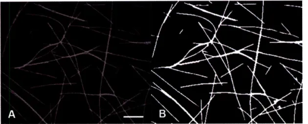
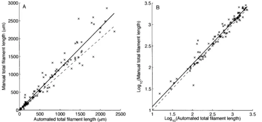
Image processing for automated cell counts Automated cell count method performance.
. . . . . 7 7 . . . . . . . . . 8 0 2-3 2-4 2-5 2-6 2-7 2-8 2-9 2-10 2-11 2-12 2-13 2-14 2-15 2-16 2-17 3-1 3-2
S... 82
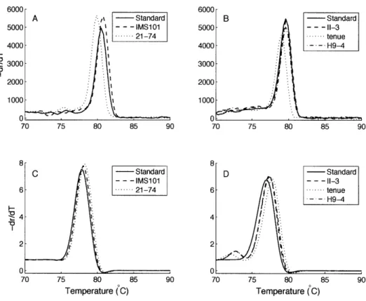
Melting curves . . . . . . .... Extraction method comparison . . . . Serial dilutions of Trichodesmium cultures Mixed culture qPCR results . . . . Maps of cruise tracks . . . .. Equatorial Atlantic data . . . .. West Pacific warm pool data . . . . South Pacific data . . . ... qPCR results versus cell counts . . . . . Clade II versus clade I . . . . Fe, P, and Trichodesmium . . . . Fe, P, and clade II:clade I . . . . Depth, temperature, and Trichodesmium Temperature and depth histograms . . . Depth, temperature, and clade II:clade I Depth of maximum concentration and mi Depth of maximum concentration histogr M elt curves ... ... qPCR product parsimony tree . . . . Histogram of temperature and depth for c Diel cycle for the west Pacific warm pool ... . 92 . ... . 96 ... . 97 . . ... . 98 .. . ... . 99 .. ... . 99 ... .. 100 ... .. 101 ... 102 ... .. 103 ... 104
xed layer depth ... 105
ams ... ... 106
... ... .... 107
... 108
lade I qPCR products ... 109
. . . ... . 111
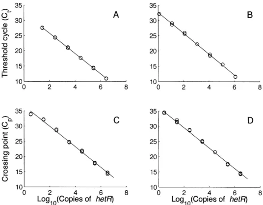
Maps of cruise tracks ... ... Hydrographic conditions of KM0701 ... ELF .. ... . ... Organisms associated with Trichodesmium colonies . . . . DIP histogram . . ... . . . 124 . . . . 127 . . . 130 . . . .13 1 . . . . 132 3-4 3-5 3-6 3-7 . . . 83 . . . 84 . . . 85 . . . 85 4-1 4-2 4-3 4-4 4-5 4-6 4-7 4-8 4-9 4-10 4-11 4-12 4-13 4-14 4-15 4-16 4-17 5-1 5-2 5-3 5-4 5-5 3-3 Standard curves .
List of Tables
1.1 Physical characteristics of Trichodesmium spp. . ... 24
1.2 Distribution of Trichodesmium spp ... ... . 25 2.1 Species names ... 38 2.2 Culture collection ... . ... 41 2.3 Summary of PCR primers. ... 44 2.4 Morphology summary ... ... 61 3.1 qPCR primers ... .. ... 74
5.1 ELF data summary ... .. .... ... 129
5.2 DIP ANOVA ... 132
CHAPTER 1
Introduction
Statement of Purpose
Trichodesmium is a colonial, N2-fixing cyanobacterium and a significant source of fixed
nitrogen (N) in oligotrophic oceans. Its roles in the biogeochemical cycling of N, phos-phorus (P), iron (Fe), and carbon (C) have made it the target of a wide variety of
physio-logical, ecophysio-logical, optical, and modeling studies. Trichodesmium spp. form colonies visi-ble to the naked eye, making field collections and observations easier than for many other phytoplankton. There is currently a genome available for Trichodesmium erythraeum
IMS 101, aiding genetic studies of this genus. Teasing out the factors that may lead to niche differentiation in Trichodesmium addresses Hutchinson's "paradox of the plank-ton," whereby many more species of plankton coexist than would be expected based on the number of limiting resources available (Hutchinson, 1961). Lessons learned from Trichodesmium can be applied to other species of phytoplankton which are not as well-studied.
There are several morphologically distinct but genetically similar species of Tri-chodesmium that coexist in tropical and subtropical waters. I hypothesized that the co-existence of several Trichodesmium species is due in part to niche differentiation. Tri-chodesmium spp. occupy different niche spaces to utilize resources such as nutrients and light and to respond to physical characteristics such as temperature and turbulence. To
explore the niche spaces occupied by Trichodesmium spp., I investigated cultured strains available in the Woods Hole culture collection and natural communities in the Atlantic and Pacific Oceans. I characterized cultured Trichodesmium strains by sequencing the het-erocyst differentiation gene hetR, 16S rDNA, and the internal transcribed spacer (ITS) re-gions; photographing and measuring trichomes (filaments); and analyzing the absorption
spectra of phycobiliproteins (PBP). I used the hetR sequences to develop a real-time quan-titative polymerase chain reaction (qPCR) method to identify and quantify the two major clades of Trichodesmium in cultures and field samples. I applied this qPCR method to
re-solve spatial distributions of Trichodesmium from samples collected with Niskin bottles across the Equatorial Atlantic, the west Pacific warm pool, and the South Pacific. Phos-phorus stress was assayed using enzyme-labeled fluorescence (ELF) of alkaline phos-phatase (AP) and N2 fixation was measured using acetylene reduction in Trichodesmium
populations across the west Pacific warm pool and the western North Atlantic.
Ecological roles of Trichodesmium
Cyanobacteria evolved 3500 Ma ago and dramatically changed the redox state of the Earth's atmosphere through oxygenic photosynthesis (Schopf, 2000). Also known as blue-green algae, cyanobacteria are found in both fresh and marine waters and a wide variety of terrestrial habitats. They are ubiquitous, prone to blooms, and can produce toxins (Cohen and Gurevitz, 2006). Trichodesmium spp. are filamentous, colonial, non-heterocystous, N2-fixing cyanobacteria belonging to the order Oscillatoriales (Waterbury,
2006). The genus name comes from the Greek root "trikh-" meaning "hair", referring to the filaments known as trichomes. Trichodesmium is found in tropical and subtropical oligotrophic waters with shallow mixed layer depths and warm temperatures.
Some cyanobacteria such as Trichodesmium are diazotrophs and contribute to new N inputs in oligotrophic systems (Capone et al., 1997). Fixed N is considered to be the proximal limiting nutrient in ocean ecosystems. For a system in steady-state, new produc-tion is stimulated by inputs of new N, resulting in excess producproduc-tion available for export
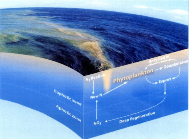
Figure 1-1: Trichodesmium and other diazotrophs fuel new production through N2
fixa-tion. Photo shows a surface slick of Trichodesmium in the western South Pacific Ocean. Inputs of new nitrogen stimulate the growth of phytoplankton. New production can be exported out of the euphotic zone, drawing down surface concentrations of carbon. Photo courtesy of Daniel Ohnemus. Illustration by E. Paul Oberlander, WHOI.
(Fig. 1-1) (Eppley and Peterson, 1979). New N comes from eddy diffusion processes, seasonal deep mixing, atmospheric deposition, lateral advection, and N2 fixation (Karl,
2002). In the North Atlantic, N2 fixation rates of Trichodesmium can equal or exceed the
vertical flux of NO3 into surface waters (Capone et al., 2005), while in the North Pacific,
N2 fixation is the source of up to half of the new N (Karl et al., 1997).
Trichodesmium is also a significant contributor of fixed C, through its own pho-tosynthesis and through stimulation of primary production of other organisms (Fig. 1-1). Trichodesmium can account for 8-47% of total primary production in the tropical North Atlantic (Carpenter et al., 2004). In a global marine ecosystem model, the presence of
N2-fixers increased primary production by diatoms and small phytoplankton through the
excretion of dissolved organic nitrogen (DON) in N-limited areas (Moore et al., 2002; Moore and Doney, 2007). Model results by Coles et al. (2004) showed that N2 fixation
increases dissolved inorganic nirogen (DIN), phytoplankton biomass, primary production and export flux in the subtropics.
Factors affecting the growth of Trichodesmium include nutrients such as P and Fe, temperature, light intensity, mixed layer depth (MLD), and turbulence (Capone et al.,
1997; Post, 2005). The amelioration of N limitation through N2 fixation can cause a
draw-down of other nutrients such as P and Fe (Karl, 2002). The North Atlantic receives more dust input than the North Pacific, and the Fe available in terrestrial dust is necessary for the enzyme nitrogenase used in N2 fixation. Low excess N relative to excess P in
dis-solved nutrients is a necessary but not sufficient condition for selection for N2-fixing
or-ganisms in oligotrophic regions; in N-limited scenarios, dominant competitors such as diatoms cannot grow well and diazotrophs or diatoms with diazotrophic symbionts can succeed (Karl, 2002). When nutrient N:P < 16, the classical Redfield ratio, diazotrophs are able to compete, drawing down P while increasing N and making the system more P-limited (Karl, 2002). Trichodesmium spp. have been shown to be able to use organic sources of P (Dyhrman et al., 2006; Sohm and Capone, 2006) and are capable of lux-ury uptake of Fe and P (Kustka et al., 2003; White et al., 2006a). In a numerical model simulating vertical migration of Trichodesmium, colonies > 1000 pm were shown to be capable of P-mining: sinking by carbohydrate ballasting below the phosphocline, tak-ing up P, and then ristak-ing back up to the euphotic zone (White et al., 2006b). Differential adaptations to nutrient limitation including alternative nutrient sources, uptake rates, half-saturation constants, luxury uptake, and P-mining can lead to niche differentiation or co-existence of species limited by different resources (Titman, 1976).
In addition to its roles in N2 and C fixation, Trichodesmium provides substrate and
shelter. A variety of organisms ranging from bacteria to crustacean larvae are enriched by 2-5 orders of magnitude in association with Trichodesmium colonies (Sheridan et al.,
2002). The copepod Macrosetella gracilis uses Trichodesmium as a substrate for juve-nile development as well as a food source (O'Neil, 1998). Trichodesmium blooms can be undesirable in regions like the Gulf of Mexico, however, because they can produce
tox-ins and contribute little to the marine food web, being grazed upon by only a few pelagic harpacticoid copepods: M. gracilis, Miracia efferata, and Oculosetella gracilis (O'Neil
and Roman, 1994; Post, 2005).
Species identifications and distributions
There are six well-described species of Trichodesmium: T erythraeum, T thiebautii, chodesmium contortum, Trichodesmium tenue, Trichodesmium hildebrandtii and Tri-chodesmium pelagicum (formerly Katagnymene pelagica and Katagnymene spiralis).
Traditionally, species of filamentous, non-heterocystous cyanobacteria such as Trichodesmium were identified using morphological characteristics including cell width, cell length, fila-ment spirality, color, gas vacuoles, presence or absence of a sheath, and colony morphol-ogy (Table 1.1) (Janson et al., 1995; Orcutt et al., 2002; Waterbury, 2006). However, these characteristics can often prove to be problematic and many studies refer to field popu-lations merely as Trichodesmium spp. or identify them by colony morphology: spheri-cal "puffs," fusiform "rafts" or "tufts," and "bowties" (Janson et al., 1995). Morpholog-ical traits used to identify Trichodesmium spp. "correlate poorly" with genetic data, as shown by the clustering of the species formerly known as K. pelagica and K. spiralis among species of Trichodesmium (Lundgren et al., 2005). The different species of Tri-chodesmium are closely related. Sequences of the nitrogenase gene nifH, the heterocyst differentiation gene hetR, 16S rRNA, and the ITS region as well as fingerprinting of HIPI show low genetic diversity among Trichodesmium (Ben-Porath et al., 1993; Orcutt et al., 2002; Lundgren et al., 2005).
Table 1.1: Physical characteristics and colony morphologies of Trichodesmium spp.(Janson et al., 1995; Orcutt et al., 2002; Lundgren et al., 2005)
Species puff tuft bowtie trichomes Cell width Cell length Other
(pm) (pm) T aureum / 20-25 gold T contortum / 30-40 T. erythraeum , 1 6-9 4-10 red-brown T. hildebrandtii I / / 14-17 5-12 T pelagicum
K. pelagica / 15-35 straight trichome
K. spiralis / 12-23 4-7 coiled trichome
T tenue / / / 5-7 10-19
T thiebautii ,/ . 7-9 6-12
The distribution of Trichodesmium is influenced by temperature. Temperature sets an overall upper limit on growth rate (Eppley, 1972). Increased temperature within a vi-able range will increase enzyme activity, hence increasing cellular functions such as res-piration; photosynthesis, particularly the Calvin Cycle; and N2 fixation. In the eastern
At-lantic, Trichodesmium is found at highest densities between 0 - 150N, with a complete absence south of 300S (Tyrrell et al., 2003). Blooms are found in surface waters with temperatures > 250C, but trichomes can be found in deeper waters with temperatures of 21 - 230C (Karl, 2002).
While often found concurrently, the species of Trichodesmium have different dis-tributions. Table 1.2 gives an overview of the dominant species found in different ocean basins. While abundance varies in time and space, T thiebautii is generally most common in the Northern Hemisphere while T erythraeum is most common in the Great Barrier Reef (Table 1.2) (Orcutt and Gundersen, 2003; Bell et al., 2005). At the Bermuda Atlantic Time-series Station (BATS), T thiebautii puffs are present year-round, but during the summer maximum of Trichodesmium abundance, T thiebautii tufts are the predominant morphology, followed by T thiebautii puffs and T erythraeum tufts (Orcutt and Gunder-sen, 2003). In addition to temporal and horizontal spatial variation, colony morphologies show different vertical distributions. In the Gulf of Aqaba, Red Sea, tuft colonies were found near the surface while puff colonies were found in the lower half of the photic zone
(Post et al., 2002). During a trans-Atlantic survey with a video plankton recorder (VPR), tufts were more abundant in the upper 50 m while puffs were abundant throughout the euphotic zone (Davis and McGillicuddy, 2006).
Table 1.2: Distribution of Trichodesmium spp.
Region Dominant Other Common Rare Species Source Species Species
N. Atlantic T thiebautii Ti erythraeum 1, 2, 3
N. Pacific T thiebautii, T contortum 4
T erythraeum
S. Pacific (Great Barrier T erythraeum 5
Reef)
S. Pacific (New Caledo- T thiebautii, T erythraeum 6, 7
nia) T tenue
S. Pacific (open ocean) T thiebautii T erythraeum, 7
T pelagicum
Red Sea T thiebautii, T tenue T hildebrandtii 8
T erythraeum
Western Indian Ocean T erythraeum T thiebautii, TI tenue, T contortum 9
Trichodesmium sp.
1 = O'Neil (1998), 2 = Carpenter et al. (2004), 3 = Capone et al. (2005), 4 = Letelier and Karl (1996), 5 = Bell and Fu (2005), 6 = Neveux et al. (2006), 7 = Chapt. 4, 8 = Post et al. (2002), 9 = Lugomela et al. (2002).
Within an ocean basin, there is high spatial and temporal variation in Trichodesmium densities (Carpenter et al., 2004; Capone et al., 2005). Mesoscale eddies can influence phytoplankton communities by upwelling or downwelling nutrients and affecting temper-ature. In the North Atlantic, diatoms and dinoflagellates bloom in upwelling mode-water eddies while cyanobacteria such as Synechococcus are prevalent in cyclones (Sweeney et al., 2003), and Trichodesmium are associated with warm, downwelling anti-cyclonic eddies (Davis and McGillicuddy, 2006). Trichodesmium are also associated with smaller physical features such as windrows and Langmuir cells (Carpenter and Price, 1977; Car-penter and Capone, 1992). Buoyant Trichodesmium colonies and trichomes accumulate at the surface in convergence zones of these features.
Competition theory
According to resource competition theory, the species which is best able to utilize and deplete the limiting resource will exclude competing species in a steady-state environ-ment (Titman, 1976). Species with different limiting resources could coexist (Tilman,
1977). An environment at steady-state is predicted to be able to hold only as many com-peting species as it has limiting resources. However, there are many more species of phy-toplankton than there are limiting resources, resulting in Hutchinson's "plankton
para-dox" (Hutchinson, 1961). Processes which support coexistence of species, and hence ad-dress the plankton paradox, include spatial and temporal heterogeneity, differing mortal-ity rates, and niche differentiation along physical gradients as well as resource gradients (Levins, 1979; Tilman et al., 1981).
Competition between two species can be illustrated with the Lotka-Volterra com-petition model: 1 dN1 = (ri -cailNl -O 12N2) (1.1) N1 dt 1 dN2 Sd (r2 - 21iNi - 2 2N2), (1.2) N2 dt
where Ni is the population size of species i, ri is the maximal growth rate for species i, cii is the intraspecific competition coefficient (aii = - where Ki is the carrying capacity), and aij is the competition coefficient parameterizing the strength of effect of species j on species i (Kot, 2001). This model incorporates both inter- and intraspecific competition for resources. The success of one species over the other depends on the relative strengths of the competition coefficients aij. Species I wins if r < 2 and 2 > rl In this case, intraspecific competition of species 1 is relatively greater than the competitive ef-fect of species 2 and the efef-fect of species 1 on 2 is greater than intraspecific competition
of species 2. Likewise, species 2 wins if - > '2 and r2 < " Coexistence occurs if
ilrl < 2 22 a 1 2
<
< r2 and r2 < r2, if intraspecific competition is greater than interspecific
tion. Either species dominates, depending on initial conditions if rI > 21 and 2 r>
O11 21 -2 2 a12
(Shigesada and Kawasaki, 1997). The Lotka-Volterra model is descriptive and assumes an environment in equilibrium where ri and aij are fixed (Tilman et al., 1981). However, in dynamic environments such as the open ocean, coexistence may occur. Trichodesmium spp. ri and aij may vary depending on the physical and nutrient environments.
A more mechanistic model of competition uses the Monod equation, similar to Michaelis-Menton enzyme kinetics. Monod's equation relates the growth rate of a popu-lation to the amount of resource available in a continuous flow environment:
1 dNi = Nin . riSj D) (1.3)
Ni dt j kij + Sj
dS n NiriSj
dS = D(Sjo-Sj) - , (1.4)
dt (kij Sj)Yij
where Sj is the external concentration of resource j, kij is the half saturation constant for species i limited by resource j, D is the dilution rate for a continuous flow system and the steady-state growth rate, Sjo is the influent concentration of resource j, n is the number of species, and Yj is the yield of species i limited by resource j measured in cells per re-source (Tilman, 1977). The growth of species i is limited by the rere-source which gives the smallest per capita growth rate. The steady-state solution to the Monod model is analo-gous to the steady-state form of the Lotka-Volterra model, but the Monod model allows explicit examination of an environment which is not in steady-state (Tilman, 1977). Co-existence is possible if each species is limited by a different resource and two species lim-ited by the same resource can coexist if rk = k21 (Tilman, 1977). For example, if one
1-D - r2-D
species of Trichodesmium is more limited by P while the other is more limited by light or Fe, they may be able to coexist.
A third model of resource competition is a variable internal stores model (Droop, 1974). This model also follows Michaelis-Menten kinetics but relates growth to internal
stores of resources, or cell quotas:
1 dNi ri(1 -gij)
min ri(l - gi D), (1.5)
N dt Qij
dQij= Vi( ri(Qij - ij),
(1.6)
dt kij + Sj
dS n S
= D(Sjo-Sj) - NiVi j (1.7)
dt i=1 kij + Sj'
where qij is the minimum internal store of resource j by species i, Qij is the cell quotient of resource j in species i, and Vij is the maximal uptake rate of resource j by species i (Tilman, 1977). The internal stores model is more complicated than the Monod model because it allows for luxury uptake of nutrients by phytoplankton. At equilibrium, the two models are equivalent, and although they showed different behavior under non-equilibrium conditions, the superior competitor in the Monod model was generally the superior com-petitor in the variable internal stores model (Grover, 1990). Variable stores predicts three outcomes depending on the flow rate D and ratio of the two nutrients: both species limited by nutrient 1, both species limited by nutrient 2, or coexistence.
The above models examine up to two limiting resources. When models include three or more limiting resources, oscillations and chaos can result in a monod-type model (Huisman and Weissing, 1999). The internal dynamics are complex enough to generate non-equilibrium conditions, and several competing species can persist on just a few re-sources (Huisman and Weissing, 1999). A variable internal stores model also had oscilla-tions and chaos that allowed more species than resources to coexist, and the oscillaoscilla-tions were damped when compared to those of a Monod model (Revilla and Weissing, 2008). These chaotic oscillations have also been observed in long-term culture experiments with multiple trophic levels and constant physical conditions (Beninch et al., 2008). Physiol-ogy and life history also play a role in competition dynamics; incorporation of a trade-off between competitive abilities and a cyclic relation between competitive ability and
enabled more than 100 species to coexist (Huisman et al., 2001). When resource limita-tion is combined with physical factors such as temperature, pH, or salinity, competitive advantages can shift from one species to another (Tilman et al., 1981). Trichodesmium
spp. compete with each other and with other phytoplankton for nutrients such as P and Fe and for light. The effects of pigmentation, temperature, and salinity can affect the kinet-ics of nutrient uptake as well as rates of N2 fixation, potentially differentiating niches for
Trichodesmium.
In addition to the internal nonequilibrium dynamics of a competitive system, the ocean itself is rarely, if ever, in steady-state when considering the space and time scales of a phytoplankter. Phytoplankton are distributed discretely, so mass conservation models
such as resource-based competition models may not always be valid (Siegel, 1998). Rates of competition increase with cell size and abundance, so in the oligotrophic ocean where abundance is low, competitive exclusion may take so many generations that other factors such as nutrient pulses or changes in physical properties become more important in pop-ulation dynamics than competition (Siegel, 1998). Stochastic perturbations (noise) from the environment can cause shifts in model dynamics and decrease the predictability of a system (Bailey et al., 2004). Episodic events such as dust deposition, mesoscale eddies, and turbulent mixing can affect community dynamics of Trichodesmium. The spatial and temporal heterogeneity of the ocean as well as relevant scales must be considered when modeling competition.
Niche differentiation in cyanobacteria
A niche is the combination of the actions and relationships of an organism in its
commu-nity (Hutchinson, 1957). In classical niche theory, overlapping niches compete strongly with each other and differentiation enables coexistence. Genomic and culture studies of unicellular cyanobacteria have shown evidence of niche differentiation. Two closely-related Synechococcus isolates from the Baltic Sea have different pigmentation, red and blue-green, which allow them to coexist by partitioning the light spectrum (Stomp et al.,
2004). Prochlorococcus has a high light adapted strain (MED4) that uses NH+ as a N
source and a low light adapted strain (MIT9313) that uses NH+ and NO- as N sources
(Rocap et al., 2003). Uncultured strains of Prochlorococcus in the North Pacific have been found to have assimilation genes for both NO3 and NO2 in both high and low light
adapted ecotypes, so N sources do not directly correspond to light adaptation (Martiny et al., 2009). Differences in the vertical distribution of Prochlorococcus ecotypes were found between the western and eastern North Atlantic using qPCR, probe hybridization, and flow cytometry (Zinser et al., 2006). A north-south transect of the Altantic Ocean
showed distributions of ecotypes of Prochlorococcus following patterns of temperature, light, and nutrients (Johnson et al., 2006). These biogeographical patterns have emerged in an ecosystem model of multiple functional groups with randomly generated parame-ters, including Prochlorococcus-like functional groups (Follows et al., 2007).
The ecology of field populations of Trichodesmium varies according to colony morphology and species. Puffs and tufts have different depth distributions (Post et al., 2002; Davis and McGillicuddy, 2006). Trichodesmium colony morphologies also respond differentially to physical forcing; at BATS, T thiebautii puffs rather than tufts become the predominant form in surface waters when the sea surface temperature is greater than 260C
and winds maintain a turbulent mixed layer (Orcutt and Gundersen, 2003). Puffs, tufts and bowties also show varying activities in enzymes such as alkaline phosphatase, an en-zyme which can cleave phosphate from organic phosphate and is expressed under P stress (Stihl et al., 2001; Webb et al., 2007; Hynes et al., 2009). N2 fixation rates can differ
sig-nificantly between colony types locally (Webb et al., 2007). Grazing rates by copepods on Trichodesmium vary with species and colony morphology, presumably due to presence of
a neurotoxin; M. gracilis and M. efferata had higher ingestion rates on T erythraeum than
on T thiebautii and higher ingestion rates on puffs than tufts of T thiebautii (O'Neil and Roman, 1994).
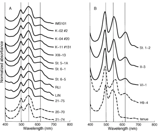
Cyanobacteria have chlorophyll a (Chla) as the primary photosynthetic pigment with phycobiliproteins (PBP) including phycoerythrin (PE), phycocyanin (PC), and
al-lophycocyanon (AP) as accessory light harvesting pigments (Waterbury et al., 1986). Species of Trichodesmium have different pigmentation and their colors range from dark red-brown (T erythraeum IMS101) to light salmon (T tenue tenue) to light green
(Tri-chodesmium sp. H9-4). T erythraeum has more PE than T thiebautii (Carpenter et al., 1993). These observations of variable characteristics across colony morphologies and species of Trichodesmium point to possible niche differentiation.
Overview of thesis chapters
Trichodesmium contributes a significant amount of fixed N to marine ecosystems (Capone et al., 1997), and measurements of Trichodesmium have been used to parameterize N2
fix-ation in a variety of models (Moore et al., 2004; Coles et al., 2004). However, much is unknown about its ecology and physiology. Mortality rates and causes of mortality are not well characterized, distributions are patchy, and potential differences among species of Trichodesmium have not been thoroughly studied and are often ignored. A vast majority of the culture experiments have used T erythraeum IMS101, which belongs to a clade that branches separately from the numerically predominant species of T thiebautii (Chaps.
2, 3). This species is also used to parameterize N2 fixation in global ecosystem models (Moore et al., 2004). To understand the roles Trichodesmium plays in the ecosystem and the factors that control its distribution, it is important to understand the differences among the species of Trichodesmium and the potential for niche differentiation. I aimed to an-swer the questions:
* What species of Trichodesmium do we have in the Woods Hole culture collection? * How are these cultured strains related to each other?
* Do the cultured strains have characteristics which might lead to niche differentia-tion?
* How are field populations of Trichodesmium distributed? * What factors control the distributions of Trichodesmium spp.?
In my PhD thesis, I characterized cultured strains of Trichodesmium, developed a molecular method to distinguish between the two clades of Trichodesmium, identified and enumerated Trichodesmium clades in the field, and investigated P stress and N2 fixation in field populations.
Chapter 2: Characterization. The Woods Hole Oceanographic Institution has
an extensive culture collection of Trichodesmium spp., established and maintained over fifteen years through the work of Dr. John Waterbury. This collection gave me the unique opportunity to take measurements from a variety of isolates. The genome of T erythraeum IMS 101 is currently available, allowing for the development of primers to target genetic markers for identification such as hetR, 16S rDNA and ITS. Species of Trichodesmium
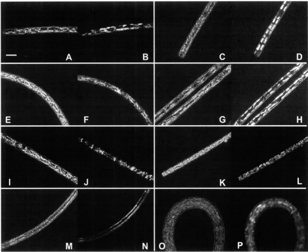
vary by cell length, cell width, and colony morphology. Using phase contrast light mi-croscopy and differential interference contrast (DIC) mimi-croscopy, I measured cell width and length and observed gas vesicles of cultured strains. I analyzed the PBP absorption spectra for the cultured strains of Trichodesmium and measured PUB:PEB and PE:PC ratios by the relative peak heights. I found that the cultured species of Trichodesmium clustered into two clades: one containing T erythraeum and T contortum, the other
con-taining T thiebautii, T tenue, T pelagicum, and TI hildebrandtii. Each clade has diverse
cell morphology, but distinct PBP absorption spectra.
Chapter 3: qPCR. The hetR gene is putatively used in cyanobacterial
hetero-cyst differentiation and may be used in diazocyte differentiation in Trichodesmium (El-Shehawy et al., 2003). Using hetR sequences from Lundgren et al. (2005) and cultured strains (Chapt. 2), I developed clade-specific primers for approximately 140 bp regions targeted for qPCR. The qPCR assay was optimized using known concentrations of cul-tured strains and standardized with cloned PCR products from each clade. I also tested a variety of DNA extraction methods to minimize PCR inhibition.
Chapter 4: Quantification of field populations. I quantified field samples of
Trichodesmium from depth profiles along horizontal gradients of the equatorial Atlantic Ocean, the west Pacific warm pool, and the South Pacific Ocean. I collected water at
var-ious depths including the surface and deep chlorophyll maximum (DCM) using a rosette of Niskin bottles. Trichodesmium clade I and II abundances and distributions were ana-lyzed with ancillary data such as temperature, salinity, depth, and nutrient concentrations. Clade II was more numerous than clade I in both the Pacific and Atlantic Oceans. Clade I had deeper and colder distributions than clade II. There was a correlation between clade I and clade II concentrations in the Pacific but not the Atlantic. There were no patterns or
Trichodesmium concentrations with respect to P or Fe.
Chapter 5: Phosphorus stress and N2 fixation. Phosphorus (P) can limit growth and N2 fixation rates of Tricodesmium. Under P stress, some phytoplankton use alkaline
phosphatase (AP) to cleave phosphate from organic sources (Stihl et al., 2001). I analyzed P stress and N2 fixation rates of Trichodesmium populations from the Atlantic and Pa-cific Oceans. I collected colonies of Trichodesmium and analyzed them for endogenous AP activity using enzyme-labeled fluorescence (ELF) and for nitrogenase activity using acetylene reduction. Both P stress and N2 fixation rates were higher in the western North
Atlantic than in the Pacific, indicating that factors other than P were constraining N2
CHAPTER 2
Diversity of the N
2
-fixing Cyanobacterium
Trichodesmium: Characterization of the Woods
Hole Culture Collection
ABSTRACT:
The filamentous, colonial cyanobacterium Trichodesmium has six well-described species, but many more names. Traditional classification was based on morphological characteristics such as cell width and length, gas vesicle distribution, and colony mor-phology, which can lead to inconsistent results. I used the Woods Hole culture collec-tion of Trichodesmium to identify cultured strains by species using cell morphology; phycobiliprotein absorption spectra; and sequences of 16S rDNA, the 16S-23S inter-nal transcribed spacer (ITS), and the heterocyst differentiation gene hetR. There were
two major clades of Trichodesmium: clade I consisting of Trichodesmium erythraeum and Trichodesmium contortum, and clade II made up of Trichodesmium thiebautii,
Tri-chodesmium tenue, TriTri-chodesmium pelagicum, and TriTri-chodesmium hildebrandtii. These clades were genetically coherent with similar phycobiliprotein composition, but morpho-logically diverse. In the continual revision of cyanobacterial taxonomy, genetic and bio-chemical information are useful and informative complements to morphology for inform-ing classification schemes.
Introduction
Identification of Trichodesmium. Trichodesmium spp. (Cyanobacteriales, order
Oscilla-toriales) live in tropical and subtropical oceans. This filamentous phytoplankter exists as both single trichomes and colonies that are visible to the naked eye, earning the nickname "sea sawdust" (Cook, 1842). They occasionally form brown or red blooms that are visible from space and gave the Red Sea its name (Ehrenberg, 1830). In addition to contributing
fixed carbon (C) to the ecosystem, Trichodesmium is a significant contributor of new ni-trogen (N) through N2 fixation in oligotrophic regions (Capone et al., 1997). While only
a few grazers feed on Trichodesmium (O'Neil and Roman, 1994), colonies offer habitat by providing an oasis in the open ocean, harboring a community of heterotrophic bacteria and invertebrates (Sheridan et al., 2002; Hewson et al., 2009).
Classical identification of Trichodesmium was based on cell width and length, sheath characteristics, distribution of gas vesicles, and colony morphology. There are six well-described species of Trichodesmium: Trichodesmium contortum, Trichodesmium erythraeum, Trichodesmium hildebrandtii, Trichodesmium pelagicum (formerly Katag-nymene spiralis and KatagKatag-nymene pelagica), Trichodesmium tenue, and Trichodesmium thiebautii. These species of Trichodesmium are genetically similar, but morphologically distinct. Classification by cell and colony morphologies can be confusing and mislead-ing due to variable and overlappmislead-ing characteristics (Anagnostidis and Komarek, 1988; Janson et al., 1995). Many genera in Oscillatoriaceae were identified by characteristics of the sheath, which can vary with environmental conditions and proved to be an unreli-able criterion (Rippka et al., 1979; Hoffmann, 1988; Anagnostidis and Komarek, 1988). Trichodesmium colonies come in a variety of morphologies including spherical puffs, fusiform tufts, and bowties. Some species can have more than one morphology, for ex-ample, TI thiebautii forms both puff and tuft colonies, and different species can have the same colony morphology: T thiebautii, Ti hildebrandtii, and Ti erythraeum all form tufts. Single colonies may also contain a variety of filaments, indicating that they are not
clonal and suggesting that colonies may coalesce from single trichomes of several species (Hynes et al., 2009).
Even more perplexing is the list of names for species currently or formerly known as "Trichodesmium." Many species originally identified as Trichodesmium were brought under the umbrella of the genus Oscillatoria and then later were separated back to Tri-chodesmium (Geitler, 1932; Rippka et al., 1979; Anagnostidis and Komirek, 1988). Species of the genus Katagnymene were found to be genetically similar to T thiebautii with
re-spect to the nitrogenase gene nifH, the 16S-23S internal transcribed spacer (ITS), and the heterocyst differentiation gene hetR, so Katagnymene spp. have been included in the genus Trichodesmium (Lundgren et al., 2001; Orcutt et al., 2002; Lundgren et al., 2005). Due to the inconsistent nature of cell morphology, a large number of synonyms have been in use (Guiry and Guiry, 2008). A selection of synonyms is summarized in Table 2.1.
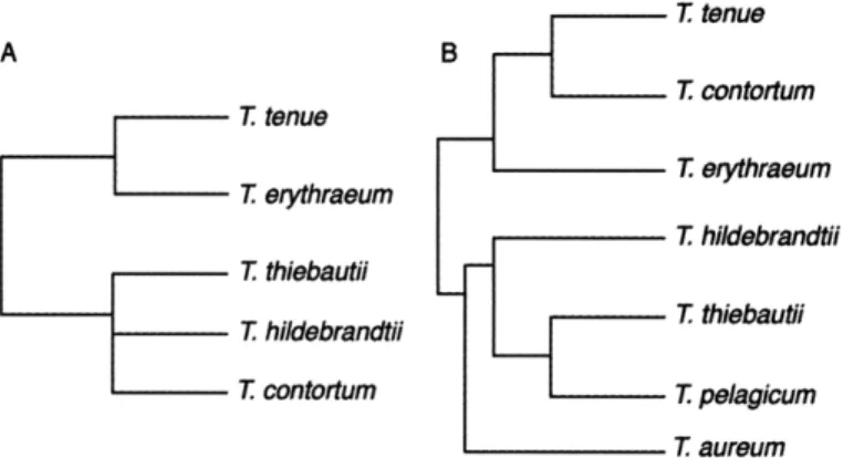
Previous studies have grouped Trichodesmium spp. into a variety of taxonomic schemes. The classification of species based on morphometric characteristics and cell structure resulted in two groups (Fig. 2-lA): (1) species with peripheral gas vesicles (T tenue, a spherical-shaped colony from an undetermined Trichodesmium sp., and T ery-thraeum); and (2) species with randomly dispersed gas vesicles (T thiebautii, T hilde-brandtii, and T contortum) (Janson et al., 1995). In phylogenetic trees of Trichodesmium,
T erythraeum branched off from all the other groups (Orcutt et al., 2002). Trees con-structed from 16S rRNA and hetR sequences of field and culture samples have shown
four clades: (1) T hildebrandii, T thiebautii, and T pelagicum; (2) a novel morphotype named Trichodesmium aureum; (3) T erythraeum and a large, dark-pigmented trichome;
and (4) i contortum and T tenue (Lundgren et al., 2005). This grouping is represented in the dendrogram in Fig. 2-1B.
Photosynthetic pigments. Members of the genus Trichodesmium have a
photosyn-thetic system typical of other cyanobacteria. Their primary photosynphotosyn-thetic pigment is chlorophyll a (Chla) with a light-harvesting phycobilisome made up of phycobiliproteins
Table 2. 1: List of species names for (2008). Species Trichodesmium aureum Trichodesmium clevei *Trichodesmium contortum *Trichodesmium erythraeum Trichodesmium flosaquae Trichodesmium havanum *Trichodesmium hildebrandtii Trichodesmium hindsii
Trichodesmium iwanoffianum Nygaard Trichodesmium lacustre
Trichodesmium lenticulare Trichodesmium maccii
Trichodesmium, taken partly from Guiry and Guiry
Synonyms
Pelagothrix clevei J. Schmidt Xanthotrichum contortum Skujaella contorta
Oscillaria erythraea (Ehrenberg) Oscillatoria erythraea (Ehrenberg) Trichodesmium ehrenbergii Montagne Trichodesmium erythraeum Ehrenberg
Oscillatoria hildenbrandtii Skujaella hildebrandtii
Trichodesmium ehrenbergiif indicum Trichodesmium hildenbrandtii Trichodesmium indicum Oscillatoria lacustris Oscillatoria lacustris Skujaella lacustris Haliarachne lenticularis Ref. 1 2, 3 4 5 6 7 5 8 5 1 7 5 2 9 5 7 2 2 5 7 10
*Trichodesmium pelagicum Katagnymene pelagica 1, 7
Katagnymene spiralis 7
Trichodesmium spiralis 11
Trichodesmium scoboideum Skujaella scoboidea 5
*Trichodesmium tenue Trichodesmium radians 2
Heliotrichum radians 12
*Trichodesmium thiebautii Oscillatoria thiebautii 7
Skujaella thiebautii 5
Heliotrichum radians 13
*Species represented in the Woods Hole culture collection (past and present).
1 = Lundgren et al. (2005), 2 = Anagnostidis and Komirek (1988), 3 = Silva et al. (1996), 4 = Schitt (1893), 5 = Drouet (1968), 6 = Kitzing (1845), 7 = Geitler (1932), 8 = Hallegraeff et al. (2003), 9 = Gomont (1892), 10 = Li et al. (1984), 11 = Dyhrman et al. (2006), 12 = Golubic (1977), 13 = Wille (1904)
T tenue T contortum T tenue ythraeum T erythraeum T erythraeum T hildebrandtii T thiebautii T hildebrandtii T thiebauti T contortum T pelagicum T aureum
Figure 2-1: Dendrograms of classification groupings of Trichodesmium in previous studies. (A) Grouping based on morphological characteristics (Janson et al., 1995). (B) Grouping based on hetR sequences (Lundgren et al., 2005).
Chi a PUB PEB PC AP Chi a
400 450 500 550 600 650 700 Wavelength (nm)
Figure 2-2: Typical absorption peak values for photosynthetic pigments in cyanobacteria: chlorophyll a (Chla), phycourobilin (PUB), phycoerythrobilin (PEB), phycocyanin (PC), allophycocyanin (AP) (Rippka et al., 1974).
(Glazer, 1987). Trichodesmium phycobilisomes are dominated by the phycobiliprotein phycoerythrin (PE), which has absorbance peaks at 490 -500 nm for its brown
phycouro-bilin (PUB) component and at 545 -565 nm for its red phycoerythrobilin (PEB)
compo-nent. The phycobilisome also contains the blue-green phycocyanin (PC), which absorbs around 620 nm, and the blue allophycocyanin (AP), which absorbs at 650 nm (Fig. 2-2). In contrast, Chla has absorption peaks at about 440 nm and 680 nm (Rippka et al., 1974).
Chromatic adaptation has been described in field populations and cultured chodesmium (Subramaniam et al., 1999; Bell and Fu, 2005). PUB:PEB ratios in Tri-chodesmium have been shown to increase at midday in the Caribbean Sea
(Subrama-niam et al., 1999). Great Barrier Reef strain T erythraeum GBRTRLI101 showed chro-matic adaptation by changing its PE:Chla ratio as well as its PE:PC ratio; PE increased under green light, PC increased under red light, and Chla increased under blue and red light (Bell and Fu, 2005). However, chromatic adaptation has not been found in other field studies (McCarthy and Carpenter, 1979; Neveux et al., 2006). Pigmentation of Tri-chodesmium in the field and in culture varies considerably with colors ranging from red to brown to green. This color variation is partly due to phycobiliprotein composition; Car-penter et al. (1993) noted that T erythraeum had more phycoerythrin per colony and a higher PE:Chla than TI thiebautii. Color may also be influenced by growth state;
senes-cent Trichodesmium cells appear more brown or green than healthy cells due to chlorosis.
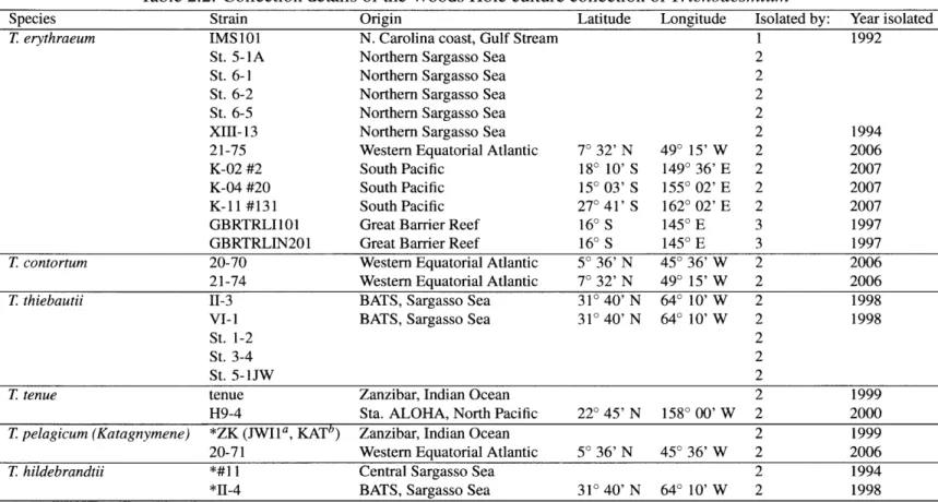
Characterization of the Woods Hole culture collection. The Woods Hole
Oceano-graphic Institution (WHOI) has an extensive collection of Trichodesmium cultures from all over the world (Table 2.2). To classify these strains and understand the relationships among the various species, I characterized the culture collection using cell morphology, phycobiliprotein spectra, and sequences of 16S rDNA, ITS, and hetR. The results of this study showed that there are two major clades of Trichodesmium based on sequence analy-sis. Surprisingly, the clades were not morphologically homogeneous, but were consistent with respect to phycobiliprotein content.
Table 2.2: Collection details of the Woods Hole culture collection of Trichodesmium
Species Strain Origin Latitude Longitude Isolated by: Year isolated
T erythraeum IMS 101 N. Carolina coast, Gulf Stream 1 1992
St. 5-1A Northern Sargasso Sea 2
St. 6-1 Northern Sargasso Sea 2
St. 6-2 Northern Sargasso Sea 2
St. 6-5 Northern Sargasso Sea 2
XIII- 13 Northern Sargasso Sea 2 1994
21-75 Western Equatorial Atlantic 70 32' N 490 15' W 2 2006
K-02 #2 South Pacific 180 10' S 1490 36' E 2 2007
K-04 #20 South Pacific 150 03' S 1550 02' E 2 2007
K-11 #131 South Pacific 270 41' S 1620 02' E 2 2007
GBRTRLI101 Great Barrier Reef 160 S 1450 E 3 1997
GBRTRLIN201 Great Barrier Reef 160 S 1450 E 3 1997
T contortum 20-70 Western Equatorial Atlantic 50 36' N 450 36' W 2 2006
21-74 Western Equatorial Atlantic 70 32' N 490 15' W 2 2006
T thiebautii II-3 BATS, Sargasso Sea 310 40' N 640 10' W 2 1998
VI-1 BATS, Sargasso Sea 310 40' N 640 10' W 2 1998
St. 1-2 2
St. 3-4 2
St. 5-1JW 2
T tenue tenue Zanzibar, Indian Ocean 2 1999
H9-4 Sta. ALOHA, North Pacific 220 45' N 1580 00' W 2 2000
T pelagicum (Katagnymene) *ZK (JWI1a, KATb) Zanzibar, Indian Ocean 2 1999
20-71 Western Equatorial Atlantic 50 36' N 450 36' W 2 2006
T hildebrandtii *#11 I Central Sargasso Sea 2 1994
*II-4 BATS, Sargasso Sea 310 40' N 640 10' W 2 1998
*Extinct strains
1 = L. Prufert-Bebout (Prufert-Bebout et al., 1993), 2 = J. B. Waterbury, 3 = P. R. E Bell (Bell et al., 2005)
Methods
Culture conditions. Batch cultures were grown at 260C, 50 pE m-2s- 1, under soft
white fluorescent bulbs with a 14:10 light-dark cycle in RMP medium made from 75% sterile Sargasso Sea water amended with trace metals and vitamin B12, as described in
Webb et al. (2001), and with 2 x 10⁻⁶ M EDTA, 15 x 10-6 M phosphoric acid, 1 x 10⁻⁷
M Fe (ferric citrate). Cultures were maintained in Citranox- and acid-washed 250 mL polycarbonate baffled flasks closed with cotton stoppers or 75 mL square polycarbonate culture bottles (Nalgene).
Micrographs. Wet mounts of culture were examined using a Zeiss Axioplan 2
micro-scope using phase contrast with a Plan-Neofluar 100 x objective lens and differential in-tereference contrast (DIC) using an x Plan-Fluar 100 x objective lens. Micrographs were taken using a Zeiss Axiocam HRC digital camera and Axiovision 4.6.3 software. Images were measured, and cell metrics were determined using the MATLAB image processing toolbox.
Phycobiliprotein extraction and absorption spectra. Trichodesmium
phycobilipro-tein extracts were prepared as described in Rippka et al. (1974) with minor modifications. Cultures were filtered on 25 mm, 5 pm polycarbonate filters and stored at -20'C. Two mL of pH 7.0, 5 mmol L-1 phosphate buffer containing 0.1 mol L- 1 NaCl were added to each sample and vortexed to remove cells from the filter. Cells were lysed at 4'C using a digital sonifier (Branson; 3 x for I min at 65% with on/off cycles for 0.5 s each), a tissue homogenizer, or a small French pressure cell (Aminco). The extracts were centrifuged in a Beckman ultracentrifuge for 45 minutes at 112,000 rcf and 4 'C. The supernatant was pipetted into a 1.5 mL microcentrifuge tube and centrifuged in an Eppendorf 581 OR cen-trifuge for 15 minutes at 20,000 rcf and 40C. Seventy-five pL of supernatant was pipetted
into a quartz microcuvette and the absorption spectra from 400 -700 nm (0.5 nm step)
phos-phate buffer alone were subtracted from the absorption spectra of the samples. Replicate spectra were averaged and normalized to the maximum absorption.
Sequences and phylogeny. Cultured strains of Trichodesmium were filtered on 25 mm,
5 pm polycarbonate filters and stored in liquid N2 until extraction. DNA was extracted
us-ing the DNeasy Tissue Kit (Qiagen), followus-ing the manufacturer's instructions for animal tissue. Extracted DNA was stored at -200C. All polymerase chain reactions (PCR) were performed on a Biorad iCycler with 25 pL reactions. Primers are summarized in Table
2.3. Amplification of 16S rDNA was performed using the high-fidelity iProof kit (Biorad) to get a 662 bp fragment. Each reaction contained 1 x HF Buffer, 200 pM dNTPs, 0.5 pM each of primers CYA-106F and CYA-781R(a) (Nibel et al., 1997), 0.02 U iProof DNA polymerase, and 4 ng of genomic DNA. Reactions were cycled using the temperature
pro-file of 980C for 30 s; 30 cycles of 980C for 10 s, 65.5'C for 20 s, and 72°C for 20s; and
one cycle at 72C for 7 min. The ITS region was amplified using the MasterTaq Kit (Ep-pendorf, 5 Prime) to get a 1026 bp fragment. Reactions consisted of 1 x TaqMaster PCR Enhancer; 1 x Taq Buffer; 3.5 mM Mg2+ (including Mg2+ in the Taq Buffer); 200 pM
dNTPs; 0.5 pM each of primer tril6S-1247F (Orcutt et al., 2002) and primer tri-23SR, modified from 23S-241R (Rocap et al., 2002) to be specific to Trichodesmium based on the T erythraeum IMS 101 genome; 1.25 U Taq DNA polymerase; and 4 ng genomic
DNA. Thermal cycling conditions for ITS were 950 for 2 min; 30 cycles of 950 for 1 min,
550 for 1 min, and 720for 1 min; and a final cycle of 720 for 10 min. Amplification of
hetR was conducted using the iProof kit (Biorad) to get a 448 bp fragment. The reac-tion mixture was made up of 1 x HF Buffer, 200 pmol L- dNTP, 0.5 pmol L- 1 each of primers PHI and PH2 (Lundgren et al., 2005), and 0.02 U pL- iProof DNA polymerase,
and 4ng of genomic DNA. Cycling conditions for hetR were 98' for 30 s; 30 cycles of 98'