HAL Id: hal-02648752
https://hal.inrae.fr/hal-02648752
Submitted on 29 May 2020
HAL is a multi-disciplinary open access
archive for the deposit and dissemination of
sci-entific research documents, whether they are
pub-lished or not. The documents may come from
teaching and research institutions in France or
abroad, or from public or private research centers.
L’archive ouverte pluridisciplinaire HAL, est
destinée au dépôt et à la diffusion de documents
scientifiques de niveau recherche, publiés ou non,
émanant des établissements d’enseignement et de
recherche français ou étrangers, des laboratoires
publics ou privés.
Copyright
Matrix Protoein from Sclerites of Red Coral, Corallium
rubrum
Julien Debreuil, Eric Tambutté, Didier Zoccola, Emeline Deleury, Jean-Marie
Guigonis, Michel Samson, Denis Allemand, Sylvie Tambutté
To cite this version:
Julien Debreuil, Eric Tambutté, Didier Zoccola, Emeline Deleury, Jean-Marie Guigonis, et al..
Molec-ular Cloning and Characterization of First Organic Matrix Protoein from Sclerites of Red Coral,
Corallium rubrum. Journal of Biological Chemistry, American Society for Biochemistry and
Molecu-lar Biology, 2012, 287 (23), pp.19367 - 19376. �10.1074/jbc.M112.352005�. �hal-02648752�
Molecular Cloning and Characterization of First Organic
Matrix Protein from Sclerites of Red Coral, Corallium rubrum
*
□SReceived for publication, February 10, 2012, and in revised form, April 12, 2012Published, JBC Papers in Press, April 13, 2012, DOI 10.1074/jbc.M112.352005
Julien Debreuil‡1, Éric Tambutté‡, Didier Zoccola‡, Emeline Deleury§¶, Jean-Marie Guigonis储**‡‡, Michel Samson§§, Denis Allemand‡, and Sylvie Tambutté‡2
From the‡Centre Scientifique de Monaco, Avenue Saint-Martin, MC-98000, Monaco, the§Institut Sophia Agrobiotech (ISA) INRA 1355, CNRS 7254, 400 route des Chappes, Sophia-Antipolis F-06903, France, the¶Université de Nice-Sophia Antipolis, Nice F-06107, France, the储Laboratoire Transporteur en Imagerie et Radiothe´rapie Oncologique, Commissariat à l’Energie Atomique, Nice F-06107, France, the **Faculté de Médecine, Université de Nice-Sophia Antipolis, Nice F-06107, France, the‡‡Centre Antoine Lacassagne, Nice F-06107, France, and the§§E´quipe Région Institut National de la Sante´ et de la Recherche Medicale 21/ E´quipe Associe´e 4319, Faculté de Médecine, Université de Nice-Sophia Antipolis, Nice F-06107, France
Background:Sclerites of alcyonarian species are biominerals formed of organic matrix molecules trapped inside a mineral inorganic fraction.
Results:Scleritin is a small basic organic matrix protein, synthetized by scleroblasts and incorporated into sclerites. Conclusion:Scleritin is the first organic matrix protein fully characterized and localized in the sclerites of C. rubrum. Significance:Scleritin provides information on the biomineralization pathway in C. rubrum.
We report here for the first time the isolation and character-ization of a protein from the organic matrix (OM) of the sclerites of the alcyonarian, Corallium rubrum. This protein named scle-ritin is one of the predominant proteins extracted from the EDTA-soluble fraction of the OM. The entire open reading frame (ORF) was obtained by comparing amino acid sequences from de novo mass spectrometry and Edman degradation with an expressed sequence tag library dataset of C. rubrum. Scleritin is a secreted basic phosphorylated protein which exhibits a short amino acid sequence of 135 amino acids and a signal peptide of 20 amino acids. From specific antibodies raised against peptide sequences of scleritin, we obtained immunolabeling of sclero-blasts and OM of the sclerites which provides information on the biomineralization pathway in C. rubrum.
Biomineralization is defined as both the study of biogically produced mineral materials (or biominerals) and the processes that lead to their formation (1). Calcium carbonate skeletal structures are the most abundant biominerals encountered in the metazoan world (2). Much of what is known about biomin-eralization is to date inferred from marine calcifying model organisms such as mollusks and echinoderms where data are available from the ultrastructural to the biochemical and molecular levels (3–5). For other organisms, data are far less abundant despite their importance for a better basic
under-standing of the biomineralization process. This is the case for cnidarians among which species from the subclass Hexacorallia are typically known for their role in producing tropical coral reefs.
This is also the case for various other taxa such as alcyonarian species (6). Among Alcyonaria many families typically contain species with skeletal structures called sclerites (or spicules) which are considered to form a fragmented skeleton located in the soft tissues (coenenchyme). Sclerites are suggested to pro-vide structural support (7) and may have a potential role of physical defense against predation (8, 9). Sclerites are also a main source of information for systematic studies because their morphology and morphometry are species-specific (10, 11).
Among alcyonarian, the precious coral Corallium rubrum is a well known sclerites-producer species. This Mediterranean species has a cultural and commercial importance, and its axial skeleton has been used for jewelry and art objects for centuries (12–14). The sclerites are distributed within the animal tissues, and they have been described in detail by means of light and scanning electron microscopy (15, 16). Their microstructure never reaches more than a few micrometers in axial or lateral directions (60 –90 m) (17). Their density can reach about 106/mg of tissue proteins (18), and they are initially formed in
intracellular vesicles within cells, named scleroblasts, present in the mesoglea (17). Sclerites are also incorporated in the axial skeleton, but the pathway of incorporation remains debated (19, 13, 20).
Sclerites of C. rubrum are, as for other biominerals, a com-posite material formed of an organic fraction called organic matrix (OM)3trapped inside a mineral inorganic fraction. This inorganic fraction of calcium carbonate is crystallized under the form of Mg-calcite, and the OM represents⬍2% of the dry
*This work was conducted as part of the Centre Scientifique de Monaco research program, supported by the Government of the Principality of Monaco.
Author’s Choice—Final version full access. □S This article containssupplemental Fig. S1.
The nucleotide sequence(s) reported in this paper has been submitted to the Gen-BankTM/EBI Data Bank with accession number(s) JQ652458.
1Supported by the Ministère Français de l’Enseignement Supérieur et de la
Recherche, Ecole Doctorale Diversité du Vivant 392, Université Pierre et Marie Curie.
2To whom correspondence should be addressed. Tel.: 37793301211; Fax:
37792167981; E-mail: stambutte@centrescientifique.mc.
3The abbreviations used are: OM, organic matrix; BisTris,
bis(2-hydroxyethyl)-iminotris(hydroxymethyl)methane; EST, expressed sequence tag; MS/MS, tandem MS; RACE, rapid amplification of cDNA ends; Tricine, N-[2-hydroxy-1,1-bis(hydroxymethyl)ethyl]glycine.
at INRA Institut National de la Recherche Agronomique on June 13, 2018
http://www.jbc.org/
weight of the skeletal structure (21). From biomimetic experi-ments, it is suggested that the OM plays a role of assembler between inorganic building blocks forming mesocrystals (22). These blocks have been recently evidenced in the sclerites of C.
rubrum(23).
Previous biochemical works on the axial skeleton and scler-ites of C. rubrum have revealed that their OM is composed of proteins, glycosaminoglycans, and proteoglycans (20, 21, 24, 25), as well as pigments such as carotenoids (26, 27) or trans-polyacetylene molecules (28). However, the most abundant lit-erature on the biochemistry of OM in Alcyonaria concerns: (i) the Alcyoniidae: Lobophytum crassum and Synularia
polydac-tyla(29 –34); (ii) the Gorgoniidae: Leptogorgia virgulata (35– 40) and Pseudoplexaura flagellosa (41). Nevertheless, to date only partial sequences of OM proteins have been obtained for alcyonarian sclerites (32, 42), and even when widening to the Anthozoa, only one OM protein has been fully characterized in a scleractinian coral (43) (for review, see also Refs. 44 – 46).
Based on the methodology of OM protein extraction and separation by gel electrophoresis that we previously set up (20, 47) and in combination with transcriptomic data from an EST library,4we have isolated and fully characterized one protein of
OM in the sclerites of C. rubrum. To our knowledge, this is the first report of a complete sequence of an organic matrix protein in the Alcyonaria subclass.
EXPERIMENTAL PROCEDURES
Organic Matrix Extraction—Sclerites from colonies of C.
rubrumcollected at 30-m depth in Marseille, Riou Island (out-side cave, Gulf of Lion; Mediterranean coast of France) were prepared as described by Debreuil et al. (20). Extraction of the organic fraction from the mineral fraction was adapted from Puverel et al. (48) and Debreuil et al. (47). Briefly, after demin-eralization of sclerites powder with 0.25MEDTA (pH 7.8, 23 h,
4 °C; Sigma), the solution was prefiltered (0.2m of polyether-sulfone) and centrifuged (10,000⫻ g, 15 min, 4 °C). The super-natant was filtered, concentrated, and rinsed thoroughly (8 times) with ultrapure water (3,500⫻ g, Centricon Plus 70, cut-off 5 kDa). Protein content was determined using a Quant-iTTM
protein assay kit (QubitTM; Molecular Probes) with bovine
serum albumin (BSA) as a standard and Qubit fluorometer apparatus (Q32857; Invitrogen).
Electrophoresis and Immunolabeling—One- and two-dimen-sional gel electrophoresis and Western blotting were per-formed as described by Debreuil et al. (20, 47). BisTris 12% polyacrylamide gels (Criterion, Bio-Rad) were used for wide range molecular weight protein; and Tris-Tricine 16.5% poly-acrylamide gels (Criterion, Bio-Rad) for small molecular weight proteins. The protein markers used were: Silver Stain Molecu-lar Weight Marker (M6539; Sigma) for the BisTris silver-stained electrophoresis gel, Kaleidoscope Polypeptide Stan-dards (Bio-Rad 161-0325) for the Tris-Tricine silver-stained electrophoresis gels, and Precision Plus Protein WesternCTM
standard (161-0376 Bio-Rad) both for the BisTris and Tris-Tri-cine gels of Western blots.
The primary antibodies used for Western blotting were anti-phosphoserine (9332; Abcam), anti-phosphothreonine (9337; Abcam), anti-phosphotyrosine (9319; Abcam), and polyclonal antibodies raised against two amino acid sequences of the OM protein identified in the present study, i.e. scleritin: NH2
-FIEL-SKRMQRESSNFC-COOH and NH2 -CNTRPVQPISRQLDDL-COOH. For these last antibodies, both peptides were mixed with Freund’s complete adjuvant and injected intraperitoneally into a rabbit (Oryctolagus cuniculus; Eurogentec). Rabbit was immunoboosted by several injections of peptides mixed with incomplete Freund’s adjuvant (at 7, 10, and 18 days after the first injection). The final bleeding was conducted 28 days after the last injection. Preimmune (from 0 day bleeding) and final sera were drawn off and stored at⫺20 °C.
Electroelution Method—After one-dimensional SDS-PAGE (16.5% polyacrylamide gel; Criterion, Bio-Rad) of the whole extract of OM of sclerites and a staining step overnight with ImperialTMProtein Stain (Thermo Scientific), the
electroelu-tion of the 18-kDa protein band was performed with an Electro-Eluter (model 422; Bio-Rad) as used by Rahman et al. (42) for the purification of OM proteins in alcyanorian sclerites. Elec-troelution was performed for 5 h at 60 mA, using dialyze caps (cutoff 3.5 kDa) in an elution buffer (25 mMTris, 192 mM
gly-cine, 0.1% SDS). The samples were then concentrated, rinsed thoroughly (eight times) by ultrafiltration using Amicon-Ultra (cutoff 5 kDa; Millipore), freeze-dried, and then used for the two-dimensional electrophoresis experiment.
Sequencing Methods—The amino acid sequence determina-tion based on Edman degradadetermina-tion was performed using an Applied Biosystems gas-phase sequencer (model 492; s/n: 9510287J; Institut de Biologie Structurale, CEA/CNRS/UJF, Grenoble, France). Phenylthiohydantoin amino acid deriva-tives generated at each sequence cycle were identified and quantified on-line with an HPLC system (Applied Biosys-tems 140C and Applied BiosysBiosys-tems 610A data analysis soft-ware 2.1) with PTH-amino acid standard kit (PerkinElmer Life Sciences P/N 4340968; according to instructions 900776 Rev D).
Mass spectrometry analysis was performed on protein spot from the two-dimensional electrophoresis gel. Proteins were digested with trypsin, blocked with 5% formic acid, and ana-lyzed by capillary-LC/ESI/MS/MS (LTQ/FT-Orbitrap mass spectrometer; Thermo Fisher) coupled with pumps and autosampler under standard conditions (275 °C; 4500 V). Helium was used as the collision gas, and experiments were performed in parallel mode (30,000 survey resolution and 5 data-dependent ion trap MS/MS scans). De novo analysis was operated on PEAKS Studio 4.5 (Bioinformatics Solutions; Waterloo, ON, Canada). Then putative amino acid sequences obtained were analyzed against nucleotide sequences data base dynamically translated in all reading frames (tBLASTn) (49) from an EST library of C. rubrum.4
Cloning Protein Gene—Total RNA extraction and cDNA synthesis from three colonies of C. rubrum (Riou Island) were, respectively performed as described by Zoccola et al. (50) and Moya et al. (51). PCR amplification was carried out using plat-inum Taq DNA polymerase (Invitrogen) and primers designed using nucleotide sequences of the contig corru5764135 from
4Centre Scientifique de Monaco, unpublished data.
at INRA Institut National de la Recherche Agronomique on June 13, 2018
http://www.jbc.org/
the EST library of C. rubrum.4 Nucleotides sequences were
chosen from the deduced consensus between amino acid sequence from Edman degradation, mass spectrometry analyses, and the translated contig corru5764135: 5 ⬘-AGGAAGGTTG-GTCAGGGCGT-3⬘ for the forward primer and 5⬘-AGCGAA-CTTCGGAGTCCTTA-3⬘ for reverse primer. PCR amplifica-tion product was ligated into a plasmid vector (pGEM-T easy vectorsystemII;Promega),andDNAsequencingofthreeindepen-dent clones was carried out on both strands with SP6 and T7 primer sequences (Macrogen).
After the sequence confirmation, RACE experiments were performed using the 5⬘/3⬘-RACE kit (Roche Applied Science) with the primers 5⬘-CATTAAGCTCTGAAGCAGTGG-3⬘ and 5⬘-AGGAAGGTTGGTCAGGGCGT-3⬘ for 5⬘-RACE; and the primers 5⬘-AAACGTATGCAACGAGAGTCA-3⬘ and 5⬘-AGCGAACTTCGGAGTCCTTA-3⬘ for 3⬘-RACE. Both ends were cloned and sequenced as mentioned above. All sequences were assembled using the Lasergene software (SEQMAN LASERGENE 9.0.4; DNASTAR) to obtain the whole open read-ing frame (ORF).
Nucleotide and Amino Acid Sequences Analysis—The theo-retical mass, isoelectric point, and amino acid composition of the protein identified in the present study were obtained with Editseq 9.0.4 (DNASTAR Inc.). The complete translated ORF was interpreted with Bioworks 3.3 (Thermo Fisher) with both b and y ion series for charge 2, 3, and 4 and valuate Xcorr for recovery confirmation (xc (⫾ 1, 2, 3) ⫽ 1.50, 2.00, 2.50); supple-mental Fig. S1a). BLASTp and tBLASTn analysis (49) were car-ried out to identify homologous sequences in UniProt (52) and GenBank database on the NCBI server (53). From the EXPASY online server, the putative glycosylation sites were identified using DictyOGlyc1.1, NetCGlyc 1.0, NetNGlyc 1.0, YinOYang1.2; phos-phorylation sites with NetPhos2.0; and the signal peptide with Sig-nalP 4.0. The subcellular localization were predicted using TargetP 1.1 (54) and PsortII prediction tool (55).
Histology and Immunolabeling on Tissue Cross-sections—To obtain a histological overview of a demineralized branch of C.
rubrum, a longitudinal section was incubated with hematoxy-lin/eosin/aniline blue to stain, respectively, nuclei and cytoplas-mic and connective regions. Then, the polyclonal anti-scleritin antibodies used for Western blotting were used for immuno-histochemistry. Briefly as described by Debreuil et al. (20), pieces of C. rubrum colonies (2–3-cm length) were fixed, demineralized, dehydrated, and embedded in paraffin wax. Deparaffinized cross-sections were mounted on a silane-coated glass slide and incubated in saturating solution. Sections were then incubated with anti-scleritin antibodies. Controls were performed with preimmune serum as primary antibodies. All samples were then incubated with biotinylated rabbit anti-bodies (Sigma) as secondary antianti-bodies, stained with streptavi-din-Alexa Fluor 568 (Molecular Probes, Invitrogen). 0.002% DAPI (4⬘6-diamidino-2-phenylindole, Sigma) was used to stain the nuclei. Samples were embedded in Pro-Long antifade solu-tion (Molecular Probes, Invitrogen), and observed with a con-focal laser scanning microscope (Leica SP5) equipped with UV and visible laser lines.
RESULTS
Separation of OM proteins of sclerites from C. rubrum by one-dimensional electrophoresis (BisTris 12% polyacrylamide gel; Fig. 1a), shows a set of protein bands distributed mainly in the range of low apparent molecular mass (ⱕ12 kDa) as in Debreuil et al. (20). To obtain a better resolution of small molecular mass proteins, we performed electrophoresis with Tris-Tricine 16.5% polyacrylamide gels (Fig. 1b) which are the preferred electrophoretic systems for the resolution of proteins smaller than 30 kDa (56). The bands previously obtained at 12, 10.5, and 10 kDa in BisTris 12% polyacrylamide gel (Fig. 1a) and in our previous work (20), respectively, give three major bands at 18, 14, and 12 kDa in Tris-Tricine 16.5% polyacrylamide gel (Fig. 1b). This difference in apparent molecular mass is com-mon when using different percentage concentration of acryl-amide in electrophoresis gels (56). We chose to study the pre-dominant 18-kDa protein band. This band was excised, electroeluted, and used for two-dimensional gel electrophore-sis (Fig. 1c). The protein spot obtained with a high isoelectric point (pI) at nearly 9.4 was excised, destained, digested, and analyzed by capillary-LC/ESI/MS/MS. Reliable peptide se-quences were obtained in the MS/MS spectra with identical y and b series ions: PLSSEAVALFFNK from 711.89 m/z and KWNTFIELSK from 633.35 m/z (supplemental Fig. S1, b and c). These amino acid sequences and the one obtained from Edman degradation (RKVGQGVIN) matched only with one translated nucleotide contig from an EST library of C. rubrum (81,497 sequences).4 This contig (corru5764135) of 368 nucleotides
long encodes for a sequence of 122 amino acids but not a com-plete ORF. From this contig, primers were designed and by 3⬘-RACE and 5-⬘RACE, a complete ORF was obtained (Fig. 2). The full-length cDNA sequence obtained is 665 bp long and contains, at 40 bp from the 5⬘-end, a methionine in a Kozak (57) cnidarian (58) context followed by an ORF of 468 bp (GenBank accession number JQ652458). This ORF encodes a protein of 155 amino acids composed of an N-terminal signal peptide of 20 amino acids and a C-terminal sequence of 135 amino acids (Fig. 2). This protein has a calculated molecular mass of 17.78 kDa and a calculated pI of 9.35 (DNASTAR Editseq 9.0.4). This protein is a secreted protein, its secretion is confirmed by the Target 1.1 (54) and PsortII (55) tools with, respectively, 86.9% and 55.6% probability. Indeed, the signal peptide is no more present once the protein is incorporated in the OM (confirmed by MS/MS recovery; supplemental Fig. S1, a and d). This secreted protein is named scleritin. Scleritin is a basic small protein (calculated molecular mass of 15.65; calculated pI of 9.39; DNASTAR Editseq 9.0.4) composed mainly of polar, hydrophobic, and amine forms of amino acids (Phe, Asn, and Gln in majority).
The analysis of the amino acid sequence of scleritin reveals several potential sites of post-translational modifications (Fig. 2) such as seven phosphorylations on Thr-24, Ser-65, Ser-72, Ser-73, Thr-113, Tyr-129, and Tyr-147 which are confirmed by the Western blotting results (Fig. 1, d–f); five O-glycosylations on Thr-24, Ser-56, Ser-57, Thr-150, and Ser-154; and one
N-glycosylation on Asp-152.
at INRA Institut National de la Recherche Agronomique on June 13, 2018
http://www.jbc.org/
We looked for homologies between scleritin and other sequences from the available public sequence databases, Gen-Bank nonredundant proteins, and nucleotide databases (NCBI-NRdb, NCBI-dbEST, UniProt). No significant hits were obtained by BLASTp and tBLASTn (e-value cutoff: 0.01) (49).
We then looked at the localization of scleritin in the tissues of C.
rubrum. First, we tested the polyclonal antibodies raised against scleritin by Western blotting on OM extract from the sclerites. The Western blotting shows that only one band at 18 kDa is labeled (Fig. 1g), confirming that the antibodies are specific for scleritin (18-kDa protein band from the OM extract; Fig. 1b).
These antibodies were then used to label the tissues after skeleton demineralization (Fig. 3). A longitudinal section of tis-sues stained with hematoxylin/eosin/aniline blue shows the typical histology of a colony of C. rubrum with oral epithelium, polyps, gastrodermic canals, and aboral epithelium surround-ing the OM of the axial skeleton (Fig. 3a). The labelsurround-ing of these
tissues (Fig. 3, b–h) with the anti-scleritin antibodies revealed by Alexa Fluor 568 appears in orange to yellow with increasing the intensity of labeling; and nuclei in cells stained with DAPI appear in blue. Z-stack reconstructions of 10 tissue sections show that: (i) the OM of the sclerites is labeled (Fig. 3, b–h); (ii) the scleroblasts but no other cell types are labeled (Fig. 3, b–f and h); (iii) labeling occurs from the apex to the base of the colony (Fig. 3, b–f); (iv) the OM of the axial skeleton is not labeled whereas labeling occurs in the OM of sclerites present in the medullar part of the axial skeleton (Fig. 3, b, e, and g). It must also be noted that most of the immunolabeling signal is observed at the periphery of the colony (i.e. near the oral epi-thelium) where sclerites are more numerous.
DISCUSSION
Molecular and Biochemical Characterization of Scleritin— By combining transcriptomic and proteomic approaches, we
FIGURE 1. Silver staining of electrophoresis gels and Western blots on organic matrix extracted from sclerites of C. rubrum. a, one-dimensional SDS-PAGE (10g protein/lane; BisTris 12% polyacrylamide gel, silver stain molecular mass marker (M6539; Sigma) as protein marker). b, one-dimensional SDS-PAGE (10g protein/lane, Tris-Tricine 16.5% polyacrylamide gel, kaleidoscope polypeptide standards (161-0325; Bio-Rad) as protein marker); c, two-dimensional electrophoresis gel (Tris-Tricine 16.5% polyacrylamide gel, kaleidoscope polypeptide standards (161-0325) as protein marker) of the major protein band (18*) excised from one dimensional SDS-PAGE. d, Western blot (5g of proteins/lane; BisTris 12% polyacrylamide gel, Precision Plus Protein WesternCTM
standard (161-0376; Bio-Rad) as protein marker) with antibodies against phosphorylated serine. e, Western blot with antibodies against the phosphorylated threonine (5g of proteins/lane; BisTris 12% polyacrylamide gel, Precision Plus Protein WesternCTMstandard (161-0376) as protein marker). f, Western blot
with antibodies against phosphorylated tyrosine (5g of proteins/lane; BisTris 12% polyacrylamide gel, Precision Plus Protein WesternCTMstandard (161-0376)
as protein marker). g, Western blot with anti-scleritin antibodies (Tris-Tricine 16.5% polyacrylamide gel, Precision Plus Protein WesternCTMstandard (161-0376)
as protein marker). MW, molecular mass; M, protein marker; and triangles show apparent molecular mass of proteins (in kDa).
at INRA Institut National de la Recherche Agronomique on June 13, 2018
http://www.jbc.org/
identified a coral OM protein in the sclerites of C. rubrum which we named scleritin. Scleritin is a basic small molecular mass protein composed mainly of polar, hydrophobic, and amine forms of amino acids (Phe, Asn, and Gln in majority). This result is not trivial because OM proteins are usually acidic (i.e. low range of isoelectric point) (59), and in C. rubrum the OM of sclerites is composed mainly of acidic and polar amino acids (Asp and Gly) (21). However, basic small molecular mass OM proteins have already been evidenced in other calcifying
organisms (for review, see Ref. 60); for mollusks, see Ref. 3; for echinoderms, see Ref. 61. In corals the only OM protein fully characterized, i.e. galaxin from the tropical hexacoral Galaxea
fascicularis, does not show any acidic domain (43).
The analysis of the amino acid sequence of scleritin reveals several potential sites of post-translational modifications such as phosphorylations and glycosylations which could explain the difference between the calculated molecular mass of scleritin (15.65 kDa) and that of the secreted form observed on the
elec-FIGURE 2. Nucleotide gene and amino acid sequences of scleritin. Nucleotide sequence above (GenBank accession number JQ652458) and deduced translation of the ORF (nucleotides 40 –507) below. The putative signal peptide is underlined, the peptide sequence obtained after Edman degradation is highlighted in gray, the two consecutive peptides obtained by MS/MS are indicated in bold letters, the potential N-glycosylation site is highlighted in black (Asp-152), the five potential O-glycosylation sites (Thr-24, Ser-56, Ser-57, Thr-150, and Ser-154) are indicated by asterisks, and the seven potential phosphory-lation sites are boxed (Thr-24, Ser-65, Ser-72, Ser-73, Thr-113, Tyr-129, and Tyr-147).
at INRA Institut National de la Recherche Agronomique on June 13, 2018
http://www.jbc.org/
trophoresis gels (18 kDa; Fig. 1, b and c). OM proteins of several calcifying models are known to be post-translationally modified (3, 60, 62– 64). Such modifications can participate in the bind-ing of calcium ions as proposed for marine calcifybind-ing
inverte-brates (see Refs. 65– 67 for phosphorylations in mollusks and crustaceans and Ref. 68 for glycosylations in mollusks). These post-translational modifications could even be a prerequisite step in the calcification process of corals. Indeed, Allemand et
at INRA Institut National de la Recherche Agronomique on June 13, 2018
http://www.jbc.org/
al.(69) have shown that the inhibition of N-glycosylations by tunicamycin reduces both the rate of calcification and the incorporation of macromolecules (OM proteins) into the skel-eton of the tropical coral Stylophora pistillata. Moreover, gly-cosylated proteins (or glycoproteins) were also reported in the OM of (i) the sclerites of L. virgulata (37, 38, 70), (ii) the skeletal structures of other alcyonarian species (3, 29, 42, 71), and (iii) the axial skeleton of C. rubrum (25).
Concerning scleritin, the potential phosphorylation sites at both extremities of the amino acid sequence (i.e. Thr-24 at the N terminus and Tyr-147 at the C terminus; Fig. 2) are certainly highly accessible and could be crucial for proper folding, cal-cium binding, and other functions in the calcification process (3, 60, 63). Scleritin also possesses six predicted sites of glyco-sylation (N-glycoglyco-sylation on Asp-152; and O-glycoglyco-sylations on Thr-24, Ser-56, Ser-57, Thr-150 and Ser-154; Fig. 2). This high ratio of predicted glycosylation sites relative to the shortness of the amino acid sequence suggests a potential activity of scleritin in playing different roles such as stabilization of the protein (as in echinoderms) (61) and/or scaffold for the control of mineral deposition as in mollusks (72–76, 4, 3, 63, 64).
The fact that we did not find any homology (insignificant e-values) between scleritin and other sequences from all sequence databases available (GenBank nonredundant proteins and nucleotide databases: NCBI-NRdb, NCBI-dbEST, Uni-Prot) could indicate that scleritin is a species-specific OM pro-tein of C. rubrum. However, this could also be because even for phylogenetically neighbor species, currently there are very few data available (i.e. approximately 1,000 EST sequences and nearly 2,600 proteins sequences for Alcyonaria).
Insights in Biomineralization Process: from Scleritin Synthesis to Incorporation—Even if we cannot precisely determine the role of scleritin, from the sequence analysis and the existing literature on alcyonarian sclerites formation (77), we can pro-pose a pathway from synthesis to incorporation within the crys-talline structure of the sclerites.
Scleroblasts are the only cells that produce scleritin because only these cells in the tissues are labeled by the anti-scleritin antibodies. Because scleritin is a secreted protein, this shows a trafficking pathway where scleritin is targeted to the endo-plamic reticulum. As for other proteins, in the endoplasmic reticulum the peptide signal is lost, and the potential N-glyco-sylations together with conformational changes in an active form (secondary and tertiary configurations) (78) can occur. Then as shown for mammal cells (79, 80), phosphorylations (Fig. 3) and potential O-glycosylations can occur in the Golgi apparatus. From the Golgi apparatus, scleritin is secreted into vesicles. From ultrastructural observations such a process has also been proposed for the formation of sclerites in L. virgulata (37) where the first step of formation of the sclerites occurs
within “intracellular vesicles probably originating from the Golgi apparatus.”
From immunolabeling experiments, some other important piece of information concerns the growth mechanism of the axial skeleton. In the literature two possibilities have been pro-posed: (i) the axial skeleton only results from the migration and the fusion of sclerites (i.e. theory of Lacaze-Duthiers) (19) and (ii) the axial skeleton results from the incorporation of sclerites at the apex allowing mainly for vertical growth (medullar part) and then a concentric secretion (annular part) by the skeleto-genic epithelium allowing mainly for centrifugal growth (i.e. theory of Allemand) (13). This last hypothesis was supported by kinetic experiments of biocalcification using45Ca as a tracer
(18, 81). In addition, Grillo et al. (17) showed that micropro-tuberances on the axial skeleton are smaller, more spinose, and less numerous per surface unit than sclerites; and later Marschal et al. (82) observed a growth ring pattern of OM in the annular part of the axial skeleton but not in the medullar part.
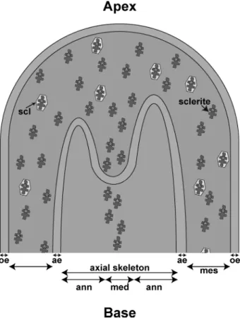
FIGURE 4. Diagram of sclerite incorporation in medullar part of axial
skel-eton at apex of a branch of C. rubrum. From the inside to the outside:
med-ullar part of the axial skeleton (med); annular part of the axial skeleton (ann); aboral epithelium (ae); mesoglea (mes); scleroblast (scl); and oral epithelium (oe). Scleritin from the organic matrix of sclerites is indicated by yellow asterisks.
FIGURE 3. Histology and immunohistochemistry of a longitudinal section of a demineralized branch of C. rubrum. a, overview of a section stained with hemalun/eosin/aniline blue which, respectively, stain nuclei and cytoplamic and connective regions observed with a bright field light microscope. b–h, labeling with antibodies against scleritin (orange to yellow) is merged with blue labeling of cell nuclei with DAPI: b, immunolabeling of apical region of the colony; c, immunolabeling of tissues close to a polyp; d, immunolabeling of oral epithelium; e, immunolabeling at the interface between aboral epithelium and organic matrix of axial skeleton; f, immunolabeling at the base of a branch; g, magnification of e showing labeling of organic matrix of sclerites inside the organic matrix of axial skeleton. h, magnification of f showing labeling of organic matrix of a sclerite inside a scleroblast. Observations from b to h were performed with a confocal microscope. SW, sea water; po, polyp; oe, oral epithelium; sc, scleroblast; gc, gastrodermic canal; ae, aboral epithelium; ax om, organic matrix of axial skeleton; scl om, organic matrix of sclerite. Each image was obtained by Z-stack reconstruction of 10 tissue sections, with 1-m step.
at INRA Institut National de la Recherche Agronomique on June 13, 2018
http://www.jbc.org/
Considering the two hypotheses for the axial skeleton mechanism of growth of C. rubrum, in a previous paper (20) we used antibodies raised against the whole OM extract of sclerites and axial skeleton. However, the antibodies cross-reacted with the two organic matrices, and we could not discriminate between labeling from sclerites or axial skele-ton. In the present study we raised antibodies against scleri-tin which is, as described above, a specific protein of the OM of sclerites. When we used these antibodies against the demineralized axial skeleton, only the scleritin from sclerites located in the medullar part of the axial skeleton was labeled (Fig. 3g). On the opposite side, the OM surrounding the medullar part of the axial skeleton (i.e. the annular part) was not labeled. This result thus confirms that the annular part results from another process than the fusion of sclerites and that the incorporation of sclerites only occurs in the medul-lar part of the axial skeleton (Fig. 4).
Scleritin is the first organic matrix protein fully characterized in an alcyonarian species. With a low molecular mass and a high isoelectric point, it presents no homology with any other sequence available in data bases. Our study illustrates the effi-ciency of combining transcriptomic and proteomic approaches for the identification of coral organic matrix proteins, opens gates for further protein characterization among alcyonarian species, and provides information on sclerites and axial skele-ton formation in C. rubrum.
Acknowledgments–We thank F. Zuberer for assistance during coral sampling and the RAMOGE Agreement for the Alain Vatrican Award; A. Bertucci, N. Techer, and N. Segonds for technical help and fruitful discussions; A. Venn for proofreading the English in the paper; and two anonymous reviewers for comments that helped to improve the paper.
REFERENCES
1. Estroff, L. A. (2008) Introduction: biomineralization. Chem. Rev. 108, 4329 – 4331
2. Lowenstam, H. A., and Weiner, S. (1989) On Biomineralization, pp. 7–16, Oxford University Press, New York
3. Marin, F., Luquet, G., Marie, B., and Medakovic, D. (2008) Molluscan shell proteins: primary structure, origin, and evolution. Curr. Top. Dev. Biol. 80, 209 –276
4. Mann, S. (2001) Biomineralization: Principles and Concepts in Bioinor-ganic Materials Chemistry(Compton, R. G., Davies, S. G., and Evans, J., eds) pp. 6 –23, Oxford University Press, New York
5. Dubois, P., and Chen, C. P. (1989) in Echinoderm Studies (Jangoux, M., and Lawrence, J. M., eds) pp. 109 –178, Rotterdam
6. Ehrlich, H., Koutsoukos, P. G., Demadis, K. D., Pokrovsky, O. S. (2009) Principles of demineralization: modern strategies for the isolation of or-ganic frameworks. Part II. Decalcification. Micron 40, 169 –193 7. Wainwright, S. A., Biggs, W. D., Currey, J. D., and Gosline, J. M. (1982)
Mechanical Design in Organisms, Princeton University Press, Princeton, NJ
8. Harvell, C. D., and Fenical, W. (1989) Chemical and structural defenses of Caribbean gorgonians (Pseudopterogorgia spp.): intracolony localization of defense. Limnol. Oceanogr. 34, 382–389
9. Lewis, J. C., and Wallis, E. V. (1991) The function of surface sclerites in gorgonians (Coelenterata, Octocorallia). Biol. Bull. 181, 275–288 10. Bayer, F. M. (1961) The Shallow-water Octocorallia of the West Indian
Region, Martinus Nijhoff, The Hague
11. Vargas, S., Breedy, O., Siles, F., and Guzman, H. M. (2010) How many
kinds of sclerite? Towards a morphometric classification of gorgoniid mi-croskeletal components. Micron 41, 158 –164
12. Tescione, G. (1973) The Italians and Their Coral Fishing, Fausto Fioren-tino, Napoli
13. Allemand, D. (1993) The biology and skeletogenesis of the Mediterranean red coral: a review. Precious Corals Octocoral Res. 2, 19 –39
14. Morel, J.-P., Rondi-Costanzo, C., and Ugolini, D. (2000) Corallo di Ieri, Corallo di Oggi(Bari, ed), Centro Universitario Europeo per i beni cul-turali, Ravello Edipuglia
15. Weinberg, S. (1976) Revision of the common octocorallia of the Mediter-ranean circalittoral. I. Gorgonacea. Beaufortia 24, 63–103
16. Mateu, G., Traveria, A., Fontarnau, R., and Masso, C. (1986) Biodiagé-nesis mineralògica del Corallium rubrum (L.). Bol. Inst. Esp. Oceanogr.
3,1–12
17. Grillo, M. C., Goldberg, W. M., and Allemand, D. (1993) Skeleton and sclerite formation in the precious red coral Corallium rubrum. Mar. Biol.
117,119 –128
18. Allemand, D., and Grillo, M.-C. (1992) Biocalcification mechanism in gor-gonians:45Ca uptake and deposition by the Mediterranean red coral
Cor-allium rubrum. J. Exp. Zool. 262, 237–246
19. Lacaze-Duthiers, H. (1864) Histoire Naturelle du Corail, pp. 110 –124, J. B. Baillière et Fils, Paris
20. Debreuil, J., Tambutté, S., Zoccola, D., Segonds, N., Techer, N., Allemand, D., and Tambutté, E. (2011) Comparative analysis of the soluble organic matrix of axial skeleton and sclerites of Corallium rubrum: insights for biomineralization. Comp. Biochem. Physiol. B 159, 40 – 48
21. Allemand, D., Cuif, J.-P., Watabe, N., Oishi, M., and Kawaguchi, T. (1994) Bulletin Institut Océanographique Monaco 14,129 –139
22. Cölfen, H., and Antonietti, M. (2005) Mesocrystals: inorganic superstruc-tures made by highly parallel crystallization and controlled alignment. Angew. Chem. Int. Ed. 44,5576 –5591
23. Floquet, N., and Vielzeuf, D. (2011) Mesoscale twinning and crystallo-graphic registers in biominerals. Am. Mineralogist 96, 1228 –1237 24. Borelli, G., Mayer-Gostan, N., Merle, P. L., De Pontual, H., Boeuf, G.,
Allemand, D., and Payan, P. (2003) Composition of biomineral organic matrices with special emphasis on turbot (Psetta maxima) otolith and endolymph. Calcif. Tissue Int. 72, 717–725
25. Dauphin, Y. (2006) Mineralizing matrices in the skeletal axes of two Corallium species (Alcyonacea). Comp. Biochem. Physiol. A 145, 54 – 64
26. Merlin, J. C., and Dele, M. L. (1983) Etude par spectroscopie Raman de résonance de la pigmentation des squelettes calcaires de certains coraux. Bull. Soc. Zool. Fr. 108,289 –301
27. Cvejic, J., Tambutté, S., Lotto, S., Mikov, M., Slacanin, I., and Allemand, D. (2007) Determination of canthaxanthin in the red coral (Corallium rubrum) from Marseilles by HPLC combined with UV and MS detection. Mar. Biol. 152,855– 862
28. Fritsch, E., and Karampelas, S. (2008) Comment on Determination of canthaxanthin in the red coral (Corallium rubrum) from Marseille by HPLC combined with UV and MS detection. Mar. Biol. 154, 929 –930 29. Rahman, M. A., and Isa, Y. (2005) Characterization of proteins from the matrix of spicules from the alcyonarian, Lobophytum crassum. J. Exp. Mar. Biol. Ecol. 321,71– 82
30. Rahman, M. A., Isa, Y., and Uehara, T. (2005) Blood-based proteomics for personalized medicine: examples from neurodegenerative disease. Pro-teomics 5,1–9
31. Rahman, M. A., Isa, Y., and Uehara, T. (2006) Studies on two closely related species of octocorallians: biochemical and molecular characteris-tics of the organic matrices of endoskeletal sclerites. Mar. Biotechnol. 8, 415– 424
32. Rahman, M. A., Isa, Y., Takemura, A., and Uehara, T. (2006) Analysis of proteinaceous components of the organic matrix of endoskeletal sclerites from the alcyonarian Lobophytum crassum. Calcif. Tissue Int. 78, 178 –185
33. Rahman, M. A., Oomori, T., and Uehara, T. (2008) Carbonic anhydrase in calcified endoskeleton: novel activity in biocalcification in alcyonarian. Mar. Biotechnol. 10,31–38
34. Rahman, M. A., and Oomori, T. (2008) Structure, crystallization, and
at INRA Institut National de la Recherche Agronomique on June 13, 2018
http://www.jbc.org/
eral composition of sclerites in the alcyonarian coral. J. Crystal. Growth
310,3528 –3534
35. Kingsley, R. J., Tsuzaki, M., Watabe, N., and Mechanic, G. L. (1990) Col-lagen in the spicule organic matrix of the gorgonian Leptogorgia virgulata. Biol. Bull. 179,207–213
36. Kingsley, R. J., Melaro, E. W., Coe, K. E., Flory, M. R., Skorupa, A. M., and Harclerode, K. A. (1996) Mechanisms of the annual cycling of organic-matrix collagen from spicules of the gorgonian Leptogorgia virgulata. In-vert. Biol. 115,89 –98
37. Kingsley, R. J., and Watabe, N. (1982) Ultrastructural investigation of spic-ule formation in the gorgonian Leptogorgia virgulata (Lamarck) (Coelen-terata: Gorgonacea). Cell Tissue Res. 223, 325–334
38. Kingsley, R. J., and Watabe, N. (1983) Analysis of proteinaceous compo-nents of the organic matrices of spicules from the gorgonian Leptogorgia virgulata. Comp. Biochem. Physiol. B 76, 443– 447
39. Kingsley, R. J., and Watabe, N. (1989) The dynamics of spicule calcification in whole colonies of the gorgonian Leptogorgia virgulata Lam. (Coelen-terata, Gorgonacea). J. Exp. Mar. Biol. Ecol. 133, 57– 65
40. Samata, T., Kingsley, R. J., and Watabe, N. (1989) Ca-binding glycoprotein from the spicules of the octocoral Leptogorgia virgulata. Comp. Biochem. Physiol. B 94,651– 654
41. Goldberg, W. M. (1988) Chemistry, histochemistry, and microscopy of the organic matrix of spicules from a gorgonian coral. Histochem. Cell Biol. 89,163–170
42. Rahman, A., Oomori, T., and Wörheide, G. (2011) Calcite formation in soft coral sclerites is determined by a single reactive extracellular protein. J. Biol. Chem. 286,31638 –31649
43. Fukuda, I., Ooki, S., Fujita, T., Murayama, E., Nagasawa, H., Isa, Y., and Watanabe, T. (2003) Molecular cloning of a cDNA encoding a soluble protein in the coral exoskeleton. Biochem. Biophys. Res. Commun. 304, 11–17
44. Tambutté, S., Tambutté, É., Zoccola, D., and Allemand, D. (2007) in Handbook of Biomineralization: The Biology of Biominerals Structure For-mation(Baeuerlein, E., ed) pp. 243–259, Wiley-VCH, New York 45. Allemand, D., Tambutté, É., Zoccola, D., and Tambutté, S. (2011) in Coral
Reefs: An Ecosystem in Transition(Dubinsky, Z., and Stambler, N., eds) pp. 119 –150, Springer, The Netherlands
46. Tambutté, S., Holcomb, M., Ferrier-Pagès, C., Reynaud, S., Tambutté, É., Zoccola, D., and Allemand, D. (2011) Coral biomineralization: from the gene to the environment. J. Exp. Mar. Biol. Ecol. 408, 58 –78
47. Debreuil, J., Tambutté, S., Zoccola, D., Segonds, N., Techer, N., Marschal, C., Allemand, D., Kosuge, S., and Tambutté, É. (2011) Specific organic matrix characteristics in skeletons of Corallium species. Mar. Biol. 158, 2765–2774
48. Puverel, S., Tambutté, E., Pereira-Mouriès, L., Zoccola, D., Allemand, D., and Tambutté, S. (2005) Soluble organic matrix of two scleractinian cor-als: partial and comparative analysis. Comp. Biochem. Physiol. B 141, 480 – 487
49. Altschul, S. F., Madden, T. L., Schäffer, A. A., Zhang, J., Zhang, Z., Miller, W., and Lipman, D. J. (1997) Gapped BLAST and PSI-BLAST: a new generation of protein database search programs. Nucleic Acids Res. 25, 3389 –3402
50. Zoccola, D., Tambutté, E., Sénégas-Balas, F., Michiels, J. F., Failla, J. P., Jaubert, J., and Allemand, D. (1999) Cloning of a calcium channel␣1 subunit from the reef-building coral, Stylophora pistillata. Gene 227, 157–167
51. Moya, A., Tambutté, S., Bertucci, A., Tambutté, E., Lotto, S., Vullo, D., Supuran, C. T., Allemand, D., and Zoccola, D. (2008) Carbonic anhydrase in the scleractinian coral Stylophora pistillata: characterization, localiza-tion, and role in biomineralization. J. Biol. Chem. 283, 25475–25484 52. Jain, E., Bairoch, A., Duvaud, S., Phan, I., Redaschi, N., Suzek, B. E,
Martin, M. J, McGarvey, P., and Gasteiger, E. (2009) Infrastructure for the life sciences: design and implementation of the UniProt website. BMC Bioinformatics 10,136
53. Geer, L. Y., Marchler-Bauer, A., Geer, R. C., Han, L., He, J., He, S., Liu, C., Shi, W., and Bryant, S. H. (2010) The NCBI BioSystems database. Nucleic Acids Res. 38,D492– 496
54. Emanuelsson, O., Brunak, S., von Heijne, G., and Nielsen, H. (2007)
Lo-cating proteins in the cell using TargetP, SignalP, and related tools. Nat. Protocols 2,953–971
55. Horton, P., Park, K. J., Obayashi, T., Fujita, N., Harada, H., Adams-Collier, C. J., and Nakai, K. (2007) WoLF PSORT: protein localization predictor. Nucleic Acids Res. 35,W585-W587
56. Schägger, H. (2006) Tricine-SDS-PAGE. Nat. Protocols 1, 16 –22 57. Kozak, M. (1984) Compilation and analysis of sequences upstream from
the translational start site in eukaryotic mRNAs. Nucleic Acids Res. 12, 857– 872
58. Mankad, R. V., Gimelbrant, A. A., and McClintock, T. S. (1998) Consensus translational initiation sites of marine invertebrate phyla. Biol. Bull. 195, 251–254
59. Weiner, S. (1986) Organization of extracellularly mineralized tissues: a comparative study of biological crystal growth. CRC Crit. Rev. Biochem.
20,365– 408
60. Treccani, L., Khoshnavaz, S., Blank, S., von Roden, K., Schulz, U., Weiss, I. M., Mann, K., Radmacher, M., and Fritz, H. M. (2005) in Biopolymers Online(Fahnestock, S. R., and Steinbu¨chel, A., eds) pp. 289 –298, Wiley-VCH, New York
61. Ameye, L., De Becker, G., Killian, C., Wilt, F., Kemps, R., Kuypers, S., and Dubois, P. (2001) Proteins and saccharides of the sea urchin organic ma-trix of mineralization: characterization and localization in the spine skel-eton. J. Struct. Biol. 134, 56 – 66
62. Luquet, G., and Marin, F. (2004) Biomineralisations in crustaceans: stor-age strategies. Comptes Rendus Palevol. 3, 515–534
63. Mann, K., Poustka, A. J., and Mann, M. (2010) Phosphoproteomes of Strongylocentrotus purpuratusshell and tooth matrix: identification of a major acidic sea urchin tooth phosphoprotein, phosphodontin. Proteome Sci. 8,1–14
64. Furuhashi, T., Schwarzinger, C., Miksik, I., Smrz, M., and Beran, A. (2009) Molluscan shell evolution with review of shell calcification hypothesis. Comp. Biochem. Physiol. B 154,351–371
65. Borbas, J. E., Wheeler, A. P., and Sikes, C. S. (1991) Molluscan shell matrix phosphoproteins: correlation of degrees of phosphorylation to shell min-eral microstructure and to in vitro regulation of minmin-eralization. J. Exp. Zool. 258,1–13
66. Hecker, A., Testenière, O., Marin, F., and Luquet, G. (2003) Phosphory-lation of serine residues is fundamental for the calcium-binding ability of Orchestin, a soluble matrix protein from crustacean calcium storage structures. FEBS Lett. 535, 49 –54
67. Testenière, O., Hecker, A., Le Gurun, S., Quennedey, B., Graf, F., and Luquet, G. (2002) Characterization and spatiotemporal expression of or-chestin, a gene encoding an ecdysone-inducible protein from a crustacean organic matrix. Biochem. J. 361, 327–335
68. Sarashina, I., and Endo, K. (2001) The complete primary structure of mol-luscan shell protein 1 (MSP-1), an acidic glycoprotein in the shell matrix of the scallop Patinopecten yessoensis. Mar. Biotechnol. 3, 362–369 69. Allemand, D., Tambutté, E., Girard, J. P., and Jaubert, J. (1998) Organic
matrix synthesis in the scleractinian coral Stylophora pistillata: role in biomineralization and potential target of the organotin tributyltin J. Exp. Biol. 201,2001–2009
70. Weiner, S., Traub, W., and Lowenstam, M. A. (1983) in Biomineralization and Biological Metal Accumulation(Westbrock, P., and De Jong, E. W., eds) pp. 205–224, Reidel Publishing Company, Dordrecht, The Nether-lands
71. Kingsley, R. J., and Watabe, N. (1982) Ultrastructure of the axial region in Leptogorgia virgulata(Cnidaria: Gorgonaceae). Trans. Am. Microsc. Soc.
101,325–339
72. Weiner, S., and Addadi, L. (1997) Design strategies in mineralized biolog-ical materials. J. Mater. Chem. 5, 689 –702
73. Marin, F., and Luquet, G. (2004) Molluscan shell proteins. Comptes Ren-dus Palevol. 3,469 – 492
74. Marie, B., Le Roy, N., Zanella-Cléon, I., Becchi, M., and Marin, F. (2011) Molecular evolution of mollusc shell proteins: insights from proteomic analysis of the edible mussel Mytilus. J. Mol. Evol. 72, 531–546 75. Marie, B., Trinkler, N., Zanella-Cleon, I., Guichard, N., Becchi, M.,
Pail-lard, C., and Marin, F. (2011) Proteomic identification of novel proteins from the calcifying shell matrix of the Manila clam Venerupis
at INRA Institut National de la Recherche Agronomique on June 13, 2018
http://www.jbc.org/
rum. Mar. Biotechnol. 13, 955–962
76. Marie, B., Zanella-Cléon, I., Guichard, N., Becchi, M., and Marin, F. (2011) Novel proteins from the calcifying shell matrix of the Pacific oyster Cras-sostrea gigas. Mar. Biotechnol. 13, 1159 –1168
77. Sethmann, I., Helbig, U., and Worheide, G. (2007) Octocoral sclerite ul-trastructures and experimental approach to underlying biomineralisation principles. Cryst. Eng. Comm. 9, 1262–1268
78. Segev, N., Tokarev, A. A., and Alfonso, A. (2009) in Trafficking Inside Cells Pathways, Mechanisms, and Regulation(Segev, N., ed) pp. 1–12, Landes Bioscience and Springer Science⫹Business Media, New York
79. Capasso, J. M., Keenan, T. W., Abeijon, C., and Hirschberg, C. B. (1989)
Mechanism of phosphorylation in the lumen of the Golgi apparatus: translocation of adenosine 5⬘-triphosphate into Golgi vesicles from rat liver and mammary gland. J. Biol. Chem. 264, 5233–5240
80. Anitei, M., and Hoflack, B. (2011) Exit from the trans-Golgi network: from molecules to mechanisms. Curr. Opin. Cell Biol. 23, 443– 451
81. Allemand, D., and Bénazet-Tambutté, S. (1996) Dynamics of calcification in the Mediterranean red coral, Corallium rubrum (Linnaeus) (Cnidaria, Octocorallia). J. Exp. Zool. 276, 270 –278
82. Marschal, C., Garrabou, J., Harmelin, J. G., and Pichon, M. (2004) A new method for measuring growth and age in the precious red coral Corallium rubrum(L.). Coral Reefs 23, 423– 432
at INRA Institut National de la Recherche Agronomique on June 13, 2018
http://www.jbc.org/
Michel Samson, Denis Allemand and Sylvie Tambutté
Julien Debreuil, Éric Tambutté, Didier Zoccola, Emeline Deleury, Jean-Marie Guigonis,
Corallium rubrum
Sclerites of Red Coral,
doi: 10.1074/jbc.M112.352005 originally published online April 13, 2012 2012, 287:19367-19376.
J. Biol. Chem.
10.1074/jbc.M112.352005 Access the most updated version of this article at doi:
Alerts:
When a correction for this article is posted •
When this article is cited •
to choose from all of JBC's e-mail alerts Click here
Supplemental material:
http://www.jbc.org/content/suppl/2012/04/13/M112.352005.DC1 http://www.jbc.org/content/287/23/19367.full.html#ref-list-1This article cites 69 references, 5 of which can be accessed free at
at INRA Institut National de la Recherche Agronomique on June 13, 2018
http://www.jbc.org/