Polymyxins: Antibacterial Activity,
Susceptibility Testing, and Resistance
Mechanisms Encoded by Plasmids or
Chromosomes
Laurent Poirel,a,b,cAurélie Jayol,a,b,cPatrice Nordmanna,b,c,d
Emerging Antibiotic Resistance Unit, Medical and Molecular Microbiology, Department of Medicine, University of Fribourg, Fribourg, Switzerlanda; French INSERM European Unit, University of Fribourg (LEA-IAME), Fribourg, Switzerlandb; National Reference Center for Emerging Antibiotic Resistance, Fribourg, Switzerlandc; University of Lausanne and University Hospital Center, Lausanne, Switzerlandd
SUMMARY . . . 558 INTRODUCTION. . . 558 Structure. . . 559 Mechanism of Action. . . 559 Spectrum of Activity. . . 560 Pharmacodynamics . . . 560
USE OF COLISTIN IN HUMAN AND VETERINARY MEDICINE. . . 560
Use in Human Medicine. . . 560
Commercial formulations. . . 561
Routes of administration . . . 561
Pharmacokinetics. . . 561
Dosing regimen . . . 562
(i) Patients with normal renal function . . . 562
(ii) Patients with renal insufficiency . . . 563
Toxicity. . . 564
Use in Veterinary Medicine . . . 564
METHODS FOR SUSCEPTIBILITY TESTING. . . 565
Dilution Methods . . . 565
Broth dilution methods. . . 565
(i) Broth microdilution method. . . 565
(ii) Broth macrodilution method (or tube dilution method) . . . 565
Agar dilution method. . . 566
Routine Susceptibility Testing Methods. . . 566
Nonautomatic systems . . . 566
(i) DD test (Kirby-Bauer procedure) . . . 566
(ii) Etest strips. . . 567
(iii) UMIC system . . . 567
Automatic systems . . . 567
(i) Vitek 2 system . . . 567
(ii) Phoenix automated microbiology system . . . 567
(iii) MicroScan system . . . 568
(iv) Sensititre system . . . 568
Impact of Materials on MIC Determination . . . 568
Impact of medium . . . 568
Impact of powder composition . . . 568
Impact of the composition and treatment of plates . . . 568
Presence or absence of P-80 . . . 569
Impacts of Subcultures and Storage on MICs . . . 569
Impact of subcultures . . . 569
Impact of storage . . . 569
Interpretive Criteria . . . 569
Quality Controls. . . 569
Correlation between MICs of Colistin and Polymyxin B . . . 570
Qualitative Detection Techniques . . . 570 (continued)
Published in "Clinical Microbiology Reviews 30(2): 557–596, 2017"
which should be cited to refer to this work.
Rapid detection of heterogeneous populations among colistin-resistant
Gram-negative bacteria by use of capillary electrophoresis . . . 570
Rapid detection of colistin-resistant A. baumannii isolates by use of the Micromax assay. . . 570
Rapid detection of colistin-resistant Enterobacteriaceae isolates by use of the Rapid Polymyxin NP test . . . 571
Selective medium . . . 571
Genotypic Methods. . . 571
RESISTANCE MECHANISMS IN ENTEROBACTERIACEAE . . . 572
Intrinsic Resistance Mechanisms in Proteus mirabilis and Serratia marcescens . . . 572
Mechanisms Responsible for Acquired Resistance in Enterobacteriaceae . . . 572
Genes encoding LPS-modifying enzymes . . . 573
(i) The pmrC gene . . . 573
(ii) The pmrHFIJKLM operon and the pmrE gene . . . 573
(iii) The pmrA and pmrB genes, which encode the PmrAB two-component system. . 573
(iv) The phoP and phoQ genes, which encode the PhoPQ two-component system. . 574
Regulators of the PmrAB and PhoPQ Two-Component Systems . . . 574
The mgrB gene . . . 574
The crrAB operon . . . 574
The Intrinsic Regulator RamA . . . 578
Plasmid-Mediated Resistance to Polymyxins . . . 578
Other Mechanisms Contributing to Polymyxin Resistance in Enterobacteriaceae. . . 580
Hyperproduction of CPS. . . 580
Role of porins . . . 583
Role of efflux pumps . . . 583
Mechanisms of Polymyxin Resistance in Pseudomonas aeruginosa and Acinetobacter baumannii . . . 583
Pseudomonas aeruginosa . . . 583
Acinetobacter baumannii. . . 583
EPIDEMIOLOGY OF RESISTANCE TO POLYMYXINS . . . 584
General Epidemiology of Resistance to Polymyxins . . . 584
North America. . . 584 South America. . . 584 Europe . . . 584 Middle East . . . 587 Africa . . . 587 Asia. . . 587 Risk Factors. . . 587
Specific Epidemiology of the Plasmid-Mediated mcr-1 Resistance Gene. . . 588
CONCLUSIONS . . . 588
ACKNOWLEDGMENTS. . . 589
REFERENCES . . . 589
AUTHOR BIOS. . . 596
SUMMARY Polymyxins are well-established antibiotics that have recently regained significant interest as a consequence of the increasing incidence of infections due to multidrug-resistant Gram-negative bacteria. Colistin and polymyxin B are being seri-ously reconsidered as last-resort antibiotics in many areas where multidrug resis-tance is observed in clinical medicine. In parallel, the heavy use of polymyxins in veterinary medicine is currently being reconsidered due to increased reports of polymyxin-resistant bacteria. Susceptibility testing is challenging with polymyxins, and currently available techniques are presented here. Genotypic and phenotypic meth-ods that provide relevant information for diagnostic laboratories are presented. This re-view also presents recent works in relation to recently identified mechanisms of poly-myxin resistance, including chromosomally encoded resistance traits as well as the recently identified plasmid-encoded polymyxin resistance determinant MCR-1. Epidemio-logical features summarizing the current knowledge in that field are presented.
KEYWORDS Gram-negative bacteria, MCR-1, lipopolysaccharide, polymyxins, toxicity INTRODUCTION
C
olistin (also known as polymyxin E) is a polypeptide antibiotic that was originally isolated in 1947 from the soil bacterium Paenibacillus polymyxa subsp. colistinus (1). Colistin and polymyxin B belong to the class of polymyxins, which is one of the primary classes of antibiotics with activity against most Gram-negative bacteria.Structure
The chemical structure of polymyxins is similar to that of cationic antimicrobial peptides (CAMPs) (defensins and gramicidins), which represent the first line of defense against bacterial colonization in eukaryotic cells (2). Polymyxins are cationic polypep-tides that consist of a cyclic heptapeptide possessing a tripeptide side chain acylated at the N terminus by a fatty acid tail (3,4) (Fig. 1). The inherent toxicity of colistin may be explained by the hydrophobic properties of the N-terminal fatty acyl segment, which also accounts significantly for its antimicrobial activity, and also by positions 6 and 7, which are very important (5,6).
Colistin and polymyxin B differ by only a single amino acid in the peptide ring, with a phenylalanine in polymyxin B and a leucine in colistin (Fig. 1) (7). Polymyxin B is administered directly as an active antibiotic, whereas colistin is administered as an inactive prodrug, colistin methanesulfonate (also known as colistimethate [CMS]) (Fig. 1) (7).
The terms “colistin” and “colistimethate” are not interchangeable, since they corre-spond to different forms of colistin available for clinical use (4). Indeed, colistimethate sodium is a polyanionic inactive prodrug that is less toxic than colistin sulfate (Fig. 1)
(4, 8). Colistimethate is formed by the reaction of colistin with formaldehyde and
sodium bisulfite (9). This prodrug is transformed in aqueous media, and also in vivo in biological fluids, and is converted into colistin and several inactive methanesulfonated compounds (10,11).
Mechanism of Action
The target of polymyxins is the outer membrane of Gram-negative bacteria. Because of an electrostatic interaction occurring between the ␣,␥-diaminobutyric acid (Dab) residue of the positively charged polymyxin on one side and the phosphate groups of the negatively charged lipid A membrane on the other side, divalent cations (Ca2⫹and Mg2⫹) are displaced from the negatively charged phosphate groups of membrane lipids (12). The lipopolysaccharide (LPS) is therefore destabilized, consequently increas-ing the permeability of the bacterial membrane, leadincreas-ing to leakage of the cytoplasmic
content and ultimately causing cell death (4,13). Note that even though the LPS is the initial target, the exact mode of action of polymyxins still remains unclear.
Another antibacterial mechanism is the endotoxin effect. The endotoxin of Gram-negative pathogens corresponds to the lipid A portion of the LPS; polymyxins have the ability to bind to and neutralize this LPS molecule released during cell lysis (14).
Finally, another mode of action of the polymyxins is the inhibition of vital respiratory enzymes (inhibition of type II NADH-quinone oxidoreductases [NDH-2]) in the bacterial inner membrane (15).
Spectrum of Activity
Polymyxins have a narrow antibacterial spectrum, mainly against common Gram-negative bacteria. They are active against most members of the Enterobacteriaceae family, including Escherichia coli, Enterobacter spp., Klebsiella spp., Citrobacter spp., Salmonella spp., and Shigella spp. Polymyxins also have significant activity against common nonfermentative Gram-negative bacteria, including Acinetobacter baumannii, Pseudomonas aeruginosa, and Stenotrophomonas maltophilia (13).
Conversely, some species are naturally resistant to polymyxins, including Proteus spp., Morganella morganii, Providencia spp., Serratia marcescens, Pseudomonas mallei, Burkholderia cepacia, Chromobacterium spp., Edwardsiella spp., Brucella, Legionella, Cam-pylobacter, and Vibrio cholerae. Polymyxins are not active against Gram-negative cocci (Neisseria spp.), Gram-positive bacteria, and anaerobic bacteria (13).
Pharmacodynamics
The antibacterial effect of colistin is concentration dependent (4, 16–18). The pharmacokinetic-pharmacodynamic (PK-PD) index that best predicts the antibacterial activity against A. baumannii and P. aeruginosa is the ratio of the area under the concentration-time curve for free drug from 0 to 24 h to the MIC (fAUC0 –24/MIC), with this index being superior to the maximum concentration of drug in serum (Cmax)/MIC relationship, suggesting that time-averaged exposure to colistin is more important than the achievement of high peak concentrations (19–21). An average steady-state plasma colistin concentration of 2g/ml has been suggested as a reasonable target value for isolates with MICs ofⱕ1g/ml, maximizing the antimicrobial activity while minimizing the risk of nephrotoxicity (22). An inadequate AUC/MIC ratio likely leads to treatment failure. The colistin antibacterial effect is extremely rapid, occurring as early as 5 min after exposure (17,18,23,24).
A postantibiotic effect was observed against Klebsiella pneumoniae, P. aeruginosa, and A. baumannii (25). However, it is important to highlight that polymyxins have minimal postantibiotic effects at clinically relevant concentrations. Despite the major initial killing rate observed against colistin-susceptible strains exposed to colistin alone, regrowth has been reported for A. baumannii (17) and K. pneumoniae (18) in static time-kill studies. Colistin heteroresistance, a phenomenon corresponding to the emer-gence of a colistin-resistant subpopulation (that can grow in the presence ofⱖ4g/ml of colistin) within a susceptible population (i.e., with a MIC of ⱕ2g/ml), has been observed for A. baumannii (26,27), K. pneumoniae (18,28), and P. aeruginosa (23). USE OF COLISTIN IN HUMAN AND VETERINARY MEDICINE
Use in Human Medicine
After its discovery in 1947, colistin was used in Japan and Europe during the 1950s (29). Then, after its approval by the U.S. FDA in 1959, colistimethate (CMS), the inactive prodrug of colistin, replaced colistin for parenteral administration (29). Colistin and CMS have been used widely for decades for treatment of infections caused by Gram-negative bacteria. However, in the 1970s, because of their toxicity, especially nephro-toxicity (30), their use was reconsidered. They were then replaced by novel, more active and less toxic antibiotics, such as aminoglycosides, quinolones, and-lactams. For 20 years, the use of colistin was restricted to ophthalmic and topical uses. Systemic or nebulized colistin was used only for cystic fibrosis patients.
However, the increasing prevalence of multidrug-resistant (MDR) Gram-negative bacteria (31), particularly K. pneumoniae, A. baumannii, and P. aeruginosa, has forced physicians to reintroduce systemic polymyxin as a valuable therapeutic option (4,
13,32).
Considering the paucity of novel antibiotics, colistin is currently often the only effective antibiotic agent against MDR organisms, particularly carbapenemase-pro-ducing bacteria.
Commercial formulations. There are more than 30 polymyxin molecules, among which there are five main chemical compounds (polymyxins A to E), each containing multiple components. Although colistin (polymyxin E) and polymyxin B are both used in clinical practice (33), colistin is the most widely used polymyxin (23). The two most common commercially available parenteral formulations of the colistin prodrug, CMS, are Colomycin (Forest Laboratories UK Limited, Dartford, United Kingdom), primarily employed in Europe, and Coly-Mycin M Parenteral (Monarch Pharmaceuticals, Inc., Bristol, TN), primilarily employed in the United States (34). Unfortunately, the vials of both formulations contain different dry powder quantities, and the two products are differently labeled, with Colomycin being labeled in international units (IU) of CMS and Coly-Mycin M Parenteral being labeled in milligrams of CMS or colistin base activity (CBA) (34). The conversion is as follows: 1 million units (MU) CMS⫽ 80 mg CMS ⫽ 30 mg CBA (35). To add to the confusion, some other brands corresponding to generic products are now available (36). The multiplicity of terms used to express contents of vials and dose regimens unfortunately creates confusion and does not allow any meaningful comparison of data collected from studies performed in different parts of the world.
Routes of administration. Colistin sulfate can be administered orally as tablets and syrup for selective digestive tract decontamination (no absorption) and topically for the treatment of bacterial skin infections (13). CMS, the less toxic prodrug, has different administration routes, i.e., parenteral (including intravenous) and intramuscular, but intrathecal or intraventricular administration is also possible (13). The intramuscular injection is rarely used in clinical practice because it may be very painful locally, and also because its absorption is variable (33). Both colistin sulfate and CMS can be delivered through inhalation by aerosol therapy, but there is a higher frequency of bronchoconstriction with colistin sulfate (33). Delivery of CMS by inhalation and by the intrathecal and intraventricular routes allows much higher concentrations in lung fluid and cerebrospinal fluid, respectively, than those seen with systemic administration. Moreover, those routes of administration lead to negligible plasma exposure and are less toxic (in particularly less nephrotoxic) (22).
In aqueous solutions, colistimethate sodium is transformed into colistin; therefore, it should be administered shortly after reconstitution to avoid the toxicity associated with colistin (37).
Pharmacokinetics. Because of their discovery and their introduction into clinical use more than 50 years ago, polymyxins were never subjected to the drug development approval process currently required by international drug regulatory authorities. Con-sequently, the PK and PD data on the rational use of polymyxins (maximizing antibac-terial activity and minimizing toxicity and development of resistance) were not avail-able until recently. The fact that, until recently, plasma concentrations of CMS and formed colistin could not be differentiated because of a lack of suitable techniques was another obstacle limiting progress in this area. The recent development of chromato-graphic methods allowing quantitative assessment of each compound separately significantly contributed to the renewed interest in prescribing colistin and colistime-thate (38, 39). It was clearly demonstrated that the observed antimicrobial activity results from the action of colistin itself, which is generated in vivo when CMS is given. For accurate PK information, a prerequisite is to quantify separately the inactive prodrug (CMS) and the active entity (colistin) (34).
After its parenteral administration, a large proportion of CMS is eliminated mainly through the kidneys by glomerular filtration and tubular secretion (Fig. 2A) (11).
Because in a healthy individual the clearance of CMS by the kidneys is much higher than its conversion clearance to colistin, no more than 20 to 25% of a CMS dose is hydrolyzed in vivo into an active colistin entity (7). Consequently, the colistin concen-trations resulting from the original CMS administration are low. In contrast to CMS, colistin is eliminated predominantly by a nonrenal way because of its extensive renal tubular reabsorption (Fig. 2A) (11,40). Although colistin is poorly excreted in urine, the urinary concentration of colistin may be relatively high after administration of CMS due to the conversion of CMS (highly excreted by the kidneys) into colistin within the urinary tract (7).
In contrast to colistin, polymyxin B is administered directly in its active antibacterial form. As for colistin formed from CMS, polymyxin B is subject to very extensive renal tubular reabsorption and is thus eliminated mainly by a nonrenal clearance mecha-nism(s) (Fig. 2B) (7).
Dosing regimen. Due to renewed interest in its use in the context of infections caused by multidrug-resistant bacteria, and considering the increasing rates of resis-tance to colistin currently observed, CMS has to be administered carefully. In particular, the regimens allowing maximal antibacterial activity and minimal development of resistance have to be defined accurately, since the regimens need to minimize adverse effects (23). A study analyzing product data characteristics of intravenous CMS revealed a lack of uniformity between manufacturers, with quite broad variations in term of indications, dose regimens (3 to 12 MU/day), and PK (36). Moreover, dosing regimens given by manufacturers are often discordant with the dosing regimens recommended by the recent literature (21,34,41).
(i) Patients with normal renal function. The currently used dosage regimens of CMS generate suboptimal exposure to colistin in many critically ill patients, in particular in renally competent patients. Two studies reported low plasma colistin Cmax values FIG 2 Overview of the pharmacokinetic pathways for colistimethate (CMS) and colistin (A) and for polymyxin B (B). The thicknesses of the arrows indicate the relative magnitudes of the respective clearance pathways when kidney function is normal. CMS includes fully and all partially methanesulfon-ated derivatives of colistin. After administration of CMS, extensive renal excretion of the prodrug occurs, with some of the excreted CMS being converted to colistin within the urinary tract. (The figure is based in part on data from reference7.)
following administration of 174 mg to 250 mg (2 to 3 MU) of CMS every 8 or 12 h, with steady-state levels of 1.15 to 5.14 g/ml or 0.68 to 4.65 g/ml, respectively (42). Moreover, a significant delay in obtaining steady-state plasma concentrations of formed colistin was reported for CMS treatment started without administration of a loading dose (43). In the latter study, concentrations of colistin in the plasma were reported to be below the MIC breakpoint (2 g/ml), which is a main drawback considering that a delayed initiation of appropriate antibiotic therapy has been shown to be associated with increased mortality rates, in particular for critically ill patients (44). In addition, low colistin concentrations may induce the amplification of colistin-resistant subpopulations (18,45). Interestingly, on consideration of the current dose range product recommendations for CMS, it was confirmed that its administration at the upper limit to patients with normal renal function resulted in low and potentially suboptimal plasma colistin concentrations, especially when the MIC for the infecting bacterial strain was in the upper range (2g/ml) or if the infection was associated with a high bacterial inoculum (21). That study also revealed that steady-state plasma colistin concentrations are highly variable, with up to a 10-fold range achieved across patients at a given creatinine clearance (21).
In contrast, there is a relatively low interpatient variability (3.3-fold) across a wide range of creatinine clearance values following administration of polymyxin B (46). Considering that polymyxin B is not given as a prodrug, it is easier to rapidly achieve a desired plasma concentration of polymyxin B (46).
There is no consensus about dosing regimens, even though recently published dosing suggestions seem to be widely accepted (19). Compared to those suggested by the manufacturers, the regimens in recent studies support the administration of a loading dose and of higher doses of CMS in order to achieve adequate colistin concentrations leading to a better therapeutic effect (21,41,47). The dosing regimen currently recommended by the recent literature (for patients with good renal function) is a loading dose of 4.5 MU of CMS followed by maintenance doses of 4.5 MU twice daily (48–50). A colistin-containing combination therapy has to be considered if the infecting pathogen shows an MIC of colistin above 1g/ml, if there is a high inoculum, or in dealing with deep-seated infections (e.g., in lungs). One therefore has to consider adding antibiotics to colistin regimens, especially for patients with relatively normal renal function (21,22).
Data about the pharmacokinetics, effectiveness, and safety of polymyxins were recently reviewed by the European Medicines Agency (EMA). There have been recom-mended changes in terms of product information in order to ensure the safer use of polymyxins (51). According to the EMA, polymyxins should be reserved for the treat-ment of serious infections due to aerobic Gram-negative pathogens with limited treatment options (51). Also, they should be given with another suitable antibiotic when possible. The recommended dose for CMS in adults is 9 MU daily in 2 or 3 divided doses as a slow intravenous infusion. For dealing with critically ill patients, a loading dose of 9 MU should be given. For patients with renal impairment, doses should obviously be reduced, with consideration of the creatinine clearance.
Because the efficacy and toxicity of colistin are dose dependent, it is crucial that optimal dose regimens be used to maximize the antimicrobial activity and to minimize adverse effects and the development of resistance. This is especially important for critically ill patients, as they are most at risk for high morbidity and mortality (52).
(ii) Patients with renal insufficiency. A study showed that colistin levels were elevated in patients with renal insufficiency, presumably due to decreased elimination of the antibiotic generating a higher rate of conversion of CMS to colistin (43). Development of nephrotoxicity is consequently higher in patients with renal insuffi-ciency than in patients with normal renal function (53).
Dalfino et al. (54) suggested a new dose adjustment for high-dose colistin therapy for patients with renal insufficiency. For patients with creatinine clearance of 20 to 50 ml/min, they recommend a loading dose of 9 MU and maintenance doses of 4.5 MU
every 24 h. For patients with creatinine clearance of⬍20 ml/min, they recommend a loading dose of 9 MU and maintenance doses of 4.5 MU every 48 h (21,55).
Toxicity. Rates of toxicity following intravenous administration of CMS are consid-ered lower today than those observed in previous studies, and it has to be mentioned that the criteria for defining toxicity have also been updated (56). The lower toxicity may be related to the fact that there are fewer chemical impurities in CMS but also to the fact that monitoring in intensive care units (ICUs) is better nowadays and the coadministration of other nephrotoxic drugs is significantly avoided (33).
Colistin has a narrow therapeutic window, and major adverse effects related to its parenteral use are neurotoxicity and nephrotoxicity. Neurotoxicity is dose dependent and reversible (55) and may cause peripheral and facial paresthesia, weakness, dizzi-ness/vertigo, visual disturbances, confusion, ataxia, and neuromuscular blockade, even leading to respiratory failure or apnea (56). The most common neurotoxicological effect is paresthesia (occurring in 27% of patients), and there is no report of neuromuscular blockade or apnea in the recent literature (56). Nephrotoxicity is the most common and concerning adverse effect, especially with the newly recommended high-dose regimen. Similarly to neurotoxicity, nephrotoxicity is dose dependent. The risk of colistin-associated nephrotoxicity increases with plasma colistin concentrations above 2.5 to 3 g/ml, as revealed by recent PK-PD analyses (57). Other risk factors for nephrotoxicity include coadministration of other drugs that are also nephrotoxic (anti-inflamatory drugs, vancomycin, or aminoglycoside antibiotics) and patient-related factors (ad-vanced age, male sex, hypoalbuminemia, hyperbilirubinemia, preexisting chronic kid-ney disease, and severity of illness) (33, 56). Nephrotoxicity is reported to be a rapid-onset effect, with most cases occurring within the first week of treatment, and is mostly reversible (33,55). Rates of nephrotoxicity in recent studies ranged from 6% to 55% (33). The large range of nephrotoxicity rates may be explained partially by different definitions of renal failure, the dosing regimens used, the concomitant administration of nephrotoxic drugs, and the use of colistin monitoring to adapt dosing regimens. The RIFLE (risk–injury–failure–loss– end-stage renal disease) classification is used to deter-mine colistin-associated nephrotoxicity (58).
Two recent comparative studies involving large numbers of patients showed that the nephrotoxicity rates were lower for polymyxin B than for CMS/colistin (59,60). Use in Veterinary Medicine
As opposed to the case in human medicine, in veterinary medicine colistin has been used extensively for decades for the treatment and prevention of infectious diseases. The majority of polymyxin consumption corresponds to orally administered forms, with differ-ent formulations (premix, powder, or oral solutions). The main usage is related to differ- entero-bacterial infections, and in particular to gastrointestinal infections caused by E. coli in poultry and pigs within intensive husbandry systems (61). Apart from this common usage for treating infections caused by Enterobacteriaceae, another usage corresponds to growth promotion, which is a common practice worldwide. Furthermore, the fact that only a thin line exists between oral metaphylactic therapy, preventive starter rations, and growth promotion adds to the problem. In 2011, polymyxins were the fifth most sold class of antimicrobials (7%) for treating food-producing animals in Europe (61).
Despite this extensive use in veterinary medicine, the resistance rate to colistin in E. coli strains recovered from healthy animals remains⬍1% in many European countries (62). However, resistance to colistin has increasingly been reported (10%) among porcine-pathogenic E. coli strains in Belgium (63), and the emergence of resistance has been described for cattle (64). Moreover, some recent data revealed the possibility of horizontal transmission from farm animals to humans in Asia (65). Given the increasing need to retain the efficacy of colistin to treat MDR infections in humans, the potential for spreading colistin-resistant isolates from animals to humans, and the recent iden-tification of colistin-resistant Enterobacteriaceae organisms harboring a plasmid-borne colistin resistance determinant in animals and food products (see below), the use of colistin in veterinary medicine is being reevaluated. As a very recent example, the
formal Ministry of Agriculture of China decided to ban colistin as a feed additive for animals (66). Also, the European Medicines Agency provided a position paper in June 2016, in which updated advice on the use of colistin products in animals within the European Union is provided (67).
METHODS FOR SUSCEPTIBILITY TESTING
Despite such a long term of clinical use (decades), the optimal method for polymyxin susceptibility testing still remains undefined. However, the recent emergence of MDR Gram-negative bacteria and the subsequent increased use of colistin prompted the scien-tific community to develop rapid and reliable methods to determine the susceptibility of isolates to polymyxins, as this is now an urgent need in clinical laboratories. Polymyxin susceptibility testing is now a major challenge, as human infections with colistin-resistant Gram-negative bacteria are associated with higher patient mortality (68). The difficulties in testing susceptibility to polymyxins are diverse, including poor diffusion of polymyxins into agar, the inherent cationic properties of polymyxins, the occurrence of heteroresistance to polymyxins in many species, and the lack of a reliable reference method that may allow reliable comparisons of commercial tests (69,70).
Dilution Methods
The aim of dilution methods is to determine the MIC, corresponding to the lowest concentration of polymyxin that inhibits visible bacterial growth after an incubation of 16 to 24 h at 35⫾ 2°C.
Broth dilution methods. Broth dilution is a technique in which a bacterial suspen-sion at a predetermined concentration is tested against various concentrations of antimicrobial agent in a liquid medium with a predetermined formulation. Two types of broth dilution methods are available: (i) the broth macrodilution method, performed with a minimum volume of 2 ml in standard test tubes; and (ii) the broth microdilution (BMD) method, performed with a volume of 0.05 to 0.1 ml in microtitration trays.
(i) Broth microdilution method. BMD is the reference susceptibility test method. It is currently the only method recommended by the Clinical and Laboratory Standards Institute (CLSI) and the European Committee on Antimicrobial Susceptibility Testing (EUCAST) (71,72) for polymyxin antimicrobial susceptibility testing.
According to CLSI recommendations, BMD is performed with cation-adjusted Mueller-Hinton broth (CA-MHB), a range of 2-fold dilutions of polymyxins (ranging from 0.12 to 512g/ml), and a final bacterial inoculum of 5 ⫻ 105CFU/ml in each well (73). BMD is considered to be the optimal method and is currently recommended for susceptibility testing in the recent document proposed by the joint CLSI-EUCAST Polymyxin Breakpoints Working Group (http://www.eucast.org/fileadmin/src/media /PDFs/EUCAST_files/General_documents/Recommendations_for_MIC_determination _of_colistin_March_2016.pdf).
However, BMD is quite laborious, and manual preparation (if the technique used is not an automated one) of antibiotic solutions may lead to significant errors. It is therefore not adaptable for most clinical microbiology laboratories. Furthermore, non-reproducible and noninterpretable MIC results have been reported due to the presence of skip wells (i.e., wells that exhibit no growth, whereas growth is observed in wells with higher antibiotic concentrations) for Enterobacter species (69), P. aeruginosa (72), and A. baumannii (73). This phenomenon might be caused by heteroresistant subpopulations for Enterobacter spp. (69). In parallel, “skip well” isolates of P. aeruginosa have been found to have increased expression of the pmrAB, phoQ, and arn genes related to changes in the LPS structure, reducing the potential binding sites of polymyxins (74). Nevertheless, BMD currently remains the reference method for determination of MICs because of its reproducibility, reliability, and possibility of automation.
(ii) Broth macrodilution method (or tube dilution method). The growth medium (CA-MHB), the inoculum bacterial suspension, the preparation of 2-fold dilutions of polymyxins, the incubation conditions, and the reading of the plate are identical to those for the broth microdilution method. The only difference is the volume of growth
medium and the use of test tubes instead of trays. When evaluated against BMD results, the results of the broth macrodilution method showed the highest agreement (83%) compared to other available methods, and no false susceptibility was observed (70).
Agar dilution method. Agar dilution is another reference method that relies on various concentrations of polymyxin molecules in Mueller-Hinton agar (usually 2-fold serial dilutions), followed by the seeding of a defined bacterial inoculum onto the agar plate. In accordance with the CLSI recommendations, the polymyxin powder is dis-solved in sterile water and added to molten MH agar to provide 2-fold dilutions (usually ranging from 0.12g/ml to 512 g/ml) (70,71). A bacterial inoculum corresponding to a 0.5 McFarland standard (approximately 108 CFU/ml) is prepared, and then 10-fold dilutions are performed. One microliter of this dilution is spotted manually or with an automated system, and each spot consequently inoculates 104CFU of bacteria.
Agar dilution may theoretically avoid the adsorption of colistin to the plates, but no study has measured the colistin concentration in agar dilution plates to confirm this hypothesis. Numerous studies have demonstrated a strong correlation between agar dilution and BMD (70,75,76), with the exception of results obtained with P. aeruginosa and S. maltophilia isolates from cystic fibrosis patients (77,78). One advantage of the agar dilution method is the ability to test multiple strains on the same plate and the possibility to semiautomate the method. However, the agar dilution method also presents some disadvantages, as it is very laborious if not automated and the plates (not available from commercial sources) must be used within a week of preparation.
Many studies have employed the agar dilution method as a standard; however, BMD remains the primary reference method for polymyxin MIC testing. In a recent document proposed by the joint CLSI-EUCAST Polymyxin Breakpoints Working Group, it is stated that agar dilution is not recommended for susceptibility testing (http://www.eucast .org/fileadmin/src/media/PDFs/EUCAST_files/General_documents/Recommendations _for_MIC_determination_of_colistin_March_2016.pdf).
Routine Susceptibility Testing Methods
Nonautomatic systems. (i) DD test (Kirby-Bauer procedure). The disk diffusion (DD) test refers to the diffusion of a given concentration of polymyxin from disks into MH agar that has been seeded with a defined bacterial inoculum. According to the CLSI and EUCAST guidelines, the disk diffusion test is performed by applying a bacterial inocu-lum corresponding to a 0.5 McFarland standard (approximately 108CFU/ml) suspended in 0.85% NaCl onto the entire surface of an MH agar plate by use of a sterile cotton swab. Paper disks impregnated with polymyxin are placed on the inoculated agar surface. Following the CLSI guidelines, the contents of colistin and polymyxin B on the paper disks are 10 g and 300 U, respectively (72), while following the EUCAST recommendations, the colistin content is 50 g (71). The growth inhibition zone diameter around the disk is measured after incubation for 16 to 24 h at 35⫾ 2°C. The diameter of the inhibition zone is proportional to the bacterial susceptibility to poly-myxins and inversely correlates with the MIC of the bacterial strain.
The DD test is easy and cheap and does not require specific equipment. These advantages explain why this method is commonly used as a primary test method to screen large numbers of isolates. However, the poor and slow diffusion of polymyxins through agar gives small zones of inhibition and limits the predictive accuracy of the DD test. In fact, many studies showed that the DD test is a nonreliable method for measuring susceptibility to colistin for Gram-negative rods, giving an unacceptable and very high rate of false susceptibility (up to 35%) compared to that with dilution methods (76,78–80). A higher concentration of colistin in the disk (50g as recom-mended by EUCAST versus 10 g as recommended by CLSI) does not improve the reliability of the test (80). Susceptible results should therefore be confirmed by dilution tests. On the other hand, no false resistance results are found with this method (80).
This method is not reliable and should be abandoned. For human medicine, EUCAST recommends that precise MIC determination be mandatory before clinical use and no longer provides disk breakpoints (71). For veterinary medicine, EUCAST recommends
precise determination of the MIC each time that the diameter of the inhibition zone is between 15 and 18 mm for a given strain.
(ii) Etest strips. Etests are thin plastic test strips impregnated with increasing antibiotic concentrations. MICs are read with the concentration scale marked on the upper surface. According to the manufacturer’s recommendations, this method is performed by applying a bacterial inoculum of approximately 108CFU/ml (turbidimetry of a 0.5 McFarland standard) suspended in 0.85% NaCl onto the entire surface of an MH agar plate by use of a sterile cotton swab. Etest strips containing a colistin concentra-tion gradient (ranging from 0.016 to 256g/ml) are placed on the inoculated agar surface, and the MIC is determined after incubation for 16 to 24 h at 35⫾ 2°C. The MIC value is defined by the intersection of the lower part of the ellipse-shaped growth inhibition area with the test strip. When the intersection occurs around the MIC endpoint, the highest MIC intersection is recorded (75). When small colonies grow within the zone of inhibition, the strain must be considered heteroresistant to colistin, and the highest MIC intersection is recorded (75,81).
Several studies, notably including few resistant isolates, found an excellent correla-tion between the Etest and reference techniques (75, 76, 80,82). However, studies including larger numbers of resistant isolates reported high rates of false susceptibility (up to 32%) for Gram-negative rods compared to those with dilution methods (69,70,
78,83). The Etest method may fail to detect resistance to colistin even when isolates
exhibit high MICs by dilution methods (70, 83). In addition, there are discrepancies between MICs measured by Etest and MICs measured by dilution methods (70,82). It has been reported that the Etest strip method underestimates the level of resistance of polymyxin-resistant strains (MIC ⱖ 4 g/ml) and overestimates the MIC values for susceptible strains (MIC⬍ 4g/ml) (70).
This method is easy to perform but is relatively expensive and does not reliably detect colistin-resistant isolates. As for the disk diffusion test, susceptibility results obtained by Etest require a 24-h delay.
(iii) UMIC system. The UMIC system (Biocentric) consists of broth microdilution unitary panels in which the wells contain prediluted lyophilized colistin at concentra-tions ranging from 0.06 to 64g/ml. The inoculation is performed manually, and the required incubation time ranges from 18 to 24 h. The performance of this system has not yet been evaluated.
Automatic systems. Use of instruments may allow susceptibility testing to be performed in a shorter period than that required for manual methods, as the sensitive optical detection systems of current instruments measure subtle changes in bacterial growth. To date, four automated instruments capable of measuring susceptibility to polymyxins are available. Two of them generate overall rapid (4 to 16 h) susceptibility test results (Vitek 2 and Phoenix), while the others (MicroScan and Sensititre) are overnight systems. These systems are associated with computer software to interpret susceptibility results.
(i) Vitek 2 system. The Vitek 2 system (bioMérieux) uses plastic reagent cards that contain microliter quantities of antibiotics and test media in wells (84). It tests colistin concentrations ranging from 0.5 to 16g/ml and monitors turbidimetry to determine bacterial growth during a period of 4 to 10 h. Compared to dilution methods, the Vitek 2 system displays a low sensitivity for detecting colistin-resistant Gram-negative isolates (83) and is not reliable for detecting heteroresistant subpopulations (76).
(ii) Phoenix automated microbiology system. The BD Phoenix automated micro-biology system (BD Diagnostics) has a large incubator reader. Panels test colistin concentrations ranging from 0.5 to 4g/ml, and the inoculation is manual or automatic (84). MIC results are generated in 6 to 16 h. No study has evaluated the performance of Phoenix for detection of colistin resistance among Gram-negative bacteria. The only published study evaluating polymyxin susceptibility by using the Phoenix system unfortunately did not include colistin-resistant strains (85). Nevertheless, we recently evaluated the accuracy of this system by testing 100 enterobacterial isolates (60 colistin-resistant and 40 colistin-susceptible isolates) and found a high rate (15%) of
false-susceptible results. We observed a low sensitivity for detecting colistin heterore-sistance in K. pneumoniae and Enterobacter cloacae isolates (our unpublished data) but a good sensitivity for detecting plasmid-mediated colistin resistance.
(iii) MicroScan system. The MicroScan system (Beckman Coulter Diagnostics) uses microdilution trays with colistin concentrations of 2 and 4 g/ml. The trays are inoculated manually and incubated in the instrument for 16 to 20 h (84). Compared to dilution methods, the categorical agreement of the MicroScan system is 87% for Acinetobacter isolates (86), and the sensitivity is 88% for detection of polymyxin B resistance in K. pneumoniae isolates (87).
(iv) Sensititre system. The Sensititre system (Thermo Fisher Scientific) is an auto-mated incubation and reading system (84). The tests are standard broth microdilution panels containing prediluted ranges of lyophilized colistin within the wells (0.12 to 128 g/ml). Inoculation may be performed by using a Sensititre autoinoculator. Growth is measured after an incubation of 18 to 24 h. A single study has evaluated the Sensititre method, and a 96% categorical agreement with BMD was found, with no false suscep-tibility results reported (70).
Impact of Materials on MIC Determination
Impact of medium. Polymyxin resistance is regulated by the two-component systems PhoP/PhoQ and PmrA/PmrB (88), which respond to cation (calcium, iron, and magnesium) concentrations and pH variations. These systems are involved in the LPS modifications leading to polymyxin resistance.
There is a high variability of cation concentrations in MH medium depending on the commercial brand, and calcium and magnesium concentrations measured for each brand tested are far below the recommendations of the CLSI (89). This is why the CLSI recommends cation-adjusted MH or supplementation of the culture medium with cations (72,73).
Iso-Sensitest agar is a well-defined medium with stabilized mineral content that was developed to overcome problems associated with traditional media used for antimi-crobial susceptibility tests. Comparison of the agar dilution and Etest methods on MH and Iso-Sentitest agar (76) revealed a lack of detection of the resistant subpopulation of heteroresistant E. cloacae isolates for the Etest performed on MH agar, while Iso-Sensitest agar was more sensitive for detecting the resistant subpopulation with both methods (76).
However, a cation-dependent inhibition of antimicrobial activity has been reported for polymyxin antibiotics (90,91). In fact, it is suspected that the colistin antimicrobial activity might be overestimated if tested using conventional cation-adjusted MH as recommended by the CLSI. Note that the calcium concentration recommended by the CLSI for determining colistin susceptibility in vitro is 2-fold higher than the concentra-tion found in human interstitial space fluid in vivo (92). A recent study revealed that the MIC of colistin might be misestimated if tested with conventional cation-adjusted growth media (overestimation for P. aeruginosa and A. baumannii and underestimation for E. coli) (92). The use of cation-adjusted or non-cation-adjusted medium therefore remains questionable, and a consensus is still needed.
Impact of powder composition. MIC testing is performed using commercially available polymyxin B and colistin sulfate powders. The variability in the relative proportions of the mixture components between powder batches and manufacturers is a potential source of variability of the results (93,94). In parallel, MICs obtained using BMD with purified forms of the major compounds of polymyxin B were within a log2 dilution of the MICs obtained using the U.S. Pharmacopoeia polymyxin B sulfate powder mixture (95). These data suggest that the powder composition may not have an impact on polymyxin susceptibility testing. Note that CMS, as a prodrug, cannot be used for susceptibility testing, as it yields erroneously high MIC values (96).
Impact of the composition and treatment of plates. Due to their cationic proper-ties, polymyxins adhere to the negative charges of the microtiter trays commonly used for BMD. Karvanen et al. (97) measured the colistin concentrations following incubation
in polypropylene, polystyrene, and glass tubes. The adsorption was proportionally higher at lower concentrations of the drug. Consequently, the results of colistin BMD measurements significantly differ if tests are conducted in microtiter plates with different coated wells (98). The amount of colistin adsorbed to the plate surface therefore depends on various factors, such as the coating applied to the plate, and is not consistent from well to well (K. Sei, presented at the January 2012 Meeting of the CLSI Subcommittee on Antimicrobial Susceptibility Testing, Tempe, AZ, 22 to 24 January 2012). Since the nature and treatment of plastics are not addressed in the CLSI recommendations, significant variability is observed between laboratories performing the reference BMD method.
Presence or absence of P-80. Polysorbate 80 (P-80 or Tween 80) is a surfactant used for the preparation of BMD panels used for susceptibility testing (99). This surfactant has been recommended by the CLSI to prevent or at least mitigate binding of lipoglycopeptides to plastics (72,73). The presence of 0.002% P-80 mitigates colistin adsorption to polystyrene microplates (Sei, presented at the January 2012 Meeting of the CLSI Subcommittee on Antimicrobial Susceptibility Testing). When P-80 is added to a final concentration of 0.002% in the well, the polymyxin MICs are 4- to 8-fold lower than those obtained without P-80 among isolates with low MICs by BMD testing (70,99).
It is noteworthy that the effect of P-80 on bacterial viability has not been well evaluated. Also, another concern is that P-80 may act synergistically with polymyxins, consequently giving artificially lower MICs (100). Also, in P. aeruginosa, P-80 increases cell permeability and lyses spheroblasts (101). On the other hand, polymyxins desta-bilize the outer membrane, allowing P-80 to access the inner membrane and induce cell lysis. Therefore, isolates resistant to polymyxins would not be affected significantly by P-80. Therefore, only isolates with polymyxin MICs ofⱕ1g/ml might be affected (94). In January 2014, the CLSI subcommittee decided to pursue a recommendation of polymyxin BMD testing without P-80. However, if a susceptibility breakpoint of ⱕ1 g/ml is chosen, the ability to detect susceptible isolates without using P-80 may be compromised (94). Since the use of P-80 is still questionable, a solution might be to determine MICs in glass plates, as colistin fixation on glass is less extensive (94). However, glass plates are fragile and expensive.
Impacts of Subcultures and Storage on MICs
Impact of subcultures. A study by Li et al. (26) revealed a loss of colistin resistance when resistant isolates were subcultured without selective pressure. For instance, about 98% of a colistin-resistant A. baumannii population lost the resistance phenotype after a single passage in a colistin-free medium.
Impact of storage. A loss of colistin resistance was also observed after 6 to 8 months of storage at⫺70°C (70). Among 25 isolates that initially tested resistant by a dilution method, five (20%) tested susceptible by the same method after freezing. The avail-ability of easy, rapid, and inexpensive techniques allowing screening of colistin resis-tance on fresh cultures in routine laboratories is consequently a real clinical need. Interpretive Criteria
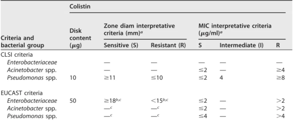
There is a lack of consensus between the two organizations setting up breakpoints for polymyxins, namely, the CLSI in the United States (72) and EUCAST in Europe (71). The zone diameter and MIC interpretive criteria given by those two organizations for colistin and polymyxin B are shown in Table 1. However, recent data related to PK suggest that the current breakpoints might be too high (21).
Quality Controls
Quality control organisms are required during susceptibility testing in order to ensure accuracy and standardization of the procedures. Quality control can be assessed using the E. coli ATCC 25922 (NCTC 12241; CIP 76.24) and P. aeruginosa ATCC 27853
(NCTC 12903; CIP 76110) reference strains. The disk diffusion and MIC quality control ranges for these strains determined by the CLSI are shown inTable 2(72).
Correlation between MICs of Colistin and Polymyxin B
Despite the high similarity of the molecular structures of colistin and polymyxin B, a recent study including 15,377 Gram-negative bacteria revealed differences between the MICs of colistin and polymyxin B (102). MIC values determined by the Sensititre system were 2-fold higher for polymyxin B than for colistin for 55% and 53% of Klebsiella species isolates (n ⫽ 4,177) and E. coli isolates (n ⫽ 6,311), respectively. However, a categorical agreement of⬎99% was obtained for enterobacterial strains when breakpoints ofⱕ2/ⱖ4 for both colistin and polymyxin B were applied. That study showed a high level of agreement between MICs of colistin and polymyxin B for P. aeruginosa and Acinetobacter spp.
Qualitative Detection Techniques
Rapid detection of heterogeneous populations among colistin-resistant Gram-negative bacteria by use of capillary electrophoresis. Sautrey et al. (103) proposed a capillary electrophoresis method for rapid detection of heterogeneous populations of colistin-resistant strains. However, further development is required for such applica-tions to be used in clinical laboratories on a daily basis.
Rapid detection of colistin-resistant A. baumannii isolates by use of the Micromax assay. The Micromax assay is based on the detection of released nucleotides, indicating cell wall damage, in the presence of colistin (104). After incubation with 0.5g/ml of colistin, strains are considered resistant to colistin ifⱕ11% of bacteria present cell wall damage. Bacteria are incubated for 90 min in Mueller-Hinton broth to achieve expo-nential growth and then incubated for 60 min with colistin at concentrations of 0 and 0.5 g/ml, respectively. Bacteria embedded in agarose are incubated with a lysis solution removing only weakened cell walls. The released fragmented DNA may be stained with the fluorochrome SYBR gold (Molecular Probes, Eugene, OR) and visualized by fluorescence microscopy (45 min to 60 min of technical processing and scoring under the microscope). This method is faster than the routine automatic microdilution TABLE 1 Colistin and polymyxin B breakpoints according to CLSI and EUCAST in 2014
Criteria and bacterial group Colistin Disk content (g)
Zone diam interpretative criteria (mm)a
MIC interpretative criteria (g/ml)a
Sensitive (S) Resistant (R) S Intermediate (I) R CLSI criteria Enterobacteriaceae — — — — — Acinetobacter spp. — — ⱕ2 — ⱖ4 Pseudomonas spp. 10 ⱖ11 ⱕ10 ⱕ2 4 ⱖ8 EUCAST criteria Enterobacteriaceae 50 ⱖ18b,c ⬍15b,c ⱕ2 — ⬎2 Acinetobacter spp. —c —c ⱕ2 — ⬎2 Pseudomonas spp. —c —c ⱕ4 — ⬎4
a—, not determined or absent.
bZone diameter interpretative criteria for Enterobacteriaceae given by EUCAST are only for veterinary medicine; M
from 15 to 18 mm.
cNo zone diameter interpretative criteria for human medicine; the MIC must be determined before use.
TABLE 2 Zone diameter and MIC quality control ranges for polymyxins according to CLSI guidelines
Strain
Zone diam (mm) range MIC (g/ml) range
Colistin Polymyxin B Colistin Polymyxin B
E. coli ATCC 25922 11–17 13–19 0.25–2 0.25–2
procedure (3 h 30 min versus 6 to 8 h) and is accurate for detecting colistin resistance in A. baumannii (100% sensitivity and 96% specificity). Another advantage is that it can be automated. However, the manual task and the cost of the materials (fluorochrome and epifluorescence microscope) are disadvantages for its routine use.
Rapid detection of colistin-resistant Enterobacteriaceae isolates by use of the Rapid Polymyxin NP test. We developed the Rapid Polymyxin NP test, which is based on the detection of bacterial growth in the presence of a defined polymyxin concen-tration (105). Detection is based on detection of glucose metabolism upon bacterial growth. Glucose metabolism induces the formation of acid, leading to a color change of the red phenol used as a pH indicator. The test is performed with a final concen-tration of bacteria of ca. 108CFU/ml in each well (or tube), and the final concentration of polymyxin is 3.75g/ml. Visual inspection of the tray is made after 10 min and then every hour for 2 h. The test is considered positive (indicating polymyxin resistance) if the isolate grows in the presence of colistin (color change from orange to yellow), while it is considered negative (indicating polymyxin susceptibility) if the isolate does not grow in the presence of polymyxin (no color change). This test is rapid (less than 2 h) and easy to perform.
By testing a total of 200 enterobacterial isolates exhibiting either resistance (intrinsic or acquired, or various) or susceptibility to polymyxins, the specificity and sensitivity of this test were evaluated at 99.3% and 95.4%, respectively, compared to BMD as the reference method (105). Note that the Rapid Polymyxin NP test identified the isolates exhibiting a heteroresistance phenotype as well as those producing the plasmid-mediated MCR-1 determinant (see below).
For the Rapid Polymyxin NP test, the adequate culture media for culturing the bacteria prior to the test were Mueller-Hinton agar, Luria-Bertani agar, Columbia agar plus 5% sheep blood, chocolate agar, UriSelect 4 agar, and eosin methylene blue agar. The Rapid Polymyxin NP test may also detect colistin-resistant Enterobacteriaceae directly from blood cultures (106). Results are obtained within 4 h.
Selective medium. So far, no selective medium allowing screening for any type of polymyxin-resistant Gram-negative isolates (with intrinsic, chromosomally encoded, or plasmid-mediated polymyxin resistance) has been available. Neither commercial nor in-house screening culture media had been designed that might permit screening of patients possibly colonized by polymyxin-resistant isolates. Therefore, we developed SuperPolymyxin, a selective culture medium for detection of any type of polymyxin-resistant Gram-negative organism (107). The SuperPolymyxin medium prevents swarm-ing of Proteus spp. (intrinsically resistant to polymyxins) and also the growth of Gram-positive bacteria and fungi, by addition of daptomycin and amphotericin B, respectively. Its base corresponds to the eosin methylene blue medium (Levine’s medium) (108) selective for Gram-negative bacteria, which differentiates lactose fer-menters (black colonies) from nonferfer-menters (colorless or light lavender colonies). In addition, differentiation of lactose fermenters is also possible to some extent. The SuperPolymyxin medium contains a colistin concentration (3.5g/ml) that allows clear categorization between polymyxin-resistant and -susceptible isolates. The sensitivity and specificity of this medium have been found to be 100% (107).
Genotypic Methods
Although the mechanisms underlying resistance to polymyxins have not all been elucidated, acquisition of colistin resistance in Gram-negative bacteria has been attrib-uted to lipopolysaccharide (LPS) modifications via diverse routes, including (i) the addition of cationic groups to the LPS reducing the overall negative charge of the LPS and consequently preventing the fixation of polymyxins; (ii) loss of the LPS and, consequently, loss of the polymyxin target; (iii) the overproduction of capsule polysac-charide (CPS) hiding polymyxin binding sites; and (iv) the release of CPS trapping polymyxins. Specific modifications of outer membrane porins and overexpression of efflux pump systems have also been described (88).
Several molecular mechanisms have been associated with colistin resistance in Gram-negative bacteria, such as alterations in the PmrA/PmrB, PhoP/PhoQ, ParR/ParS, ColR/ColS, and CprR/CprS two-component systems and alterations in the mgrB gene, which encodes a negative regulator of PhoPQ. Mutations leading to the addition of cationic groups on lipid A result in a less anionic lipid A and, consequently, to less fixation of polymyxins (88).
Similarly, alterations in the lpxA, lpxC, and lpxD genes of A. baumannii result in inactivation of lipid A biosynthesis, leading to a complete loss of LPS and, consequently, to a loss of the polymyxin target (109).
The mechanisms of polymyxin resistance can be identified by sequencing those specific genes. However, molecular techniques cannot be envisioned in the near future considering that (i) many chromosomally encoded mechanisms of resistance remain to be identified, (ii) it is difficult to extrapolate whether some substitutions identified in proteins known to be involved in LPS biosynthesis lead to resistance, and (iii) the levels of expression of the corresponding genes may vary and consequently influence the level of resistance to polymyxins.
There is an exception that corresponds to the recent identification of the plasmid-borne mcr-1/mcr-2 genes, whose products confer resistance to polymyxins (see below). According to the current knowledge on the topic, identification of these genes may be considered a signature of resistance or reduced susceptibility to polymyxins. This is why identifying the gene makes sense in this case, since qualitative genetic results may be translated directly into a nonsusceptibility phenotype. Screening of both mcr-1 and mcr-2 can be performed by using a standard PCR protocol using the primers MCR-1/2-Fw (5=-TAT CGC TAT GTG CTA AAG CC-3=) and MCR-1/2-Rv (5=-TCT TGG TAT TTG GCG GTA TC-3=), giving rise to a 715-bp amplicon. Also, a SYBR green-based real-time PCR assay that provides a simple, specific, sensitive, and rapid molecular tool for detection of mcr-1-positive isolates was recently published (110). That technique was validated on human and animal isolates and may be applied to extensive surveillance studies.
Porin mutations and overexpression of efflux pump systems may also be involved in colistin resistance (88), and it is very likely that phenotypic resistance to polymyxins in clinical isolates often results from combined resistance mechanisms (e.g., defects in outer membrane proteins combined with structural modification of the LPS). Phenotypic meth-ods, such as the Rapid Polymyxin NP test, are consequently very relevant for determining the subsequent therapeutic decision, since they actually concretely determine the suscep-tibility or lack thereof of isolates, in contrast to genotypic methods, which detect only potential resistance and require sequencing of multiple genes.
RESISTANCE MECHANISMS IN ENTEROBACTERIACEAE
Intrinsic Resistance Mechanisms in Proteus mirabilis and Serratia marcescens In P. mirabilis and S. marcescens, naturally occurring resistance to polymyxins is linked to the constitutive expression of the arnBCADTEF operon and/or the eptB gene, causing addition of phosphoethanolamine (pEtN) and/or 4-amino-4-deoxy-L-arabinose
(L-Ara4N) cationic groups to the LPS. This modification increases the charge of the LPS,
which is the initial target of the polymyxins, and therefore decreases polymyxin binding, giving rise to intrinsic resistance of these species (111–113).
Mechanisms Responsible for Acquired Resistance in Enterobacteriaceae
Acquired resistance to polymyxins has been identified in several genera of the Enterobacteriaceae, such as Klebsiella, Escherichia, Enterobacter, and Salmonella. Colistin resistance mechanisms remain unknown for some bacterial species, but several molecular mechanisms have been identified. The most common is modification of the LPS via cationic substitution, similar to that observed in bacteria with intrinsic resistance to polymyxins. A single transferable mechanism of resistance has been identified so far (see below), with most of the resistance mechanisms being encoded chromosomally.
Similar to what is observed in strains that are naturally resistant to colistin, addition of cationic groups (L-Ara4N and pEtN) to the LPS is responsible for acquisition of colistin
resistance in Enterobacteriaceae. A large panel of genes and operons are involved in qualitative modification of the LPS (Fig. 3), including genes and operons coding for enzymes that are directly involved in LPS modifications (genes responsible for synthesis of cationic groups and/or their addition to the LPS), i.e., the pmrC gene, the pmrE gene, and the pmrHFIJKLM operon; regulatory genes, such as those encoding proteins involved in the PmrAB and PhoPQ two-component systems; and the regulators of these two-component systems, i.e., the mgrB gene, which negatively regulates the PhoPQ system, and the newly described crrAB two-component regulatory system, which regulates the PmrAB system.
Genes encoding LPS-modifying enzymes. (i) The pmrC gene. The pmrCAB operon codes for three proteins, namely, the phosphoethanolamine (pEtN) phosphotransferase PmrC, the response regulator PmrA (also called BasR), and the sensor kinase protein PmrB (also called BasS) (114). The phosphoethanolamine phosphotransferase PmrC adds a pEtN group to the LPS (Fig. 3) (114).
(ii) The pmrHFIJKLM operon and the pmrE gene. The pmrHFIJKLM operon (also called the arnBCADTEF or pbgPE operon) codes for a total of seven proteins (115). The pmrE gene and the pmrHFIJKLM operon are responsible for the synthesis of the
L-aminoarabinose group (L-Ara4N) and its fixation to lipid A (Fig. 3) (115).
(iii) The pmrA and pmrB genes, which encode the PmrAB two-component system. Environmental stimuli, such as macrophage phagosomes, ferric (Fe3⫹) iron, aluminum (Al3⫹), and low pH (e.g., pH 5.5), mediate activation of PmrB through its periplasmic domain (114). The PmrAB and PhoPQ two-component systems are normally activated when bacteria are phagocytized into macrophages, allowing bacterial survival (114).
PmrB is a protein with tyrosine kinase activity that activates PmrA by phosphoryla-tion. PmrA in turn activates the transcription of the pmrCAB operon, the pmrHFIJKLM operon, and the pmrE gene involved in LPS modification (pEtN andL-Ara4N addition)
(Fig. 3) (114).
Specific mutations within the pmrA and pmrB genes have been described as being responsible for acquired colistin resistance in K. pneumoniae (105,116–120),
ter aerogenes (121), and Salmonella enterica (122,123) (Table 3). These mutations are responsible for constitutive activation of the PmrAB two-component system, leading to upregulation of the pmrCAB operon, the pmrHFIJKLM operon, and the pmrE gene, and thus to the synthesis of pEtN andL-Ara4N and their transfer to lipid A (Fig. 3).
Some polymorphism in the pmrAB genes of colistin-resistant E. coli has been reported (124,125), but the involvement of these mutations in the colistin resistance phenotype has not formally been demonstrated, since no complementation or site-directed mutagenesis has been performed.
(iv) The phoP and phoQ genes, which encode the PhoPQ two-component system. The phoPQ operon codes for two proteins, namely, the regulator protein PhoP and the sensor protein kinase PhoQ. Environmental stimuli, such as macrophage phagosomes, low magnesium (Mg2⫹), and low pH (e.g., pH 5.5), mediate activation of PhoQ through its periplasmic domain (114). The PhoPQ two-component system allows the expression of genes that code for magnesium transport, enzymes that modify the LPS to allow resistance to cationic antimicrobial peptides, and enzymes that decrease the cell stress caused by acidic pH or some virulence factors (126,127). The PhoPQ two-component system therefore allows bacterial survival under conditions of low magnesium or acidic pH or in the presence of cationic antimicrobial peptides.
PhoQ is a protein with tyrosine kinase activity that activates PhoP by phosphoryla-tion. PhoP in turn activates the transcription of the pmrHFIJKLM operon, involved in the addition of L-Ara4N to the LPS (Fig. 3) (126, 127). PhoP can also activate the PmrA
protein, either directly or indirectly via the PmrD connector protein, causing the addition of pEtN to the LPS.
Several mutations in the phoP and phoQ genes are responsible for acquired resis-tance to polymyxins in K. pneumoniae (81, 105, 117, 118, 120, 128) (Table 3). One mutation potentially involved in colistin resistance in E. coli has also been described (65). These mutations are responsible for constitutive activation of the PhoPQ two-component system, leading to upregulation of the pmrHFIJKLM operon and thus to the synthesis ofL-Ara4N and its transfer to lipid A (Fig. 3).
Regulators of the PmrAB and PhoPQ Two-Component Systems
The mgrB gene. MgrB (also called YobG) is a small transmembrane protein of 47 amino acids (129). Upon activation of PhoP, the mgrB gene is upregulated. The MgrB protein in turn represses the expression of the PhoQ-encoding gene, leading to negative regulation of the PhoPQ two-component system (Fig. 3) (129). Inactivation of the mgrB gene (the negative regulator of the PhoPQ two-component system) leads to overexpres-sion of the phoPQ operon, thus causing pmrHFIJKLM operon activation, leading to the production ofL-Ara4N responsible for the acquisition of colistin resistance.
Several missense mutations resulting in amino acid substitutions and nonsense mutations and therefore leading to a truncated MgrB protein may be responsible for acquired resistance to colistin in K. pneumoniae (Table 3). Other alterations, such as insertions or deletions of small nucleotide sequences in the mgrB gene, or even some complete deletions of the mgrB locus, have been reported (120,130,131). Insertional inactivation caused by diverse insertion sequences (IS), belonging to several families and inserted at different locations within the mgrB gene, is often responsible for acquired resistance to colistin in K. pneumoniae (105,117,120,130–132) and Klebsiella oxytoca (133,134). Recently, the transposition of genes encoding extended-spectrum -lactamases (ESBLs) or carbapenemases, leading to disruption of the chromosomal mgrB gene, was reported as a source of resistance to colistin (135, 136). Notably, selective pressure with-lactams leading to the acquisition of -lactamase genes may therefore be responsible for coselection of colistin resistance. Despite the high homol-ogy observed among mgrB gene sequences of Enterobacteriaceae organisms (129), disruption of this gene has so far not been found to be responsible for acquired resistance to colistin in genera other than Klebsiella.
The crrAB operon. The crrAB (colistin resistance regulation) operon codes for two proteins, namely, the regulatory protein CrrA and the sensor protein kinase CrrB. The
TABLE 3 Chromosomal mutations and amino acid deletions responsible for acquired colistin resistance in Klebsiella pneumoniae, Enterobacter aerogenes, Escherichia coli, Salmonella enterica, P. aeruginosa, and A. baumannii isolates
Bacterial group and species Protein (normal length [aa]) Domain involved (residues)a,b Amino acid changed Reference(s) Enterobacteriaceae
K. pneumoniae PmrA (223) REC (1–112) S42N 120
G53C 105,120 G53S 105 Trans_reg_C (145–216) PmrB (365) TM (13–35) ΔR14 118 L17Q 105 HAMP (90–142) L82R 116 S85R 120 T140P 120 HisKA (143–203) T157P 117–119 S208N 118 ΔY209 118 HATPase_c (250–358) R256G 117 PhoP (223) REC (1–112) V3F 117 L26Q 120 S86L 117 Trans_reg_C (145–220) D191Y 81
PhoQ (488) PhoQ sensor (10–189) R16C 105
L26P 117 L96P 120 D150G 117 S174N 118 HAMP (195–263) V258F 117 HisKA (267–330) L348Q 120 HATPase_c (375–482) G385S 120 D434N 128 MgrB (47) K3* 105 L9* 120 I13* 120 A14S 120 W20R 105 L24H 130 V26* 120 M27K 105 C28F 120 C28Y 117,120,128,130 C28* 105,120 Q30* 105,120 D31N 120 Q33* 105 F35I 120 G37S 130 C39Y 105 N42Y/K43I 105 I45T 105 W47R 105 W47* 105 *48Y 117 CrrB (353) Q10L 128,137 TM (12–34) Y31H 137 HAMP (81–135) L94 M 128 HisKA (136–200) W140R 137 N141I 137 P151S 137 S195N 137 E. aerogenes PmrA G53S 121
E. coli PmrA (222) REC (1–112) R81Sc 125
Trans_reg_C (145–216)
PmrB (363) Δ7–12c 124
TM1 (15–37) TM2 (69–91)
TABLE 3 (Continued) Bacterial group and species Protein (normal length [aa]) Domain involved (residues)a,b Amino acid changed Reference(s) HAMP (92–144) HisKA (145–205) T156Kc 124 A159Vc 124 V161Gc 125 HATPase_c (252–360) PhoQ_sensor (10–189) HAMP (195–263) HisKA (267–330) PhoP (223) REC (1–112) Trans_reg_C (145–220) PhoQ (486) PhoQ_sensor (10–189) HAMP (195–263) HisKA (267–330) HATPase_c (374–480) E375Kc 65
S. enterica PmrA (222) REC (1–112) G15Rc 123
G53Ec 123 G53Rc 123 R81Cc 123 R81Hc 123 Trans_reg_C (145–216) PmrB (356) TM (13–35) Δ11–14c 122 L14Fc 123 L14Sc 123 M15Lc 122 L22Pc 123 S29Rc 123 HAMP (89–141) T92Ac 123 P94Qc 123 E121Ac 123 S124Pc 123 N130Yc 123 HisKA (142–202) T147Pc 123 R155Pc 123 T156Mc 123 T156Pc 123 V161Gc 123 V161Lc 123 V161Mc 123 E166Kc 123 M186Ic 123 HATPase_c (249–356) G206Rc 123 G206Wc 123 S305Rc 123 Nonfermentative bacilli
P. aeruginosa PmrA (221) REC (1–112)
Trans_reg_C (145–216) L157Q 166 PmrB (477) L14P 167 TM1 (15–37) PD (38–160) ΔD45 74,167 A54V 167 TM2 (161–183) L167P 166 HAMP (186–238) G188D 167 F237L 118 HisKA (239–304) L243Q 167 A247T 168 A248V 167 S257N 167 M292I 167 M292T 169 HATPase_c (348–459) PhoQ (448) R6C 170 TM1 (7–29)
TABLE 3 (Continued) Bacterial group and species Protein (normal length [aa]) Domain involved (residues)a,b Amino acid changed Reference(s) ΔV57–Q332 170 PD (30–166) N104I 118 K123Q 166 K123E 118 Q133E 118 A143V 166 V152* 168 TM2 (167–189) V184G 118 A207R 118 R214H 118 H223R 168 HisKA (238–300) V260G 163,247 HATPase_c (343–448) ΔL364–G365170 I421* 170 Fr at I421 170 D433* 170 R444C 170
ParR (235) REC (7–117) L18I 118
N24S 118 S24N 118 M59I 171 Trans_reg_C (152–228) E156K 171 ParS (428) TM1 (5–27) L14Q 171 PD (28–131) V101 M 171 TM2 (132–154) L137P 171 HAMP (155–207) HisKA (208–273) Q232E 118 HATPase_c (318–428) G361R 118 H398R 247 ColS A106V 172 CprS R241C 172
A. baumannii PmrA (224) REC (2–112) E8D 177,180
M12I 174 P102H 173 S119T 174 Trans_reg_C (150–221) PmrB (444) TM1 (10–29) T13N 173 S14L 175 S17R 177 Fr at F26 176 PD (30–141) ΔA32–E35 174 D64V 174 A80V 174 L87F 175 Y116H 177 I121F 178 TM2 (142–164) M145K 175 ΔL160 174 P170L 174,179 P170Q 174 A183T 178 A184V 178 P190S 178 T192I 178 L208F 174 HisKA (218–280) A226V 174 A227V 173,175,176 Q228P 178 R231L 174 T232I 177 P233S 118,173–176,179 P233T 173 T235I 174 N256I 174

![TABLE 3 (Continued) Bacterial group and species Protein (normallength [aa]) Domain involved(residues)a,b Amino acidchanged Reference(s) ΔV57–Q332 170 PD (30–166) N104I 118 K123Q 166 K123E 118 Q133E 118 A143V 166 V152* 168 TM2 (167–189) V184G 118 A207R 118](https://thumb-eu.123doks.com/thumbv2/123doknet/14935873.665806/21.892.208.692.152.1103/continued-bacterial-protein-normallength-involved-residues-acidchanged-reference.webp)
