S782
SESSION I: THEORETICAL AND PRECLINICAL EXPERIMENTAL
BASES OF PROPHYLAXIS
Perioperative Antibiotic Prophylaxis of Wound and Foreign
Body Infections: Microbial Factors Affecting Efficacy
F. A. Waldvogel, P. E. Vaudaux, D. Pittet, and P. D.Lew
From the Infectious Disease Division and the Clinique Medicale Therapeutique, University Hospital, Geneva, Switzerland
Numerous microbial factors are responsible for perioperative infections and influence the efficacy of antibiotic prophylaxis. These factors include the staphylococcal carrier state, bacterial adher-ence to a number of host proteins, the production of glycocalyx by sessile bacteria, and shifts in antibiotic resistance. A full understanding of the mechanisms involved will lead to further reductionsinthe number of postoperative infections. Unfortunately, the microbial factors affect-ing prophylaxis cannot be evaluated separately under clinical conditions; they are easier to study under circumstances whose bacteriologic features are well defined and in which the presence offoreign materials (e.g., sutures) greatly potentiates pathogenic mechanisms. Such circumstances exist, for example, in infections developing after "clean" surgery and in experimental models. Since even clean wounds are found tobe contaminated when sampled carefully, the control of infection is more a quantitative than a qualitative problem. The critical period for the develop-ment of infection is short: an antibiotic course not exceeding 24 hours seems effective in prevent-ing infection.
Wounds produced by surgical intervention, their sponta-neoushealing,their complications (including infections), and methods to accelerate their closure have been described for thousandsof years. The use of sutures, for example, is clearly depicted in the Smithpapyrus, which datesback to 4000B.C. [1]. Postoperative infectionevidently has always been a feared complication of surgical procedures, and many techniques and devices have been developed throughout history to prevent such infection. Withthe advent of antibiotics, it was believed that the prevalence of this type of infection would decrease drastically. This was not the case. For instance, in a study on herniorrhaphy between.1937and1957,the incidence of postoperativeinfection hoveredsteadilyaround4
%
[2]. Over the past15years, however, considerable workhas shownthe benefitof shortcoursesof antimicrobial agents for the preven-tion of infecpreven-tion in a widevarietyof surgical procedures[3-5].The factthat skinincision, organ manipulation,or the con-sequences thereof increase the incidence of local infection points toward alteration of local host factors influencing microbial elimination. Demonstrations of the efficacy of an-timicrobial agents are indicative of the impact ofbacterial
Grant support: Swiss National Research Foundation (3.829.0.87) and Beecham Pharmaceuticals, Bern, Switzerland.
Reprints and correspondence: Dr.F.A. Waldvogel, Clinique Medicale Therapeutique, University Hospital, 24, rue Micheli-du-Crest, 1211Geneva 4, Switzerland.
Reviews of Infectious Diseases 1991;13(Suppl lO):S782-9
© 1991by The University of Chicago. All rights reserved. 0162-0886/91/1305-0002$02 .00
factors on the postsurgical evolution of wounds. The role of bacterial factors is further underscored by the clear demon-strationthat prophylacticantibioticsare effective in contami-natedand clean-contaminated surgery, whereas their efficacy is more controversial in clean surgery- a situation in which the bacterial load is very low and therefore more difficultto evaluate.
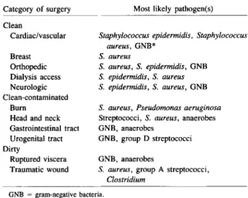
It hasbecomealmostaxiomatic to categorize surgical proce-dures as clean, clean-contaniinated, contaminated,and puru-lent. Each category probably has common as well as specific risks and pathogenicfactors [1]. Each also has its owntypical group of pathogenic organisms, among which Staphylococ-cus aureus is found at a rate close to 50% [6] and gram-negative enteric bacteria almostas often (table 1). In a repre-sentativestudy extendingover5 years and encompassing 247 postoperative infections, S. aureuswas responsible for138 infections and gram-negative enteric organisms for the majority of the others [7]. Although recent studies have de-scribed the growing role of many other microorganisms, includinganaerobes, the control of S.aureusand coagulase-negative staphylococci would reducethe prevalence ofpostsur-gical infectionsby >50
%.
The present review will therefore concentrate essentially on these organisms and their patho-genic factors.Itis difficult to delineate the roleof a singlepathogenic factor leading to postoperative infection, since host factors and microbial factors are in a state of perpetual interaction. Nevertheless, three important observations at least help to define the boundaries of this problem. First, bacterial infec-tion of a surgical woundnecessarily starts by the
contamina-GNB= gram-negative bacteria.
Table 1. Most likely pathogensinpostoperative wound infections.
tion of the wound with the microorganism. Second, this contamination cannot be totally prevented even under the most stringent aseptic conditions; this point is demonstrated by the observation of bacterial growth in 68%of 350 wounds after clean operations [8]. Thus a prerequisite for the prevention of the observation of bacterial postoperative staphylococcal infections is a better understanding of the colonization/anchor-ing mechanisms of these organisms. Moreover, the control of infection is more a quantitative than a qualitative problem, since, after all, most wounds are contaminated to a greater or lesser extent. Third, prophylactic antibiotic therapy has been shown to be effective even if given over a short period. This observation applies not only to clean orthopedic proce-dures but also to abdominal surgery [9, 10]. It can therefore be concluded that the pathogenic mechanisms - both host-me-diated and microbial- are operational over a brief period dur-ing and after surgery. The data published most recently by Platt et al. [11] on two types of surgery (mastectomy and her-niorrhaphy), with similar suppression of infection by short-term antibiotic therapy, suggest that the pathophysiologic mechanisms are the same despite major differences in the sur-gical procedures.
In clean surgery the risk of infection essentially depends on the contamination of the wound during the procedure [12] and on the presence of sutures, as has been repeatedly demon-strated [13]. No specifi c stimulation of bacterial pathogenic-ity can be ascribed to a particular type of suture [14]. Most infections after clean surgery are due to S.aureusand coagu-lase-negative staphylococci. The growing importance of the latter group of organisms has become clear in recent years.
Category of surgery Clean Cardiac/vascular Breast Orthopedic Dialysis access Neurologic Clean-contaminated Bum
Head and neck Gastrointestinal tract Urogenital tract Dirty
Ruptured viscera Traumatic wound
Most likely pathogen(s)
Staphylococcus epidermidis, Staphylococcus aureus, GNB*
S.aureus
S. aureus, S. epidermidis, GNB
S.epidermidis, S.aureus S. epidermidis, S. aureus, GNB
S.aureus, Pseudomonas aeruginosa Streptococci, S. aureus, anaerobes GNB, anaerobes
GNB, group D streptococci
GNB, anaerobes
S. aureus, group A streptococci, Clostridium
technical problems. Several factors in surgical intervention, all known to increase the prevalence of infection, may affect the microbial flora directly or indirectly, qualitatively or quan-titatively; however, the individual factors are difficult to as-sess. Variations in surgical technique limit the feasibility of multicenter studies. Whereas the duration of an operation can be clearly determined, other important factors are not only variable but difficult to quantify, such as trauma, ischemia, tissue hypoxia, and edema. Moreover, hypotension and shock (also difficult to quantitate) have been associated with an in-creased risk of infection. Finally, emergency surgery- an elu-sive term - is associated with increased risk. It is particularly difficult to assess the importance and possible interaction of various microbial factors in mixed infection, such as that fol-lowing abdominal surgery. Microbial factors due to organ-isms colonizing or infecting a site during surgery may be difficult to differentiate from those originating from microor-ganisms acquired in the intensive care unit or from those linked to invasive procedures or therapeutic devices. Finally, if these factors are assessed indirectly by perioperative prophylaxis, the dosage and the pharmacokinetic profile of the antibiotic are variables that cannot be standardized under all circum-stances.
All of these difficulties can be overcome to some extent by the use of animal models [15], whose development has been rather slow. More specifically, evaluation of the efficacy of preventive antibiotics can be facilitated through an analysis of the microbial factors leading to infection and/or inhibited by perioperative antibiotics in the simplified model system of clean surgery, in which the epidemiology and bacteriology of infection are relatively well defined [16] and staphylococci are the organisms primarily implicated. Several experimen-tal models are available for the study of host or microbial fac-tors in this setting [17].
The presence of foreign material- including sutures, in-dwelling devices, and prostheses - has been clearly documen-ted as a major pathogenic factor, overshadowing the majority of other pathophysiologic influences [18]. For example, Elek and Cohen [19] and later James and MacLeod [20] and No-ble [21] showed that the presence of subcutaneous foreign ma-terial resulted in a decrease of the minimal infecting dose of staphylococci from >1Q6 to <103 •In experiments using poly-methacrylate or Teflon tissue cages in guinea pigs, Zimmerli et al. [22] demonstrated a similar potentiation of bacterial in-fectivity and quantitatively assessed both bacterial growth and the inflammatory reaction. As we shall see, this animal model has proven to be of great help in the assessment of bacterial pathogenic factors influencing antibacterial prophylaxis.
Problems in the Evaluation of Microbial Factors Clinical evaluation of microbial factors affecting perioper-ative antibiotic prophylaxis is hampered by many logistic and
Bacterial Factors Influencing the Efficacy of Prophylaxis
Important microbiologic determinants of the efficacy of an-tibiotic prophylaxis in clean surgery will now be discussed
S784 Waldvogel et al. RID 1991;13 (Suppl 10)
in light of some general principles of postoperative wound infection. First, the wound must be contaminated by the offending organism; anchoring and adherence are initial man-datory steps in the development of infection. Second, on care-ful microbiologic assessment, most apparently clean wounds are found to be contaminated; this observation implies a quan-titative rather than a qualitative microbe-host interaction in the development of wound infection. Third, antibiotic pro-phylaxis (i.e., activity against pathogenic bacterial mecha-nisms) is effective even if the regimen is given briefly, but only if the timing is right; thus these microbial factors appear to act over a brief period.
Presence ofbacteria. Wound contamination with S.aureus
starts often with a nasal carrier state and more rarely with a vaginal or rectal carrier state [23]. Treatment with systemic rifampin [24], local mupirocin [25], or other agents has been shown to reduce the rate of infection of dialysis access sites and of wound contamination, most probably via a reduction in the degree and duration of bacterial contamination, since the prophylactic effect seems to be short-lived. The problem is more complicated in the case of coagulase-negative staphylococci, since these organisms are ubiquitous skin con-taminants and since culture techniques sample only part of the complex skin flora, potentially missing small, possibly infectious subpopulations [26]. Although the data from clin-ical studies on clean operations are still subject to controversy, prophylactic antibiotics eradicating coagulase-negative staphy-lococci (among other organisms) have been shown to be effec-tive in well-defined operations such as aortocoronary bypass [27, 28]. The microbiologic price paid for such successful treatment is high, however. Coagulase-negative staphylococci carrying the gene for methicillin resistance increased dramat-ically in number when cultured quantitatively from the nares and the subclavian and inguinal areas of patients given high-dose prophylactic antibiotics during cardiac surgery [29]. In addition, the use of broad-spectrum antibiotics (e.g., nafcil-lin and rifampin) among a similar group of cardiovascular pa-tients led to an important increase in rifampin-resistant organisms [30].
In conclusion, whereas colonization with S.aureuscan be suppressed for short periods by antibiotics without a major risk of resistance, simultaneous multiplication of
Staphylo-coccusepidermidisoccurs readily and resistance develops
fre-quently when broad-spectrum antibiotic prophylaxis is used. There is therefore a definite need to define the shortest effec-tive prophylactic treatment period under these circumstances in order to minimize both proliferation and resistance of coagulase-negative staphylococci.
In situ multiplication ofbacteria. Itwould be of great
in-terest to assess the rate of bacterial multiplication after coloni-zation as well as the effects that endogenous or exogenous substances or factors may have upon that rate. In this regard, it is worth mentioning the remarkable studies performed by Miles in 1956 [31]. Evaluating bacterial multiplication
in-directly by measuring the diameter of skin lesions after sub-cutaneous inoculation ofPseudomonas aeruginosa,he showed that a full-blown lesion could be obtained in <24 hours. In addition, he demonstrated the effect of hypovolemic shock on the diameter of the lesions and brilliantly showed that adrenalin increased their size. Liquoid (polyanethol sulfonate) also in-creased the size of the bacterial lesions by 240-fold but ex-erted its effect for only 5 hours. Most important, an antibiotic (in this case, streptomycin forP.aeruginosalesions) decreased the size of the lesions by a factor of 45 but exerted a protec-tive effect for only 3 hours. Miles defined the period during which the experimental lesions could be modulated by sev-eral factors as the decisive period. Although considerable progress has been made in this field, the early concept of a decisive period is most useful, possibly defining the time necessary for bacteria to adhere, to multiply, and to become sheltered from host defenses and antibiotics.
Microbial multiplication in a surgical wound associated with a foreign device has been evaluated in two recent studies. Zim-merli et al. have shown that, even with small inocula of S.
aureus, a latency period precedes active microbial
multipli-cation [22]. The importance of this latency (decisive) period is illustrated by a study evaluating the timing of prophylactic antibiotic therapy [32]: with a delay of>6hours between terial inoculation and the beginning of antibiotic therapy, bac-terial growth occurred unabated despite treatment. Thus, at least in staphylococcal infections, it can be generally accepted that the decisive period during which microbial pathogenic effects are potentially reversible is~6 hours.
Antibiotic susceptibilityandresistance. Although an
effec-tive program of perioperaeffec-tive antibiotic prophylaxis obviously should be chosen in light of the sensitivities of the potential pathogens, this task is far from easy. For example, the devel-opment of resistance in staphylococci during prophylactic treatment with rifampin is impossible to circumvent [33]. A quantitative increase of resistant coagulase-negative staphylococci during broad-spectrum antibiotic treatment [29] and the emergence of resistant strains under similar condi-tions [31] have been documented [34].
Of even greater concern is the transfer of resistance between
S.epidermidisand S.aureus;this problem has been well
ex-plored in gram-negative bacteria but has only recently received attention in staphylococci. Thus, gentamicin-resistant strains of S.aureusand S.epidermidisisolated during a nosocomial outbreak in a nursery showed single plasmid identity by all test methods, including restriction endonuclease analysis [35]. The emergence of antibiotic resistance in S.aureusduring another epidemic was shown to result from genetic transfer of a plasmid from S.epidermidis[36]. That skin-inhabiting coagulase-negative staphylococci can be the source of inter-genetic plasmid transfer of resistance has been clearly estab-lished by Naidoo and Noble [37].
Other mechanisms of bacterial resistance must be consid-ered. Although disputed for years, the role of "foreign:'
solu-ble, bacterial {j-Iactamase in the hydrolysis of ampicillin, which favors the multiplication of a non-d-lactamase-producing strain ofStreptococcus pneumoniaein an animal model [38], has recently been established. This mode of in-teraction is probably of little clinical significance, since most antimicrobial agents used as prophylactic agents are stable to {j-Iactamases.
Finally, an important type of phenotypic bacterial resistance deserves discussion. In 1985 Sheth et al. presented convinc-ing data pointconvinc-ing to an increase in antibiotic MBCs for coagulase-negative staphylococci when the organisms were cultured in vitro in the presence of a catheter [39]. Moreover, organisms adherent to catheters could survive bactericidal con-centrations of antibiotics for prolonged periods. Similar results reported by Nickel et al. [40] were ascribed to the protective effect of an extracellular matrix secreted by many organisms. These phenomena are reversible and are probably due to genetic regulatory events or phenotypic changes [41]. They may involve a variety of mechanisms, possibly specific for each drug, but are of sufficient general importance to explain the persistence of organisms in many chronic infections as-sociated with tissue destruction or the presence ofmechanical devices. Whether this modulation of antibiotic susceptibility is associated with (or the consequence of) surface adhesion, surface growth, production of slime or other extracellular pro-ducts, changes in cellwall permeability, or effectson the target structures for antibiotics warrants further investigation.
Bacterialadherence. Adherence is a prerequisite for
bac-terial multiplication and invasion, according to our present concepts of infection. The adherence of S.aureusand coagu-lase-negative staphylococci to wounds, various sutures, and prosthetic materials has been widely investigated, and only some aspects pertinent to perioperative antibiotic prophylaxis will be discussed here. The adherence of coagulase-negative staphylococci varies greatly with the influence of encapsulation and other surface events [42]. Pretreatment with a variety of antimicrobial agents has a pronounced impact on staphylococ-cal adherence to plastic, as has recently been demonstrated by Schadow and associates [43] and as has previously been established for other microorganisms[44-46].These results indicate that antibiotics modify the interaction of microor-ganisms with natural or foreign surfaces, but the respective mechanisms responsible for these modifications remain un-known.
Some general comments can be made regarding the inter-action of S.aureusand coagulase-negative staphylococci with foreign surfaces in light of work performed in our laboratory and elsewhere. First of all, foreign surfaces rapidly become coated with a variety of host proteins, which play an impor-tant role in bacterial adhesion [47]. Many of these proteins carry domains of attachment for S. aureusand coagulase-negative staphylococci as well as for other microorganisms. A comprehensive report on these adherence domains for staphylococci has been published by Christensen et al. [48].
The question at this stage is not so much how these host pro-teins express their affinity for microorganisms, but rather how the organisms interact with these domains. Kinetic studies have shown this interaction to be rapid and irreversible. Thus clinical isolates of S.aureusbound avidly to fibronectin and fibrinogen, and binding to laminin occurred at a lower level [49]. These results were less striking with coagulase-negative staphylococci. When strains were categorized according to their origin (intravenous device infection, septicemia, healthy carriers), no differences were found except for the adherence of a subpopulation of coagulase-negative staphylococci to fibrinogen.
Thus there does not seem to be one bacterial strain - or sev-eral strains - with surface properties particularly suited to the colonization of foreign material. This point was well illus-trated in a study by Vaudaux et al. [50], in which three staphylococcal strains (S. aureusstrain Wood46,S.aureus
strain Cowan I, and S.epidermidisstrain Rp 12) showed differ-ent degrees of binding to fibrinogen- or fibronectin-coated poly methacrylate or to catheters inserted in patients (figure 1). No quantitative prediction can therefore be made for a clin-ical situation since bacterial binding will depend on the num-ber of receptors on the bacterial surface (one fibronectin receptor has now been identified in S. aureus[51, 52]) and on the amount of fibronectin, fibrinogen, and other host pro-teins deposited on the foreign surface. In addition, recent work in our laboratory suggests that fibronectin molecules unfold differently depending on the nature of the surfaces, thereby liberating various binding sites for S. aureus[53].
In conclusion, interaction between foreign material and bac-teria (at least in the case of staphylococci) presupposes the deposition of a host protein layer, which plays the role of a ligand with a binding domain on the bacteria. Some of the characteristics of the bacterial sites are now well known, but little information is available on whether antibiotic treatment or other measures can modulate these sites. Recent work has suggested that, even in the absence of foreign material, staphylococcal binding in surgical wounds may be mediated via binding to fibronectin and collagen [54] or to other ex-tracellular matrix proteins.
Slimeproductionand its effects on postoperative infection.
Most microorganisms, when grown under sessile conditions, produce extracellular polysaccharides that are deposited as an intercellular matrix. The available information on this ma-terial, calledglycocalyxorslime; has been reviewed exten-sively by Costerton and colleagues [55] and by Gristina et al. [56]. The production of capsular polysaccharide material, in correlation with bacterial growth, has also been demon-strated by chemical [57] and morphologic [58] criteria in an experimental model. Recent data obtained by Nickel et al. [40] suggest that glycocalyx decreases bacterial susceptibil-ity to antibiotics.Itis difficult, however, to dissociate glycoca-lyx production from other events such as slow growth and adhesion to a surface; critical experiments-e.g., the
addi-8786 Waldvogel et al. RID 1991;13 (Suppl 10)
Sa.iIWlWI Sa.eDidermidis
Cowan I Wood 46 Rp12
•
•
•
'- # If -e-# #•
•
-
•
•
'-••
,
I
•
•
•
•
~..-
#•
0 0•
0•
0!
••
.A-....0-•
8 0 '- 0•
-0 I-
•
0 ~•
0 0PeO.COl PeO.Ol PeO.C02
~epjdermjdjs Rp12
»>
..
...-i
I
tI
Sa.auIIWl Wood 46 ~iUDWi CowanI 106-.-- - - - - -r-- - - - - --.-- - "---- - --, n----.Fibrinogen-eoated~
.M_M_-.
Fibronectin-eoated"
..
, 105 !:a
~ .!!! Qj U III .Cl 'E ~ 104 Q) s: "0 III '0 CD .Cl E ::::l Z Concentration (J.l.glrnl) of soluble Fn or Fg 24 8 16 3224 8 16 3224 8 16 32 Un-inserted Inserted Un-insertedInserted Un- Inserted
inserted
Figure 1. Promotion of adherence of three laboratory strains of staphylococci by in vitro coating of polymethacrylate coverslips with purified fibrinogen (Fg) or fibronectin (Fn) at indicated concentrations(left)or by blood components deposited in vivo on inserted polyvinyl chloride catheters (right). Bars indicate median values for each group. Reprinted with permission from [50].
tion of increasing concentrations of glycocalyx to adherent bacteria, with concomitant measurements of bacterial suscep-tibilities-have not, to our knowledge, been performed yet. Glycocalyx affects other mechanisms linked with periopera-tive infections, as summarizedbyJohnson et al. [59] and by Gray and associates [60]. Elucidation of the direct or indirect effects of glycocalyx on perioperative antimicrobial prophy-laxis (except for the demonstrated effect on microbial perme-ability [40]) waits novel methods of investigation.
TimingandPharmacokinetics of Antibiotic Prophylaxis
Clinical and experimental data suggest that there is a "deci-sive period;' as Miles called it, during which wound infec-tion can be modulated, i.e., during which microbial and host factors are still in a reversible stage [31]. At present, the only way to measure the reversibility of the microbial factors is to determine the optimal timing of antibiotic prophylaxis. As has been mentioned, we showed that, in the presence of for-eign material, a 6-hour lapse between bacterial inoculation
and the initiation of short-term antibiotic prophylaxis led to the failure of prophylaxis [32]. These first 6 hours seem, there-fore (in such an experimental model, at least), to represent the decisive period describedbyMiles [31]. Thus the estab-lishment of adequate antibiotic levels (exceeding bactericidal concentrations) in a wound during the first 6 hours after bac-terial inoculation and the maintenance of these levels for an adequate period (here: 24 hours) should constitute efficient prophylaxis.
One example of this technique that has been shown to be effective (as studied in tissue cages in guinea pigs) consists of a single dose (30 mg/kg) of vancomycin or fleroxacin, a long-acting quinolone [61]. When, however, either of the lat-ter prophylactic drugs was compared with equal single doses of ciprofloxacin or ofloxacin - two quinolones with a shorter half-life in tissues-the results differed quite strikingly (table 2). Protection against infection was incomplete with the short-acting quinolones, even at a bacterial inoculum as low as lQ2 cfu of S.aureus strain Wood 46. Moreover, levels of ofloxa-cin and ciprofloxaofloxa-cin decreased rapidly (figure 2), while those of vancomycin and fleroxacin remained predictably >0.7
+---.----,---...---'0.'- f - - - , - - - . - - - , - - - '
o 1
Time after administration (hours)
Figure 2. Pharmacokinetics of twoa-quinolonesin serum and tis-sue cage fluid after intraperitoneal administration of 30 mg/kg.
Analysis is based on three principles. First, the wound must be contaminated, and adherence mechanisms are therefore of paramount importance. Second, most "clean" wounds are found to be contaminated when sampled carefully, and the control of infection is therefore more a quantitative than a qualitative problem. Finally, the critical period for the devel-opment of infection is short: antibiotic prophylaxis of
<
24 hours' duration seems to be effective both clinically and ex-perimentally.With these principles as a framework, the effects of an-timicrobial agents on microbial factors must be evaluated. The staphylococcal carrier state (whether involving S.aureusor coagulase-negative staphylococci) influences the frequency of wound infection and can be modified in a positive or nega-tive mannerbyantibiotics. Bacterial adherence certainly plays a crucial role in postoperative infection and is favoredby bac-terialligands interacting specifically with host proteins such as fibrinogen, fibrin, fibronectin, and laminin. Bacterial mul-tiplication is influenced by many factors during the first few hours of wound infection - a decisive period that offers many therapeutic possibilities. The production of glycocalyx by ses-sile bacteria alters the susceptibility of many bacteria to anti-biotics, although the mechanisms involved have not yet been elucidated. Antibiotic susceptibilities are evidently critical de-terminants of the efficacy of perioperative antibiotic prophy-laxis: initial resistance and the emergence of resistance by selection or genetic alteration are well known phenomena, and plasmid transfer among species of staphylococci is a newly observed mechanism of potentially great danger. Variation in the susceptibility of adherent vs. fluid-phase bacteria may be a fundamental biologic phenomenon that explains many in-stances of treatment failure in chronic infection.
A full understanding of these mechanisms should allow us to further reduce the prevalence of postoperative infections.
Clprofloxacln
...:
/""''','0..._._._._..._.
()o·· ..Oserum _ tissue cageflUid
mean±SD 10r - - - , OIloxacln 10 E ~ 'C ~ Q)
!f
s
~ c: <.l '5 ~ -cNo. of tissue cages yielding <10 cfu per mLino. analyzed
Drug Inoculum (%)
(no. of doses*) (cfu) At 48 h At 7 d
Ofloxacin (1) 102 7/8 (88) 4/8 (50)t 103 2/8 (25)+ 2/8 (25)+ Ciprofloxacin (1) 102 4/8 (50)t 4/8 (50)t 103 1/8 (l3)§ 0/8 (. . . )§ Fleroxacin (1) 102 18/18 (100) 18/18 (100) 103 6/6 (100) 5/5 (l00) 104 10/10 (l00) 4/9 (44) Ofloxacin (2) 102 4/4 (100) 4/4 (l00) 103 3/4 (75) 3/4 (75)
Table 2. Comparison of ofloxacin, ciprofloxacin, and fleroxacin in the prophylactic treatment of tissue cage infection due to S.aureus
strain Wood 46 in guinea pigs.
- - -
-NOTE. Data are from [61]. * Each dose was 30 mg/kg.
tp <.02 vs. fleroxacin.
+
P<.05 vs. fleroxacin (Fisher's 2 x 2 exact test, two-tailed).§P<.01 vs. fleroxacin.
pg/mLfor 24 hours [61]. A second injection of the shorter-acting quinolone ofloxacin was necessary and sufficient at 3 hours to produce adequate tissue levels and protection (table 2). These and similar studies all indicate that microbial fac-tors are operational- and can be brought under control-during the first 24 hours after surgery in animal models.
Conclusion
Several microbial factors are responsible for perioperative infections and influence the efficacy of antibiotic prophylaxis. Unfortunately, clinical studies do not allow the evaluation of each factor separately, and experimental studies in this field are limited. The problem can be partly solved by the exclu-sive study of infection after clean surgery, since in these cir-cumstances bacteriologic features are well defined, the presence of foreign materials greatly potentiates pathogenic mechanisms, and experimental models are readily available.
References
1. Majno G. The healing hand. Man and wound in the ancient world. Cam-bridge, MA: Harvard University Press, 1975:1-571
2. Postlethwait MD. Principles of operative surgery: antisepsis, technique, sutures, and drains. In: Sabiston DC Jr, ed. Textbook of surgery. Philadelphia: WB Saunders, 1986:317-32
3. Hirschmann JV, Inui TS. Antimicrobial prophylaxis: a critique of re-cent trials. Rev Infect Dis 1980;2:1-23
4. Conte JE, Jacob LS, Polk HC Jr. Antibiotic prophylaxis in surgery: a comprehensive review. Philadelphia: JB Lippincott, 1984 5. DiPiro JT, Bivins BA, Record KE, Bell RM, Griffen WO Jr. The
prophylactic use of antimicrobials in surgery. Curr Probl Surg 1983;20:69-132
6. Dineen P. A critical study of 100 consecutive wound infections. Surg Gynecol Obstet 1961;113:91-6
7. Maitland AIL. Postoperative infection. Br J Surg 1965;52:931-40 8. HoweCWoBacterial flora of clean wounds and its relation to subsequent
sepsis. Am J Surg 1964;107:696-700
9. Doyon F, Evrard J, MazasF. Evaluation des essais therapeutiques pu-blies sur l'antibioprophylaxie en chirurgie orthopedique. Rev Chir Orthop 1989;75:72-6
S788 Waldvogel et al. RID 1991;13 (Suppl 10)
10. DiPiroIT, Cheung RPF, Bowden TA Jr, Mansberger JA. Single dose systemic antibiotic prophylaxis of surgical wound infections. Am J Surg 1986;152:552-9
11. Platt R, Zaleznik DF, Hopkins CC, Dellinger EP, Karchmer AW,Bryan CS, Burke JF, Wikler MA, Marino SK, Holbrook KF, Tosteson TD, Segal MR. Perioperative antibiotic prophylaxis for herniorrhaphy and breast surgery. N Engl J Med 1990;322:153-60
12. Wiley AM, Ha'eri GB. Routes ofinfection: a study of using ''tracer par-ticles" in the orthopedic operating room. Clin Orthop 1979;139:150-5 13. Simchen E, Stein H, Sacks TG, Shapiro M, MichelJ. Multivariate anal-ysis of determinants of post-operative wound infection in orthopedic patients. J Hosp Infect 1984;5:137-46
14. Katz S, Izhar M, Mirelman D. Bacterial adherence to surgical sutures: a possible factor in suture induced infection.Ann Surg 1981;194:35-41 15. Zimmerli W, Waldvogel FA. Models of foreign-body infections. In: Zak
0, Sande MA, eds. Experimental models in antimicrobial chemo-therapy. Vol 1. London: Academic Press, 1986:295-317 16. Mayhall CG. Surgical infections including burns. In: Wenzel RP, ed.
Prevention and control of nosocomial infections. Baltimore: Williams & Wilkins, 1987:344-84
17. Kinsman OS. Models for staphylococcal colonization and skin infec-tions. In:Zak0,Sande MA, eds. Experimental models in antimicrobial chemotherapy. Vol 2. London: Academic Press, 1986:135-46 18. Bisno AL, WaldvogelFA, eds. Infections associated with indwelling
med-ical devices. Washington, DC: American Society for Microbiology,
1989
19. Elek SD, Conen PE. The virulence ofStaphalococcus pyogenesfor man: a study on the problems of wound infection. Br J Exp Pathol 1957;38:573-86
20. James RC, MacLeod C1. Induction of staphylococcal infections in mice with small inocula introduced on sutures. Br J Exp PathoI1961;42: 266-77
21. Noble WC. The production of subcutaneous staphylococcal skin lesions in mice. Br J Exp Pathol 1965;46:254-62
22. Zimmerli W, Waldvogel FA, Vaudaux P, NydeggerVE.Pathogenesis of foreign body infection: description and characteristics of an ani-mal model. J Infect Dis 1982;146:487-97
23. WaldvogelFA.Staphylococcus aureus(including toxic shock syndrome). In: Mandell GL, Douglas RG Jr, Bennett JE, eds. Principles and prac-tice of infectious diseases. 3rd ed. New York: Churchill Livingstone, 1990: 1489-510
24. Yu VL, Goetz A, Wagener M, Smith PB, Ribs JD, Hanchett J, Zuravleff 11.Staphylococcus aureusnasal carriage and infection in patients on hemodialysis. N Engl J Med 1986;315:91-6
25. Casewell MW, Hill RLR. Elimination of nasal carriage of Staphylococ-cus aureuswith mupirocin ("pseudo monic acid") - a controlled trial. J Antimicrob Chemother 1986;17:365-72
26. Lilly HA, Lowbury EJL, Wilkins MD. Limits to progressive reduction of resident skin bacteriabydisinfection. J Clin PathoI1979;32:382-5 27. Fong IW, Baker CB, McKee DC. The value of prophylactic antibiotics in aorta-coronary bypass operations: a double-blind randomized trial. J Thorac Cardiovasc Surg 1979;78:908-13
28. Penketh ARL, Wansbrough-Jones MH, Wright E, Imrie F, Pepper JR, Parker D1. Antibiotic prophylaxis for coronary artery bypass graft surgery [letter]. Lancet 1985;1:1500
29. Kernodle DS, Barg NL, Kaiser AB. Low-level colonization of hospital-ized patients with methicillin-resistant coagulase-negative staphylo-cocci and emergence of the organisms during surgical antimicrobial prophylaxis. Antimicrob Agents Chemother 1988;32:202-8 30. Archer GL, Armstrong BC. Alteration of staphylococcal flora in cardiac
surgery patients receiving antibiotic prophylaxis. J Infect Dis 1983; 147:642-9
31. Miles AA. Nonspecific defense reactions in bacterial infections. Ann N Y Acad Sci 1956;66:356-69
32. Tshefu X, Zimmerli W, Waldvogel FA. Short-term administration of ri-fampin in the prevention or eradication of infection due to foreign bodies. Rev Infect Dis 1983;5:474-80
33. Sande MA, ed. The use of rifampin in the treatment of nontuberculous infections. Rev Infect Dis 1983;5(Suppl 3):S399-632
34. Roberts NJ Jr, Douglas RG Jr. Gentamicin use andPseudomonasand
Serratiaresistance: effect of a surgical prophylaxis regimen. An-timicrob Agents Chemother 1978;13:214-20
35. Jaffe HW, Sweeney HM, Nathan C. Identity and interspecific transfer of gentamicin-resistance plasmids inStaphylococcus aureusand Staph-ylococcus epidermidis.J Infect Dis 1980;21:2105-215
36. Cohen ML, Wong ES, Falkow S. Common R-plasmids in Staphylococ-cusaureusandStaphylococcus epidermidisduring a nosocomial Staph-ylococcusaureusoutbreak. Antimicrob Agents Chemother 1982;21: 210-5
37. Naidoo J, Noble WC. Skin as a source of transferable antibiotic resis-tance in coagulase-negative staphylococci. Zentralbl Bakteriol Mikro-bioI Hyg [A] 1987;Suppl 16:225-34
38. Renneberg J, Walder M. The role of .8-lactamase in mixed infections in mice in relation to treatment with ampicillin. J Infect Dis 1989;160:337-41
39. Sheth NK, Franson TR, Sohnle PG. Influence of bacterial adherence to intravascular catheters on in-vitro antibiotic susceptibility. Lancet 1985;2:1266-8
40. Nickel JC, Ruseska I, WrightJB. Costerton Jw. Tobramycin resistance ofPseudomonas aeruginosacells growing as a biofilm on urinary catheter material. Antimicrob Agents Chemother 1985;27:619-24 41. Bryan LE. Two forms of antimicrobial resistance: bacterial persistence and positive function resistance. J Antimicrob Chemother 1989;23: 817-20
42. Hogt AH, Dakert J, Feijen 1. Adhesion of coagulase-negative staphy-lococci onto biomaterials. Zentralbl Bakteriol Mikrobiol Hyg [A] 1987;Suppl 16:113-31
43. Schadow KH, Simpson WA, Christensen GD. Characteristics of adher-ence to plastic tissue culture plates of coagulase-negative staphylococci exposed to subinhibitory concentrations of antimicrobial agents. J Infect Dis 1988;157:71-7
44. Eisenstein BI, Ofek I, Baechey EH. Interference with the mannose binding and epithelial cell adherence ofEscherichia colibysublethal con-centrations of streptomycin. J Clin Invest 1979;63:1219-28 45. Stephens DS, Krebs JW, McGee ZA. Loss of pili and decreased
attach-ment to human cells byNeisseria meningitidisand Neisseriagonor-rhoeaeexposed to subinhibitory concentrations of antibiotics. Infect Immun 1984;46:507-13
46. Tylewska S, Hjerten S, Wadstrom T. Effect of subinhibitory concentra-tions of antibiotics on the adhesion ofStreptococcus pyogenesto pha-ryngeal epithelial cells. Antirnicrob Agents Chemother 1981;20:563-6 47. Vaudaux PE, Lew PD, Waldvogel FA. Host factors predisposing to for-eign body infections. In: Bisno AL, Waldvogel FA, eds. Infections associated with indwelling medical devices. Washington, DC: Amer-ican Society for Microbiology, 1989:3-26
48. Christensen GD, Baddour LM, Hasty DL, Lowrance JH, Simpson WA. Microbial and foreign body factors in the pathogenesis of medical device infections. In: Bisno AL, Waldvogel FA, eds. Infections as-sociated with indwelling medical devices. Washington, DC: Ameri-can Society for Microbiology, 1989:27-59
49. Herrmann M, Vaudaux PE, Pittet D, Auckenthaler R, Lew PD. Schumacher-Perdreau F, Peters G, WaldvogelFA. Fibronectin, fibrino-gen, and laminin act as mediators of adherence of clinical staphylococ-cal isolates to foreign material. J Infect Dis 1988;158:693-701 50. Vaudaux P, Pittet D, Haeberli A, Huggler E, NydeggerVE, Lew DP,
Waldvogel FA. Host factors selectively increase staphylococcal ad-herence on inserted catheters: a role for fibronectin and fibrinogen or fibrin. J Infect Dis 1989;160:865-75
51. Flock J-I, Froman G, Jonsson K, Guss B, Signas C, Nilsson B, Raucci G, Hook M, Wadstrom T, Lindberg M. Cloning and expression of the gene for a fibronectin-binding protein fromStaphylococcus aureus.
EMBO J 1987;6:2351-7
52. Froman G, Switalski LM, Speziale P, Hook M. Isolation and charac-terization of a fibronectin receptor fromStaphylococcus aureus.J Biol Chern 1987;262:6564-71
53. Vaudaux P, Clivaz X, Emch R, Descouts P, Lew D. Heterogeneity of antigenic and proadhesive activity of fibronectin adsorbed on various metallic or polymeric surfaces. In: Heimke G, Soltesz V, LeeAIC,
eds. Clinical implants. Advances in biomaterials. Vol 9. Amsterdam: Elsevier Science Publishers, 1990:31-6
54. Waldstrom T, Speziale P, Rozgonyi F, Ljungh A, Maxe I, Ryden C. In-teractions of coagulase-negative staphylococci with fibronectin and collagen as possible first step of tissue colonization in wounds and other tissue trauma. Zentralbl Bakteriol Mikrobiol Hyg [A] 1987;Suppl 16:83-91
55. Costerton JW, Irwin RT, Cheng K-J. The bacterial glycocalyx in nature and disease. Annu Rev Microbiol 1981;35:299-324
56. Gristina AG, Oga M, Webb LX, Hobgood CD. Adherent bacterial coloni-zation in the pathogenesis of osteomyelitis. Science 1987;228:990-3 57. Arbeit RD, Dunn RM. Expression of capsular polysaccharide during experimental focal infection withStaphylococcus aureus.J Infect Dis 1987;156:947-52
58. Falcieri E, Vaudaux P, Huggler E, Lew0, WaldvogelF.Role of bac-terial exopolymers and host factors on adherence and phagocytosis ofStaphylococcus aureusin foreign body infections. J Infect Dis 1987;155:524-31
59. Johnson GM, Regelmann WE, Gray ED, Peters G, Quie PG. Staphylococ-cal slime and host defenses: effects on polymorphonuclear granulo-cytes. Zentralbl Bakteriol Mikrobiol Hyg [A] 1987;SuppI16:33-44 60. Gray Ed, Regelmann WE, Peters G. Staphylococcal slime and host defenses: effectsof lymphocytes and immune function. Zentralbl Bak-teriol Mikrobiol Hyg [A] 1987;Suppl 16:45-54
61. Bouchenaki N, Vaudaux PE, Huggler E, Waldvogel FA, Lew PD. Suc-cessful single-dose prophylaxis ofStaphylococcus aureusforeign body infections in guinea pigsbyfleroxacin. Antimicrob Agents Chemo-ther 1990;34:21-4