Mutagenesis vol.11 no.4 pp.315-325, 1996
Induction of somatic mutation and recombination by four
inhibitors of eukaryotic topoisomerases assayed in the wing spot
test of Drosophila melanogaster
Hansjfirg Frei1 and Friedrich E.Wtirgler
Institute of Toxicology, Swiss Federal Institute of Technology (ETH) and University of Zurich, Schorenstrasse 16, CH-8603 Schwerzenbach, Switzerland
'To whom correspondence should be addressed
Four inhibitors of eukaryotic topoisomerases were investi-gated for genotoxic effects in the wing spot test of Drosophila
melanogaster. As a somatic mutation and recombination
test (SMART) this assay assesses mitotic recombination and mutational events of various kinds. We studied campto-thecin as a topoisomerase I inhibitor, as well as ellipticine as an intercalating inhibitor and teniposide and etoposide as two non-intercalating inhibitors of topoisomerase II. Wing spots were induced in flies trans-heterozygous for the recessive wing cell markers multiple wing hairs (mwh)
and flare (flr3) as well as in flies heterozygous for mwh and the multiply inverted TM3 balancer chromosome. All four compounds proved significantly genotoxic in this test The spot induction frequencies formally standardized to the millimolar unit of exposure dose decreased in the order camptothecin > teniposide > ellipticine S etoposide in the mwhJflr3 inversion-free genotype. In the mwh/TM3 genotype, in which mitotic crossing over is suppressed because of the inversion-heterozygosity, the observed spot frequencies were considerably reduced, but to different extents. In this genotype, spot induction by ellipticine was not statistically significant, and it was determined that >99% of the spots are due to mitotic recombination in
mwh/flr3 flies. For the other compounds, spot induction in the inversion-heterozygous genotype was significant The relative contribution of recombination to total spot induc-tion in the inversion-free genotype was 88% for campto-thecin. It was significantly lower for the two epipodo-phyllotoxins teniposide (71%) and etoposide (59%). Only suggestions can be proffered at present as to how these proportions could be related to the primary damage pro-duced by the respective compounds on the chromosomes.
Introduction
The somatic mutation and recombination tests (SMART) of
Drosophila melanogaster developed in recent years are
sensit-ive and inexpenssensit-ive in vivo assays capable of detecting a great variety of genotoxins (WUrgler and Vogel, 1986). The in vivo assays with the insect Drosophila may be viewed as a link between micro-organismal in vitro and mammalian in vivo genotoxicity test systems.
The wing spot test is one of the two SMART assays currently in use. Essentially, it is based on the induction of mutant spots (clones) that arise from loss of heterozygosity in the cells of developing animals which are heterozygous for recessive wing cell marker mutations. Such mutant spots can be due to different mutation and recombination mechanisms (Graf et al.,
© UK Environmental Mutagen Society/Oxford University Press 1996
1984). To evaluate to what extent the mechanism of mitotic recombination contributes to wing spot induction, we rely upon the analysis of the two genotypes of which the fly progeny recovered in the wing spot test are composed, i.e. marker heterozygotes for the wing cell marker mutations
multiple wing hairs (mwh) and flare-3 (flr3) and inversion
heterozygotes mwh/TM3 in which mitotic crossing over is suppressed (Frei et aL, 1992a).
Chemicals which produce genotoxic effects by mechanisms other than covalent binding to DNA are of particular interest in the evaluation of genotoxicity test procedures as such compounds may act as inhibitors of enzymes and thereby cause genetic damage in an indirect way. Because enzyme inhibition may depend on a specific affinity between the inhibiting and the inhibited molecules there is the possibility of a species-specific response due to macromolecular evolutionary divergence between homologous proteins in different organ-isms. Within the frame of a large validation programme for the wing spot test, we set out to investigate particularly chemicals which interact with enzymes of DNA metabolism and replication for possible genotoxic activity. In this study, we report on topoisomerase inhibitors.
Topoisomerases are important for various DNA transactions, such as replication, transcription and recombination (cf. reviews by Liu, 1989; Osheroff, 1989a; Ferguson and Baguley, 1994). By their capacity of cleaving transiently the phosphodiester bonds in DNA strands they allow for single- or double-strand passage, auto-rotation of the double helix and relief from torsional constraints in DNA coiling arising, for instance, from helical unwinding during DNA or RNA synthesis. Topoisomer-ase II has also functions in mitosis and meiosis—in mitosis particularly for chromosome condensation and the separation of interlocked double-stranded DNA at segregation (Buchenau
et al., 1993; Ferguson and Baguley, 1994).
We were interested to compare inhibitors of topoisomerase I (single-strand cleaving enzyme) and topoisomerase II (double-strand cleaving enzyme) with respect to possible genotoxic effects.
Both types of topoisomerase are well characterized in the test organism D.melanogaster. Topoisomerase I shows extensive homology to the corresponding polypeptides of yeast species and man, but differs from these by a serine- and histidine-rich 200 bp N-terminal stretch (Hsieh et al, 1992). Like that in yeast and man, topoisomerase II of Drosophila is a homodimer and has subunits of comparable size and shape (Shelton et al, 1983). The homology between D.melanogaster and yeast species is almost as close as that between the two yeast species Saccharomyces cerevisiae and S.pombe (Osheroff, 1989a). There are correspondences of the
Drosophila topoisomerase II to gyrase (the heterotetrameric
bacterial type II topoisomerase), but the homologies are regionally very restricted and loose (Wyckoff et al, 1989).
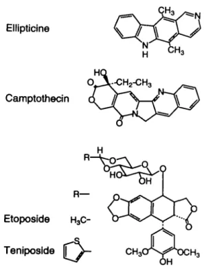
The chemicals investigated in this study (Figure 1) include three topoisomerase II inhibitors: the intercalating agent ellipticine, as well as the two non-intercalating podophyllotoxin 315
Hansjdrg Frei and Friedrich E.Wflrgler
H3
Ellipticine
Camptothecin
Fig. 1. Chemical structures of the four topoisomerase inhibitors studied.
derivatives teniposide and etoposide. The fourth compound is camptothecin, which is well known as a prototype topoisomer-ase I inhibitor. We opted for chemicals which differed in their DNA and enzyme binding (Liu, 1989; Ferguson and Baguley, 1994). Different genotoxic actions may lead to different reaction patterns in the wing spot test, since this SMART assay is considered sensitive to the induction of various types of genetic endpoints, such as mitotic recombination, deletion, segmental aneuploidy and point mutation (Haynie and Bryant, 1977; Graf et al, 1984). We wanted to know whether these agents showed genotoxic activity in the cells of the proliferating wing primordia of Drosophila and, if so, to what extent they induced mutational and recombinational events.
Materials and methods
Chemicals
Ellipticine (CAS no. 519-23-3) and camptothecin (CAS no. 7689-03-4) were purchased from Sigma (St Louis, MO). Etoposide (CAS no 33419-42-0) was a gift from Dr R.D.Snyder (Stauffer Chemicals, Farmington, CT)- Teniposide (CAS no. 29767-20-2) was tested as the clinical preparation Vumon® (Bristol-Myers, Princeton, NJ) containing 10 mg/ml of teniposide; the two tested concentrations of 0.1 and 0.05 mg/ml correspond to 0.152 and 0.076 mM respectively. Solutions and dilutions of the compounds were made with 5% ethanol and 5% Tween-80 in water except for Vumon®, which was diluted with water.
Wing spot test
For the wing spot test, we adopted the methods and screening criteria of Graf
et al. (1984), except that other strains were used for the outcross. Eggs were
collected for 8 h from a cross between mwh females and fl^/TMl, ri pP sep
l(3)89Aa bx34* e B(f males [for genetic symbols and descriptions see Lindsley
and Zimm (1992)]. Progeny were raised at 25°C. For the first 72 h they were kept on standard commeal medium copiously enriched with baker's yeast and were then fed with the chemicals in the experimental vials during the remainder of larval life (-48 h). Vials contained 1.5 g of Drosophila Instant Medium (Carolina Biological Supply, Burlington, N Q rehydrated with 5 ml of the test solutions.
The wings of the hatched flies were examined at X400 magnification for the occurrence of mutant spots (clones) on the phenotypically wildtype wing blade. Wings of mwh/flr3 marker heterozygous flies were screened for single
spots (phenotype mwh or flr3) and twin spots (phenotypes mwh and flr3 in adjacent clones), while on the inversion-heterozygous mwh/TM3 wings, only
mwh single spots can be found, as the multiply inverted TM3 balancer chromosome does not carry flr3 or any other suitable wing cell marker mutation.
Statistical evaluation
For an unbiased comparison of the induction frequencies in the two genotypes, only mwh clones in mwh single spots and in twin spots were taken into consideration (cf. Frei et al., 1992a). The mwh clone induction frequencies per cell and per cell division were calculated with and without clone size corrections according to the latter reference.
For statistical testing, the decision procedure of Frei and Wiirgler (1988) was applied. As customary for the evaluation of wing spot data (Graf et al., 1984), the following classes among the total of spots are distinguished: small single spots (1-2 cells in size), large single spots (3 cells or larger) and twin spots. This classification is biologically meaningful (Frei and WUrgler, 1988). From the statistical point of view, it should be pointed out, however, that the three spot categories are not equivalent with respect to statistical power, i.e. if used in significance testing, particularly to evaluate chronic exposure data, small single spots represent the most sensitive and twin spots the least sensitive of the three criteria (Frei and WUrgler, 1995).
Results
The results of wing analysis are shown in Table I. Historical and pooled controls show that water and the Tween-80/ethanol solvent used as carriers in the experiments give very similar spontaneous spot frequencies and that in mwh/TM3 inversion-heterozygotes the recovery of mwh clones is reduced to about one-third of the frequency found in mwh/flr3 inversion-free
marker heterozygotes. Because concurrent controls showed only a few spots, the data from the pooled large controls were used for control corrections in the determination of clone induction frequencies to minimize the influence of stochastic fluctuations.
For significance testing, however, the spot scores in treated groups were always compared with the corresponding concur-rent control. In the mwh/flr3 genotype, all four compounds
tested showed clear positive effects, with significant and exposure dose-related induction for all spot categories con-sidered.
For ellipticine, exceptionally, we found a reduced frequency of spots at the highest exposure concentration tested (8 mM) compared with the next highest concentration (4 mM), but in the range of 0.5-4 mM there was a steady increase with dose in the frequency of all spot categories over almost an order of magnitude in exposure concentration. The reduction at the highest concentration could be a selection effect due to toxicity of the compound since ~50-60% of the animals did not reach the adult stage at the 8 mM exposure level. However, a repellent effect or growth inhibition could also be responsible for the decline, because we noted with this compound and at this concentration that the majority of the pupae were underdeveloped in size. This was not the case at lower concentrations, whereas some pupal lethality was still observed (20-50% approximately in the exposure concentration range 0.5-4 mM).
With teniposide, all animals died at exposure concentrations of 0.25-1 mg/ml, while at the two genotoxic exposure concen-trations of 0.05 and 0.1 mg/ml the pupal lethalities were estimated as 20 and 70% respectively.
Etoposide was not visibly toxic in the tested concentration range of 0.5-2 mM. Solubility problems were encountered with this compound which had to be tested as a suspension. It is possible that the comparatively strong genotoxic response recorded at the 2 mM exposure in comparison with the lower exposure levels reflects an unequal exposure of the growing larvae owing to a non-homogeneous distribution of the com-pound in the test medium.
Solubility problems were also encountered with campto-316
Topolsomerase inhibitors in the Drosophila wing spot test
thecin, but the five highest concentrations assayed (0.5-7 mM) were so toxic that flies barely survived. Only wings from lower exposure levels could be analysed; at 0.25 mM concentra-tion there were roughly 70% surviving flies, while at 0.05 and 0.1 mM concentrations survival was similar to the controls. Particularly striking with this compound, a dose-dependent delay in larval development was observed virtually throughout the whole exposure range, but particularly at the strongly toxic exposure levels. As large clones are considered to reflect induction early in ontogeny, developmental retardation in combination with chemical instability may explain the rela-tively large size of the clones induced with this chemical (camptothecin produced the largest clones; see below).
If possible, we test potential mutagens with a geometric series of dilutions, try to attain the maximum tolerated dose (MTD), and examine MTD, MTD/2, MTD/4 etc. for genotoxic effects. In the present study, the toxic limit was reached with teniposide (>0.15 mM), camptothecin (>0.2 mM) and ellipticine (>8 mM). General toxicity can be quite unrelated to genotoxicity: for instance, at the non-toxic 2 mM exposure concentration, etoposide was more effective in inducing wing spots than teniposide at its 0.15 mM MTD. The genotoxic efficiency (denned here as genotoxicity at the MTD) was lowest for teniposide, significantly higher for camptothecin and in turn significantly higher for ellipticine (Table I, Figure 2). Because etoposide was not toxic at the concentrations tested, we do not know the MTD for this compound, but on account of the present data (Figure 2) it can be concluded that its genotoxic efficiency must be significantly higher than that of the analogue teniposide.
Genotoxicity and recombinagenicity
As a significant induction of twin spots was obtained for all four compounds, it is proved that they are recombinagenic. While this criterion is valid for qualitative judgements, the relative frequency of twin spots identified among the total of spots recovered does not seem a suitable basis for the quanti-tative estimation of the relative recombinagenic potency of genotoxic compounds. It is well known that flr mutations are not or not fully expressed phenotypically in small clones (Szabad et al, 1983). This makes the correct identification of small twin spots difficult.
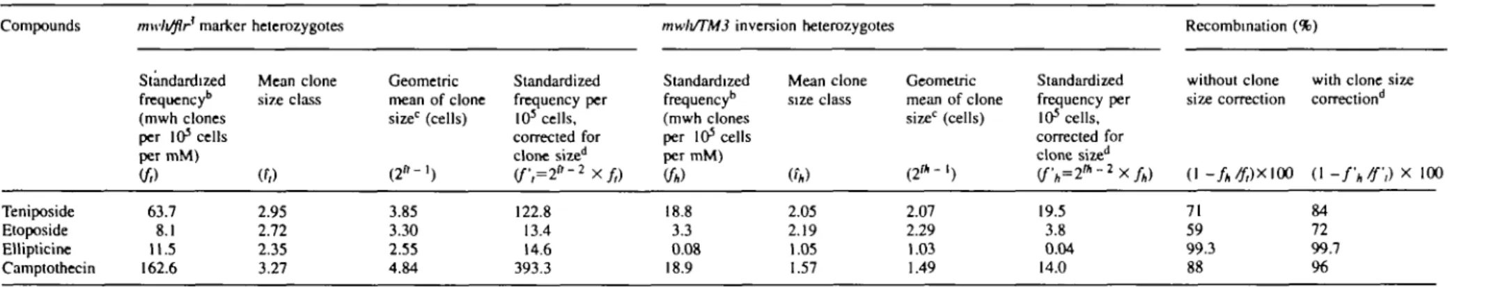
In the present experiments, the identification of twin spots was clearly correlated with spot size. In Figure 3 we used the geometric mean of mwh clone size as a measure of average spot size. The graph shows that in controls, clones are small on average and that only ~10% of the spots recovered are identified as twin spots. In the treatment series, clones were larger and the proportion of twin spots scored was greater.
Because of the uncertainties which arise from the spot size dependence of twin spot identification, it is far more preferable to estimate the relative contribution of recombination to spot induction from a comparison of the two genotypes mwh/flr3
and mwh/TM3, which constitute the two types of offspring from the outcross used in the wing spot test. In the latter genotype recombination is suppressed due to inversion-heterozygosity and therefore the difference in mwh clone induction frequencies between the two genotypes can be used to estimate the contribution of recombination to clone induction in the former, inversion-free genotype.
Owing to the reduced spot induction frequency in mwh/
TM3 flies compared with mwh/flr3 ones, there were in general
more inconclusive results at the lower exposure concentrations in the mwh/TM3 genotype than in the mwh/flr3 flies (Table I).
is
I
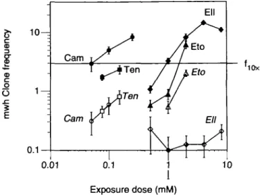
CD O O SI E '10X 0.01 Exposure dose (mM)Fig. 2. Double logarithmic plot of mwh clones recovered per wing at
different exposure concentrations: camptothecin (Cam, # O ) ; ellipticine (Ell, • O ) ; teniposide (Ten, • • ) ; etoposide (Eto, AA). Frequencies (±SE) are shown for mwh/flr3 marker-heterozygous flies (full symbols) and mwh/TMi
inversion-heterozygous flies (italics, open symbols). About 3 mwh clones per wing correspond to a 10-fold increase (/IOX) over the spontaneous level in mwh/flr3 flies.
a
0.20 . 1 0 -• T • • Camptothecin Teniposide Etoposide Ellipticine Controls • • i • • T " T • • • • I I I I IGeometric mean of clone size (mwh cells)
Fig. 3. Correlation between twin spot recovery and clone size. The plot is
based on uncorrected observation data in the different exposure series as reported in Table I.
At the higher exposure levels, however, etoposide, teniposide and camptothecin showed positive results, although for campto-thecin the result was significant only for the small single spots (1-2 cells in size) and for the total of spots, while the large single spots (3 cells or larger in size) were more frequent than in the concurrent control but not significantly so.
For ellipticine, by contrast, no significant spot induction was found in the inversion-heterozygous flies at any of the five concentrations tested which had given clearly positive results in the inversion-free genotype. Therefore, it appears that with ellipticine all or nearly all spots recovered in inversion-free mwh/flr3 flies are due to reciprocal
recom-bination.
For a quantitative comparison of spot induction by the four compounds in the two genotypes we unified the data to 317
TaMe I. Statistical significance of wing '•Controls and compounds Genotypes 'Water (historical mwh/flr3 mwh/TM3 25% Tween-80 + mwlt/flr3 mwlt/TM3 Cone. (mM) No. of wings (N)
and pooled controls) 790 504 5% ethanol (historica 'Teniposide (Vumon) mwlt/flr3 mwh/TM3 2Etoposide mwh/flr3 mwlt/TM3 2Ellipticine mwlt/flr3 mwli/TM3 ^Camptothecin mwlt/flr3 mwlt/TM3 0 0.076 0.152 0 0.076 0.152 0 0.5 1 2 0 1 2 0 0.5 1 2 4 8 0 0.5 1 2 4 8 0 0.05 0.10 0.25 0 0.05 0.10 382 100 48 48 38 48 48 26 48 48 48 48 48 42 48 48 48 48 48 44 20 28 22 20 40 78 48 48 48 48 40 24 16 22
spot induction by teniposide, etoposide, ellipticin and camptothecin after feeding Spots per wing (no. of spots) statistical diagnosis"
Small single spots (l-2cells)b
m = 2
0.24(189) 0.11 (53) and pooled controls)
0.21 (80) 0.08 (8) 0.17 (8) 0.96 (46) + 0.84 (32) + 0.06 (3) 0.44 (21) + 0.50 (13) + 0 19 (9) 0.35 (17) i 0.52 (25) + 2.83 (136) + 0.08 (4) 0.45 (19) + 1.38 (66) + 0.19 (9) 0.42 (20) + 1.65 (79) + 4.83 (232) + 8.66 (381) -f-7.70(154) + 0.07 (2) 0.23 (5) . 0.10 (2) i 0.08 (3) i 0.13 (10) i 0.19 (9) i 0.19 (9) 1.04 (50) + 2.33 (112) + 2.60(104) + 0.08 (2) 0.25 (4) i 0.45 (10) +
Large single spots (>2 cells)6 m = 5 0.03 (27) 0.01 (3) 0.04 (14) 0.03 (3) 0.02 (1) 0.38 (18) + 1.00 (38) + 0.06 (3) 0.02 ( 1 ) -0.31 (8) + 0.02 (1) 0.15 (7) + 0.23 (11) + 2.15 (103) + 0.04 (2) 0 . 1 0 ( 4 ) i 0.67 (32) + 0.06 (3) 0.38 (18) + 1.21 (58) + 2.23 (107) + 4.00 (176) + 2.00 (40) + 0.04 (1) 0.00 (0) i 0.00 (0) i 0.05 (2) i 0.08 (6) i 0.02 (1) i 0.02 (1) 1.29 (62) + 1.75 (84) + 3.78 (151) + 0.00 (0) 0.06 ( l ) i 0.14 (3) i Twin spots m = 5 0.03 (20) g 0.03 (10) * 0.02 (1) 0.44(21) + 0.55 (21) + i 0.04 (2) 0.08 (4) i 0.15 (7) i 1.35 (65) + g 0.02 (1) 0.29 (14) + 0.54 (26) + 1.35 (65) + 2.23 (98) + 1.50(30) + t 0.00 (0) 0.65 (31) + 1.02 (49) + 2.15 (86) + g Total spots m = 2 0.30 (236) 0.11 (56) 0.27 (104) 0.11 (11) 0.21 (10) 1.77 (85) + 2.39 (91) + 0 13 (6) 0.46 (22) + 0.81 (21) + 0.25 (12) 0.58 (28) + 0.90 (43) + 6.33 (304) + 0.13 (6) 0.55 (23) + 2.04 (98) + 0.27 (13) 1.08 (52) + 3.40(163) + 8.42 (404) + 14.89 (655) + 11.20(224) + 0.11 (3) 0.23 (5) i 0.10 (2) i 0.13 (5)i 0.21 (16) i 0.21 (10) i 0.21 (10) 2.98(143) + 5.10(245) + 8.52(341) + 0.08 (2) 0.31 (5) i 0.59 (13) + to mwh/flr3 Total mwh clones0 (n) 234 56 103 11 9 83 89 6 22 21 12 28 43 300 6 23 98 12 52 158 395 641 221 3 5 2 5 16 10 10 142 241 336 2 5 13
marker heterozygotes and Mean clone mwh size class"1 (0 1.94 1.57 2.01 2 45 2.33 2.66 2.92 4.00 1.50 2.43 2.08 2.71 2.30 2.69 3.00 1.96 2.28 2.42 2.73 2.50 2.33 2.33 2.16 2.00 1.20 1.50 2.40 2.25 1.50 1.50 3.18 2.84 3.49 1.50 2.00 1.69 [2.81] [3.06] [1.48] [2.57] [3.32] [2.43] [2.72] [1.83] [2.27] [2.97] [2.54] [2.34] [2.34] [2.17] [0.02] [12.00] [2.00] [2.01] [0.43] [3.30] [2.89] [3.54] [1.75] [1.52] Clone (per 1 mwh/TM3 induction inversion heterozygotes frequencies
03 cells per cell division/
Without size correctionde With t
(n/NC 1.21 0.46 1.11 0.45 0.77 7.09 9.60 0.51 1.88 3.31 1.02 2.39 3.67 25.61 0.51 2.24 8.37 1.02 4.44 13.49 33.73 59.71 45.29 0.44 0.93 0.41 0.51 0.84 0.85 0.85 12.12 20.58 34.43 0.34 1.28 2.42 ) [5.87] [8.38] [1.42] [2.85] [1.29] [2.57] [24.51] [1.79] [7.92] [3.33] [12.39] [32.62] [58.60] [44.18] [0.48] [-0.04] [0.06] [0.39] [0.40] [11.02] [19.47] [33.32] [0.83] [1.97] (2f-2 1.16 0.34 l.ll 0.62 0.97 11.22 18.18 2.05 1.33 4.46 1.09 3.92 4.53 41.32 1.02 2.18 10.13 1.37 7.37 19.08 42.37 75 17 50.70 0.44 0.53 0.29 0.68 1.00 0.60 0.60 27.53 36.79 96.57 0.24 1.28 1.96 lone-size correction'1* )X(nWQ [10.32] [17.53] [0.99] [4.22] [3.21] [3.45] [40.39] [1.60] [9.52] [6.53] [18.05] [41.29] [74.09] [49.59] [0.12] [-41.97] [0.06] [0.39] [0.14] [27.15] [35.97] [96.70] [0.70] [1.41] X T re i an d "*]
I
3.s-Table I. Continued
'•^Superscript numbers refer to the corresponding pooled and historical controls.
•"Statistical diagnoses according to Frei and WUrgler (1988): + , positive; -, negative; 1, inconclusive, m, multiplication factor for the assessment of significantly negative results. Significance levels a 0.05.
blncluding rare flr3 single spots.
Considering mwh clones from mwh single spots and from twin spots.
^Numbers in square brackets are induction frequencies corrected for spontaneous incidence estimated from the large histoncal and pooled controls.
CC 24 400, i.e. approximate number of cells examined per wing.
'Calculated according to Frei et al. (1992a).
''Only mwh single spots can be observed in mwh/TM3 heterozygotes as the balancer chromosome TM3 does not carry a flr mutation.
Table II. Standardized mwh clone induction frequencies per millimolar unit of exposure concentration and the prevalence of recombination events*
Compounds Teniposide Etoposide Ellipticine Camptothecin mwli/flr marker Standardized frequency*1 (mwh clones per 103 cells per mM) {ft) 63.7 8.1 11.5 162.6 heterozygotes Mean clone size class (f/> 2.95 2.72 2.35 3.27 Geometric mean of clone size0 (cells) ( 2 " - ' ) 3.85 3.30 2.55 4.84 Standardized frequency per 103 cells, corrected for clone sized (f;=2"-2xf,) 122.8 13.4 14.6 393.3
mwli/TM3 inversion heterozygotes
Standardized frequency* (mwh clones per 105 cells per mM)
to)
18.8 3.3 0.08 18.9 Mean clone size class «*) 2.05 2.19 1.05 1.57 Geometric mean of clone size0 (cells) ( 2 * -1) 2.07 2.29 1.03 1.49 Standardized frequency per 105 cells. corrected for clone sized (fh=2n -2Xfh) 19.5 3.8 0.04 14.0 Recombination without clone size correction (1 -fh/f,)X\(X 71 59 99.3 88 (%)with clone size correction"1 ) (1 -fh/f,) X 100 84 72 99.7 96
aAll values arc control corrected. Frequencies in mwh/flr3 marker-heterozygotes and mwh/TM3 inversion-heterozygotes are calculated with and without clone size correction; accordingly, somewhat different estimates are obtained for the relative contributions of recombination to the totals of clone induction.
kClone frequencies per wing divided by the number of cells examined per wing (24 400) estimate frequencies per cell and per cell division in chronic exposure experiments (Frei and WUrgler, 1988).
c Geometric mean and ^corrections calculated according to Frei et al. (1992a).
S
HansjSrg Frei and Friedrich E.WOrgler
formally equimolar standardized values. The observed mwh clone numbers in the two genotypes at each exposure level (Table I) were corrected by subtraction of the estimated number of spontaneous clones, so that the corrected frequencies corresponded to estimated frequencies of induced spots. The respective pooled and historical control frequencies and the numbers of wings analysed in the different treatment series served as the basis for this correction. An approximate average induction frequency per millimolar unit of exposure concentra-tion was then calculated by linear extrapolaconcentra-tion combining the results from the different exposure levels for each compound. The results are shown in Table II as standardized mwh clone induction frequencies per millimolar concentration, per cell and per cell division.
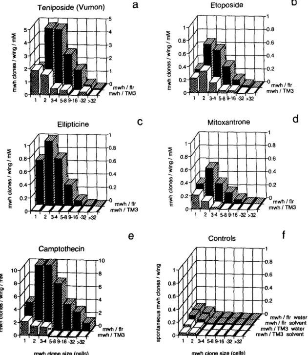
The same correction procedure was also used for the data underlying Figure 4a-e, except that the clones were arranged into size classes and the standardized frequencies in this case are given as clone induction frequencies per wing. In Figure 4f the spontaneous clone frequencies are given for the pooled controls. Figure 4a and e could not be drawn to the same scale as the other illustrations, because camptothecin and teniposide were genotoxic at comparatively low exposure concentrations. For comparison, the data for mitoxantrone are redrawn from Frei et al. (1992a) in Figure 4d.
The standardized clone induction frequency per millimolar exposure unit was highest for camptothecin. With teniposide it was ~3 times, with ellipticine about 15-25 times and with etoposide ~2O-3O times lower than with camptothecin. For
O> Teniposide (Vumon) mwh/fir mwfi/TM3 Etoposide 1 2 3-4 5-8 9-16 -32 >32 1 2 34 5-8 9-16 -32 >32 0.8 mwti/flr mwh/TM3 Ellipticine Mitoxantrone 2 o •s 1 2 3-4 5^9-16-32 >32 Camptothecin mwh/fir mwh/TM3 mwti/flr mwti/TM3 1 2 3-4 5-8 9-16-32 >32
mwh clone size (cells)
O) c 8
0.8-I
I
o & 0.6- 0.4- 0.2-mwh/fir mwh/TM3 mwh/fir water mwh/fir solvent mwh/TM3 water mwh/TM3 solvent mwh clone size (cells)Fig. 4. Clone size distributions and mwh clone induction frequencies standardized to the millimolar exposure concentration unit in mwh/flr3
marker-heterozygous flies and in mwh/TM3 inversion-marker-heterozygous flies. Frequencies are control-corrected except in (f). Mitoxantrone data (d) are redrawn from Frei
et al. (1992a).
Topoisomerase inhibitors in the Drosophila wing spot test
equal effect exposures similar relationships are found. Frequen-cies of ~3 mwh clones per wing correspond to an ~ 10-fold increase (fiox) o v e r the spontaneous control frequency. For
the / | ox level, the respective approximate exposure doses are 0.05 mM for camptothecin, 0.15 mM for teniposide, 1 mM for ellipticine and 1-2 mM for etoposide, as shown in Figure 2. The graph also illustrates that at the/|Ox and other exposure
levels ellipticine, camptothecin and teniposide differ signifi-cantly among each other in their respective reductions in clone frequency (mwh/TM3 < mwh/flr3). However, on account of
means and standard errors, the difference in reduction between the epipodophyllotoxins teniposide and etoposide is not statistically significant and may be fortuitous.
The comparison of the two genotypes demonstrates that mitotic recombination is the prevalent mechanism of wing spot induction. Ellipticine and camptothecin appear as exclusive or strong recombinagens respectively. Teniposide and etoposide are preferentially recombinagenic, but some 30—40% of the spots are of non-recombinational origin. In Table II the estimations of the proportions of clone induction attributed to recombination differ somewhat, depending on whether clone size corrections are applied or not in the calculations, but the general conclusions remain unaffected by this.
Spot size
The spot size distributions in Figure 4a-e show that in mwh/
flr3 flies, clones are larger than expected (mean mwh clone
size > 2 cells) and therefore clone size corrections in Table II lead to increased standardized frequencies compared with the estimates obtained without size correction [for theoretical considerations see Frei et al. (1992a)]. This was most pro-nounced for the relatively large clones induced by campto-thecin, which is known to be chemically unstable in its lactone ring and for which this type of correction seems particularly appropriate. But also for the other compounds the clone size correction leads to increased estimates. This reflects probably a reduced exposure of the cells towards the end of imaginal disk development. Shortages relative to theoretical expectation in the first size class (comprising <50%) and to some extent in the second clone size class (comprising <25% of the total of mwh clones) probably mean that the clones in the small size classes (1-2 cells) have reduced frequencies, because for these the time of induction is during the last and penultimate mitotic divisions in the pupa when the drug uptake has ceased. For the compounds which are able to induce wing spots in the mwh/TM3 inversion-heterozygous genotype (Figure 4a-e), mwh clones are smaller on average than in the inversion-free
mwh/flr3 marker heterozygotes. It has been postulated for
mitoxantrone that a majority of the small clones recovered in
mwh/TM3 flies may represent cases of induced segmental
aneuploidy with reduced proliferation capacity (Frei et al, 1992a). The reduction in clone induction frequency in the
mwh/TM3 flies compared with the mwh/flr3 flies is also less
pronounced for the two smallest clone size classes than for the larger size classes, which is consistent with the idea that inversion-heterozygosity only suppresses mitotic crossing over but not the induction of deletions and other segmentally deficient chromosomal aberrations, enabling a hemizygous mwh manifestation owing to the uncovered recessive mwh mutation.
Discussion
The SMART tests of Drosophila are versatile genotoxicity test systems detecting a great variety of genotoxins belonging to
different chemical classes, including promutagens requiring enzymatic bioactivation. Some promutagens, such as procarba-zine, hexamethylphosphoramide (HMPA) or the group of pyrrolizidine alkaloids, which are genotoxic in mammalian cells but whose detection poses problems in bacterial genotox-icity test systems, are readily detected by the SMART assays in Drosophila (for refs see Wurgler and Vogel, 1986; Frei
et al, 1992b). Here we study four topoisomerase inhibitors
for their effects in the wing spot test. This seemed an interesting group of compounds, because their effects depend on enzymes for which there is evidence of significant evolutionary diver-gence particularly between prokaryotes and eukaryotes, but possibly also between lower and higher eukaryotes. The four topoisomerase inhibitors studied proved genotoxic in the
Drosophila wing spot test in agreement with genotoxicity in
mammalian and human cells, where they induce chromosome aberrations and sister chromatid exchanges (Bhuyan et al, 1972; Huang et al, 1973; Chen et al, 1984; DeMarini et al, 1987a; Moore et al, 1987; Maraschin et al, 1990; Agarwal
et al, 1994).
Molecular effects of topoisomerase poisons
For the type of inhibitors functioning as topoisomerase poisons (Ferguson and Baguley, 1994), it appeared that an early event of drug action is the formation of an intermediate called the trapped cleavable complex, which represents the drug-stabilized ternary complex between DNA, the covalently bound topoisomerase and the inhibitor. While in the non-inhibited state the strand breaks catalysed by the enzymes are transient, they are not religated in the trapped complex (cf. Liu, 1989; Ferguson and Baguley, 1994; Ralph et al, 1994). The cleavage can be visualized by molecular biological methods following treatment of the complexes with rapid protein denaturants which reveal DNA single- and double-strand breaks associated with bulky protein (i.e. topoisomerase) adducts.
Topoisomerase inhibitors genotoxic in Drosophila
The wing spot test of Drosophila apparently detects this condition adequately and therefore seems to be a good predictor for this type of genotoxic activity. Besides teniposide, etopo-side, ellipticine and camptothecin, other topoisomerase inhib-itors have been studied previously in the eye and wing SMART assays of Drosophila. The intercalating agents adriamycin (doxorubicin), daunomycin (daunorubicin) and mitoxantrone gave positive results and were recombinagenic as they signifi-cantly increased the frequency of twins spots (Frei etal, 1985, 1992a; Clements and Vogel, 1988).
Mitotic recombination, and single and twin spots in the wing spot test
The relative frequency of twin spots may give some idea of the recombinagenicity of a compound. Genotoxic chemicals can give quite different results in this respect (Ramel and Magnusson, 1992). The marker gene mwh is located at the tip of the left arm of chromosome 3 (section 61E2-62A3). The cytological locations of neighbouring genes place fir roughly to the middle (section 69) of the left arm of chromosome 3 [cf. Lindsley and Zimm (1992) for map positions]. Under the assumption that mitotic crossing over is proportional to the physical distance on the chromosome between centromere and marker genes (Becker, 1976), one would expect ~50% twin spots (recombination between flr and the centromere) and 50% mwh single spots (recombination between mwh and flr) if all spots were due to mitotic crossing over. For chemicals such as chromium(VI)oxide or ethylene oxide, twin spot frequencies
Hansjbrg Frei and Fried rich E.WUrgler
of 40—45% have been reported and for the former it was determined that >90% of the wing spots are due to mitotic recombination (Graf et al, 1992; Ramel and Magnusson, 1992). However, with the four topoisomerases studied, twin spot recovery was lower and not much different between the compounds. On average 26, 22, 16 and 23% of the induced spots were recovered as twin spots following exposure to teniposide, etoposide, ellipticine and camptothecin respectively (summarized from Table I).
Quantitation of recombinagenicity
The present data have shown that twin spot identification depends on clone size. Ramel and Magnusson (1992) have already pointed out that for chemicals producing predominantly small spots, the lack of expression of the fir genotype in small clones can lead to serious biases, because small twin spots cannot be readily identified.
For an unbiased evaluation of recombinagenicity, we there-fore prefer to compare the mwh clone frequencies in the two genotypes mwh/flr and mwh/TM3. The difference in clone induction between the two genotypes is a quantitative measure of recombinagenicity. In this way, the relative contribution of mitotic crossing over to total spot induction has been deter-mined previously for mitoxantrone (Frei et al, 1992a). It was estimated that 68% of the clones resulted from mitotic crossing over (cf. Figure 4d). The four topoisomerase inhibitors pres-ently studied differ from each other. In a comparison of recombinational to non-recombinational origin of induced clones, ellipticine ranks highest (almost 100% clones by recombination), followed by camptothecin (88%), teniposide (71%), and etoposide (59%). The difference between teniposide and etoposide was not statistically significant, however.
Surprisingly, the two intercalating topoisomerase n inhib-itors ellipticine (this study) and mitoxantrone (Frei et al, 1992a) are dissimilar in this respect. Whereas ellipticine appears as an almost exclusive recombinagen, mitoxantrone is preponderantly recombinagenic but with a significant propor-tion of spots arising from mechanisms other than recombinapropor-tion (cf. Figure 4c and d).
The clone induction pattern of mitoxantrone resembles more that of the two non-intercalating topoisomerase II inhibitors teniposide and etoposide. Notwithstanding that the treatments revealed differences in genotoxic effectiveness, the clone size distributions are similar for the three compounds and the proportions of clone induction in the two genotypes indicate that ~60-70% of the clones are due to recombination (cf. Figure 4a, b and d).
Non-disjunction in SMART analysis
As topoisomerase II has functions in mitosis which are important for chromosome segregation (cf. Ferguson and Baguley, 1994) the question may be raised as to whether some of the wing spots recovered from treatment with topoisomerase II inhibitors could be due to induced non-disjunction and represent clones monosomic for recessively marked chro-mosomes.
There is evidence that chemicals able to interfere with the mitotic apparatus can induce wing spots at low but significant frequencies. This was found previously with the spindle poisons vinblastine (Graf et al., 1984) and chloral hydrate (Zordan
et al., 1994), as well as with the chelators nitrilotriacetic acid
and (ethylenedinitrilo)tetraacetic acid (Zordan et al, 1990, 1991). Vogel and Nivard (1993) report positive results obtained in the wAv+ eye spot test with five spindle poisons. However,
as a rule, the aneuploidogenic compounds found positive in SMART assays have typically shown weak effects. Therefore, the clear-cut and strong genotoxic effects produced by the topoisomerase inhibitors in the SMART assays are better explained by structural DNA damage rather than by impaired chromosome segregation. In the mwh/TM3 genotype, more-over, inversion-heterozygosity suppresses reciprocal mitotic recombination, but would not prevent mitotic non-disjunction. As wing spot induction in this genotype was not significant with ellipticine, non-disjunction due to topoisomerase II inhibition seems not to play a major role in wing spot induction, at least with respect to this latter compound. However, very minor contributions by non-disjunction cannot be ruled out com-pletely in view of the weak but positive responsiveness of the SMART assays to induced somatic monosomy.
Performance of the SMART method compared with bacterial, fungal and mammalian test systems
Whereas the SMART assays of Drosophila seem to be good predictors of mammalian and human genotoxicity of topoiso-merase inhibitors, there is evidence that bacterial test systems are not (Gocke, 1991), and that test systems with lower eukaryotes, such as moulds and yeasts, may have reduced specificity.
The mould Neurospora crassa, in spite of being a eukaryote, has shown a type of response more similar to bacteria than to mammalian cells for compounds such as amsacrine (m-AMSA), etoposide, teniposide and ellipticine (Gupta, 1990). These compounds are specific for the mammalian topoisomerase II and show less interaction with gyrase and therefore are much less potent in bacterial assays (Gupta et al, 1987; Liu, 1989; Gocke, 1991). Inactivity or low activity seems particularly characteristic for etoposide and teniposide in bacterial and fungal genotoxicity assays. For instance, Nakanomyo et al. (1986) have reported very weak mutagenic effects for these two non-intercalating agents in frameshift strains (sic!) but not in base substitution strains of the
Salmonella/microsomes test.
The intercalating topoisomerase II inhibitors m-AMSA, adriamycin and daunorubicin, as well as the non-intercalating etoposide, have been studied in strain D5 of the yeast
S.cerevis-iae. This test system assesses mitotic crossing over, but also
other endpoints such as conversion and mutation at the
adenine-2 locus. Under growing conditions, the intercalating agents
gave positive results with respect to the total of aberrant colonies as well as to twin colonies indicative of mitotic crossing over, but overall the frequency of production of aberrant colonies was low (Ferguson and Turner, 1988). However, among the aberrant colonies those due to mitotic crossing over were relatively frequent compared with those obtained with other types of genotoxin. Therefore, but particu-larly also because the non-intercalating etoposide was negative in this assay, the authors suggested that intercalation is the activity which is involved in the recombination process. However, as the two epipodophyllotoxins assayed in the
Drosophila wing spot test were positive for recombinant as
well as non-recombinant spot induction, it rather seems that insensitivity of yeast, Neurospora and bacteria to the effects of these compounds is a specific property of the test organisms.
Mechanistic considerations
Recently Ferguson et al (1993) found evidence that in
S.cerevi-siae recombinational repair of damage by intercalating topoiso-322
Topoisomerase inhibitors in the Drosophila wing spot test
merase II inhibitors depends on three repair genes. Recombinational repair of damage produced by the strong recombinagen DACA particularly required the RAD52 gene product known to be involved in the recombinational repair of double-strand breaks.
By analogy we tnay associate the >99% recombinational origin of wing spots recovered in Drosophila following expo-sure to ellipticine with the production of double-strand breaks, which this compound appears to produce almost exclusively (Pommier et al, 1984). Other compounds which are also able to induce significant numbers of wing spots of non-recombinational origin may in part produce other types of damage. It has been reported for etoposide, for instance, that it produces a mixture of single- and double-strand breaks, with a moderate predominance of single-strand breaks (Osheroff, 1989b). Inhibitors show preferences for particular base pair sequences and stabilize cleavable complexes at compound-specific sites. The bias for compound-specific bases on the cleaved strand is generally stronger than for the complementary strand, so that many sites probably are of the single-strand cleaved type (Ralph et al, 1994), even in the case of topoisomerase II inhibition. In contrast to the ellipticines, mitoxantrone has site specificities similar to those of teniposide (cf. refs in Ferguson and Baguley, 1994; Ralph et al, 1994).
The topoisomerase I inhibitor camptothecin showed 88% of the wing spots to be due to recombination and appears as the next strongest recombinagen after ellipticine among the four agents investigated in this study (cf. Figure 4c and e). In yeast, too, camptothecin induces high levels of homologous recombination and is weakly mutagenic; moreover, cytotoxicity is more pronounced in strains carrying a defective rad52 repair gene (Nitiss and Wang, 1988).
There is some similarity in the planar aromatic ring systems in the chemical structures of ellipticine and camptothecin (Figure 1), but this feature is probably not the common determinant cause of high recombinagenicity characterizing the two compounds. Whereas ellipticine appears to be a perfect intercalator with specificity for topoisomerase II inhibition, camptothecin was reported not to bind to DNA and to inhibit exclusively topoisomerase I in cell-free in vitro reaction systems (Hsiang et al, 1985). However, more recent studies suggest that camptothecin nevertheless might have at least some potential for intercalation but rather into the less common Z- than B-DNA (Fukada, 1985; Wittig et al, 1989).
Camptothecin trapping topoisomerase I was found to pro-duce large amounts of DNA single-strand damage in cell-free
in vitro reaction systems (Hsiang et al, 1985), which seems
to contradict the double-strand break hypothesis for the induc-tion of mitotic crossing over (Ferguson et al, 1993). However, in cultured human and Drosophila cells, prominent double-strand as well as single-double-strand breaks were observed following treatment with camptothecin (Zhang et al, 1988; Kroeger and Rowe, 1989). The more pronounced cytotoxicity of campto-thecin in mutant rad52 yeast strains (Nitiss and Wang, 1988) may also be related to induced double-strand damage.
It is most conceivable in addition that the relationship between spot induction by recombinational or other mechan-isms in the SMART assays is not solely determined by the nature of the trapped complex but also by additional factors such as the stability of the ternary complex and the dynamics of processing of the damage, e.g. during repair. Although we cannot tell actually how the clones of non-recombinational origin are produced, we may conclude from the present data
that in this respect there are differences in action between the topoisomerase inhibitors.
Topoisomerase drugs non-genotoxic in the Drosophila wing spot test
Not all topoisomerase inhibitors are genotoxic in Drosophila and induce wing spots. While the poisons of eukaryotic topoisomerases (Ferguson and Baguley, 1994; Ralph et al, 1994) discussed so far do not prevent cleavage of the DNA but inhibit religation, other topoisomerase-directed drugs interfere with cleavage. Certain gyrase-directed bactericidal agents such as novobiocin or nalidixic acid are weak inhibitors in eukary-otes but are able to act on eukaryotic topoisomerase n . They inhibit enzyme-catalysed ATP hydrolysis and prevent formation of the cleavable complex (Robinson et al, 1993; Ferguson and Baguley, 1994). Novobiocin is highly bactericidal (Hussy
et al, 1986), but it was not mutagenic in genotoxicity assays
with Salmonella typhimurium or Escherichia coli (McCoy
et al, 1980; Gocke, 1991). When applied alone, this drug did
not induce spots in the wing spot test of Drosophila but did have modulating antagonistic effects when combined with monofunctional alkylating agents (Ramel and Magnusson, 1992). In mammalian cells, too, novobiocin alone was unable to induce enzyme-mediated DNA strand breaks, but did have cell-cycle-dependent increasing or decreasing modulatory effects in combination with ionizing radiation or clastogenic chemicals (Marshall et al, 1983; Takahashi et al, 1985, 1986; DeMarini et al, 1987b; Dillehay et al, 1987).
The benzo[c]phenantridine alkaloid fagaronine and its con-gener nitidine have been identified as intercalators (Pezzuto
et al, 1983) inhibiting both types of topoisomerase, but mainly
the type I enzyme (Larsen et al, 1993; Wang et al, 1993). However, despite intercalation, fagaronine was not genotoxic in the 5a/mon€//o/microsomes test (strain TM677; Pezzuto
et al, 1983). In the Drosophila wing spot test, fagaronine,
nitidine and several derivatives were negative (P6rez-Chiesa and Narvaez, 1993; P6rez-Chiesa and Rodriguez, 1993). The mechanism of topoisomerase inhibition by fagaronine and congeners is not known, but it may be recalled that there are other DNA intercalating drugs, such as 9-aminoacridine, which may interfere with topoisomerase II attachment to DNA because of inducing helix unwinding and extension. 9-Ami-noacridine does not function as a topoisomerase II poison, but on the contrary can act antagonistically against a variety of topoisomerase II poisons (Ferguson and Baguley, 1994). On account of the present results we would predict that fagaronine and its congeners are not potent clastogens or mutagens in mammalian cells if active at all.
Concluding remarks
With respect to the performance of the SMART methods of
Drosophila, particularly also in genotoxicity testing of agents
which do not react directly with DNA, it is notable that the wing spot test is sensitive to the genotoxic mammalian topoisomerase I and topoisomerase II inhibitors, especially also to the non-intercalating epipodophyllotoxins. As with other genotoxic agents, the somatic mutant spots induced are mainly, but not exclusively, due to mitotic recombination. In this respect, ellipticine appears to be somewhat exceptional, because it is not common in the wing spot test that genotoxic agents induce only or almost exclusively recombination events.
Acknowledgements
Thanks are due to Dr Ulrich Graf for critical reading of the manuscript. We thank Mss Doris Singer, Muriel Badoux and Heidi Bolliger for skilful technical 323
Hansjdrg Frd and Friedrtch E.WOrgler
assistance. We also acknowledge the help of Ms Rehana Ahmed (Karachi, Pakistan), who received support as a research trainee from the Food and Agricultural Organization of the United Nations. We are grateful to two anonymous reviewers of the manuscript for most valuable comments and suggestions.
References
Agarwal.K., Mukherjee,A. and Sen.S. (1994) Etoposide (VP 16): cytogenetic studies in mice. Environ. Mol. Mutagen., 23, 190-193.
Becker,HJ. (1976) Mitotic recombination. In Ashburner.M. and Novitski.E. (eds), The Genetics and Biology of Drosophila. Academic Press, London, vol. lc, pp. 1020-1087.
Bhuyan.B.K., Fraser.TJ. and Li.L.H. (1972) Cell cycle phase specificity and biochemical effects of ellipticine on mammalian cells. Cancer Res., 32, 2538-2544.
Buchenau.P., Saumweber.H. and Amdt-Jovin.DJ. (1993) Consequences of topoisomerase II inhibition in early embryogenesis of Drosophila revealed by in vivo confocal laser scanning microscopy. J. Cell Sci., 104, 1175-1185. Chen.G.L., Yang.L., Rowe.T.C, Halligan.B.D., Tewey.K.M. and Liu.L.F. (1984) Non-intercalative antitumor drugs interfere widi the breakage-reunion reaction of mammalian DNA topoisomerase II. J. Biol. Chem.,
259, 13560-13566.
Clements J. and Vogel.E.W. (1988) Somatic recombination and mutation assays in Drosophila: a comparison of the response of two different strains to four mutagens. Mutat. Res., 209, 1-5.
DeMarini,D.M., Brock.K.H., Doerr.C.L. and Moore,M.M. (1987a) Mutagenicity and clastogenicity of teniposide (VM-26) in L5178Y/TK"1
"'"-3.7.2C mouse lymphoma cells. Mutat. Res., 187, 141-149.
DeMariniJD.M., Doerr.C.L., Meyer,M.K., Brock.K.H., HozierJ. and Moore.M.M. (1987b) Mutagenicity of m-AMSA and o-AMSA in mammalian cells due to clastogenic mechanism: possible role of topoisomerase. Mutagenesis, 2, 349-355.
Dillehay.L.E., Denstman,S.C. and Williams J.R. (1987) Cell cycle dependence of sister chromatid exchange induction by DNA topoisomerase II inhibitors in Chinese hamster V79 cells. Cancer Res., 47, 206-209.
Ferguson.L.R. and Baguley.B.C. (1994) Topoisomerase II enzymes and mutagenicity. Environ. Mol. Mutagen., 24, 245-261.
Ferguson,L.R. and Tumer.P.M. (1988) Mitotic crossing-over by anticancer drugs in Saccharomyces cerevisiae strain D5. Mutat. Res., 204, 239-249. Ferguson.L.R., Turner,P.M. and Baguley,B.C. (1993) Induction of mitotic
crossing-over by the topoisomerase II poison DACA (W-[2-dimethyl-aminoethyl]acridine-4-carboxamide) in Saccharomyces cerevisiae. Mutat.
Res., 289, 157-163.
Frei.H. and Wiirgler.F.E. (1988) Statistical methods to decide whether mutagenicity test data from Drosophila assays indicate a positive, negative, or inconclusive result Mutat. Res., 203, 297-308.
Frei.H. and Wtirgler.F.E. (1995) Optimal experimental design and sample size for the statistical evaluation of data from somatic mutation and recombination tests (SMART) in Drosophila. Mutat. Res., 334, 247-258.
Frei,H., WUrgler.F.E., Juon.H., Hall,C.B. and Graf.U. (1985) Anstolochic acid is mutagenic and recombinogenic in Drosophila genotoxicity tests. Arch.
Toxicoi, 56, 158-166.
Frei,H., ClementsJ., Howe.D. and Wlirgler,F.E. (1992a) The genotoxicity of the anti-cancer drag mitoxantrone in somatic and germ cells of Drosophila
melanogaster. Mutat. Res., 279, 21-33.
Frei,H., UlthyJ., Brauchli J., Zweifel.U., WUrgler.F.E. and Schlatter.C. (1992b) Structure/activity relationships of the genotoxic potencies of sixteen pyrrolizidine alkaloids assayed for the induction of somatic mutation and recombination in wing cells of Drosophila melanogaster. Chem.-Biol.
Interact., 83, 1-22.
Fukadajvl. (1985) Action of camptothecin and its derivatives on deoxyribonucleic acid. Biochem. Pharmacol., 34, 1225-1230.
Gocke,E. (1991) Mechanism of quinolone mutagenicity in bacteria. Mutat.
Res., 248, 135-143.
Graf,U., WUrgler^.E., Katz^U., Frei,H., Juon,H., Hall.C.B. and Kale.P.G. (1984) Somatic mutation and recombination test in Drosophila melanogaster.
Environ. Mutagen., 6, 153-188.
Graf.U., Ok-Soon,H. and Olvera Ramirez,O. (1992) The genotoxicity of chromium(VI) oxide in the wing spot test of Drosophila melanogaster is over 90% due to mitotic recombination. Mutat. Res., 266, 197-203. Gupta,R. (1990) Tests for the genotoxicity of m-AMSA, etoposide, teniposide
and ellipticine in Neurospora crassa. Mutal. Res., 240, 47-58.
Gupta,R.S., Bromke,A., Bryant,D.W., GuptaJ*., Singh.B. and McCalla,D.R. (1987) Etoposide (VP16) and teniposide (VM26): novel anticancer drugs, strongly mutagenic in mammalian but not prokaryotic test systems.
Mutagenesis, 2, 179-186.
HaynieJ.L. and Bryant.PJ. (1977) The effects of X-rays on the proliferation dynamics of cells in the imaginal wing disc of Drosophila melanogaster.
Wilh. RouxArch. Develop. Biol., 183, 85-100.
Hsiang,Y.-H., Hertzberg,R., HechtS. and Liu.L.F. (1985) Camptothecin induces protein-linked DNA breaks via mammalian DNA topoisomerase I.
J. Biol. Chem., 260, 14873-14878.
Hsieh,T.-S., Brown.S.D., Huang.P. and FosteU. (1992) Isolation and characterization of a gene encoding DNA topoisomerase I in Drosophila
melanogaster. Nucleic Acids Res., 20, 6177-6182.
Huang,C.C, Hou,Y. and WangJJ. (1973) Effects of a new antitumor agent epipodophyllotoxin, on growth and chromosomes in human hematopoietic cell lines. Cancer Res., 33, 3123-3129.
Hussy.P, Maass.G., TUmmler.B., Grosse.F. and Schomburg.U. (1986) Effect of 4-quinolones and novobiocin on calf thymus DNA polymerase a primase complex, topoisomerases I and II, and growth of mammalian lymphoblasts.
Antimicrob. Agents Chemother., 29, 1073-1078.
Kroeger.P.E. and Rowe.T.C. (1989) Interaction of topoisomerase I with the transcribed region of the Drosophila HSP 70 heat shock gene. Nucleic
Acids Res., 17, 8495-8509.
Larsen,A.K., Grondard.L., CouprieJ., Desoize.B., Comoe.L., JardilherJ.-C. and RiouJ.-F. (1993) The antileukemic alkaloid fagaronine is an inhibitor of DNA topoisomerases I and II. Biochem. Pharmacol., 46, 1403-1412. Lindsley.D.L. and Zimm.G.G., eds. (1992) The Genome of Drosophila
melanogaster. Academic Press, San Diego, CA.
Liu.L.F. (1989) DNA topoisomerase poisons as antitumor drugs. Annu. Rev.
Biochem., 58, 351-375.
Maraschino, Dutrillaux.B. and Aurias.A. (1990) Chromosome aberrations induced by etoposide (VP-16) are not random. Int. J. Cancer, 46, 808—812. Marshall,B., Darkin.S. and Ralph.R.K (1983) Evidence that mAMSA induces
topoisomerase action. FEBS Lett., 161, 75-78.
McCoy.E., Petrullo.L.A. and Rosenkranz,H.S. (1980) Non-mutagenic genotoxicants: novobiocin and nalidixic acid, 2 inhibitors of DNA gyrase.
Mutat. Res., 79, 33-^3.
Moore.M.M., Brock,K.H., Doerr.C.L. and DeMarini.D.M. (1987) Mulagenesis of L5178Y/TK+/~-3.7.2C mouse lymphoma cells by the clastogen ellipticine.
Environ. Mutagen., 9, 161-170.
Nakanomyo.H., Hiraoka,M. and Shiraya,M. (1986) Mutagenicity tests of etoposide and teniposide. J. Toxicoi. Sci., ll(Suppl. 1), 301-310. NitissJ. and WangJ.C. (1988) DNA topoisomerase-targeting antitumor drugs
can be studied in yeast Proc. Natl. Acad. Sci. USA, 85, 7501-7505. Osheroff.N. (1989a) Biochemical basis for the interactions of type I and type
II topoisomerases with DNA. Pharmacol. Then, 41, 223-241.
Osheroff.N. (1989b) Effect of antineoplastic agents on the DNA cleavage/ religation reaction of eukaryotic topoisomerase II: inhibition of DNA religation by etoposide. Biochemistry, 28, 6157-6160.
P6rez-Chiesa,Y. and Narvaez,Z. (1993) Evaluation of genotoxicity of the indenoisoquinohne analogues of fagaronine and nitidine in Drosophila
melanogaster. Mutat. Res., 301, 207-212.
P£rez-Chiesa,Y. and Rodriguez^. (1993) Absence of mutagenicity of benzo[c]phenantridine alkaloids in somatic cells of Drosophila
melanogaster. comparison with 7,12-dimethylbenz(a]anthracene and
chrysene. Mutat. Res., 298, 277-283.
PezzutoJ.M., Antosiak.S.K., Messmer.W.M., Slaytor.M.B. and Honig.G.R. (1983) Interaction of the antileukemic alkaloid, 2-hydroxy-3,8,9-trimethoxy-5-methylbenzo[c]phenantridine (fagaronine), with nucleic acids.
Chem.-Biol. Interact., 43, 323-339.
Pommier.Y, Schwartz,R.E., Kohn.K.W. and ZwelIing,L.A. (1984) Formation and rejoining of deoxyribonucleic acid double-strand breaks induced in isolated cell nuclei by antineoplastic intercalating agents. Biochemistry, 23, 3194-3201.
Ralph.R.K., Judd.W., Pommier.Y. and Kohn.K.W. (1994) DNA topoisomerases. In Neidle.S. and Waring.M. (eds). Molecular Aspects of Anticancer
Drug-DNA Interactions. Macmillan, London, vol. 2, pp. 1-95.
Ramel.C. and MagnussonJ. (1992) Modulation of genotoxicity in Drosophila.
Mutat. Res., 267, 221-227.
Robinson.MJ., Corbett.A H. and Osheroff.N. (1993) Effects of topoisomerase H-targeted drugs on enzyme-mediated DNA cleavage and ATP hydrolysis: evidence for distinct drag interaction domains on topoisomerase II.
Biochemistry, 32, 3638-3643.
Shelton.E.R., Osheroff.N. and Bratlag.D.L. (1983) DNA topoisomerase II from Drosophila melanogaster; purification and physical characterization.
J. Biol. Chem., 258, 9530-9535.
SzabadJ., Soos.I., PoIgar.G. and Hejja,G. (1983) Testing the mutagenicity of malondialdehyde and formaldehyde by the Dwsophila mosaic and the sex-linked recessive lethal tests. Mutat. Res., 113, 117-133.
Takahashi.K., Kaneko.I., Nishiyama,C. and Nakano.K. (1985) Effect of novobiocin on the frequencies of chromatid-type aberrations and
Topoisomerase inhibitors in the Drosophila wing spot test
chromatid exchanges following gamma-irradiation. Mutat. Res., 144, 265-270.
Takahashi,K., Kaneko.I. and Nishiyama.C. (1986) Increase in the frequency of y-ray induced chromosomal aberrations in mammalian cells by post-treatment with novobiocin. Int. J. Radiat. Bioi, 49, 657-663.
Vogel.E.W. and Nivard.MJ.M. (1993) Performance of 181 chemicals in a Drosophila assay predominantly monitoring interchromosomal mitotic recombination. Mutagenesis, 8, 57-81.
Wang,L.-K., Johnson.R.K. and Hecht,S.M. (1993) Inhibition of topoisomerase I function by nitidine and fagaronine. Chem. Res. Toxicoi, 6, 813-818. Wittig.B., Dorbic.T. and Rich,A. (1989) The level of Z-DNA in metabolically
active, permeabilized mammalian cell nuclei is regulated by torsional strain.
J. Cell Biol., 108, 755-764.
Wurgler.F.E. and Vbgel.E.W. (1986) In vivo mutagenicity testing using cells of Drosophila melanogaster. In De Serres.FJ. (ed.), Chemical Mutagens,
Principles and Methods for their Detection. Plenum Press, New York, vol.
10, pp. 1-72.
Wyckoff.E., Natalie.D., Nolan J.M., Lee.M. and Hsieh,T.-S. (1989) Structure of the Drosophila DNA topoisomerase II gene; nucleotide sequence and homology among topoisomerases II. J. Mol. Biol., 205, 1-13.
Zhang.H., WangJ.C. and Liu.L.F. (1988) Involvement of DNA topoisomerase I in transcription of human nbosomal RNA genes. Proc. Natl. Acad. Sci.
USA, 85, 1060-1064.
Zordan,M., Russo,A., Costa,R., Bianco.N., Beltrame.C. and LevisAG. (1990) A concerted approach to the study of aneuploidogenic properties of two chelating agents (EDTA and NTA) in the germ and somatic cell lines of
Drosophila and the mouse. Environ. Mol. Mutagen., 15, 205-213.
Zordan.M., Graf.U., Singer.D., Beltrame.C, Dalla ValleX-, Osti.M., Costa.R. and Levis.A.G. (1991) The genotoxicity of nitrilotriacetic acid (NTA) in a somatic mutation and recombination test in Drosophila melanogaster. Mutat.
Res., 262, 253-261.
Zordan.M., Osti.M., Pesce.M. and Costa,R. (1994) Chloral hydrate is recombinogenic in the wing spot test in Drosophila melanogaster. Mutat.
Res., 322, 111-116.
Received on August 30, 1995; accepted on March II, 1996