Lycopene isomerisation takes place within enterocytes during absorption in
human subjects
Myriam Richelle*, Bele´n Sanchez, Isabelle Tavazzi, Pierre Lambelet, Karlheinz Bortlik and
Gary Williamson
Nestle´ Research Center, Nestec Ltd, PO Box 44, CH-1000 Lausanne 26, Switzerland
(Received 18 September 2009 – Revised 6 January 2010 – Accepted 7 January 2010 – First published online 9 March 2010)
Lycopene in fruits and vegetables occurs mostly (80 – 97 %) in the all-E configuration, whereas a considerable proportion of lycopene in the human body is present as Z-isomers. The Z-isomers offer potentially better health benefits and show improved antioxidant activity in vitro when compared with the all-E-isomer. The absorption of dietary lycopene is a complex process involving transfer of the carotenoid from the food matrix into micelles, uptake by enterocytes, packaging into chylomicrons and finally secretion into plasma. Isomerisation could take place at any of these individual steps. By exploiting in vitro and in vivo models, we traced lycopene isomerisation during absorption using various methods to mimic gastric and duodenal conditions, incorporation into mixed micelles, absorption and metabolism by various Caco-2 cell clones, and performed a postprandial study in human subjects to identify the profile of lycopene isomers in plasma chylomicrons. We demonstrate that all-E-lycopene remains unchanged during its passage in the gastrointestinal tract, including its incorporation into mixed micelles. The key site of lycopene isomerisation is inside the intestinal cells resulting in 29 % of lycopene as Z-isomers. Lycopene isomerisation in the various Caco-2 cell clones is consistent with that observed in human chylomicrons formed in a postprandial state. There is no selection in the release of lycopene isomers from enterocytes. Although there is a huge inter-individual variability of total lycopene absorption reported both in in vitro intestinal cell lines as well as in human chylomicrons, the lycopene isomer profile is quite similar.
Z-lycopene: Intestinal absorption: Isomerisation: Bioavailability: Carotenoids
Lycopene is an acyclic C40 non-polar carotenoid, present in several dietary sources such as tomato, watermelon, guava and apricot(1). Although lycopene is not a provitamin A, studies have shown multiple biological activities including decreased risk of prostate cancer(2), inhibition of cell prolifer-ation, migration and invasion in breast, endometrial and liver carcinoma cells(3 – 8), and prevention of mutagenesis and chromosome instability(9,10). In addition, a variety of epide-miological trials indicated that high intakes of lycopene-containing foods (primarily tomato products) or blood lycopene concentrations are associated with decreased risk of CVD and prostate cancer(11 – 15).
Lycopene has eleven conjugated double bonds and each of them could be either in an E or Z configuration. All-E-lycopene is the predominant isomer in plants, representing about 80 – 97 % of total lycopene in tomatoes and related products(16). In human body fluids and tissues such as plasma, breast milk, prostate, testis and skin, 25 – 70 % of lycopene is found in various Z forms(17 – 24). The high concentrations of Z-isomers in vivo triggered the hypothesis that they may be more bioavail-able and/or have a different specific bioactivity. In terms of bioefficacy, Shi & Le Maguer(16)indicated that the biological potency of Z-lycopene isomers is different from that of the all-E form. Bo¨hm et al.(25) found that some Z-isomers had
a stronger in vitro antioxidant activity than the all-E form. For these reasons, Z-lycopene isomers are regarded as offering potentially better health benefits than the all-E-isomer.
Although Z-lycopene is the main component in human plasma and tissues, it is still not known if Z-isomers are absorbed preferentially from the food and/or if isomerisation occurs in humans. According to Tyssandier et al.(26), lycopene appears in plasma chylomicrons mostly as all-E-lycopene after consumption of a tomato-rich meal. In contrast, Gustin et al.(27) and Unlu et al.(28) reported that about 40 % of lycopene is present in chylomicrons as Z-lycopene isomers, indicating that lycopene isomerisation could take place during absorption. When a tomato-based meal rich in the all-E-isomer is consumed daily over a few weeks, plasma lycopene concentration increases, where 60 % is as Z-lycopene isomers and 40 % is as all-E-lycopene(29,30).
In the present study, we investigated the isomerisation of lycopene during the absorption process. This evaluation has been performed at different stages of absorption: under con-ditions in the gastrointestinal tract using simple in vitro models such as gastric and duodenal juices, incorporation into mixed micelles and using the more sophisticated gastro-intestinal model (TIM-1), under conditions found in enterocytes using Caco-2 cells as a model of human intestinal epithelium,
* Corresponding author: Dr Myriam Richelle, fax þ 41 21 785 85 44, email myriam.richelle@rdls.nestle.com
Abbreviations:LC, liquid chromatography; TIM-1, computer-controlled dynamic gastrointestinal model; TRL, TAG-rich lipoproteins. qThe Authors 2010
British
Journal
of
and in vivo, in chylomicrons released during the postprandial phase in humans.
Materials and methods Tomato products
Tomato paste, used for the TIM-1 model and for the human clinical trial, was obtained from Thomy (Vevey, Switzerland). It contained 0·0751 g lycopene per 100 g. Tomato oleoresin, used for the in vitro experiments, was purchased from Indena (Milan, Italy). It was prepared by ethyl acetate extrac-tion of tomatoes and its lycopene content was 8·8 g per 100 g. Both tomato extracts had an identical profile of lycopene iso-mers consisting of about 94 % all-E- and 5 % 5-Z-isoiso-mers.
In addition to lycopene, these tomato extracts contained phytoene, phytofluene and b-carotene, but in lower amounts. Typically, in tomato paste lycopene, phytoene, phytofluene and b-carotene contributed 79, 12, 6 and 3 % of total caroten-oids, respectively.
Gastric and duodenal juices
Gastric juice (100 ml) was prepared using 0·3 g pepsin and 0·5 g NaCl dissolved in 80 ml distilled water; pH was adjusted with 1M-HCl. The solution was transferred into a volumetric flask and the volume adjusted to 100 ml. Before every exper-iment, gastric juice was mixed with sodium chloride (0·5 %) with a ratio 20:6 (v/v). Final pH was in the range of 2 – 4.
Duodenal juice was prepared by mixing a water solution of 4·9 % porcine bile with a solution of 2·42 % pancreatin (in water) and sodium phosphate buffer of pH 7·0 (39 mM
-NaH2PO4 and 62·5 mM-Na2HPO4H2O) with a ratio of 1:1:7 (by vol.). All the chemicals and digestive enzymes used to prepare the gastric and duodenal juices were from Sigma (Basel, Switzerland).
For every test on gastric or duodenal juice, 2 mg tomato oleor-esin were weighed into a dark Pyrex tube and incubated with 0·8 g groundnut oil under agitation at room temperature for 90 min. After this period, 4 ml gastric juice or 4 ml duodenal juice or a combination of 3 ml of each were added to the tube and incubated at 378C. Samples were collected after 15 min, 30 min, 1 h and 2 h for the gastric juice and after 30 min, 1 h and 2 h for the duodenal juice and stored at 2 808C until analysis of their total lycopene content and their lycopene isomer profile.
Mixed micelles preparation
Artificial mixed micelles were used to screen for isomerisation within the gastrointestinal tract and also to deliver lycopene to Caco-2 cells. Tomato oleoresin, as the lycopene source, was dissolved in dichloromethane. The preparation of mixed micelles was performed according to Sugawara et al.(31).
Dynamic gastrointestinal model
The dynamic gastrointestinal model (TIM-1) was from TNO (Wageningen, The Netherlands) as described in detail by Minekus et al.(32). The digestive enzymes used with this model are pepsin, porcine bile, trypsin and (-amylase from Sigma (Basel, Switzerland), Pancrex V powder from
Paines & Byrne (Staines, Middlesex, UK) and lipase from Amano (Chipping Norton, Oxon, UK). Tomato paste (5 g) present in a 125 g portion of the standard meal was tested in duplicate. The standard meal consisted of semolina (70 g) cooked in 200 ml hot water, white bread (40 g), egg whites (60 g), groundnut oil (40 g), natural yoghurt (125 g), sugar (5 g) and water (330 g) and was of the same composition as the one used in the human intervention study.
Tissue culture
Caco-2 cell monolayers are a model of the human intestinal epithelium. Caco-2 cells spontaneously differentiate into polarised absorptive cell monolayers and, after differentiation, display morphological and biochemical characteristics similar to human enterocytes. This model gives reproducible values that closely correlate with in vivo data and has been used to study the molecular mechanisms involved in the absorption of carotenoids(33 – 35). For the present study we used three different Caco-2 cell clones: Caco-2 SM, a generous gift of Dr Shubha Murthy (University of Iowa, USA), Caco-2 and Caco-2 HTB37 which were obtained from the America Type Culture Collection (ATCC; Rockville, MD, USA). For main-tenance, Caco-2 and Caco-2 HTB37 cells were seeded at a density of 40 £ 103 cells/cm2, and cultured in a humidified
incubator at 10 % CO2 and 378C in Dulbecco’s modified
Eagle’s medium containing 4·5 g glucose/l, 20 % fetal bovine serum, 1 % non-essential amino acids, gentamicin (150 mg/ml), fungizon (1 mg/ml), penicillin (100 U/ml) and streptomycin (100 mg/ml). Caco-2 SM cells were seeded at a density of 6·7 £ 103 cells/cm2 and cultured in a humidified
incubator at 5 % CO2 and 378C in Dulbecco’s modified
Eagle’s medium containing 4·5 g glucose/l, 10 % fetal bovine serum, gentamicin (150 mg/ml), penicillin (100 U/ml),
streptomycin (100 mg/ml) and 2 mM-L-glutamine. The
medium was changed every 2 d. In order to obtain differen-tiated monolayers, Caco-2 and Caco-2 HTB37 cells were seeded at a density of 1 £ 106 cells/well and cultured for 21 d. Caco-2 SM cells were seeded at a density of 1·5 £ 105 cells/well and cultured for 14 d.
Cellular lycopene uptake
Medium (2 ml) containing lycopene-rich mixed micelles (2 mM
of total lycopene) was incubated with the different Caco-2 cell clones for periods of 2 or 6 h. After the incubation, the super-natant fraction of the wells was collected on dry ice and stored at 2 808C pending analysis of total lycopene content as well as lycopene isomer profile. The cell monolayers were washed twice with PBS and cells were lysed with 5 % sodium dodecyl sulfate. The lysate was also collected on dry ice and stored at 2 808C pending analysis of the total lycopene content and isomer profile.
Human intervention study
Subjects. There were thirty healthy men enrolled in the study. A total of twenty-seven subjects, aged 24 (SEM 1) years, completed the study. Their mean starting body weight was 70 (SEM1) kg and BMI was 22·5 (SEM0·3) kg/m2. The present study was conducted according to the guidelines laid down
British
Journal
of
in the Declaration of Helsinki and all procedures involving human subjects were approved by the ethical committee of Marseille (Marseille, France). All subjects received informa-tion on the background and design of the study and gave written informed consent before participation. They were free to withdraw from the study at any time.
Study design. Subjects were asked to refrain for 48 h before the postprandial test from eating tomato (fresh or sauce including ketchup and harissa), pizza, ratatouille, lasagna, pasta including tomato sauce, watermelon, pink grapefruits and guava. In addition to this dietary restriction, the subjects ate a standard meal the evening before the post-prandial test consisting of green vegetables, a source of cereals (paste, bread or rice), lean meat or fish, one low-fat natural yoghurt, one fruit and one mineral water. They should have consumed this dinner in the evening between 19.00 and 20.00 hours. These recommendations were checked by the investigator on the postprandial test day. After an overnight fast, each subject consumed a standard meal (as previously described in the TIM section). This meal was consumed within 30 min. No other food was allowed over the following 6 h, but the subjects were allowed to drink bottled water. Blood samples were drawn before administration of the stan-dard meal as well as at 2, 3, 4, 5 and 6 h post-absorption and introduced into evacuated tubes containing K-EDTA. The tubes were immediately placed into an ice-water-bath.
TAG-rich lipoprotein isolation. On the test day, plasma (6 ml) was overlaid with 0·9 % NaCl solution and centri-fuged for 28 min at 32 000 rpm at 108C in an SW41Ti rotor (Beckman, Fullerton, CA, USA) in an L7 ultracentrifuge (Beckman). The upper phase containing TAG-rich lipo-proteins (TRL), i.e. mainly chylomicrons with low amounts of VLDL, was collected. Immediately after recovery, TRL were divided into samples and immediately stored at 2 808C. Total lycopene content and lycopene isomer profile were determined within 10 d.
Lycopene determination
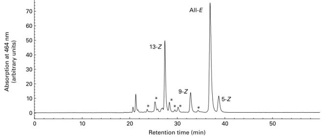
Total lycopene content and the profile of lycopene isomers were determined according to the method described previously
by Schierle et al.(21) for lycopene products (Fig. 1) and in vitro models, and according to the method of Ferruzzi et al.(36) for TRL. The main lycopene isomers identified are 5-Z-, 9-Z-, 13-Z- and all-E-lycopene. Minor compounds (shown with an asterisk in Fig. 1) consisted of other Z-lyco-pene isomers. They were characterised as Z-lycoZ-lyco-pene with liquid chromatography (LC) – MS/MS using an Applied Biosystems APIC 4000 LC – MS/MS (Foster City, CA, USA): isocratic flow 1 ml/min; declustering potential, 130 V; 60 psi (414 kPa) N2; capillary voltage 22 V; vaporiser tempera-ture, 4008C; corona needle 5 mA; the fragmentation conditions used were as described by dos Anjos Ferreira et al.(22). The peak areas of unidentified Z-lycopene isomers were summed and reported as x-Z-lycopene (Table 1).
Expression of the results
Total lycopene is the sum of all lycopene isomers, i.e. all-E-and Z-isomers. Results of in vitro experiments are expressed as mean values and standard deviations. Results of TRL from the human intervention study are expressed as mean values with their standard errors.
Results
A simplified scheme of the absorption process of carotenoids in humans is depicted in Fig. 2. We used several models (in vitro and in vivo) to determine at which stage lycopene can undergo isomerisation: (1) within the lumen of the gastrointestinal tract; (2) incorporation into mixed micelles; (3) within the small intestine using three Caco-2 cell clones and (4) during release from enterocytes in vivo into human blood plasma (chylomicrons or TRL).
Lycopene isomerisation within the gastrointestinal tract Lycopene in either tomato paste or tomato oleoresin consists of 94 % all-E- and 5 % 5-Z-isomers.
Step 1 in Fig. 2. We assessed lycopene isomerisation
within the gastrointestinal tract using various in vitro models. Lycopene did not undergo Z isomerisation in the
0 10 20 30 40 50 0 10 20 30 40 50 60 70 13-Z All-E 5-Z 9-Z * * * * * *
Retention time (min)
Absorption at 464 nm
(arbitrary units)
Fig. 1. Representative HPLC chromatogram of lycopene isomers. * Presence of other Z-lycopene isomers as confirmed by liquid chromatography – MS/MS.
British
Journal
of
presence of gastric juice at pH values between 2 and 4, nor in the presence of duodenal juice at pH 7 and also not when incu-bated sequentially in both conditions (data not shown).
Step 2 in Fig. 2. Incorporation of lycopene into mixed micelles did not induce any degradation of lycopene (data not shown), or any change of lycopene isomer profile (Table 1).
Steps 1 and 2 in Fig. 2. In the gastrointestinal model, TIM-1, mimicking both conditions in the stomach and the small intestine, the lycopene isomer profile of the tomato pro-duct remained unchanged, confirming that lycopene isomerisa-tion did not take place in the gastrointestinal lumen (Table 1). Lycopene isomerisation within the intestine
Step 3 in Fig. 2. The next step in the lycopene absorption process is the transfer from the mixed micelles into human enterocytes. As a model for human enterocytes, differentiated Caco-2 cells (Caco-2, Caco-2 SM and Caco-2 HTB37) were incubated with mixed micelles containing lycopene (2 mM).
The content as well as the isomer profile of lycopene present in mixed micelles were stable for at least 6 h under cell-culture conditions in the absence of cells.
Cells efficiently took up lycopene from mixed micelles in a time-dependent manner leading to a 13 % transfer of lycopene from the mixed micelles into Caco-2 or Caco-2 SM cells at the end of the 6 h incubation, while this transfer was about four times lower (3 %) for the Caco-2 HTB37 clone. The intracellular lycopene isomer profile was similar in the three cell clones and consisted of a significantly higher proportion of Z-isomers than in the starting material: 5-Z (12·1 (SD
1·6) % total lycopene) . 13-Z (5·6 (SD 1·2) % total
lycopene) . 9-Z (2·9 (SD 0·5) % total lycopene) . x-Z (2·7 (SD 4·7) % total lycopene). The lycopene isomer profile in
the cells was similar after 2 and 6 h incubation (data not shown), even though the total lycopene content was lower after 2 h. The absence of Z-lycopene isomers in the incubation medium indicates that lycopene isomerisation took place within the cells.
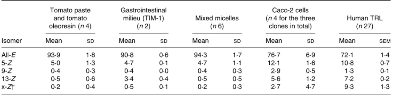
Table 1. Lycopene isomer profiles measured in tomato paste and tomato oleoresin, in gastrointestinal milieu (TIM-1), in mixed micelles, in Caco-2 cells as well as in human TAG-rich lipoproteins (TRL) secreted postprandially*
(Mean values and standard deviations or standard errors) Tomato paste and tomato oleoresin (n 4) Gastrointestinal milieu (TIM-1) (n 2) Mixed micelles (n 6) Caco-2 cells (n 4 for the three
clones in total)
Human TRL (n 27)
Isomer Mean SD Mean SD Mean SD Mean SD Mean SEM
All-E 93·9 1·8 90·8 0·6 94·3 1·7 76·7 6·9 72·1 1·4
5-Z 5·0 1·3 4·7 0·1 4·7 1·1 12·1 1·6 10·8 0·7
9-Z 0·4 0·3 0·4 0·0 0·4 0·3 2·9 0·5 1·3 0·1
13-Z 0·5 0·6 3·4 0·4 0·5 0·5 5·6 1·2 7·2 0·2
x-Z† 0·2 0·4 0·5 0·1 0·2 0·3 2·7 4·7 9·3 1·3
* Results are expressed as percentage of total lycopene. † x-Z ¼ sum of unidentified Z-lycopene isomers.
Chylomicron Lumen Blood Food Stomach 1. Stimulated gastric and duodenal juice Oil droplets Small intestine VLDL LDL HDL Enterocytes Enterocytes Enterocytes 3. Caco-2 cells 4. Post-prandial test Small intestine 2. Incorporation into mixed micelles Mixed micelles
Fig. 2. Simplified scheme of the absorption of lycopene in human subjects. The in vitro and in vivo model systems mimicking different stages of lycopene digestion and absorption that were used during the work are indicated as 1 – 4.
British
Journal
of
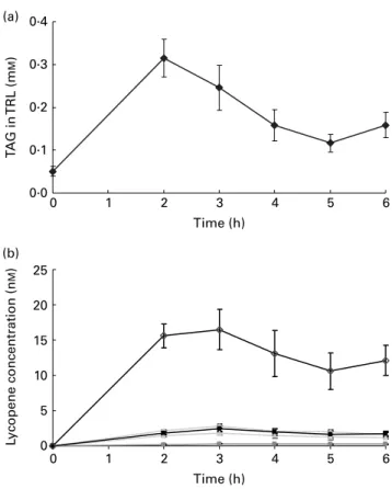
Lycopene isomerisation during absorption in humans Step 1 to step 4 in Fig. 2. We performed a postprandial test in healthy males to allow characterisation of the lycopene appearing in intestinal secreted lipoproteins, i.e. chylomicrons. Subjects consumed a standard meal containing lycopene pre-sent in tomato paste, i.e. providing mainly all-E-lycopene (Table 1). This standard meal contained 40 g groundnut oil to ensure that subjects would secrete chylomicrons into the blood circulation. Consumption of the standard meal led to a marked production of chylomicron particles in TRL as charac-terised by the increase of plasma TAG concentration (Fig. 3(a)). Lycopene was efficiently absorbed and secreted in chylomicrons as shown by the rise of TRL-lycopene concentration (Fig. 3(b)). The profile of lycopene isomers appearing in TRL consisted of 29 % of Z-lycopene isomers with the proportion decreasing in the following order: 5-Z (10·8 (SD 0·7) %); x-Z (9·3 (SD 1·3) %); 13-Z (7·2 (SD 0·2) %); 9-Z (1·3 (SD0·1) %). This profile of Z-lycopene isomers
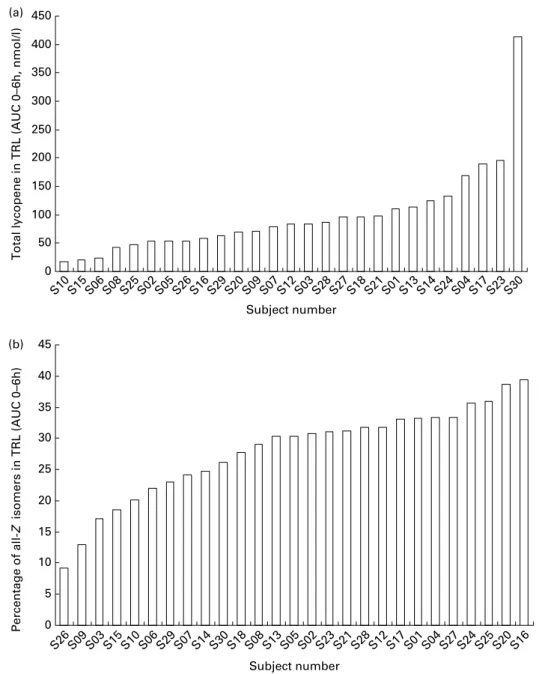
was similar between 2 and 6 h post-absorption. In contrast to the 23-fold inter-individual variation observed for total lycopene bioavailability, the proportion of Z-lycopene isomers in TRL only showed an inter-subject variation of 4 (Fig. 4). This indicates that ‘low’ and ‘high’ lycopene absorbers(19 – 21) exhibit quite similar ability to isomerise lycopene.
Discussion
The absorption of lycopene requires lycopene partitioning into bile salt micelles during small intestine digestion, uptake across the brush-border membrane of enterocytes and incorporation into chylomicron particles for secretion into lymph (Fig. 2).
Lycopene isomerisation did not take place in the gastrointes-tinal lumen either in the stomach, in the duodenum, or during transfer into mixed micelles, as assessed with various in vitro models. In contrast, all-E-lycopene was isomerised into various Z-lycopene isomers when in contact with various clones of Caco-2 cells. Lycopene absorption with a meal led to the secretion of chylomicrons present in TRL containing about 29 % of lycopene as Z-lycopene isomers. The proportion of individual lycopene isomers in Caco-2 cell monolayers, and in human TRL, was quite consistent, and decreased in the following order: 5-Z . 13-Z ¼ x-Z . 9-Z. The presence of 9-Z-, 13-Z- and x-Z-isomers indicates lycopene isomerisation rather than preferential uptake of Z-lycopene, since these lyco-pene isomers were absent in the test lycolyco-pene preparations which contained about 94 % all-E- and about 5 % 5-Z-lycopene. However, this does not exclude a preferential uptake of 5-Z-lycopene by cells, since its proportion was higher in the Caco-2 cell monolayers and TRL than in the lycopene preparation.
Given that isomerisation of all-E-lycopene into Z-isomers is promoted by contact with acids(37), our first goal was to assess whether there was any isomerisation of lycopene in the acidic environment of the stomach and the small intestine. Our find-ing showfind-ing an absence of lycopene isomerisation in the stomach is in agreement with results obtained in vitro(38) and in healthy subjects(26). However, they disagree with in vitro results showing an isomerisation of lycopene that has been reported in vitro in a ‘gastric milieu’ set at pH 1·6(26,39). This discrepancy might be due to the use of a low pH in this in vitro model, which mimics the stomach pH in the fasting stage only. Indeed, the stomach pH, which is about 1·8 in the fasting state, sharply increases to 5·4 – 6·2 after intake of a meal, and then continuously decreases to reach 1·8 – 2·9 after 3 h digestion. The duodenal pH, which is about 5 in the fasting state, increases to pH 6·1 – 6·6 after intake of a meal and remains constant during digestion. In the present results, we did not observe isomerisation of lycopene in the in vitro model mimicking this duodenal con-dition. In the gastrointestinal lumen, the final step before absorption is the transfer of lycopene into mixed micelles. The isomer profile of lycopene in the mixed micelles was similar to the one of the tomato product, which indicates no lycopene isomerisation, but also no preferential transfer of Z-lycopene (in our particular case, 5-Z-lycopene). This is in disagreement with results of Boileau et al.(40) showing a higher incorporation of Z-lycopene into mixed micelles than the all-E counterpart. Using the more sophisticated TIM-1 model, which is an in vitro model that allows the closest simu-lation of in vivo dynamic physiological processes occurring in the lumen of the stomach currently available, we confirm that lycopene does not undergo isomerisation in the gastrointesti-nal tract. These results are in agreement with those recently reported by Blanquet-Diot et al.(41). Although the main para-meters of digestion, such as pH, body temperature, peristaltic mixing and transport, gastric, biliary and pancreatic secretions 0 5 10 15 20 25 L ycopene concentration (n M ) 0·0 0·1 0·2 0·3 0·4 (a) (b) 0 1 2 3 4 5 6 Time (h) 0 1 2 3 4 5 6 Time (h) T A G in TRL (m M )
Fig. 3. TAG (a) and lycopene isomer (b) concentrations in TAG-rich lipopro-teins (TRL)-containing chylomicrons of subjects having consumed a standard meal. Results are expressed in mMfor TAG and in nMfor lycopene. ( – S–),
All-E-lycopene; ( – – ), 5-Z-lycopene; ( – – ), 9-Z-lycopene; ( – – ), 13-Z-lycopene; ( – B –), sum of unidentified Z-lycopene isomers. Values are means (n 27), with standard errors represented by vertical bars.
British
Journal
of
and passive absorption of small molecules and water are reproduced, the actual limitations of the TIM-1 model reside in the absence of a cellular system and a feedback mechanism. To mimic enterocyte transport and metabolism, we used three clones of Caco-2 cells. Cell culture condition did not affect either lycopene content or isomer profile. Lycopene uptake by either Caco-2 or Caco-2 SM was similar, but markedly lower by Caco-2 HTB37 cells. However, lycopene isomerised in all these three cell clones and the profile of lycopene isomers were similar. The results in Caco-2 cells matched in vivo results. Lycopene appearing in chylomicrons (TRL) and in Caco-2 cells consisted of 29 and 24 %, respect-ively, as a mixture of Z-lycopene isomers. This is in agree-ment with results described by Gustin et al.(27) and Unlu et al.(28). In contrast, Tyssandier et al.(26) did not detect
Z-lycopene isomers in chylomicrons isolated from plasma from most individuals. Although a large inter-individual vari-ation of lycopene bioavailability is in agreement with data reported by several authors(42 – 44)and is characteristic of the presence of low and high absorbers in the population, a low inter-individual variation of the proportion of Z- to E-lycopene is noteworthy. These results indicate that whatever the lyco-pene absorption efficiency, lycolyco-pene isomerisation is quite similar and suggest that the factors participating in lycopene absorption are different from those involved in lycopene isomerisation.
In most human tissues, more than 50 % of the lycopene is a mixture of Z-isomers(23,28,44,45). The major Z-isomers in plasma were, in decreasing order of abundance: 5-Z . 13-Z . 9-Z. In humans, the Z-lycopene concentration is high in 0 50 100 150 200 250 300 350 400 450 (a) (b) S10 S15 S06 S08 S25 S02 S05 S26 S16 S29 S20 S09 S07 S12 S03 S28 S27 S18 S21 S01 S13 S14 S24 S04 S17 S23 S30
Total lycopene in TRL (AUC 0–6h, nmol/l)
0 5 10 15 20 25 30 35 40 45 S26 S09 S03 S15 S10 S06 S29 S07 S14 S30 S18 S08 S13 S05 S02 S23 S21 S28 S12 S17 S01 S04 S27 S24 S25 S20 S16 Subject number Subject number Percentage of all-Z isomers in TRL (AUC 0–6h)
Fig. 4. Inter-individual variation: total lycopene concentration (a) and percentage of all-Z-lycopene isomers (b) in TAG-rich lipoproteins (TRL) of each individual subject having consumed a standard meal containing lycopene (n 27). Results are expressed in nMfor total lycopene and as percentage of total lycopene for the all-Z-lycopene isomers. The all-Z-lycopene isomers consist of the sum of 5-Z-, 9-Z-, 13-Z- and unidentified Z-lycopene isomers. AUC, area under the curve.
British
Journal
of
the liver, adrenal glands, testes, skin and prostate(18,46,47). The 29 % Z-lycopene in human chylomicrons present in TRL suggests that, in the human body, additional mechanisms are involved which increase markedly the proportion of Z-lycopene by processes such as isomerisation, preferential uptake or reduced catabolism of the Z-lycopene isomers. The high proportion of Z-lycopene isomers in human tissues could conceivably be a conversion to a more biologically effective form. It is not feasible to compare the efficacy of Z- and E-lycopene in vivo, owing to the metabolic conversion of one form to another. However, Bo¨hm et al.(25) demon-strated that Z-lycopene isomers exhibit higher antioxidant capacity than their all-E counterpart in vitro. Thus it is tempt-ing to speculate that the E- to Z-lycopene conversion in vivo is a metabolic activation of this carotenoid.
Acknowledgements
We thank Dr Karen Cooper for organising the in vitro gastro-intestinal TIM-1 trials at TNO and Dr Francesca Giuffrida for the LC – MS/MS identification of the Z-isomers of lycopene.
The present study was funded in full by the Nestle´ Research Center, Lausanne, Switzerland.
M. R., B. S., P. L., K. B. and G. W. conceived of and designed the present study. B. S., I. T., P. L. and K. B. achieved the design and production of formulations of the lycopene products. M. R. coordinated the trial and supervised the analytic aspects. B. S., I. T., P. L. and K. B. contributed to the development of analytic methods, lycopene analysis and data collection. M. R. wrote the manuscript, and all authors were involved in interpreting the results and in critical revision of the paper.
No author has any advisory board affiliations. There is no conflict of interest.
References
1. Boileau TWM, Boileau AC & Erdmea JW Jr (2002) Bioavail-ability of all-trans and cis-isomers of lycopene. Exp Biol Med 227, 914 – 919.
2. Gann PH, Ma J, Giovanucci E, et al. (1999) Lower prostate cancer risk in men with elevated plasma lycopene levels: results of a prospective analysis. Cancer Res 59, 1225 – 1230. 3. Blaner WS & Olson JA (1994) Retinol and retinoic acid
meta-bolism. In The Retinoids: Biology, Chemistry and Medicine, 2nd ed., pp. 229 – 255 [MB Sporin, AB Roberts and S Goodman, editors]. New York: Raven Press.
4. Chalabi N, Delort L, Le Corre L, et al. (2006) Gene signature of breast cancer cell lines treated with lycopene. Pharmacoge-nomics 7, 663 – 672.
5. Chalabi N, Le Corre L, Maurizis JC, et al. (2004) The effect of lycopene on the proliferation of human breast cells and BRCA1 and BRCA2 gene expression. Eur J Cancer 40, 1769 – 1775. 6. Chalabi N, Satih S, Delort L, et al. (2007) Expression profiling
by whole genome microarray hybridization reveals differential gene expression in breast cancer cell lines after lycopene exposure. Biochim Biophys Acta 1796, 124 – 130.
7. Huang CS, Shih MK, Chuang CH, et al. (2005) Lycopene inhibits cell migration and invasion and upregulates Nm23-H1 in a highly invasive hepatocarcinoma, SH-Hep-1 cells. J Nutr 135, 2119–2123. 8. Nahum A, Zeller L, Danilenko M, et al. (2006) Lycopene inhibition of IGF-induced cancer cell growth depends on the level of cyclin D1. Eur J Nutr 45, 275 – 282.
9. Liu C, Russell RM & Wang XD (2006) Lycopene supplemen-tation prevents smoke induced changes in p53 phosphorylation, cell proliferation, and apoptosis in the gastric mucosa of ferrets. J Nutr 136, 106 – 111.
10. Russell RM (1998) Physiological and clinical significance of carotenoids. Int J Vitam Nutr Res 68, 349 – 353.
11. Rao AV (2002) Lycopene, tomatoes, and the prevention of coronary heart disease. Exp Biol Med (Maywood) 227, 908 – 913. 12. Levy J, Bosin E, Feldman B, et al. (1995) Lycopene is a more potent inhibitor of human cancer cell proliferation than either a-carotene or b-carotene. Nutr Cancer 24, 257 – 266.
13. Giovannucci E, Ascherio A, Rimm EB, et al. (1995) Intake of carotenoids and retinol in relation to risk of prostate cancer. J Natl Cancer Inst 87, 1767 – 1776.
14. Etminan M, Takkouche B & Caaman˜o-Isorna F (2004) The role of tomato products and lycopene in the prevention of prostate cancer: a meta-analysis of observational studies. Cancer Epidemiol Biomark Prevent 13, 340 – 345.
15. Erdman JW Jr, Ford NA & Lindshield BL (2009) Are the health attributes of lycopene related to its antioxidant function? Arch Biochem Biophys 483, 229 – 235.
16. Shi J & Le Maguer M (2000) Lycopene in tomatoes: chemical and physical properties affected by food processing. Crit Rev Food Sci Nutr 40, 1 – 42.
17. Alien CM, Smith AM, Clinton SK, et al. (2002) Tomato consumption increases lycopene isomer concentrations in breast milk and plasma of lactating women. J Am Diet Assoc 102, 1257 – 1262.
18. Clinton SK, Emenhiser C, Schwartz SJ, et al. (1996) Cis-trans lycopene isomers, carotenoids and retinol in the human prostate. Cancer Epidemiol Biomarkers Prev 5, 823 – 833.
19. Hadley CW, Clinton SK & Schwartz SJ (2003) The consump-tion of processed tomato products enhances plasma lycopene concentrations in association with a reduced lipoprotein sensi-tivity to oxidative damage. J Nutr 133, 727 – 732.
20. Holloway DE, Yang M, Paganga G, et al. (2000) Isomerization of dietary lycopene during assimilation and transport in plasma. Free Radic Res 32, 93 – 102.
21. Schierle J, Bretzel W, Bu¨hler I, et al. (1997) Content and isomeric ratio of lycopene in food and human plasma. Food Chem 59, 459 – 465.
22. dos Anjos Ferreira AL, Yeum K-J, Russell RM, et al. (2004) Enzymatic and oxidative metabolites of lycopene. J Nutr Biochem 15, 493 – 502.
23. Stahl W & Sies H (1992) Uptake of lycopene and its geometrical isomers is greater from heat-processed than from unprocessed tomato juice in humans. J Nutr 122, 2161 – 2166. 24. Wu K, Schwartz SJ, Platz EA, et al. (2003) Variations in plasma
lycopene and specific isomers over time in a cohort of U.S. men. J Nutr 133, 1930 – 1936.
25. Bo¨hm V, Puspitasari-Nienaber NL, Ferruzzi MG, et al. (2002) Trolox equivalent antioxidant capacity of different geometrical isomers of a-carotene, b-carotene, lycopene, and zeaxanthin. J Agric Food Chem 50, 221 – 226.
26. Tyssandier V, Reboul E, Dumas JF, et al. (2003) Processing of vegetable-borne carotenoids in the human stomach and duodenum. Am J Physiol Gastrointest Liver Physiol 284, G913 – G923.
27. Gustin DM, Rodvold KA, Sosman JA, et al. (2004) Single-dose pharmacokinetic study of lycopene delivered in a well-defined food-based lycopene delivery system (tomato paste oil mixture) in healthy adult male subjects. Cancer Epidemiol Biomarkers Prev 13, 850 – 860.
28. Unlu NZ, Bohn T, Francis DM, et al. (2007) Lycopene from heat-induced cis-isomer-rich tomato sauce is more bioavailable than from all-trans-rich tomato sauce in human subjects. Br J Nutr 98, 140 – 146.
British
Journal
of
29. Richelle M, Bortlik K, Liardet S, et al. (2002) A food-based formulation provides lycopene with the same bioavailability to humans as that from tomato paste. J Nutr 132, 404 – 408. 30. Burri BJ, Chapman MH, Neidlinger TR, et al. (2009) Tangerine
tomatoes increase total and tetra-cis-lycopene isomer concen-trations more than red tomatoes in healthy adult humans. Int J Food Sci Nutr 60, 1 – 16.
31. Sugawara T, Kushiro M, Zhang H, et al. (2001) Lysophospha-tidylcholine enhances carotenoid uptake from mixed micelles by Caco-2 human intestinal cells. J Nutr 131, 2921 – 2927. 32. Minekus M, Marteau P, Havenaar R, et al. (1995) A
multicom-partmental dynamic computer-controlled model simulating the stomach and small intestine. Altern Lab Anim 23, 197 – 209. 33. Reboul E, Abou L, Mikail C, et al. (2005) Lutein transport by
Caco-2 TC-7 cells occurs partly by a facilicitated process invol-ving the scavenger receptor class B type I (SR-BI). Biochem J 387, 455 – 461.
34. During A, Dawson HD & Harrison EH (2005) Carotenoid trans-port is decreased and expression of the lipid transtrans-porters SR-BI, NPC1L1, and ABCA1 is downregulated in Caco-2 cells treated with ezetimibe. J Nutr 135, 2305 – 2312.
35. Moussa M, Landrier J-F, Reboul E, et al. (2008) Lycopene absorption in human intestinal cells and in mice involves scavenger receptor class B type I but no niemann-pick C1-like 1. J Nutr 138, 1432–1436.
36. Ferruzzi MG, Nguyen ML, Sander LC, et al. (2001) Analysis of lycopene geometrical isomers in biological microsamples by liquid chromatography with coulometric array detection. J Chromatogr B Biomed Sci Appl 760, 289 – 299.
37. Rodriguez-Amaya DB (1999) Changes in carotenoids during processing and storage foods. Arch Latinoam Nutr 49, Suppl. 3, 38S – 47S.
38. Failla ML, Chitchumroonchokchai C & Ishida BK (2008) In vitro micellarization and intestinal cell uptake of cis-isomers of lycopene exceed those of all-trans lycopene. J Nutr 138, 482 – 486. 39. Moraru C & Lee T-C (2005) Kinetic studies of lycopene isomerization in a tributyrin model system at gastric pH. J Agr Food Chem 53, 8997 – 9004.
40. Boileau AC, Merchen NR, Wasson K, et al. (1999) Cis-lyco-pene is more bioavailable than trans-lycoCis-lyco-pene in vitro and in vivo in lymph-cannulated ferrets. J Nutr 129, 1176 – 1181. 41. Blanquet-Diot S, Soufi M, Rambeau M, et al. (2009) Digestive
stability of xanthophylls exceeds that of carotenes as studied in a dynamic in vitro gastrointestinal system. J Nutr 139, 876 – 883. 42. Borel P, Grolier P, Mekki N, et al. (1998) Low and high responders to pharmacological doses of b-carotene: proportion in the population, mechanisms involved and consequences on b-carotene metabolism. J Lipid Res 39, 2250 – 2260.
43. Cardinault N, Tyssandier V, Grolier P, et al. (2003) Comparison of the postprandial chylomicron carotenoid responses in young and older subjects. Eur J Nutr 42, 315 – 323.
44. O’Neill ME & Thurnham DI (1998) Intestinal absorption of b-carotene, lycopene and lutein in men and women following a standard meal: response curves in the triacylglycerol-rich lipoprotein fraction. Br J Nutr 79, 149 – 159.
45. Stahl W, Schwarz W, Sundquist AR, et al. (1992) Cis-trans isomers of lycopene and b carotene in human serum and tissues. Arch Biochem Biophys 294, 173 – 177.
46. Clinton SK (1998) Lycopene: chemistry, biology, and implications for human health and disease. Nutr Rev 56, 35 – 51. 47. Khachik F, Carvalho L, Bernstein PS, et al. (2002) Chemistry, distribution, and metabolism of tomato carotenoids and their impact on human health. Exp Biol Med (Maywood) 227, 845 – 851.