Sl52
Nitric Oxide and Infection: Another View
G. Schoedon, M. Schneemann, R. Walter, N. Blan,S. Hofer, and A. Schaffner
From the Division of Infectious Diseases, Clinical Mycology Laboratory. Department of Medicine. University of Zurich Medical School. Zurich. Switzerland
Nitric oxide (NO) has been nicknamed "murderer" and "mediator" because it has toxic and signaling properties. We review these two aspects of NO synthesis from the perspective of the clinicalinfectious disease specialist by considering the potential of NO as an endothelium-derived relaxing factor (EDRF) in inflammation and sepsis and its potential as an antimicrobial system. We deviate from observations in recent authoritative reviews and point to important speciesdiffer-ences that make it unlikely that NO serves as anEDRFmediating inflammatory vasodilatation in humans or that NO synthesized by human phagocytes has an antimicrobial function. We propose that in humans, NO synthesis is more confined and compartmentalized than in certain other animal species, and therefore, unwelcome toxicity, vasodilatation, or disturbance of paracrine signaling mechanisms(i.e.,modulation of phagocytic cell functions) are avoided during inflammation.
Nitric oxide (NO) has received much attention over the last few years and was named molecule of the year in 1992[1]. The recent scientific literature is replete with articles on the many aspects of biological and pathological processes involv-ing NO (reviewed in [2-4]), suggestinvolv-ing at times that NO might be a jack-of-all-trades. Two aspects of the generation of NO are of particular interest to infectious disease specialists:(I) the potential role of NO as a cytotoxic molecule contributing to the antimicrobial armature of phagocytes and (2) the role of NO as a vasodilator of inflammation and mediator of septic shock. We outline our view on the role of NO in infectious processes in the human system. Our view deviates in some aspects from recent authoritative analyses [2-4] and takes into account evolutionary adaptations favorable to a utilization of this highly reactive and toxic molecule, nicknamed "mur-derer" [3],as a mediator.
NO is produced in human and laboratory animals by NO synthases (NOS). NOS isoenzymes [5]have been categorized into inducible and constitutive isoforms (table I). However, it is apparent that activity of inducible NOS (iNOS) is also constitutively present in many tissues, and that a constitutively present activity of iNOS is further upregulated by enzyme-inducing signals in these cell types [6-8].Ithas also become clear that the expression and activity of the constitutive NOS (eNOS) are modulated as well [9].
NO as Antimicrobial Product of Mononuclear Phagocytes NO derived in mononuclear phagocytes by a high-output iNOS from L-arginine has been proposed to have antibacterial,
Reprints or correspondence: Dr. A. Schaffner, Clinical Mycology Labora-tory, Division of Infectious Diseases, Department of Medicine, University Hospital, CH-8091 Zilrich, Switzerland.
CUnleaJ Infeetlous Diseases 1995;21(Suppl 2):8152-7
@ 1995byThe University of Chicago. All rights reserved.
1058-4838/95/2104-0003$02.00
antifungal, and antiprotozoal activity [2-4]. In animal macro-phages, notably murine macromacro-phages, the pathway of NO syn-thesis and its regulation have been well characterized (reviewed in[10]).This includes consumption of the substrates t-arginine and O2 ,production of the co-product L-citrulline, and the need
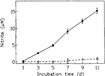
of cofactors reduced nicotinamide adenine dinucleotide phos-phate (NADPH), flavin adenine dinucleotide (FAD), and tetra-hydrobiopterin (B14). Most studies regarding the role of NO formation, including its role as an antimicrobial product, de-pend on quantitation of nitrite, an oxidation product formed spontaneously from NO in biological systems. The main reason for this is that nitrite can be measured by a convenient and technically simple test that can be performed in any laboratory. However, there are many potential pitfalls in correlating nitrite concentrations to NO production (figureI,table 2). Artificial accumulation of nitriteby an unknown mechanism occurs in culture media incubated for7or more days without ever chang-ing the medium. Nitrite formation is independent of the type of medium, the presence of living cells, or the presence of chemicals like sodium azide, and is significantly higher in the presence ofIL-4. In media incubated at room temperature with-out CO2 , nitrite forms even more readily (our unpublished
observation).
To demonstrate convincingly the production of NO, it is necessary either to use cumbersome assays measuring NO by chemoluminescence or to show that L-arginine is converted into L-citrulline along with nitrite production [II]. To date there is no convincing evidence that human phagocytes produce NO in amounts that could account for antimicrobial activity. Most, if not all, of the discrepancies in the scientific literature in regard to NO production by human phagocytes ensue from the confinement of these studies to the measuring of nitrite in biological systems. In addition, as of this writing no single laboratory has reported (based on stringent biochemical meth-ods) that human phagocytes dispose of high-output NOS activ-ity. Studies depending on L-arginine analogues used as "inhibi-tors" have also not been helpful in establishing a role for NOS
em1995;21 (SuppI2) Nitric Oxide and Infection S153
Table 1. Tissue distribution of nitric oxide synthases in animal models.
NOTE. eNOS = constitutive nitric oxide synthase; iNOS = inducible nitric oxide synthase; LPS= lipopolysaccharide.
* In animal and human cells.
tIn animal but not in human cells.
INot defined in human cells.
as a killing system of human phagocytes. In such experiments, complex biological systems such as macrophages and target cells were exposed for prolonged times to these amino acid analogues; no appropriate control experiments were performed, In particular, the potential effects of these antimetabolites on protein synthesis of phagocytes or target cells and, above all, an effect on the growth phase of microorganisms that critically affects susceptibility to killing [12] were not addressed in such studies.
On the basis of these reflections, it cannot be overemphasized that, in contrast with animal studies [10] or studies on human hepatocytes [13], scientists have failed to demonstrate, by bio-chemical methods other than mere nitrite measurement, high-output iNOS activity in human mononuclear phagocytes, in-cluding tissue macrophages and microglia [11, 14, 15], as well as in neutrophils [16]. Another basic issue related to this is the fact that there are fundamental biochemical differences be-tween human mononuclear phagocytes and phagocytes from certain animal species. Human mononuclear phagocytes lack relevant arginase or urea cycle activity, and therefore, they do not metabolize L-arginine by this pathway [8, 11].Ithas also been shown that human mononuclear phagocytes lack the en-zyme systems required for the synthesis of the BH4 ,an essential
cofactor required for NO synthesis (table 3).Itshould be inci-dentally noted that exogenous BH4can not support NO
synthe-sis in human macrophages [11].
iNOS has been cloned, and the regulation of its expression has been studied in human, murine, and rat mononuclear phago-cytes by northern analysis, which permitted quantitative
assess-Figure 1. Spontaneous formation of nitrite in cell culture medium (RPMI 1640) supplemented with 5% fetal bovine serum, human re-combinant IL-4 (200V/mL),and humanrecombinantIFN-y(500VI mL). Cell-free medium was incubated (without changing) at 3TC, 5% CO2 for the times indicated(e). In the absence of lL-4/1FN-y,
nitriteaccumulation wasminimal(0).Thisreproducible phenomenon wasalso seenwithothercell culture mediasupplemented accordingly. None of the supplements contained sodium azide. Nitrite concentra-tion= mean± SO from triplicate wells. Medium with and without supplements,P< .01 for all values 2:3 days of incubation (Student's z-test).
/!
/ !
./
. /
'~9-
- - - 9- - - -2- - - -
0 - - - - 01
3
5
7
9
11
Incubation time (d)
5
15
10
QJ +J L +JZ
ment of specific cellular mRNA levels. While lipopolysaccha-ride (LPS) and IFN-y were able to induce within hours long-lasting synthesis of iNOS mRNA in phagocytes from rats and mice, no iNOS mRNA was detectable by this method in stimu-lated and unstimustimu-lated human phagocytes [6]. When a much more sensitive reverse-transcriptase PCR (RT-PCR) method was used, minute amounts of iNOS mRNA were transiently detected upon stimulation withLPS/IFN-yin human peripheral blood mononuclear cells that had been purified of monocytes (but nevertheless contained 1%- 2% contaminating cells of other types).Itis noteworthy that no NOS activity was detect-able in these experiments. The minute amounts of iNOS mRNA detected cannot be weighed against amounts found in mononu-clear phagocytes from mice or rats [6]. In-depth studies of this phenomenon indicate that minute amounts of iNOS message can be detected by RT-PCR in apoptotic human mononuclear phagocytes, but that this message is not translated into a func-tional NOS enzyme [17, 18]. These observations confirm by molecular methods the results of antecedent biochemical stud-ies: human mononuclear phagocytes do not transcribe or trans-late iNOS message at a level that could be compared with that in phagocytes from mice or rats. Genomic analyses that so far show similarities in the promoter regions of the rodent and human iNOS gene, particularly in respect to IFN-y and LPS-dependent regions, cannot explain these important species dif-ferences[19,20].The similarities in LPS and IFN-y-dependent promoter regions of the NOS gene in humans and mice also speak somewhat against the requirement for occult cytokine IFN-'}', LPS, in combinations:
TNF-a:, IL-l, IFN-a: Endotheliumt
Hepatocytes* Macrophagest
Smooth muscle cells* Carcinoma cell lines" Neutrophils" Islet cells! Mesangium cells* Cardiomyocytes! Fibroblastst Bronchial epithelium" iNOS
Activators Calcium ionophore Excitatory amino acids Acetylcholine, bradykinin Cellular source Endothelium"
Neurons* Epithelial cells* Astrocytes* Neutrophils' Thrombocytes' Cellular source and activator(s) eNOS
SI54 Schoedon et at.
em
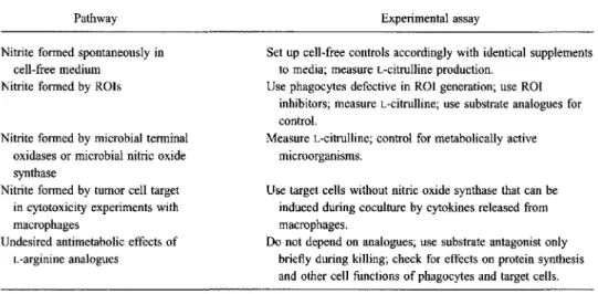
1995;21 (SuppI2)Table 2. Postulated pathways for nitric oxide production and appropriate experimental assessment. Pathway
Nitrite formed spontaneously in cell-free medium
Nitrite formed by ROIs
Nitrite formed by microbial terminal oxidases or microbial nitric oxide synthase
Nitrite formed by tumor cell target in cytotoxicity experiments with macrophages
Undesired antimetabolic effects of L-arginine analogues
NOTE. ROI= reactive oxygen intermediate.
Experimental assay
Set up cell-free controls accordingly with identical supplements to media; measure L-citrulline production.
Use phagocytes defective in ROI generation; use ROI inhibitors; measure i-citrulline; use substrate analogues for control.
Measure L-citrulline; control for metabolically active microorganisms.
Use target cells without nitric oxide synthase that can be induced during coculture by cytokines released from macrophages.
Do not depend on analogues; use substrate antagonist only briefly during killing; check for effects on protein synthesis and other cell functions of phagocytes and target cells.
Table 3. Metabolic differences between murine and human macrophages concerning the L-arginine/ nitric oxide pathway.
Parameter
L-arginine consumption Arginase activity Ornithine production Urea production
Nitric oxide synthase activity Citrulline production Cytosolic BH, Murine macrophages 600-800 J.tmo1/106cells Yes 500-700 J.tmo1/106 cells 800-1,100 J.tmo1/106 cells 1.4-2.2 J.tmol/(h·106 ) cells 60 J.tmo1/106cells 10-90 pmol/mg protein Human macrophages 10-20 J.tmo11l06cells No <0.1 J.tffio1/106cells <0.1 J.tmo1l106 cells <0.3 J.tmoll(h·106 ) cells <0.1 J.tffio1/106cells <0.03 pmol/mg protein NOTE. Table was adapted from references [8, II]. BH, = tetrahydrobiopterin.
brews that would instruct viable human macrophages to express iNOS, a concept recommended by those unwilling to accept the undeniable species differences.
Taken together, these findings show that observations made on the role of iNOS in the antimicrobial or antitumoral function of phagocytes from mice, rats, or other species can not be simply transferred to human biology. To date, there is no indi-cation that an inducible high-output NOS is antimicrobially operative in human phagocytes (or, incidentally, in rabbit mac-rophages) [11].
We have previously proposed that one reason for these spe-cies differences might be redundancy of phagocytic killing systems that would allow for species variability [II]. Because species differences regarding L-arginine metabolism of phago-cytes are not restricted to iNOS but include arginase and other enzymes of the urea cycle (table 3), these disparities must perhaps be seen in a broader context that includes other meta-bolic pathways. Finally, the question occurs as to whether it was advantageous, in an evolutionary sense, to limit the synthe-sis of NO, which might not only have antimicrobial functions but could also become injurious to the host, mediating disas-trous host responses such as vasodilatation during septic shock.
The Role of NO in Inflammatory Vasodilatation
In view of the major species differences in the regulation and expression of iNOS in phagocytes, the question arises as to whether comparable differences exist in NOSs of blood vessels. The role of NO in the control of the vascular tone has recently been extensively reviewed [2-4]. There is solid evidence that NO is an endothelium-derived relaxing factor (EDRF) mediating physiological vasodilatation in animals and humans. In addition, vasodilatation during pathological states such as sepsis and inflammation appears to be mediated by a nitrovasodilator system. In several animal species, iNOS activ-ity is induced by inflammatory mediators in endothelial cells, an activity that is comparable to that of the high-output iNOS in murine macrophages [2].Itis therefore conceivable that NO also plays an important role as an inflammatory EDRF in these species [2-4]. Comparable to observations made in macro-phages, however, no high-output iNOS activity can be induced in human endothelial cells by LPS, IFN-y, TNF-a, or IL-I [21, 22], mediators that induce high-output NOS activity in macrophages and endothelial cells from rats, mice, and cattle or in human hepatocytes [13]. Furthermore, eNOS activity
de-em1995;21(SuppI2)
Mouse
,
Rat
Constitutive
& inducible NOS
Smooth InIScle
Nitric Oxideand Infection
Human
Constitu
tive
N
OS
Smooth muscleInducible
NOS
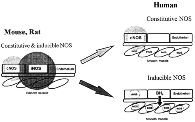
Smoothmuscle S155Figure 2. Evolutionary compartmentalizationof vascular nitric oxide (NO)synthesis. In mice and rats,endothelial inducible nitric oxide
synthase(iNOS) present along with the constitutiveenzyme(eNOS) hasthe potential to secrete NO in amounts comparableto toxic amounts
produced by rodent macrophages. In contrast, by compartmentalization of NOsynthesis in the humanvascular system into constitutive endothelialcell (physiological,light grayarrow)and inducible smooth musclecell (inflammatory,dark grayarrow)isoenzymes,the secretion
of large amounts of potentially toxic and randomly diffusing NOsynthesized during inflammatory response can be avoided.We propose that endothelial cells regulate NO synthesisin smooth muscle cells through tetrahydrobiopterin (BIL) to conserve inflammatory endothelium
-derived relaxing factor function through the nitrovasodilator system,Shadedareas:correspond to random NOdiffusion.
creases in endothelial cells under the influence of inflammatory stimuli, and mRNA for cNOS accordingly decreases after expo-sure toIFN-yand/or LPS without de novo expression of iNOS mRNA in human endothelial cells [21). Therefore, NO, in contrast to its role in physiology, appears not to be an endothe-lial mediator (EDRF) of inflammatory vasodilatation in the human system.The question then arises as to where the produc-tion of NO occurs that is responsible for the inflammatory vasodilatation attributed to the nitrovasodilator system in hu-mans. Because inflammatory mediators induce iNOS mRNA synthesis in human vascular smooth muscle cells (SMCs), it appears plausible that inflammatory vasodilatationis prompted in humans by NO synthesized within the SMCs themselves. NO activates soluble cytosolic guanylate cyclase in SMCs,
resulting in an increase of the intracellular cyclic guanosine monophosphate (cGMP) level,which inturn mediates muscle relaxation [23, 24). Synthesis of NO at the very site of its action within SMCs appears advantageous over a more distant synthesis in endothelial cells. This advantage might be particu-larly important in relation to inflammatory vasodilatation caused by high-output iNOS, which is apt to produce large amounts of NO. Randomly diffusing NO produced by an
endo-thelial iNOS could lead to considerable tissue damage ( fig-ure 2).
All observations made in human cell systems point out that NO is not the inflammatory EDRF.How then is vasodilatation controlled under inflammatory conditions? As mentioned ear-lier, inflammatory stimuli such asLPS and IFN-y induce syn
-thesisof iNOS in vascular SMCs,but endothelial factors must
alsobeconsidered for the regulation ofinflammatoryv asodila-tation.Wehave recently shown that BH4,an obligatory
cofac-tor ofNOS, is a highlyregulated secretory product of human endothelial cells [25). In cellculture experiments, exogenous
BH4has been foundto haveregulatory effects on NO synthesis and to augmentiNOS activityasa limitingfactor [8,23). Upon inflammatory stimuli, BH4 synthesis precedes an increase in
iNOS activitybyseveral hours.In addition , simultaneous inhi-bitionof BH4synthesis prevents increasediNOS activity [26].
In isolated rat aorta it has been shown that BH4 induces a
marked and long-lasting vasodilatation [27] . These findings made us consider the possibilitythat BH4is an EDRF mediating
inflammatory vasodilatationin the human vascular system. BH4
secretion by human endothelialcellsis markedly regulated by a factor of 70 by activating and deactivating cytokines [25].
SI56 Schoedon et a!. em 1995;21 (Suppl 2)
Inflammatory Stimuli:
LPS, IFN-y, IL-I, TNF-a
Endothelium
eNOS
GTP Relaxation+
~ CG~'--oOIIIIIIIltf;t.---
.,.,NO~~
Smooth muscleFigure 3. Proposal for regulation of nitric oxide (NO) synthesis in smooth muscle cells (SMCs) by endothelial tetrahydrobiopterin (BH4)
functioning as an endothelium-derived relaxing factor. While in-flammatory stimuli downregulate the expression of constitutive NO synthase (eNOS) in human vascular endothelial cells, they concomi-tantly induce in these cells the synthesis and vectorial secretion ofBH4
in the direction of SMCs. In parallel, inflammatory stimuli upregulate expression of inducible NO synthase (iNOS) in human vascular SMCs.B~is a limiting factor for NO synthesis by iNOS in inflam-matory stimulated SMCs. Endothelium-derived BH4acts as first
mes-senger modulating NO production in the SMCs, which as second messenger activates soluble guanylate cyclase (sOC) to synthesize cyclic guanosine monophosphate (cOMP), which mediates relaxation as third messenger.
In addition, endothelial cells secrete BH4vectorially into the
direction of vascular SMCs [23]. Furthermore, we found that intraarterial infusion of B~ results in a local vasodilatation that is accompanied by local consumption of L-arginine and production of cGMP [23], strong indications that BH4can act
as a mediator of vasodilatation through the nitrovasodilator pathway. In this way, BH4 acts as first messenger controlling
NO production in SMCs, which as second messenger controls the synthesis of cGMP, which regulates vasodilatation as third messenger. Based on these observations we propose that not NO but BH4is an EDRF in the human vascular system inducing
inflammatory vasodilatation (figure 3).
These details are of interest because BH4synthesis is
amena-ble to pharmacological interventions that are aimed at reducing inflammatory vasodilatation. Accordingly, inhibition of BH4
synthesis by coumarins in murine endothelial cells results in a reduction of NO synthesis [26]. Furthermore, there are indica-tions that coumarins can protect animals from experimental septic shock by decreasing NO synthesis [28]. This approach does not necessarily affect the regulation of the vascular tone by NO synthesized by endothelial cNOS under physiological
conditions. In septic shock, however, the selective inhibition ofB~synthesis might be advantageous over a global inhibi-tion of NO synthesis by L-arginine analogues affecting both iNOS and cNOS, which has a detrimental outcome in a canine model of septic shock [29].
Conclusions and Possible Significance of Evolutionary Variations in the Tissue Localization of iNOS
In conclusion, we propose that NO synthesis in humans is more restricted than in other mammalian species such as the rat and mouse and that not all human tissues dispose of iNOS activity. In humans, prevention of an unhindered redundant expression of iNOS in phagocytes circumvents undesired va-sodilatatory signaling and toxic effects. Accordingly, compart-mentalization of iNOS activity in human vessel walls with restriction of NO synthesis to SMCs during inflammation could be advantageous because it would avoid autotoxicity of NO. Furthermore, restricted compartmentalized NO synthesis may permit, even during inflammation, paracrine signaling that modulates phagocyte functions [30]. Taken together, the two observations on iNOS in human phagocytes and the vascular system could be viewed as an evolutionary refinement in the use of this intriguing molecule, which can be both a murderer and an essential mediator of biological responses.
References
I. Koshland DE Jr. The molecule of the year [editorial]. Science 1992;258:1861.
2. Moncada S, Higgs A. The L-arginine-nitric oxide pathway. N Engl J Med 1993;329:2002-12.
3. Anggard E. Nitric oxide: mediator, murderer, and medicine. Lancet 1994;343:1199-1206.
4. Vallance P, Collier J. Biology and clinical relevance of nitric oxide. BMJ 1994;309:453-7.
5. Nathan C. Nitric oxide as a secretory product of mammalian cells. FASEB J1992; 6:3051-64.
6. Chesrown SE, Monnier J, Visner G, Nick HS. Regulation of inducible nitric oxide synthase mRNA levels by LPS, lNF-y,TGF-{3, and IL-I 0 in murine macrophage cell lines and rat peritoneal macrophages. Biochem Biophys Res Commun 1994;200:126-34.
7. Bernhardt J, Tschudi MR, Dohi Y, Gut I, Urwyler B, Buhler FR, Luscher TF. Release of nitric oxide from human vascular smooth muscle cells. Biochem Biophys Res Commun 1991; 180:907-12.
8. Schoedon G, Schneemann M, Hofer S, Guerrero L, Blau N, SchaffnerA.
Regulation of the L-arginine-dependent and tetrahydrobiopterin-depen-dent biosynthesis of nitric oxide in murine macrophages. Eur J Biochem 1993;213:833-9.
9. Rosenkranz-Weiss P, Sessa WC, Milstien S, Kaufman S, Watson CA, Pober JS. Regulation of nitric oxide synthesis by proinflammatory cyto-kines in human umbilical vein endothelial cells. Elevations in tetrahy-drobiopterin levels enhance endothelial nitric oxide synthase specific activity. J Clin Invest 1994;93:2236-43.
10. Nathan CF, Hibbs 18 Jr. Role of nitric oxide synthesis in macrophage antimicrobial activity. Curr Opin Immunol 1991;3:65-70.
11. Schneemann M, Schoedon G, Hofer S, Blau N, Guerrero L, Schaffner A. Nitric oxide synthase is not a constituent of the antimicrobial armature of human mononuclear phagocytes. J Infect Dis 1993; 167:1358-63.
eID1995;21 (Suppl2) Nitric Oxide and Infection SI57
12. Lehrer RI, Ganz T, Szklarek D, Selsted ME. Modulation of the in vitro candidacidal activity of human neutrophil defensins by target cell metab-olism and divalent cations. J Clin Invest 1988; 81: 1829-35. 13. Geller DA, Nussler AK, Di Silvio M, et al. Cytokines, endotoxin, and
glucocorticoids regulate the expression of inducible nitric oxide syn-thase in hepatocytes. Proc Natl Acad Sci USA 1993;90:522-6. 14. Cameron ML, Granger DL, Weinberg JB, Kozumbo WJ, Koren HS.
Human alveolar and peritoneal macrophages mediate fungistasis inde-pendently of L-arginine oxidation to nitrite or nitrate. Am Rev Respir Dis 1990;142:1313-9.
15. Peterson PK, Hu S, Anderson WR, Chaocc.Nitric oxide production and neurotoxicity mediated by activated microglia from human versus mouse brain. J Infect Dis 1994; 170:457-60.
16. Yan L, Vandivier RW, Suffredini AF, Danner RL. Human polymorpho-nuclear leukocytes lack detectable nitric oxide synthase activity. J Im-munol 1994; 153:1825-34.
17. Kolb-Bachofen V, Alsdorf K, Fehsel K. Expression of inducible NO synthase mRNA in human monocytes [abstract no. OVIII]. In: The Macrophage, Proceedings of the Eighth Annual Conference of the Euro-pean Macrophage Study Group. Lausanne, Switzerland; 1994:57. 18. Reiling N, Ulmer AJ, Duchrow M, Ernst M, Flad HD, Hauschildt S.
Nitric oxide synthase: mRNA expression of different isoforms in human monocytes/rnacrophages. Eur J Immunoll994;24:1941 -4.
19. Nunokawa Y, Ishida N, Tanaka S. Promoter analysis of human inducible nitric oxide synthase gene associated with cardiovascular homeostasis. Biochem Biophys Res Commun 1994;200:802-7.
20. Chartrain NA, Geller DA, Koty PP, et al. Molecular cloning, structure, and chromosomal localization of the human inducible nitric oxide syn-thase gene.J BioI Chern 1994;269:6765-72.
21. MacNaul KL, Hutchinson NI. Differential expression of iNOS and eNOS mRNA in human vascular smooth muscle cells and endothelial cells under normal and inflammatory conditions. Biochem Biophys Res Com-mun 1993; 196:1330-4.
22. Werner-Felmayer G, Werner ER, Fuchs D, et al. Pteridine biosynthesis in human endothelial cells. Impact on nitric oxide-mediated formation of cyclic GMP. J Bioi Chern 1993;268:1842-6.
23. Schaffner A, Blau N, Schneemann M, Steurer J, Edgell C-JS, Schoedon G. Tetrahydrobiopterin as another EDRF in man. Biochem Biophys Res Commun 1994;205:516-23.
24. Ignarro LJ, Byrns RE, Buga GM, Wood KS. Endothelium-derived re-laxing factor from pulmonary artery and vein possesses pharmacologic and chemical properties identical to those of nitric oxide radical. Circ Res 1987;61:866-79.
25. Schoedon G, Schneemann M, Blau N, Edgell C-JS, Schaffner A. Modula-tion of human endothelial cell tetrahydrobiopterin synthesis by activat-ing and deactivatactivat-ing cytokines: new perspectives on endothelium-derived relaxing factor. Biochem Biophys Res Commun 1993; 196: 1343-8.
26. Schoedon G, Siau N, Schneemann M, Flury G, Schaffner A. Nitric oxide production depends on preceding tetrahydrobiopterin synthesis by endothelial cells: selective suppression of induced nitric oxide pro-duction by sepiapterin reductase inhibitors. Biochem Biophys Res Com-rnun 1994; 199:504 10.
27. Van Amsterdam JOC, Werner J. Tetrahydrobiopterin induces vasodilata-tion via enhancement of cGMP level. Eur J Pharmacol 1992;215: 34950.
28. Zingarelli B, Camuccio R, Di Rosa M. Cloricromene inhibits the induc-tion of nitric oxide synthase. Eur J Pharmacol 1993;243: 107-11. 29. Cobb JP, Natanson C, Hoffman WD, et al. N'<amino-t-arginine, an
inhibitor of nitric oxide synthase, raises vascular resistance but increases mortality rates in awake canines challenged with endotoxin. J Exp Med 1992; 176:1175--82.
30. IgnarroLJ. Haem-dependent activation of guanylate cyclase and cyclic OMP formation by endogenous nitric oxide: a unique transduction mechanism for transcellular signaling. Pharmacol Toxicol 1990; 67:1-7.