doi: 10.1093/intimm/dxh015
Chicken BAFFÐa highly conserved cytokine
that mediates B cell survival
Kirsten Schneider
1, Sonja Kothlow
2, Pascal Schneider
3, Aubry Tardivel
3,
Thomas GoÈbel
2, Bernd Kaspers
2and Peter Staeheli
11Abteilung Virologie, Institut fuÈr Medizinische Mikrobiologie und Hygiene, University of Freiburg, 79104
Freiburg, Germany
2Institut fuÈr Tierphysiologie, University of Munich, 80539 Munich, Germany 3Institute of Biochemistry, University of Lausanne, 1066 Epalinges, Switzerland
Keywords: B lymphocyte, chicken, cytokine, hematopoiesis Abstract
Members of the tumor necrosis factor (TNF) family play key roles in the regulation of in¯ammation, immune responses and tissue homeostasis. Here we describe the identi®cation of the chicken homologue of mammalian B cell activating factor of the TNF family (BAFF/BLyS). By searching a chicken EST database we identi®ed two overlapping cDNA clones that code for the entire open reading frame of chicken BAFF (chBAFF), which contains a predicted transmembrane domain and a putative furin protease cleavage site like its mammalian counterparts. The amino acid identity between soluble chicken and human BAFF is 76%, considerably higher than for most other known cytokines. The chBAFF gene is most strongly expressed in the bursa of Fabricius. Soluble
recombinant chBAFF produced by human 293T cells interacted with the mammalian cell-surface receptors TACI, BCMA and BAFF-R. It bound to chicken B cells, but not to other lymphocytes, and it promoted the survival of splenic chicken B cells in culture. Furthermore, bacterially expressed chBAFF induced the selective expansion of B cells in the spleen and cecal tonsils when
administered to young chicks. Our results suggest that like its mammalian counterpart, chBAFF plays an important role in survival and/or proliferation of chicken B cells.
Introduction
Members of the tumor necrosis factor (TNF) family and their receptors are important regulators of the immune system [reviewed in (1)]. B cell activating factor of the TNF family (BAFF, also known as BLyS, TALL-1, THANK, zTNF4 or TNFSF13b) (2±5) is a new member of the TNF family of cytokines that plays a major role in B cell survival, proliferation and differentiation (6). Ligands of the TNF superfamily are type II single transmembrane proteins that form biologically active trimers (7). The TNF family de®ning amino acids are located in the C-terminal extracellular receptor-binding domain of the uncleaved precursor. BAFF has 33% amino acid identity to APRIL (a proliferating inducing ligand) and ~20% to TNF-a (2). The C-terminal extracellular domain can be cleaved by a furin-like protease resulting in release of the biologically active soluble cytokine (2). BAFF is produced by macrophages, dendritic cells and some T cells (2,3,8).
BAFF can bind three receptors of the TNF receptor family, TACI (transmembrane activator, calcium modulator and
cyclophilin ligand interactor), BCMA (B cell maturation anti-gen) and BAFF-R (BAFF receptor). Interestingly, BCMA and TACI also serve as receptors for APRIL (9±12). TACI is expressed on B cells and activated T cells, whereas BAFF-R and BCMA are expressed mainly or exclusively on B cells (13± 16). Recent data suggest that BAFF-R may be the primary receptor responsible for BAFF-mediated function (13). BAFF promotes B cell survival (17) and, together with signals from the BCR, BAFF also leads to proliferation of B lymphocytes (2±5). Treatment of mice with recombinant BAFF increased the B cell frequency in the spleen and elevated the serum Ig levels (3,18). The same effects were seen in BAFF transgenic mice (12,19,20). In addition these mice showed signs of auto-immune diseases such as systemic lupus erythematosus (SLE) and, at a later stage, SjoÈgren's syndrome (SS) (12,19± 21), suggesting that BAFF is a key player in human autoimmune disease. Indeed, some SLE and SS patients have increased levels of BAFF in the serum (21±23), and BAFF
The ®rst two authors contributed equally to this work
Correspondence to: P. Staeheli; E-mail: [email protected]
null mice display dramatically reduced peripheral B cell numbers and Ig levels (24,25). Treatment of animals with a decoy receptor for BAFF reduced the mature B cell pool and prevented disease in mouse models of SLE and collagen-induced arthritis (12,26±28).
Little information is available on TNF family members in the chicken. The coding sequences of the chicken homologues of CD40 ligand (GenBank accession no. AJ243435), TRAIL (TNF-related apoptosis inducing ligand; GenBank accession no. AY057941) and Fas (GenBank accession no. AF296874) have been reported. Fragments of other putative family members can be found in EST databases, but their identity remains questionable. Here we report the cDNA cloning and biological characterization of the chicken homologue of mammalian BAFF, designated chBAFF.
Methods Animals
H.B19 (B19/B19haplotype) chickens were raised at the Institute
of Animal Physiology, University of Munich. Fertilized eggs of M11 (B2/B2 haplotype) chickens were obtained from the
Institute for Animal Science, Mariensee (29) and hatched at the Institute of Animal Physiology, Munich. M11 birds were used for the experiments described in Fig. 6(B and C). Cells
Human embryonic kidney 293T cells were maintained in DMEM supplemented with 10% FBS. Chicken splenocytes, cells of the bursa of Fabricius, the cecal tonsils and the thymus were separated by density centrifugation on Ficoll-Paque (Amersham Pharmacia Biotech, Freiburg, Germany). Peri-pheral blood lymphocytes were prepared by slow speed centrifugation as previously described (30). Cells were main-tained in RPMI 1640 medium with Glutamax (Gibco, Paisley, UK) supplemented with 10% FCS at 40°C in a CO2incubator. Cloning of chBAFF cDNA
The overlapping bursal EST database clones dkfz426_2f7r1 and riken1_33a18r1 (sequence data available at http:// swallow.gsf.de/dt40Est.html) contained the sequence infor-mation of the 3¢ and 5¢ portion of the chBAFF cDNA respectively. PCR with the oligonucleotides 5¢-AGCAACGA-GTAACATGGATTGCAC-3¢ and 5¢-GCTTCAGAGGAGTCTAA-CTGCAC-3¢ using a 855-23 T cell cDNA library as template (31) resulted in the ampli®cation of the complete open reading frame of chBAFF. The PCR product was cloned into pcDNA3.1 (Invitrogen, Karlsruhe, Germany). The sequence has been assigned the GenBank accession no. AF506010.
RNA isolation and northern blot analysis
RNA was derived from various organs of 3-week-old chickens using TRIzol reagent (Gibco, Karlsruhe, Germany) according to the manufacturer's protocol. RNA samples (15 mg per lane) were size-fractionated by electrophoresis through agarose gels containing 4% formaldehyde using standard procedures before blotting onto nylon membranes and hybridization with the indicated cDNA probes that were radiolabeled with32P.
Expression of recombinant chBAFF
For expression of chBAFF in eukaryotic cells, PCR was performed on cDNA of the chicken T cell line 855-23 (31) with primers 5¢-CAAATGCATTCTATTGTCAACGCAGAAGAA-AC-3¢ and 5¢-CCGGAATTCTCAGAGGAGTCTAACTGC5¢-CAAATGCATTCTATTGTCAACGCAGAAGAA-AC-3¢ that introduced unique NsiI and EcoRI restriction sites near the ends of the fragment. The resulting PCR product was digested with NsiI and EcoRI, and ligated behind the hemagglutinin signal peptide and Flag sequence of a modi®ed PCR-3 vector (2). 293T cells were transfected with the resulting construct encoding the 152 C-terminal amino acids of chBAFF or with empty vector using Metafectene (Biontex, Munich, Germany) according to the manufacturer's protocol. At 72 h post-transfection, the supernatants were harvested and cleared of cell debris by centrifugation. For expression in Escherichia
coli, PCR was performed with primers
CGCGGATCCTCTATTGTCAACGCAGAAGAAACA-3¢ and 5¢-CCCAAGCTTTCAGAGGAGTCTAACTGCACC-3¢ that intro-duced unique BamHI and HindIII restriction sites. The result-ing DNA product was cloned between the BamHI and HindIII restriction sites of the prokaryotic expression vector pQE9 (Qiagen, Hilden, Germany). Expression of this construct in E. coli strain M15 yielded His-chBAFF with an N-terminal exten-sion consisting of the peptide Met-Arg-Gly-Ser-(His)6-Gly-Ser. His-chBAFF was puri®ed by af®nity chromatography on a nickel chelate agarose column. Puri®cation conditions were essentially as described previously for chicken IFN-g (31). Induction was at OD600= 0.5 for a period of 1 h using an isopropylthiogalactose concentration of 1.5 mM.
Co-immunoprecipitation and western blotting
To detect the interaction between BAFF and various TNF receptors, 293T cells were transfected with eukaryotic expres-sion plasmids for either chBAFF, huBAFF (2), Flag-huEDA1, huBCMA:Fc, muBCMA:Fc, huTACI:Fc, muTACI:Fc or huBAFFR:Fc (13,32). Cell culture supernatants were collected at 72 h post-transfection. Samples of the various receptor:Fc fusion proteins (~500 ng) were mixed with Flag ligands (200 ng), adjusted to 1 ml with PBS and incubated for 2 h at 4°C on a wheel with 5 ml of Protein A±Sepharose (Amersham Pharmacia Biotech, Freiburg, Germany). Beads were harvested, loaded in empty mini-columns, washed 3 times with 200 ml of PBS, eluted with 15 ml of 0.1 M citrate NaOH, pH 2.7, neutralized and analyzed by western blotting with anti-Flag M2 antibody (Sigma, Taufkirchen, Germany). Membranes were subsequently reprobed with horseradish peroxidase-coupled goat anti-human IgG antibodies. Receptor±ligand interaction ELISA
Recombinant receptor:Fc fusion proteins and Flag-tagged soluble ligands were produced in 293T cells as described above. Speci®c receptor±ligand interactions were detected by ELISA as described (13). Brie¯y, plates were coated with mouse anti-human IgG. Plates were then incubated sequen-tially with receptor:Fc, Flag ligands, biotinylated anti-Flag M2 mAb, horseradish peroxidase-coupled streptavidin and o-phenylenediamine. To ascertain the functionality of the recombinant receptors when no speci®c ligand was available,
100 ml of horseradish peroxidase-coupled rabbit anti-human IgG (1:4000) was used to demonstrate coating of receptor:Fc. In vitro survival assay
Cells were cultured in standard RPMI 1640 medium with Glutamax supplemented with 10% FCS at 5 3 106cells/well in
24-well plates at 40°C. Supernatants of 293T cells transfected with either the Flag-chBAFF expression construct or with empty vector were added to the culture medium at a ®nal concentration of 5% or at the indicated concentrations for dose±response experiments. Mammalian BAFF was used at 2 mg/ml, a concentration previously shown to induce maximal B cell responses in the murine system (17). Absolute numbers of viable cells in the cultures were determined by Trypan blue exclusion and the frequency of living B cells [L chain+/
propidium iodide (PI)±] was then analyzed by ¯ow cytometry
as described below. The number of viable B cells was eventually calculated from these data. To investigate whether chicken BAFF induces proliferation of B-lymphocytes, puri®ed splenocytes were resuspended in PBS containing 2% chicken serum and 2.5 mmol/l carboxy-¯uorescein diacetate succini-midyl ester (CFSE) stock solution was prepared as described by the manufacturer (Molecular Probes, Leiden, The Netherlands) and incubated at 37°C for 30 min in a shaking water bath. Labeled cells were washed twice in warm RPMI 1640, adjusted to a ®nal concentration of 13107cells/ml and
cultured in the presence or absence of chBAFF as described above. As a control, cells were stimulated with 10 mg/ml of concanavalin A (ConA). After 48 h cells were stained with antibody AV-20 speci®c for the BU-1 antigen (Southern Biotechnology Associates, Birmingham, AL) and goat anti-mouse-PE (Southern Biotechnology Associates). Dead cells were stained with 7-amino-actinomycin D (7-AAD, Sigma) at 25 mg/ml, and FACS analysis was performed on the living cell population. Splenic B cell preparations of >93% purity were
obtained by magnetic cell sorting on MACS separation columns LS (Miltenyi Biotec, Bergisch Gladbach, Germany) using a chicken Ig light chain (L chain)-speci®c antibody (Southern Biotechnology Associates, Birmingham, AL), anti-mouse Ig±FITC (Sigma) and anti-FITC Micro Beads (Miltenyi Biotec).
In vivo activity of chBAFF
Young chicks (3 days old) were given i.p. injections of either puri®ed recombinant His-chBAFF or His-MxA (2 mg protein/kg body mass twice a day) over a period of 7 days. Spleens were removed and the spleen/body weight index was calculated. Lymphocytes from the spleen and cecal tonsils were separ-ated on Ficoll (Amersham Pharmacia Biotech), and subjected to analysis by ¯ow cytometry. In order to obtain suf®cient numbers of cecal tonsil lymphocytes, organs from all birds in each group were pooled prior to lymphocyte isolation. Flow cytometry
Staining of the cells for ¯ow cytometric analysis was performed according to standard procedures. Cells were stained with a mAb against IgM (M1) (33), L chain (Southern Biotechnology Associates, Birmingham, AL), Bu-1b (34), CD3 (CT3) (35), CD4 (36), CD8 (37) and TCR2 (38) followed by an anti-mouse Ig±FITC conjugate (Sigma). Staining with 293T supernatants transfected with either an expression construct for Flag-chBAFF or empty vector (dilution 1:10) was followed by staining with anti-Flag mAb (M2) (Sigma) and anti-mouse Ig± phycoerythrin (PE) conjugate (Southern Biotechnology Associates, Birmingham, AL). For two-color immuno¯uores-cence staining, cells were ®rst incubated with the CD3 or Bu-1b antibodies followed by anti-mouse Ig±FITC and normal mouse serum. The cells were subsequently stained with Flag-chBAFF, Flag-huBAFF or Flag-muBAFF followed by an anti-Flag±biotin conjugate (Sigma) and streptavidin±PE (Southern
Fig. 1. Comparison of BAFF protein sequences from chicken, man and mouse. Sequences of chBAFF (GenBank accession no. AF506010), huBAFF (GenBank accession no. AF116456) and muBAFF (GenBank accession no. AF119383) were aligned using the CLUSTAL method. Amino acid residues are represented by the one-letter code, with columns indicating identical amino acids. Dots indicate residues showing similarity, de®ned as acidic (D or E), basic (H, K or R), hydrophobic (A, F, I, L, M, P, V or W) or polar (C, G, N, Q, S, T or Y). The TMD is shaded and the furin cleavage site is boxed.
Biotechnology Associates). Dead cell discrimination for in vitro survival assays was done through staining with PI (2.5 mg/ml). Analysis was performed with a FACScan (Becton Dickinson, Heidelberg, Germany) using CellQuest software.
Results
Identi®cation of a cDNA for chBAFF
Analysis of a chicken bursal EST database (http://swal-low.gsf.de/dt40Est.html) indicated that clone dkfz426_2f7r1 and clone riken1_33a18r1 contained sequence information of a polypeptide with high similarity to human and murine BAFF. A cDNA encoding the complete protein was obtained by combining the sequence information of the two overlapping
EST clones (Fig. 1). chBAFF comprises 271 amino acids, contains a predicted transmembrane domain (TMD) and also a putative furin protease cleavage site like its mammalian counterparts. Sequence identity at the amino acid level between the soluble parts of chBAFF (starting with a serine residue at position 136) and human or murine BAFF was 76 and 70% respectively.
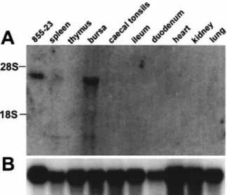
The expression pro®le of chBAFF was assessed by northern blot analysis of RNA from various chicken tissues and of the chicken T cell line 855-23 (31) using a radiolabeled PCR fragment derived from the chBAFF sequence. This cytokine seemed to be encoded by an mRNA of ~3 kb length that was mainly found in the bursa of Fabricius. The transcript was also present in cells of the 855-23 T cell line (Fig. 2).
Recombinant chBAFF binds to mammalian BAFF-R
Three different receptors of the TNF receptor family, i.e. TACI, BCMA and BAFF-R, can bind mammalian BAFF. The chicken homologues of these receptors have not been identi®ed to date. Therefore, by co-immunoprecipitation analysis we tested whether chBAFF would bind to the mammalian BAFF-R. To generate an appropriate chBAFF expression construct, we
Fig. 2. chBAFF gene expression pro®le. (A) chBAFF cDNA was used to probe a northern blot containing RNA from various chicken tissues and of the chicken T cell line 855-23 (31). (B) Re-probing of the membrane was performed with chicken GAPDH cDNA (41) to estimate the relative amounts of RNA loaded in each lane. The gel positions of the 28S rRNA and 18S rRNA are indicated.
Fig. 3. chBAFF binds mammalian BAFF-R. The receptor:Fc fusion proteins huTACI:Fc, muTACI:Fc, huBCMA:Fc, muBCMA:Fc, huBAFF-R:Fc and huEDAhuBAFF-R:Fc were mixed with supernatants of 293T cells transfected with expression plasmids encoding either Flag-huBAFF, Flag-chBAFF or Flag-huEDA, precipitated with Protein A±Sepharose and detected by western blotting using anti-Flag mAb M2 (bottom panel). The presence of receptor:Fc proteins was visualized with antibodies to human IgG (top panel). IP: immunoprecipitation; WB: western blot.
Fig. 4. chBAFF is a ligand for the mammalian receptors TACI, BCMA and BAFF-R. Binding of Flag-chBAFF and Flag-huBAFF to various TNF family receptor:Fc fusion proteins was assessed by ELISA. The plates were coated with the indicated receptor:Fc fusion proteins, ligand was added and binding was revealed using anti-Flag M2 mAb.
performed PCR on cDNA from the chicken T cell line 855-23 (31) to amplify a fragment that encoded the putative soluble form of chBAFF containing all amino acids located down-stream of the furin cleavage site. To produce a secreted form of chBAFF, the resulting cDNA fragment was cloned down-stream of the hemagglutinin signal peptide and the Flag sequence of a modi®ed version of the PCR-3 vector which permits ef®cient expression of cDNAs in eukaryotic cells (32). Supernatants of transfected 293T cells containing either Flag-chBAFF or Flag-huBAFF were used to perform co-immuno-precipitations with soluble fusion proteins consisting of the Fc domain of human IgG1 fused to the N-terminal extracellular domains of huTACI, muTACI, huBCMA, muBCMA and
huBAFF-R (huTACI:Fc, muTACI:Fc, huBCMA:Fc,
muBCMA:Fc or huBAFFR:Fc) respectively. The receptor:Fc fusion protein of the human TNF receptor family member ectodysplasin A receptor (EDAR:Fc) which binds the human TNF family ligand EDA1 and conditioned medium from 293T cells transfected with a expression construct for huEDA1 served as controls. Complexes were precipitated with Protein A±Sepharose beads, and bound proteins were analyzed by western blotting using antibodies to human IgG (Fig. 3, top) and a mAb against the Flag epitope (Fig. 3, bottom). chBAFF strongly interacted with human and murine TACI:Fc, BCMA:Fc and human BAFF-R:Fc, and did not interact with human EDAR:Fc (Fig. 3). To further demonstrate the speci®city of chBAFF, we tested its ability to bind several additional human and mouse TNF family receptors in a receptor±ligand inter-action ELISA. chBAFF and huBAFF interacted with human and mouse TACI, BCMA and BAFF-R, and both proteins did not bind to the other TNF family receptors that we tested (Fig. 4). The functionality of the various plate-coated recombinant receptor:Fc fusion proteins was demonstrated using the cognate ligands (data not shown). When no speci®c ligand was available, horseradish peroxidase-coupled rabbit anti-human IgG was used to demonstrate successful coating of receptor:Fc (data not shown).
chBAFF-R are expressed exclusively on B cells
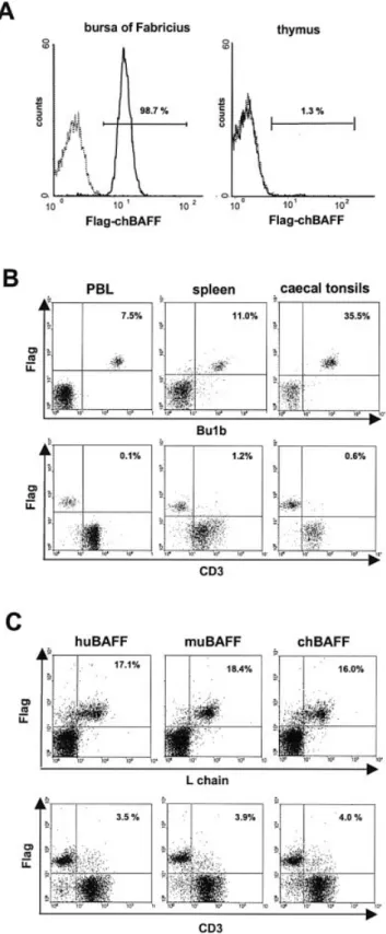
Mammalian BAFF-R are expressed mainly on B cells, sug-gesting that chBAFF-R might also be expressed on B cells of lymphoid tissues such as bursa of Fabricius, spleen, cecal tonsils and peripheral blood lymphocytes. Using recombinant Flag-chBAFF as a probe, we determined receptor expression by ¯ow cytometry. We found that most cells of the bursa of Fabricius expressed receptors for chBAFF (Fig. 5A, left). In contrast, thymocytes mostly did not bind Flag-chBAFF (Fig. 5A, right), indicating that chBAFF-R are preferentially expressed on B cells. This conclusion was con®rmed by two-color immuno¯uorescence staining (Fig. 5B). chBAFF-R expression was restricted to the Bu-1b+B cell population in
peripheral blood lymphocytes, spleen cells and cecal tonsils. ChBAFF-R expression was absent from CD3+cells.
Since chicken and mammalian BAFF show a high degree of homology, we also investigated whether mammalian BAFF binds to chicken lymphocytes. As shown in Fig. 5(C), selective binding of human and murine BAFF to chicken B cells could be demonstrated. The staining patterns and the staining inten-sities were comparable to those observed with chBAFF.
Fig. 5. Binding of chicken and mammalian BAFF to chicken lymphocytes. Binding of Flag-chBAFF to isolated cells from (A) bursa of Fabricius or thymus and (B) cecal tonsils, spleen or peripheral blood lymphocytes from 7-week-old birds was analyzed by ¯ow cytometry. (C) The ability of ¯agged huBAFF, muBAFF or chBAFF to recognize binding sites on isolated chicken splenocytes was compared by ¯ow cytometry.
chBAFF promotes the survival of splenic B cells
Mammalian BAFF is a potent survival factor for B cells. To test whether this also holds true for the chicken system, spleno-cytes were isolated and treated for 72 h with either recombinant Flag-chBAFF or control 293T cell supernatant. Analysis with a mAb speci®c for the B cell-surface marker Bu-1b combined with PI staining revealed that the B cell frequency in BAFF-treated samples was ~3-fold higher com-pared to the controls, whereas the frequency of CD3+T cells
was not affected (Fig. 6A). To further investigate this effect, splenocytes were cultured in the presence of different
concentrations of chBAFF. After 48 h, the number of viable B cells was determined. We observed a dose-dependent response to cytokine treatment. Saturation was reached if the BAFF-containing supernatant was used at concentrations >1% (Fig. 6B). To exclude the possibility that secondary effects in¯uenced the results in our mixed cell preparations, several experiments were performed using splenic B cells enriched to > 93% purity. Although cell death occurred rapidly in all cultures, the absolute numbers of living cells were higher in Flag-chBAFF-treated cultures compared to control cultures at 24, 48 and 72 h (Fig. 6C). On average, viable B cell numbers
Fig. 6. chBAFF promotes the survival of splenic B cells in culture. (A) Splenocytes were cultured for 72 h with 5% Flag-chBAFF-containing medium or with 5% medium of control cells transfected with the empty plasmid and analyzed by ¯ow cytometry. Cells were stained with PI to determine the percentage of dead cells, and with mAb to the B cell-speci®c marker Bu-1b and the T cell marker CD3. (B) Splenocytes (5 3 106cells) were treated with different concentrations (10±0.0001%) of Flag-chBAFF-containing medium or with 10% medium of control cells
transfected with the empty plasmid. After 48 h, the number of viable B cells was determined by Trypan blue exclusion and FACS analysis. Data represent mean 6 SD of three independent experiments. (C) Kinetics of cell survival. Puri®ed splenic B cell preparations (5 3 106cells,
purity >93%) were cultured for the indicated times in the presence of 5% Flag-chBAFF-containing medium or 5% medium of control cells transfected with the empty plasmid. Data represent mean 6 SD of three independent experiments. (D) CFSE-labeled splenocytes were cultured for 48 h in the presence or absence of chBAFF as described in (A) or as positive control with ConA, and analyzed by ¯ow cytometry. Cells were stained with 7-AAD, and two color FACS analysis was performed on the living cell population.
were nearly 3-fold higher after 48 h and ~5-fold higher after 72 h, strongly indicating that recombinant chBAFF promoted B cell survival in these cultures. To de®nitely prove that this effect was not a consequence of B cell proliferation, we cultured CFSE-labeled splenocytes with chBAFF or ConA. ConA-stimulated cultures showed evidence for two to three rounds of cell division after 48 hours. Under these conditions no cell proliferation was observed in the chBAFF-treated culture or in the untreated control, although the number of living B cells increased from 13.5% to 26.5% in response to chBAFF (Fig. 6D). The almost threefold decrease in the number of living B cells in the ConA-treated culture can probably be explained by the strong increase of T cells.
Since human and mouse BAFF ef®ciently bound to chicken B cells, we next determined whether they are also biologically active on chicken cells. Splenocytes were treated with ¯agged human or mouse BAFF (2 mg/ml) for 48 h before the number of viable B cells was determined. Cells treated with Flag-chBAFF (5%) or control supernatant (5%) served as positive and negative controls. We found that the mammalian BAFF preparations were nearly as potent in preventing premature death of chicken splenocytes as chBAFF (Table 1).
Increased B cell numbers in chicks treated with recombinant chBAFF
To determine the in vivo activity of chBAFF, 3-day-old chicks (seven animals per group) were injected i.p. twice a day for 7 days with 2 mg/kg body weight of E. coli-derived chBAFF
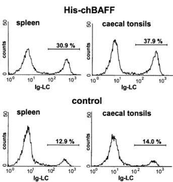
(His-chBAFF) or with the same amount of control protein (His-MxA). The spleen/body weight index was signi®cantly increased (P < 0.05, in the Wilcoxon test and Student's t-test) in chBAFF-treated birds (Table 2), indicating an increase of total lymphocyte numbers in the spleen. Flow cytometric analysis of spleens and cecal tonsils with various mAb revealed that the percentage of B cells, but not T cells, was ~2-fold higher in chBAFF-treated animals as compared to control animals (Table 3). The Ig light chain (L chain) staining pattern of a representative pair of chBAFF- and control-treated animals is shown in Fig. 7. Since splenic lymphocyte numbers and B cell frequencies were higher in chBAFF-treated animals, it can be concluded that chBAFF enhances survival or induces select-ive proliferation of B cells in spleen and cecal tonsils of cytokine-treated animals.
Discussion
Here we report the identi®cation and biological characteriza-tion of the chicken homologue of mammalian BAFF. By analyzing the BLAST search results of two chicken EST databases, we identi®ed overlapping cDNA clones that code for the complete open reading frame of chBAFF. Like its mammalian counterparts, chBAFF exhibits a type II trans-membrane protein organization. Mammalian BAFF is pro-cessed by a furin-like protease after a characteristic sequence motif which results in the release of biologically active soluble protein (2). chBAFF contains a putative furin cleavage site at the corresponding position, indicating that it might be Table 1. Effect of human and murine BAFF on chicken
splenocytes in vitro
No. of viable B cells (3105) Mean 6 SD
1 2 3
huBAFF 0.91 1.90 1.72 1.51 6 0.4
muBAFF 1.39 1.59 2.10 1.69 6 0.3
chBAFF 1.46 2.28 2.04 1.93 6 0.4
Control 0.26 0.76 0.92 0.65 6 0.3
Splenocytes were cultured for 48 h at 5 3 106 cells per well in
24-well plates with either Flag-chBAFF (5%), control supernatant (5%), Flag-huBAFF (2 mg/ml) or Flag-muBAFF (2 mg/ml). Three independent experiments were performed.
Table 2. chBAFF-dependent increase in spleen weight
Spleen weight
(mg) Body weight(g) Spleen/bodyweight index (mg/g)a
chBAFF 76.8 6 24.0 64.2 6 2.1 1.2 6 0.3 control 55.3 6 22.8 66.5 6 11.9 0.8 6 0.2 Seven animals per group were treated with either chBAFF or control protein (2 mg/kg body mass twice a day) for 7 days. Spleen and body weights were determined, and the spleen/body weight index of each animal was calculated. Mean values 6 SD are given for each group.
aThese values are signi®cantly different (P < 0.05; Wilcoxon t-test).
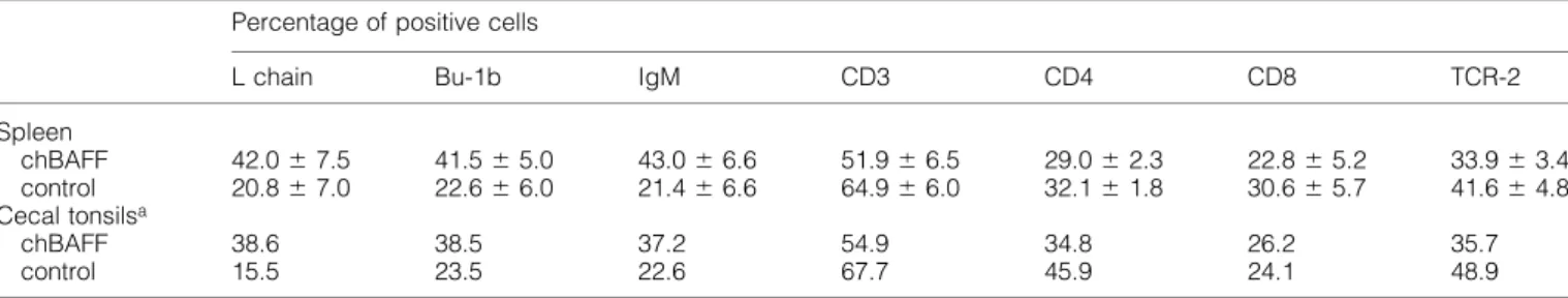
Table 3. Selective expansion of the B cell population in spleen and cecal tonsils of chBAFF-treated chicks
Percentage of positive cells
L chain Bu-1b IgM CD3 CD4 CD8 TCR-2
Spleen chBAFF 42.0 6 7.5 41.5 6 5.0 43.0 6 6.6 51.9 6 6.5 29.0 6 2.3 22.8 6 5.2 33.9 6 3.4 control 20.8 6 7.0 22.6 6 6.0 21.4 6 6.6 64.9 6 6.0 32.1 6 1.8 30.6 6 5.7 41.6 6 4.8 Cecal tonsilsa chBAFF 38.6 38.5 37.2 54.9 34.8 26.2 35.7 control 15.5 23.5 22.6 67.7 45.9 24.1 48.9
Five animals per group were treated with either chBAFF or control protein (2 mg/kg body mass twice a day) for 7 days. The average percentages of cells expressing the various surface markers (6 SD) are shown.
processed similarly. The predicted soluble forms of chicken and mammalian BAFF are surprisingly similar. At the amino acid level, soluble chBAFF is 70 and 76% identical to mouse and human BAFF respectively. Most other avian cytokines show <30% identity to their mammalian counterparts (39).
The high sequence conservation suggested that chBAFF might bind to mammalian BAFF-R. By performing appropriate co-immunoprecipitation experiments and by using a receptor± ligand interaction ELISA we were able to demonstrate that this was indeed the case. Like human BAFF, chBAFF bound speci®cally to the mammalian receptors TACI, BCMA and BAFF-R. The receptors for BAFF in mammals are expressed on B cells and on some T cells (12±14). Our ¯ow cytometric analyses revealed that Flag-tagged chBAFF bound a popu-lation of cells which was abundantly present in chicken spleen, bursa of Fabricius, peripheral blood lymphocytes and cecal tonsils, but which was virtually absent in the thymus. Since ~98% of the lymphocytes present in the bursa of Fabricius are B cells, our data strongly suggested that the receptors for BAFF in the chicken are expressed predominantly if not exclusively on B cells, which was con®rmed by two-color immuno¯uorescence staining.
As expected from the known activity of mammalian BAFF, chBAFF did not induce B cell proliferation but was clearly able to prolong the survival of splenic B cells in cell culture. Mammalian BAFF also bound to chicken B cells and promoted their survival, indicating functional cross-reactivity between mammalian and avian BAFF. chBAFF further promoted the
speci®c expansion of the B cell population in the spleen and cecal tonsils of young chicks that were treated with recombi-nant cytokine. Further studies are needed to determine whether the in vivo expansion of B cells resulted from enhanced survival or from increased proliferation of this lymphocyte population. In contrast to spleen and cecal tonsils, the number of B cells in the bursa of Fabricius of treated chicks was not increased and the size of the organ remained unchanged. This differential response to chBAFF of B cells in various lymphoid organs is not really surprising. Indeed, studies with transgenic and knockout mice have clearly demonstrated that mammalian BAFF is critical for B cell maturation in peripheral lymphoid organs, but is not required for B cell development in the bone marrow, despite the fact that BAFF mRNA is moderately expressed in this compartment (3,17,24). In birds, early development of B cells does not take place in the bone marrow, but in the bursa of Fabricius (40). By analogy with the mammalian system, the ®rst steps of B cell development in the chicken are not expected to be dependent on BAFF. However, in view of the fact that the chBAFF gene is expressed most strongly in the bursa, it may well be that BAFF is important for bursal B cell survival. chBAFF could play a role in a late B cell differentiation step, shortly before the B cells exit from the bursa. Since administration of exogenous BAFF did not positively affect the number of B cells in the bursa, at ®rst glance, this result seems to argue against this view. It is possible, however, that the level of endogenous BAFF is already very high in the bursa and that administration of exogenous BAFF has no additive effect simply because the BAFF-R on bursal B cells are already saturated with ligands.
In mammals, BAFF is expressed by cells of myeloid origin and, to a lesser extent, by T cells, but not by B cells (2,3,8). Since spleen did not contain detectable levels of BAFF transcripts, mature chicken B cells cannot be the major producers of BAFF. Thus, most likely, BAFF is synthesized by either bursa stromal cells or immature B cells. To further investigate the role of this cytokine in bursal B cell develop-ment, the activity of endogenous chBAFF needs to be blocked in vivo. Since the generation of knockout birds is still impossible, alternative strategies aimed at neutralizing this cytokine with antagonistic antibodies or with soluble decoy receptors such as BAFF-R:Fc should be considered. Results presented here (Figs 3 and 4) strongly indicate that BAFF-R:Fc has the potential to neutralize the activity of chBAFF.
In summary, by identifying chicken BAFF as well as suitable decoy receptors we paved the way for new experimental approaches aimed at a better understanding of B cell development in the chicken. Furthermore, our work is the ®rst functional characterization of a chicken TNF family member.
Acknowledgements
We thank Drs C. Ambrose and J. Thompson (Biogen, Boston, MA) for providing the cDNA for BAFF-R, and Dr E. Duda for help with the in vivo experiment. This work was supported by grants from the Deutsche Forschungsgemeinschaft, the European Community, the Swiss National Research Foundation and Intervet Int., BV, Boxmeer, The Netherlands.
Fig. 7. Increased B cell numbers in chicks treated with recombinant chBAFF. Effect of i.p. administration of His-chBAFF or control protein (His-MxA) on the frequency of various cell types in the spleen and cecal tonsils of 10-day-old chicks at 7 days post-initiation of treatment. Cells were stained with the indicated mAb and analyzed by ¯ow cytometry. A representative data set of spleen and cecal tonsils from a pair of chBAFF- and control-treated animals stained for the Ig light chain (L chain) is shown.
Abbreviations
APRIL a proliferating inducing ligand
BAFF B cell activating factor of the TNF family BAFF-R BAFF receptor
BCMA B cell maturation antigen PE phycoerythrin
PI propidium iodide SS SjoÈgren's syndrome
SLE systemic lupus erythematosus
TACI transmembrane activator, calcium modulator and cyclophilin ligand interactor
TMD transmembrane domain TNF tumor necrosis factor
TRAIL TNF-related apoptosis inducing ligand
References
1 Locksley, R. M., Killeen, N. and Lenardo, M. J. 2001. The TNF and TNF receptor superfamilies: integrating mammalian biology. Cell 104:487.
2 Schneider, P., MacKay, F., Steiner, V., Hofmann, K., Bodmer, J. L., Holler, N., Ambrose, C., Lawton, P., Bixler, S., Acha-Orbea, H., Valmori, D., Romero, P., Werner-Favre, C., Zubler, R. H., Browning, J. L. and Tschopp, J. 1999. BAFF, a novel ligand of the tumor necrosis factor family, stimulates B cell growth. J. Exp. Med. 189:1747.
3 Moore, P. A., Belvedere, O., Orr, A., Pieri, K., LaFleur, D. W., Feng, P., Soppet, D., Charters, M., Gentz, R., Parmelee, D., Li, Y., Galperina, O., Giri, J., Roschke, V., Nardelli, B., Carrell, J., Sosnovtseva, S., Green®eld, W., Ruben, S. M., Olsen, H. S., Fikes, J. and Hilbert, D. M. 1999. BLyS: member of the tumor necrosis factor family and B lymphocyte stimulator. Science 285:260. 4 Shu, H. B., Hu, W. H. and Johnson, H. 1999. TALL-1 is a novel
member of the TNF family that is down-regulated by mitogens. J. Leukoc. Biol. 65:680.
5 Mukhopadhyay, A., Ni, J., Zhai, Y., Yu, G. L. and Aggarwal, B. B. 1999. Identi®cation and characterization of a novel cytokine, THANK, a TNF homologue that activates apoptosis, nuclear factor-kappaB, and c-Jun NH2-terminal kinase. J. Biol. Chem.
274:15978.
6 Rolink, A. G. and Melchers, F. 2002. BAFFled B cells survive and thrive: roles of BAFF in B-cell development. Curr. Opin. Immunol. 14:266.
7 Bodmer, J. L., Schneider, P. and Tschopp, J. 2002. The molecular architecture of the TNF superfamily. Trends Biochem. Sci. 27:19. 8 Nardelli, B., Belvedere, O., Roschke, V., Moore, P. A., Olsen, H. S., Migone, T. S., Sosnovtseva, S., Carrell, J. A., Feng, P., Giri, J. G. and Hilbert, D. M. 2001. Synthesis and release of B-lymphocyte stimulator from myeloid cells. Blood 97:198.
9 Rennert, P., Schneider, P., Cachero, T. G., Thompson, J., Trabach, L., Hertig, S., Holler, N., Qian, F., Mullen, C., Strauch, K., Browning, J. L., Ambrose, C. and Tschopp, J. 2000. A soluble form of B cell maturation antigen, a receptor for the tumor necrosis factor family member APRIL, inhibits tumor cell growth. J. Exp. Med. 192:1677.
10 Yu, G., Boone, T., Delaney, J., Hawkins, N., Kelley, M., Ramakrishnan, M., McCabe, S., Qiu, W. R., Kornuc, M., Xia, X. Z., Guo, J., Stolina, M., Boyle, W. J., Sarosi, I., Hsu, H., Senaldi, G. and Theill, L. E. 2000. APRIL and TALL-I and receptors BCMA and TACI: system for regulating humoral immunity. Nat. Immunol. 1:252.
11 Marsters, S. A., Yan, M., Pitti, R. M., Haas, P. E., Dixit, V. M. and Ashkenazi, A. 2000. Interaction of the TNF homologues BLyS and APRIL with the TNF receptor homologues BCMA and TACI. Curr. Biol. 10:785.
12 Gross, J. A., Johnston, J., Mudri, S., Enselman, R., Dillon, S. R., Madden, K., Xu, W., Parrish-Novak, J., Foster, D., Lofton-Day, C., Moore, M., Littau, A., Grossman, A., Haugen, H., Foley, K., Blumberg, H., Harrison, K., Kindsvogel, W. and Clegg, C. H. 2000. TACI and BCMA are receptors for a TNF homologue implicated in B-cell autoimmune disease. Nature 404:995.
13 Thompson, J. S., Bixler, S. A., Qian, F., Vora, K., Scott, M. L., Cachero, T. G., Hession, C., Schneider, P., Sizing, I. D., Mullen, C., Strauch, K., Zafari, M., Benjamin, C. D., Tschopp, J., Browning, J. L. and Ambrose, C. 2001. BAFF-R, a newly identi®ed TNF receptor that speci®cally interacts with BAFF. Science 293:2108.
14 Yan, M., Brady, J. R., Chan, B., Lee, W. P., Hsu, B., Harless, S., Cancro, M., Grewal, I. S. and Dixit, V. M. 2001. Identi®cation of a novel receptor for B lymphocyte stimulator that is mutated in a mouse strain with severe B cell de®ciency. Curr. Biol. 11:1547. 15 Gras, M. P., Laabi, Y., Linares-Cruz, G., Blondel, M. O., Rigaut, J.
P., Brouet, J. C., Leca, G., Haguenauer-Tsapis, R. and Tsapis, A. 1995. BCMAp: an integral membrane protein in the Golgi apparatus of human mature B lymphocytes. Int. Immunol. 7:1093. 16 von Bulow, G. U. and Bram, R. J. 1997. NF-AT activation induced by a CAML-interacting member of the tumor necrosis factor receptor superfamily. Science 278:138.
17 Batten, M., Groom, J., Cachero, T. G., Qian, F., Schneider, P., Tschopp, J., Browning, J. L. and Mackay, F. 2000. BAFF mediates survival of peripheral immature B lymphocytes. J. Exp. Med. 192:1453.
18 Parry, T. J., Riccobene, T. A., Strawn, S. J., Williams, R., Daoud, R., Carrell, J., Sosnovtseva, S., Miceli, R. C., Poortman, C. M., Sekut, L., Li, Y., Fikes, J. and Sung, C. 2001. Pharmacokinetics and immunological effects of exogenously administered recombinant human B lymphocyte stimulator (BLyS) in mice. J. Pharmacol. Exp. Ther. 296:396.
19 Mackay, F., Woodcock, S. A., Lawton, P., Ambrose, C., Baetscher, M., Schneider, P., Tschopp, J. and Browning, J. L. 1999. Mice transgenic for BAFF develop lymphocytic disorders along with autoimmune manifestations. J. Exp. Med. 190:1697. 20 Khare, S. D., Sarosi, I., Xia, X. Z., McCabe, S., Miner, K., Solovyev,
I., Hawkins, N., Kelley, M., Chang, D., Van, G., Ross, L., Delaney, J., Wang, L., Lacey, D., Boyle, W. J. and Hsu, H. 2000. Severe B cell hyperplasia and autoimmune disease in TALL-1 transgenic mice. Proc. Natl Acad. Sci. USA 97:3370.
21 Groom, J., Kalled, S. L., Cutler, A. H., Olson, C., Woodcock, S. A., Schneider, P., Tschopp, J., Cachero, T. G., Batten, M., Wheway, J., Mauri, D., Cavill, D., Gordon, T. P., Mackay, C. R. and Mackay, F. 2002. Association of BAFF/BLyS overexpression and altered B cell differentiation with SjoÈgren's syndrome. J. Clin. Invest. 109:59. 22 Zhang, J., Roschke, V., Baker, K. P., Wang, Z., Alarcon, G. S., Fessler, B. J., Bastian, H., Kimberly, R. P. and Zhou, T. 2001. Cutting edge: a role for B lymphocyte stimulator in systemic lupus erythematosus. J. Immunol. 166:6.
23 Cheema, G. S., Roschke, V., Hilbert, D. M. and Stohl, W. 2001. Elevated serum B lymphocyte stimulator levels in patients with systemic immune-based rheumatic diseases. Arthritis Rheum. 44:1313.
24 Schiemann, B., Gommerman, J. L., Vora, K., Cachero, T. G., Shulga-Morskaya, S., Dobles, M., Frew, E. and Scott, M. L. 2001. An essential role for BAFF in the normal development of B cells through a BCMA-independent pathway. Science 293:2111. 25 Gross, J. A., Dillon, S. R., Mudri, S., Johnston, J., Littau, A., Roque,
R., Rixon, M., Schou, O., Foley, K. P., Haugen, H., McMillen, S., Waggie, K., Schreckhise, R. W., Shoemaker, K., Vu, T., Moore, M., Grossman, A. and Clegg, C. H. 2001. TACI±Ig neutralizes molecules critical for B cell development and autoimmune disease. impaired B cell maturation in mice lacking BLyS. Immunity 15:289.
26 Xia, X. Z., Treanor, J., Senaldi, G., Khare, S. D., Boone, T., Kelley, M., Theill, L. E., Colombero, A., Solovyev, I., Lee, F., McCabe, S., Elliott, R., Miner, K., Hawkins, N., Guo, J., Stolina, M., Yu, G., Wang, J., Delaney, J., Meng, S. Y., Boyle, W. J. and Hsu, H. 2000. TACI is a TRAF-interacting receptor for TALL-1, a tumor necrosis factor family member involved in B cell regulation. J. Exp. Med. 192:137.
27 Thompson, J. S., Schneider, P., Kalled, S. L., Wang, L., Lefevre, E. A., Cachero, T. G., MacKay, F., Bixler, S. A., Zafari, M., Liu, Z. Y., Woodcock, S. A., Qian, F., Batten, M., Madry, C., Richard, Y., Benjamin, C. D., Browning, J. L., Tsapis, A., Tschopp, J. and Ambrose, C. 2000. BAFF binds to the tumor necrosis factor receptor-like molecule B cell maturation antigen and is important
for maintaining the peripheral B cell population. J. Exp. Med. 192:129.
28 Wang, H., Marsters, S. A., Baker, T., Chan, B., Lee, W. P., Fu, L., Tumas, D., Yan, M., Dixit, V. M., Ashkenazi, A. and Grewal, I. S. 2001. TACI±ligand interactions are required for T cell activation and collagen-induced arthritis in mice. Nat. Immunol. 2:632. 29 Hartmann, W. 1997. Evaluation of major genes affecting
resistance to disease in poultry. World's Poult. Sci. J. 53:231. 30 Martin, A., Lillehoj, H. S., Kaspers, B. and Bacon, L. D. 1993.
Antigen-speci®c T cell proliferation following coccidia infection. Poult. Sci. 72:2084.
31 Weining, K. C., Schultz, U., Munster, U., Kaspers, B. and Staeheli, P. 1996. Biological properties of recombinant chicken interferon-gamma. Eur. J. Immunol. 26:2440.
32 Schneider, P. 2000. Production of recombinant TRAIL and TRAIL receptor: Fc chimeric proteins. Methods Enzymol. 322:325. 33 Erhard, M. H., Von Quistorp, I., Schranner, I., Jungling, A.,
Kaspers, B., Schmidt, P. and Kuhlmann, R. 1992. Development of speci®c enzyme-linked immunosorbent antibody assay systems for the detection of chicken immunoglobulins G, M, and A using monoclonal antibodies. Poult. Sci. 71:302.
34 Tregaskes, C. A., Bumstead, N., Davison, T. F. and Young, J. R. 1996. Chicken B-cell marker chB6 (Bu-1) is a highly glycosylated protein of unique structure. Immunogenetics 44:212.
35 Chen, C. L., Ager, L. L., Gartland, G. L. and Cooper, M. D. 1986.
Identi®cation of a T3/T cell receptor complex in chickens. J. Exp. Med. 164:375.
36 Luhtala, M., Salomonsen, J., Hirota, Y., Onodera, T., Toivanen, P. and Vainio, O. 1993. Analysis of chicken CD4 by monoclonal antibodies indicates evolutionary conservation between avian and mammalian species. Hybridoma 12:633.
37 Luhtala, M., Koskinen, R., Toivanen, P. and Vainio, O. 1995. Characterization of chicken CD8-speci®c monoclonal antibodies recognizing novel epitopes. Scand. J. Immunol. 42:171. 38 Cihak, J., Ziegler-Heitbrock, H. W., Trainer, H., Schranner, I.,
Merkenschlager, M. and Losch, U. 1988. Characterization and functional properties of a novel monoclonal antibody which identi®es a T cell receptor in chickens. Eur. J. Immunol. 18:533. 39 Staeheli, P., Puehler, F., Schneider, K., Gobel, T. W. and Kaspers,
B. 2001. Cytokines of birds: conserved functions-a largely different look. J. Interferon Cytokine Res. 21:993.
40 McCormack, W. T., Tjoelker, L. W. and Thompson, C. B. 1991. Avian B-cell development: generation of an immunoglobulin repertoire by gene conversion. Annu. Rev. Immunol. 9:219. 41 Panabieres, F., Piechaczyk, M., Rainer, B., Dani, C., Fort, P.,
Riaad, S., Marty, L., Imbach, J. L., Jeanteur, P. and Blanchard, J. M. 1984. Complete nucleotide sequence of the messenger RNA coding for chicken muscle glyceraldehyde-3-phosphate dehydrogenase. Biochem. Biophys. Res. Commun. 118:767.