Diversity and expression of different forms of RubisCO genes in
polluted groundwater under different redox conditions
Albin Alfreider1, Mario Schirmer2 & Carsten Vogt3
1Institute of Ecology, University of Innsbruck, Innsbruck, Austria;2Eawag– Swiss Federal Institute of Aquatic Science and Technology, Department Water Resources and Drinking Water (W+T), Du¨bendorf, Switzerland; and3Department of Isotope Biogeochemistry, Helmholtz Centre for Environmental Research– UFZ, Leipzig, Germany
Correspondence: Albin Alfreider, Institute of Ecology, University of Innsbruck, Technikerstr. 25, 6020 Innsbruck, Austria. Tel.:
+43 512 5076122; fax: +43 512 5072930; e-mail: [email protected]
Received 13 June 2011; revised 17 October 2011; accepted 2 November 2011. Final version published online 28 November 2011.
DOI: 10.1111/j.1574-6941.2011.01246.x Editor: Tillman Lueders
Keywords
groundwater; chemolithoautotrophs; RubisCO genes.
Abstract
Groundwater polluted with methyl-tert-butyl ether (MTBE) and ammonium was investigated for chemolithoautotrophic CO2 fixation capabilities based on
detailed analyses of ribulose-1,5-bisphosphate carboxylase/oxygenase (RubisCO) large subunit genes. Samples retrieved from a groundwater conditioning unit, characterized by different redox conditions, were examined for the presence of form IA, form IC (cbbL) and form II (cbbM) RubisCO genes and transcripts obtained from DNA- and RNA-extracts. Form IA RubisCO sequences, which revealed a complex and distinct variety in different sampling stations, were expressed in the original groundwater and in samples amended with oxygen, but not in the aquifer groundwater enriched with nitrate. Form IC RubisCO genes were exclusively detected in groundwater supplied with oxygen and sequences were affiliated with cbbL genes in nitrifying bacteria. cbbM genes were not expressed in the oxygen-amended groundwater, probably due to the low CO2/O2 substrate specificity of this enzyme. Most form II RubisCO
tran-scripts were affiliated with RubisCO genes of denitrifiers, which are important residents in the groundwater supplied with nitrate. The distinct distribution pattern and diversity of RubisCO genes and transcripts obtained in this study suggest that the induction of different RubisCO enzymes is highly regulated and closely linked to the actual environmental conditions.
Introduction
The abundance and productivity of prokaryotes in groundwater systems is generally dependent on organic matter availability, which is strongly limited by the lack of photosynthesis and the reduced supply of organic car-bon originating from biological processes in surface eco-systems (Baker et al., 2000; Foulquier et al., 2010). Consequently, on one hand, heterotrophic prokaryotes adapted to the oligotrophic conditions are considered to dominate microbial life in aquifers. On the other hand, reactive rock surfaces and mineral-rich groundwater are ideal environments providing a variety of potential elec-tron donors that enable chemolithoautotrophic metabo-lism (Engel, 2007), although our understanding of the ecological role of chemolithoautotrophic prokaryotes in
groundwater systems is still very limited. The importance of chemolithoautotrophy was first recognized in karst/ cave systems and in the deep subsurface. Subsequently, geochemical and biological prerequisites for the chemo-lithoautotrophic lifestyle were intensively discussed (e.g. Stevens & McKinley, 1996; Anderson et al., 1998; Neal-son et al., 2005). Not until recent years has there been growing evidence that chemolithoautotrophic bacteria are also frequent residents in shallow groundwater systems, although their actual activity remains an open issue (Alfreider et al., 2009). An autotrophic lifestyle is energet-ically expensive, and one of the most fundamental ques-tions in subsurface research is still how the microbial communities are supplied with energy (Adhikari & Kall-meyer, 2010). Autotrophic physiology is slow and under strict metabolic control, especially in pristine groundwater
MICR
systems, which are often characterized by electron donor-and electron acceptor-limiting conditions.
CO2 fixation in chemolithoautotrophic bacteria is
fre-quently catalyzed via the Calvin–Benson–Bassham cycle, with ribulose-1,5-bisphosphate carboxylase/oxygenase (RubisCO) as principal enzyme for the assimilation of CO2. From the structural viewpoint there are several
types of RubisCO, with bacteria using form I and form II RubisCO (Tabita et al., 2007; Badger & Bek, 2008). Recent studies have even described RubisCO form III proteins in anaerobic Archaea that are phylogenetically well separated from known RubisCO forms I and II (Mueller-Cajar & Badger, 2007; Tabita et al., 2008). Molecular investigation based on highly conserved large subunit RubisCO genes of form I (cbbL) and form II (cbbM) provide an excellent instrument to explore the diversity and ecology of chemolithoautotrophic bacteria in environmental samples, and they have also been used to investigate the diversity of autotrophic bacteria in groundwater systems (Lawrence et al., 2000; Alfreider et al., 2003, 2009; Nedelkova, 2005; Kellermann, 2008; Waldron et al., 2009).
The most comprehensive study in this context was per-formed by Alfreider et al. (2009), including 48 sampling stations obtained from a variety of pristine and polluted, shallow and deep groundwater systems located in Ger-many and Austria. The widespread occurrence of different forms of RubisCO detected in that study suggested that chemolithoautotrophic bacteria with the potential to assimilate CO2 via the Calvin cycle pathway are
impor-tant members of the microbial community in groundwa-ter systems.
However, what was not addressed by previous studies is the physiological significance of autotrophy in ground-water ecosystems, because all investigations were accom-plished with RubisCO sequence analysis based on DNA extracts. Consequently, former studies only hypothesized about the ecological role and activity of the RubisCO gene-harbouring bacteria and their potential involvement in important biological processes in different groundwater habitats.
The main objective of this study was to determine the CO2 fixation potential and capabilities of
chemolitho-autotrophic bacteria based on detailed analyses of Rubi-sCO DNA and messenger RNA (mRNA). Gene transcripts provide a reliable indication of specific micro-bial activities under in situ conditions and the response of microbial organisms to changes in their environment. The investigations were accomplished with samples retrieved from a groundwater conditioning unit as model system, where polluted groundwater was exposed to dif-ferent treatments altering the chemical and redox condi-tions of the original groundwater. To examine the
phylogenetic diversity of form IA, form IC and form II RubisCO genes and gene transcripts, products from PCR and reverse transcriptase PCR (RT-PCR) were subjected to construct clone libraries with selected clones investi-gated by sequence analysis.
Materials and methods
Site description and sampling location
The sampling site is located at a reference test site for the implementation of an enhanced natural attenuation approach, located close to the city Leuna, 50 km west of Leipzig, Germany. The groundwater of the study area is in the catchment area of an old industrial site and is mainly contaminated with methyl-tert-butyl ether (MTBE), ben-zene, toluene, ethylbenben-zene, xylene (BTEX) compounds and ammonium, the latter derived from a former ammo-nium production site (Martienssen et al., 2006). The aqui-fer sediments are heterogeneous and mostly composed of fine to coarse sand and gravel. The average groundwater flow velocity varies between 0.3 and 1.0 m day 1based on calculations derived from water-level data, pumping and tracer tests (Martienssen et al., 2006).
One project at the site was aimed at stimulating natural attenuation processes at a so-called conditioning facility. Five separated channels (2 m high, 1 m wide and 10 m long) were placed in the aquifer, allowing a controlled flow of contaminated groundwater and the addition of nutrients, electron acceptors, catalysts and microorgan-isms. The upstream groundwater was captured by sheet piles followed by passive flow through the conditioning chambers, which are closed systems. The groundwater was subsequently re-infiltrated into the down-gradient aquifer.
In our study, groundwater samples were taken from the groundwater effluent of two conditioning units, Leuna Oxygen (LO) and Leuna Nitrate (LN), and from the original groundwater Leuna Reference (LR), which is the groundwater inflow of the conditioning facility. Channel LO was filled with expanded clay and supplied with technical oxygen (30–40 L m 3 groundwater, depending on the solubility) and phosphate (27 g K2HPO4m 3). Groundwater in channel LN was supplied
with nitrate (34.26 g NaNO3m 3 groundwater) and
phosphate (27 g K2HPO4 m 3). This channel was filled
with an equal mixture of activated carbon and gravel, leading to sorption of MTBE and other organic com-pounds within the channel. The groundwater flow rate in the individual channels was 3.5 m³ day 1
. The physico-chemical characteristics of the groundwater samples taken from the original groundwater and the effluent of the different conditioning units are shown in Table 1.
Chemical analysis
MTBE was analyzed by gas chromatography as described elsewhere (Martienssen et al., 2006). Certain compounds were analyzed by the following certified methods: ben-zene, DIN 38407-F9-1; ammonium, DIN 38406-E5; nitrate, DIN 38405-29; sodium, potassium, magnesium, calcium, manganese, EN ISO 11885:1997; sulphate, EN ISO 10304-1; sulphide, DIN 38405-D26; bicarbonate, DIN 38409-D8; dissolved organic carbon (DOC), DIN 38409 H3; total phosphate, DIN EN 1189-D11; ortho-phosphate, EN ISO 6878:2004; total iron, ferrous iron, DIN 38406-E1. Oxygen, pH and redox potential were determined by portable electrodes (WTW, Germany).
DNA and RNA extraction
Groundwater samples for DNA and RNA analysis were collected in sterile bottles and 300–500 mL were
concen-trated on filters (pore size 0.22lm; Durapore, Millipore, Bedford, MA). The filters were immediately frozen and stored at 20°C (for DNA analysis) or ~80 °C in dry ice (for RNA analysis) until extraction.
To extract the groundwater samples, the filters were cut into small pieces using a sterile scalpel. DNA extraction of sediment samples was performed with the FastDNA®Spin Kit for soil. RNA extraction was accomplished using Fast-Prep FastRNA® Pro Blue Kit (Qbiogene Inc., Carlsbad, CA). The filter pieces were placed in a tube containing extraction buffer and beads as provided by the kits. After processing the samples in a bead beater (Retsch MM200, Retsch Inc., Haan, Germany), total genomic DNA and RNA was extracted according to the manufacturer’s proto-cols. Extracted DNA was stored at 20°C and RNA was stored at 80°C until further processing.
PCR, RT-PCR and cloning
Three sets of oligonucleotide primers were used for PCR and RT-PCR amplification of RubisCO form IA, form IC and form II gene fragments as described in Alfreider et al. (2003, 2009). PCR amplification was carried out in 50-lL reaction mixtures, containing HotStarTaq PCR Master Mix (Qiagen GmbH, Valencia, CA), 10 pmol of each pri-mer and< 1 lg template DNA. The thermal cycle param-eters for the amplification of RubisCO fragments are published elsewhere (Alfreider et al., 2003, 2009). Reverse transcription and subsequent PCR amplification were per-formed using a one-step reaction scheme carried out sequentially in the same tube (Qiagen OneStep RT-PCR Kit; Qiagen Inc., Valencia, CA). Prior to RT-PCR, aliqu-ots of the RNA extracts were digested with DNase I using the standard procedure recommended by the manufac-turer (Sigma-Aldrich Inc., St. Louis, MO). RT-PCR com-ponents for 50-lL reactions contained 19 Qiagen OneStep RT-PCR Buffer, 400lM of each dNTP, 0.6 lM of each primer, 1 UnitlL 1 RNase inhibitor, 2lL Qia-gen OneStep RT-PCR Enzyme Mix and between 0.02– 2lg of template RNA. The thermal cycle parameters for RT-PCR, performed with a Thermal Cycler Techne PHC-3 (Techne Inc., Burlington, NJ), were as follows: PHC-30 min at 55°C (form IA/IC) or 57 °C (form II) for the reverse-transcription reaction followed by a 15-min step at 95°C to inactivate the reverse transcriptase and activate the HotStartTaq DNA polymerase. For the PCR of RubisCO genes, 35–40 cycles of 30 s at 95 °C, 45 s at 55 °C (form IA/IC) or 57°C (form II) and 1 min 30 s at 72 °C were applied. Cycles were followed by a 5-min incubation step at 72°C. Potential contamination of RT-PCR reaction by genomic DNA was detected using PCR control reactions in which the reverse-transcriptase activity was inhibited. PCR and RT-PCR products were separated on 1.5%
Table 1. RubisCO-PCR amplicons derived from DNA extracts, RT-PCR products derived from mRNA-extracts and chemical characteristics of the groundwater samples
Parameter Sampling stations* LR LO LN Form IA DNA/mRNA +/+ +/+ +/ Form IC DNA/mRNA / +/+ / Form II DNA/mRNA +/+ +/ +/+ Redox potential (mV) 110 137 130 pH 7.2 6.9 7.1 Oxygen (mg L 1) 0.4† 1.59 0.51† MTBE (mg L 1) 42.89 46.1 2.58 Benzene (lg L 1 ) 179 b.d. b.d. Ammonium (mg L 1) 59.6 45.6 8.1 Nitrate (mg L 1) b.d. 34.9 b.d. Nitrite (mg L 1) 0.069 0.780 0.073 Sodium (mg L 1) 206.5 205.8 218.8 Potassium (mg L 1) 13.96 29.00 30.94 Magnesium (mg L 1) 55.1 52.0 52.3 Calcium (mg L 1) 312.8 301.3 299 Sulphate (mg L 1) 600.5 499.9 472.5 Sulphide (mg L 1) 0.01 b.d. 0.01 HCO3(mg L 1) 16.8 13.2 14.7 DOC (mg L 1) 26.4 21.7 2.2 DOC (MTBE/TBA; mg L 1) 29.4 31.5 1.9 Phosphor total (mg L 1) 0.24 1.46 0.93 o-Phosphate (mg L 1) 0.17 1.03 0.71 Iron total (mg L 1) 16.04 b.d. b.d Iron II (mg L 1) 14.98 b.d. b.d. Manganese (mg L 1) 1.36 1.071 1.054
+, (RT )PCR product detected; , no (RT )PCR product detected; b. d., below detection limit.
*Sample designation: LR, original (inflowing) groundwater; LO, addi-tion of oxygen; LN, addiaddi-tion of nitrate.
†Oxygen values in samples LN and LR probably reflect the detection limit of the method and may also be influenced by sampling artefacts.
agarose gels and visualized with SYBR Green stain (Invi-trogen, Carlsbad, CA).
Selected RubisCO PCR and RT-PCR products with the expected size range were cut out of the gels and purified (Wizard SV Gel and PCR clean up system; Promega, Madison, WI). Purified nucleic acid fragments were cloned using a PCR cloning kit (Qiagen Inc.) according to the protocols provided by the manufacturer. Single clone colonies were transferred into PCR water and 40 clones for each library were screened for the presence of RubisCO gene inserts by PCR using vector-specific prim-ers (M13). Positive amplicons of proper length were selected for sequencing analysis.
DNA sequencing and analysis
Sequencing was carried out with a capillary genetic ana-lyzer (ABI 3730; Applied Biosystems, Foster City, CA) using dye terminators performed by a sequencing service enterprise (Macrogen, Seoul, Korea). The closest relatives to RubisCO nucleotide sequences and deduced amino acid sequences were obtained using NCBI sequence simi-larity search toolsBLASTNandBLASTP(basic local alignment
search tool, Altschul et al., 1990) and microbial IMG/M tool BLAST for microbial community genomes hosted at
the Joint Genome Institute (Markowitz et al., 2008). Deduced amino acids were aligned using CLUSTAL W as
provided by MEGA 4.0 software (Tamura et al., 2007).
Neighbour-joining trees applying gamma distribution as the distance method were computed with the MEGA 4.0
software package. Bootstrap analysis (1000 replicates) was used to obtain confidence estimates for tree topology. The phylogenetic tree was optimized with the MEGA tool
Tree Explorer. Due to the absence of an agreement on the definition of an operational taxonomic unit (OTU) based on RubisCO nucleotide or amino acid sequence identities, statistical analyses based on OTUs were not performed.
RubisCO partial sequences data from transcripts (from samples LR, LO, LN) and from DNA (from samples LR and LN) have been submitted to GenBank database under accession numbers JF414941–JF415078 (see also Supporting Information, Table S1). RubisCO sequences obtained from DNA extracts of sampling station LO have already been analyzed and published by Alfreider et al. (2009).
Results and discussion Form I RubisCO
Phylogenetic analyses based on large subunit genes and deduced amino acid sequences divide form I RubisCO
into two groups (‘green’ and ‘red’), which may be further subdivided into types IA, IB, IC and ID. Whereas type IB RubisCO enzymes are mostly found in cyanobacteria, green algae and type ID in non-green algae, chemolitho-autotrophic bacteria are known to contain types IA and IC of RubisCO. Specific information on metabolic and ecological properties affecting the occurrence and distri-bution of both bacterial forms in the environment are limited (Badger & Bek, 2008). Generally, bacteria contain-ing form IC RubisCO are known for facile genetic trans-fer (Horken & Tabita 1999). It has also been suggested that obligate chemolithoautotrophs often possess form IA RubisCO, and form IC enzymes are often associated with facultative autotrophs (Badger & Bek, 2008). Exceptions are several ammonium-oxidizing bacteria affiliated with different Nitrospira species and the Gammaproteobacteria Nitrosococcus oceani, which are obligate autotrophs oxi-dizing ammonium.
In the current study, amplification of DNA (by PCR) and mRNA (by RT-PCR) coding for RubisCO genes revealed a distinct pattern in the individual samples (Table 1). Form IA RubisCO PCR and RT-PCR prod-ucts were obtained from the original (inflowing) groundwater (LR) and groundwater amended with oxy-gen (LO). In groundwater amended with nitrate (LN) and active coal, RubisCO genes were amplified only from DNA extracts. Form IC RubisCO genes were exclusively detected in DNA and RNA extracted in sam-ples obtained from the oxygenated groundwater. To evaluate the specificity of the PCR approach and to obtain information on the diversity and phylogenetic affiliation of putative RubisCO genes, amplification products obtained from all three groundwater sampling stations were cloned and selected clones were sequenced. Initial analysis of all sequenced clone inserts was accom-plished by comparison with public databases based on
BLAST search algorithm (see Materials and methods),
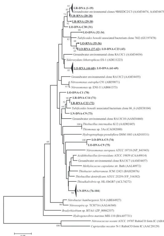
which confirmed the specificity of our approach because all sequences were found to be affiliated with the tar-geted genes. Phylogenetic trees based on deduced amino acid sequences of RubisCO form IA and IC sequences derived from this study and public databases are pre-sented in Figs 1 and 2.
The original groundwater sample revealed two phyloge-netically clearly separated clusters of RubisCO form IA sequences, which were numerically dominated by (DNA and RNA based) sequences closely related to environmen-tal RubisCO sequences obtained from anoxic and BTEX-contaminated groundwater aquifer sediments analyzed in a former study (groundwater environmental clones 9BSED C2 and C3; Alfreider et al., 2003). The closest RubisCO sequence (96% amino acid similarity) from cultivated bacteria is Sideroxydans lithotrophicus ES-1, a
microaerobic ferrous iron-oxidizing Betaproteobacterium that was isolated from groundwater and grows at circum-neutral pH (Emerson & Moyer, 1997; Druschel et al.,
2008). Two clones (LR-RNA-C22 and LR-DNA-C14) were related to a DNA sequence obtained from ground-water supplied with nitrate in the current study and to
LR-DNA (1-19)
Groundwater environmental clones 9BSEDC2/C3 (AAM34474, AAM34475) LR-RNA (20-28)
LR-RNA (29-30)
LO-DNA-C30 (31) LO-DNA (32-34)
Tubificoides benedii associated bacterium clone 76I2 (GU197478) LO-RNA (35-36)
LO-RNA (37-42); LO-DNA-C23 (43)
Groundwater environmental clone RA13C1 (AAM34454) Sideroxydans lithotrophicus ES-1 (ADE11223)
LO-RNA (44-60); LO-DNA (61-69)
Groundwater environmental clone RA13C2 (AAM34455) Nitrosomonas eutropha C91 (ABI59071)
Nitrosomonas sp. ENI-11 (AB061373) LO-DNA-C1 (70)
LR-DNA-C14 (71) LR-RNA-C22 (72)
Tubificoides benedii associated bacterium clone 86_6 (ADJ38104) LN-DNA-C9 (73)
Groundwater environmental clone RA13C10 (AAM34460) Thiobacillus intermedius K12 (AAD02445) Thiomonas sp. 3As (CAO82080)
Hydrogenophaga pseudoflava DSM 1083 (AAD10331) LO-DNA-C5 (74)
LO-DNA-C9 (75)
Nitrosomonas europaea ATCC 19718 (NP_841943) Acidithiobacillus ferrooxidans ATCC 19859 (CAA49814)
Groundwater environmental clone RA13C7 (AAM34457) Methylococcus capsulatus str. Bath (AAL40972) Thiobacter subterraneus JCM 12421 (BAH28876) Thiobacillus denitrificans ATCC 25259 (YP_316382)
Thioalkalivibrio sp. HL-EbGR7 (ACL74272) LN-DNA (76-104)
Nitrobacter hamburgensis X14 (ABE64927) Nitrosospira sp. TCH716 (AAL66360) Bradyrhizobium sp. BTAi1 (ZP_00862357)
Hydrogenovibrio marinus MH-110 (BAA07731)
Nitrosococcus oceani ATCC 19707 RubisCO form IC (ABA56859) Cupriavidus necator N-1 RubisCO form IC (AAC28129)
100 100 100 99 99 65 68 92 98 97 94 52 64 69 82 85 92 73 85 61 0.05
Fig. 1. Neighbour-joining tree calculated from deduced amino acid sequences of form IA RubisCO genes obtained from sampling stations LO, LR and LN and sequences retrieved from NCBI database. DNA-based sequences obtained from this study are indicated in bold; transcripts are indicated in bold and are underlined. Consecutive numbers in parentheses following the clone sequences refer to information provided in Supporting Information Table S1, including all clone designations and their corresponding accession numbers. Accession numbers of reference sequences are also given in parentheses. The bootstrap consensus tree is inferred from 1000 replicates. Bootstrap values below 50% are not shown.
RubisCO genes present in bacterial ectosymbionts of the shallow-water marine worm Tubificoides benedii (Rueh-land & Dubilier, 2010). Another Tubifex-associated clone sequence (clone 76I2), obtained from the same study, was the closest relative of several sequences (from DNA as well as RNA extracts) from oxygenated groundwater of sampling station LO (Fig. 1). In this sample, a major cluster of highly similar sequences obtained from DNA (nine sequences) and transcripts (14 sequences) was affili-ated with form IA RubisCO identified in S. lithotrophicus ES-1 (97% amino acid sequence similarity). Twenty-nine closely related sequences retrieved from DNA extracts of
sampling station LN formed a clearly separated cluster in the phylogenetic tree (Fig. 1). Based on amino acid sequence identity, the closest relatives are the obligately chemolithoautotrophic and facultatively anaerobic Thio-bacillus denitrificans (96% identity) and the haloalkaliphil-ic sulphur-oxidizing bacteria Thioalkalivibrio sp. HL-EbGR7 (95% similarity); the latter is also closely related to T. denitrificans. Other RubisCO form IA sequences were distantly related to cultivated representatives depos-ited in public databases. Therefore there is a lack for inferring ecophysiological characteristics from these sequences.
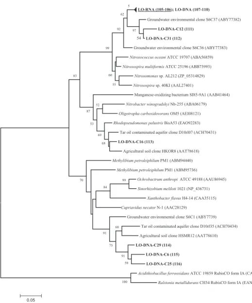
LO-RNA (105-106); LO-DNA (107-110)
Groundwater environmental clone S6C37 (ABY77382)
LO-DNA-C12 (111) LO-DNA-C31 (112)
Groundwater environmental clone S6C36 (ABY77383)
Nitrosococcus oceani ATCC 19707 (ABA56859) Nitrosospira multiformis ATCC 25196 (ABB73993)
Nitrosomonas sp. AL212 (ZP_05314829) Nitrosospira sp. 40KI (AAL27401)
Manganese-oxidizing bacterium SI85-9A1 (AAB41464)
Nitrobacter winogradskyi Nb-255 (ABA06179) Oligotropha carboxidovorans OM5 (AEI08121) Rhodopseudomonas palustris BisA53 (EAO92283)
Tar oil contaminated aquifer clone D10rl07 (ACH70431)
LO-DNA-C16 (113)
Agricultural soil clone HKOR8 (AAT78618)
Methylibium petroleiphilum PM1 (ABM94440) Methylibium petroleiphilum PM1 (ABM95736)
Ochrobactrum anthropi ATCC 49188 (AAU86945) Sinorhizobium meliloti 1021 (NP_436731)
Xanthobacter flavus H4-14 (CAA35115) Cupriavidus necator N-1 (AAC28129)
Groundwater environmental clone S6C1 (ABY7739) Tar oil contaminated aquifer clone D10rl35 (ACH70434) Agricultural soil clone HSMR12 (AAT78610)
LO-DNA-C29 (114) LO-DNA-C6 (115) LO-DNA-C25 (116)
Acidithiobacillus ferrooxidans ATCC 19859 RubisCO form IA (CAA49814) Ralstonia metallidurans CH34 RubisCO form IA (EAN48604)
100 99 59 91 68 75 84 91 70 68 69 52 53 87 83 54 97 55 60 99 92 62 5 0.05
Fig. 2. Neighbour-joining tree calculated from deduced amino acid sequences of form IC RubisCO genes obtained from sampling station LO and sequences retrieved from NCBI database. DNA-based sequences obtained from this study are indicated in bold; transcripts are indicated in bold and are underlined. Consecutive numbers in parentheses following the clone sequences refer to information provided in Table S1, including all clone designations and their corresponding accession numbers. Accession numbers of reference sequences are also given in parentheses. The bootstrap consensus tree was inferred from 1000 replicates. Bootstrap values below 50% are not shown.
Form IC RubisCO sequences were exclusively detected in groundwater supplied with oxygen (LO, Fig. 2). The majority of the sequences obtained from RNA- and DNA-extracts clustered with clone sequences (S6C36) obtained from a non-contaminated shallow aquifer inves-tigated in a former study (Alfreider et al., 2009). The addition of oxygen in an ammonium-rich environment is an ideal habitat for nitrifying bacteria. Sequence analysis of form IC transcripts revealed their affiliation to mem-bers of the Nitrosospira lineage and N. oceani (Fig. 2), indicating their potential role in the nitrification process at this sampling station. In a study of Uta˚ker et al. (2002) it was ascertained that the majority of ammonium-oxidi-zing bacteria possess form IC RubisCO; therefore, the absence of RubisCO form IA sequences affiliated with ammonium-oxidizing bacteria at sampling station LO is not peculiar. All other form IC RubisCO clone sequences obtained from sample LO originated from DNA extracts and showed the highest amino acid similarities with sequences obtained by cultivation-independent studies from agricultural soils and a tar oil-contaminated aquifer (Selesi et al., 2005; Kellermann, 2008).
Nigro & King (2007) suggested that the distribution of form IA- and IC-containing chemolithoautotrophic bacte-ria corresponds to functional distinctions of both forms and is associated with the relative distribution of the availability of sulphide. In fact, the ability to use sulphide as electron donor is known for a number of form IA-con-taining bacteria – a physiological feature never observed in RubisCO form IC chemoautotrophs. This concept was supported by the results of this study. Form IC RubisCO DNA and mRNA were exclusively detected in groundwa-ter samples supplied with oxygen (sampling station LO) but not in the original groundwater or the groundwater supplied with nitrate, which were characterized by very low sulphide concentrations.
Form II RubisCO
The form II RubisCO enzyme in Proteobacteria is mark-edly different from that of form I with regard to sequence similarity and kinetic properties. An essential biochemical characteristic of form II enzymes is the poor affinity for CO2 and a low discrimination against O2 (Tabita, 1999).
From the viewpoint of RubisCO ecology, it has been sug-gested that form II enzymes are adapted to low-O2 and
high-CO2 environments (Badger & Bek, 2008). Form II
RubisCOs are found in two gene arrangements, which are well correlated with the metabolic functioning of the organisms in which they occur (Badger & Bek, 2008). An interesting feature of form II RubisCO is that it is often found in organisms that also contain form I. Chemoauto-trophic bacteria that have acquired the genes encoding
both forms of RubisCO may have an advantage in ecosys-tems where O2and CO2concentrations vary considerably,
because the dissimilar kinetic properties of the enzymes would allow efficient CO2assimilation under both aerobic
and anaerobic conditions (Alfreider et al., 2003).
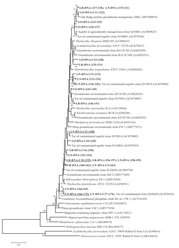
Form II RubisCO from RNA and DNA were success-fully amplified from samples LR and LN but not in oxy-gen-amended groundwater (sampling station LO), where cbbM genes detected in bacterial DNA were not expressed (Table 1). This distribution pattern corresponds well with the kinetic properties known for RubisCO form II (see above). Sequence analysis of clone libraries revealed that cbbM sequences were widely distributed in the phyloge-netic tree (Fig. 3). In the original groundwater sample LR, a single cbbM sequence (clone C10) was detected from RNA extracts, which was identical with RubisCO from DNA extracts obtained from sampling station LN. Form II RubisCO transcripts from LN were represented by two phylotypes: a single cbbM sequence (L6-RNA-C21) and a cluster of 10 almost identical sequences. Both phylotypes were closely related or identical to RubisCO clone sequences obtained from a tar oil-contaminated aquifer (Kellermann, 2008).
The affiliation of sequences obtained from groundwater samples to known cbbM sequences of cultured bacterial strains was wide ranging, including numerous obligate and facultative chemolithoautotrophs (Fig. 3). The closest relatives for cbbM transcripts obtained from sampling stations LN and LR include RubisCO cbbM analyzed for T. denitrificans, S. lithotrophicus ES-1 and Accumulibacter phosphatis clade IIA with amino acid sequence similarities ranging between 92% and 96%. In contrast to the distri-bution and diversity patterns observed with form IA Ru-bisCO sequences, cbbM sequences were often represented by identical or closely related form II sequences retrieved from all three sampling stations, LR, LO and LN. For example, one distinct cluster with DNA sequences obtained from all samples showed a high degree of sequence similarity; these sequences were affiliated to dif-ferent environmental clones obtained from polluted and pristine groundwater or soils (Fig. 3, sequences at the top of the tree).
Biogeochemical considerations
The original groundwater (LR) was characterized by vir-tually anoxic conditions with elevated concentrations of ammonium and ferrous iron (Table 1). Nitrate was below the detection limit; in contrast, sulphate was present in significant amounts. Bacteria using form IC RubisCO for CO2 fixation, which are often represented by facultative
autotrophs or mixotrophs, were not detected in the origi-nal sampling station. It has been suggested that facultative
autotrophic bacteria can be found in environments where inorganic and organic compounds are available (Badger & Bek, 2008). In sample LR, DOC consists mainly of
MTBE and tert-butyl alcohol (TBA), which is recalcitrant under the in situ conditions and therefore does not serve as an organic carbon source (Table 1).
LR-DNA (117-118); LN-DNA (119-121) LO-DNA-C11 (122)
Oak Ridge pristine groundwater metagenome (IMG: 2007500839) LO-DNA (123-125)
LO-DNA (126-127)
Aquifer in agriculturally managed area clone Sy5M02 (ACH69823) Tar oil contaminated aquifer clone D10M01 (ACH70386) Thiobacillus thioparus DSM 505 (ACF06647)
Acidithiobacillus ferrooxidans ATCC 23270 (ACK78027) Groundwater environmental clone RA13C2II (AAM26289)
Groundwater environmental clone RA13C10II (AAM26291) LO-DNA-C14 (128)
LR-DNA (129-131)
Halothiobacillus neapolitanus ATCC 23641 (AAD02442) LN-DNA-C31 (132)
LN-DNA (133-135)
LN-DNA (136-142); Tar oil contaminated aquifer clone D15M18 (ACH70404) LO-DNA (143-145)
Groundwater environmental clone ZZ15C8II (AAM26293) Tar oil contaminated aquifer clone D15M16 (ACH70403)
LR-DNA (146-147)
Thiobacillus intermedius K12 (AAC24964) Dechloromonas aromatica RCB (AAZ48366)
Groundwater environmental clone ZZ15C5II (AAM26292) Rhodoferax ferrireducens DSM 15236 (EAO41516) Deep groundwater environmental clone P3C1 (ABY77473) LN-RNA-C21 (148)
Tar oil contaminated aquifer clone D15M14 (ACH70402) LO-DNA-C10 (149)
Tar oil contaminated aquifer clone D12M02 (ACH70393) LR-DNA-C16 (150)
LN-DNA (151-154)
LR-RNA-C10 (155); LR-DNA (156-157); LN-DNA (158-159)
LR-DNA (160-162); LN–DNA-C5 (163)
Tar oil contaminated aquifer clone D12M30 (ACH69798) Groundwater environmental clone S6C2 (ABY77449) Sideroxydans lithotrophicus ES-1 (ADE10264)
Thiobacillus denitrificans ATCC 25259 (AAZ98591) LN-DNA (164-165)
LN-RNA (166-175); LN-DNA-C27 (176); Tar oil contaminated clone D24M20 (ACH70416) Candidatus Accumulibacter phosphatis clade IIA str. UW-1 (ACV34169)
Polaromonas naphthalenivorans CJ2 (ZP_01020427) Deep groundwater clone V4C1 (ABY77454)
Magnetite-containing magnetic vibrio MV-1 (AAL76921) Magnetospirillum magneticum AMB-1 (YP_422059) Rhodobacter sphaeroides 2.4.1 (ABA80879)
Hydrogenovibrio marinus MH-110 (BAA05677)
Acidithiobacillus ferrooxidans ATCC 19859 RubisCO form I (CAA49814) Nitrosococcus oceani ATCC 19707 RubisCO form I (ABA56859)
99 51 69 99 92 89 61 73 91 88 97 98 62 92 86 98 94 69 97 50 71 92 83 52 59 56 62 83 50 81 0.1
Fig. 3. Neighbour-joining tree calculated from deduced amino acid sequences of form II RubisCO genes obtained from sampling stations LO, LR and LN and sequences retrieved from IMG and NCBI database. DNA-based sequences obtained from this study are indicated in bold, transcripts are indicated in bold and are underlined Consecutive numbers in parentheses following the clone sequences refer to information provided in Table S1, including all clone designations and their corresponding accession numbers. Accession numbers of reference sequences are also given in parentheses. The bootstrap consensus tree was inferred from 1000 replicates. Bootstrap values below 50% are not shown.
Sample LO, which was obtained from the conditioning unit supplied with oxygen, showed in comparison with the reference site a decline in ammonium concentration and the occurrence of nitrate (Table 1), which can be explained by nitrification of ammonium to nitrate. Rubi-sCO transcripts affiliated with ammonium-oxidizing bac-teria confirm their active role in the nitrogen cycle at this sampling station. Recent studies suggest that aerobic ammonium oxidation by autotrophic Archaea is of major significance in marine and soil ecosystems (e.g. Zhang et al., 2010; Pratscher et al., 2011; Yakimov et al., 2011). CO2 fixation of autotrophic thaumarchaeal ammonium
oxidizers is accomplished via the 3-hydroxypropionate/ 4-hydroxybutyrate cycle, which was not investigated in this study. Consequently, future studies should also include the analysis of genes coding for key enzymes in this pathway in order to assess whether inorganic carbon fixation by Archaea is also associated with ammonium oxidation in groundwater systems. The oxidation of ferrous iron is another source for the consumption of oxygen. The analysis of RubisCO genes and transcripts revealed sequences affiliated with the iron-oxidizing Beta-proteobacteria S. lithotrophicus. The concentration of DOC (in the form of MTBE/TBA) was not affected by the sup-ply with oxygen (Table 1), indicating that MTBE was not degraded under aerobic conditions.
The addition of nitrate and active coal at sampling sta-tion LN caused a significant reducsta-tion of ammonium and MTBE in the effluent (Table 1). Furthermore, nitrate was completely consumed, suggesting the importance of den-trification and/or anammox (anaerobic ammonium oxi-dation) activities. The ecological role of anammox bacteria was not covered by our investigations because autotrophy in these microorganisms is based on the reductive acetyl-CoA pathway for carbon fixation (Scho-uten et al., 2004; Strous et al., 2006). RubisCO form I obtained from DNA was closely related to several denitri-fying bacteria including T. denitrificans, which is capable of oxidizing inorganic sulphur compounds or ferrous iron using nitrate as electron acceptor (Beller et al., 2006). Thiobacillus denitrificans is also able to use sul-phur/iron minerals, for example pyrite, as electron donors, which is an important physiological trait for adaptation to groundwater systems. Some sulphate might be reduced to sulphide, as the sulphate concentrations slightly decreased (Table 1). However, on the one hand, the question is whether indeed sufficient amounts of reduced sulphur compounds or iron-sulphur minerals were available for reducing the added nitrate. On the other hand, the inflowing groundwater sample (LR) con-tained ferrous iron in significant amounts (Table 1). Thus, the denitrification process might be driven partly by autotrophic ferrous iron-oxidizing phylotypes related
to T. denitrificans using nitrate as electron acceptor. The presence of cbbM transcripts affiliated with RubisCO genes hosted in dentrifiers, including A. phosphatis and T. denitrificans, was detected at sampling station LN (Fig. 3). Accumulibacter phosphatis is well known to be primarily responsible for biological phosphorus removal in waste water and sludge, suggesting the preference for a heterotrophic lifestyle in an environment with high amounts of readily available organic carbon. In a metage-nomic study, the detection of key genes of the Calvin cycle including phosphoribulokinase and RubisCO is evi-dence of the ability of A. phosphatis to fix CO2 (Garcia
Martin et al., 2006). These findings indicate that Accumu-libacter clades are also adapted to carbon limited habitats which was verified in a recent study by Peterson et al. (2008), which included the investigation of lakes, rivers and springs. Although the denitrification capabilities in different clades of A. phosphatis strains remains to be clarified (Zeng et al., 2003; Flowers et al., 2009), the high similarity with A. phosphatis sequences of the majority of cbbM transcripts obtained from sample LN suggests the presence of autotrophic bacteria that are actively involved in dissimilatory nitrate reduction. In this context it should be noted that the groundwater at sampling sta-tions LO and LN was supplied with the same amount of phosphate (27 g K2HPO4m 3), but the actual phosphate
concentration was lower in sampling station LN. Phos-phate is probably metabolized by bacterial populations affiliated with Accumulibacter spp., which are well known to accumulate inorganic phosphate efficiently (Hessel-mann et al., 1999; Flowers et al., 2009).
Besides ferrous iron, MTBE and the related DOC might be other important electron donors for nitrate reduction at sampling station LN, as MTBE and DOC were significantly reduced during the passage through the conditioning unit LN (Table 1). It cannot be excluded, however, that MTBE was almost completely adsorbed by the active coal used as a filling material of this channel. Indeed, MTBE oxidation with nitrate or ferric iron (which probably accumulated in the channel due to the constant oxidation of ferrous iron with nitrate) as elec-tron acceptors is rarely observed in the environment (Bradley et al., 2001; Somsanak et al., 2001). Thus, the results suggest the existence of two main biological sinks for nitrate in the form of anoxic, nitrate-dependent microbial oxidation of ferrous iron and ammonium. Nitrite formed during the ferrous iron-dependent nitrate reduction might be channelled in the anammox process (Kuenen, 2008).
Although the role of facultative autotrophic prokary-otes for the degradation of MTBE was not particularly investigated in this study, their potential importance at the sampling station should be noted. For example the
methylotrophic bacterium Methylibium petroleiphilum PM1 is known to play a key role for aerobic MTBE deg-radation in contaminated aquifers (Wilson et al., 2002; Smith et al., 2005). Methylotrophic autotrophy as an alternative type of nutrition based on RubisCO pathway was demonstrated for the methylotrophic bacterium Bei-jerinckia mobilis (Dedysh et al., 2005). Although an auto-trophic metabolism for strain M. petroleiphilum PM1 has not yet been confirmed, a whole genome analysis study of strain PM1 revealed two sets of genes coding for form I RubisCO and associated enzymes necessary for CO2
assimilation via the Calvin cycle (Kane et al., 2007). Whereas the activity of PM1 at the Leuna site has not been proven yet, the closely related (95.6% 16S rRNA gene sequence similarity) MTBE-degrading bacterium Aquincola tertiaricarbonis str L108 has been isolated from Leuna groundwater (Rohwerder et al., 2006; Lechner et al., 2007) and its activity in aerated Leuna groundwater trenches was recently demonstrated (Jechalke et al., 2011). The genome of strain L108 has been partly sequenced and genes coding for the small and large sub-unit of RubisCO were identified (T. Rohwerder, pers. commun.). Preliminary sequence analysis showed that the cbbL gene of strain L108 is affiliated with RubisCO large subunit genes in M. petroleiphilum PM1 (93 and 83% amino acid identity; data not shown). An explanation as to why MTBE was not degraded at the sampling station supplied with oxygen (LO) is provided by the presence of a metabolically active nitrifying microbial community. The competitive effect of ammonium oxidizers, which are characterized by a higher growth rate than MTBE degrad-ers, was shown in a model that was developed for an experimental packed bed reactor (Waul et al., 2008).
Acknowledgements
We thank Joerg Ahlheim and Ralf Trabitzsch (both UFZ) for the logistic support and technical assistance in the field. This study was funded by the Austrian Science Fund to A.A. (Project # FWF P17649).
References
Adhikari RR & Kallmeyer J (2010) Detection and
quantification of microbial activity in the subsurface. Chem Erde-Geochem70: 135–143.
Alfreider A, Vogt C, Hoffmann D & Babel W (2003) Diversity of ribulose-1,5-bisphosphate carboxylase/oxygenase large-subunit genes from groundwater and aquifer
microorganisms. Microb Ecol45: 317–328. Alfreider A, Vogt C, Kaiser M & Psenner R (2009)
Distribution and diversity of autotrophic bacteria in
groundwater systems based on the analysis of RuBisCO genotypes. Syst Appl Microbiol32: 140–150.
Altschul SF, Gish W, Miller W, Myers EW & Lipman DJ (1990) Basic local alignment search tool. J Mol Biol215: 403–410.
Anderson RT, Chapelle FH & Lovley DR (1998) Evidence against hydrogen-based microbial ecosystems in basalt aquifers. Science281: 976–977.
Badger MR & Bek EJ (2008) Multiple Rubisco forms in proteobacteria: their functional significance in relation to CO2acquisition by the CBB cycle. J Exp Bot59: 1525–1541. Baker MA, Valett HM & Dahm CN (2000) Organic carbon
supply and metabolism in a shallow groundwater ecosystem. Ecology81: 3133–3148.
Beller HR, Chain PSG, Letain TE, Chakicherla A, Larimer FW, Richardson PM, Coleman M, Wood AP & Kelly DP (2006) The genome sequence of the obligately
chemolithoautotrophic, facultatively anaerobic bacterium Thiobacillus denitrificans. J Bacteriol188: 1473–1488. Bradley PM, Chapelle FH & Landmeyer JE (2001) Methyl
t-butyl ether mineralization in surface-water sediment microcosms under denitrifying conditions. Appl Environ Microbiol67: 1975–1978.
Dedysh SN, Smirnova KV, Chmelenina VN, Suzina NE, Liesack W & Trotsenko YA (2005) Methylotrophic autotrophy in Beijerinckia mobilis. J Bacteriol187: 3884– 3888.
Druschel GK, Emerson D, Sutka R & Luther GW (2008) Low oxygen and chemical kinetic constraints on the geochemical niche of neutrophilic iron(II) oxidizing microorganisms. Geochim Cosmochim Acta72: 3358–3370.
Emerson D & Moyer C (1997) Isolation and characterization of novel iron-oxidizing bacteria that grow at circumneutral pH. Appl Environ Microbiol63: 4784–4792.
Engel AS (2007) On the biodiversity of sulfidic karst habitats. J Cave Karst Stud69: 187–206.
Flowers JJ, He S, Yilmaz S, Noguera DR & McMahon KD (2009) Denitrification capabilities of two biological phosphorus removal sludges dominated by different ‘Candidatus Accumulibacter’ clades. Environ Microbiol Rep1: 583–588.
Foulquier A, Simon L, Gilbert F, Fourel F, Malard F & Mermillod-Blondin F (2010) Relative influences of DOC flux and subterranean fauna on microbial abundance and activity in aquifer sediments: new insights from13C-tracer experiments. Freshw Biol55: 1560–1576.
Garcia Martin H, Ivanova N, Kunin V et al. (2006) Metagenomic analysis of two enhanced biological phosphorus removal (EBPR) sludge communities. Nat Biotechnol24: 1263–1269.
Hesselmann RP, Werlen C, Hahn D, van der Meer JR & Zehnder AJ (1999) Enrichment, phylogenetic analysis and detection of a bacterium that performs enhanced biological phosphate removal in activated sludge. Syst Appl Microbiol 22: 454–465.
Horken KM & Tabita FR (1999) Closely related form I ribulose bisphosphate carboxylase/oxygenase molecules that possess different CO2/O2substrate specificities. Arch Biochem Biophys361: 183–194.
Jechalke S, Rosell M, Martı´nez-Lavanchy PM, Pe´rez-Leiva P, Rohwerder T, Vogt C & Richnow HH (2011) Linking low stable isotope fractionation to expression of the cytochrome P450 monooxygenase encoding ethB gene for proving MTBE biodegradation in aerated treatment pond systems. Appl Environ Microbiol77: 1086–1096.
Kane SR, Chakicherla AY, Chain PSG et al. (2007) Whole-genome analysis of the methyl tert-butyl ether-degrading beta-proteobacterium Methylibium petroleiphilum PM1. J Bacteriol189: 1931–1945.
Kellermann C (2008) Autotrophy in Groundwater Ecosystems. PhD Thesis, Ludwig-Maximilians-Universita¨t Mu¨nchen, Munich.
Kuenen JG (2008) Anammox bacteria: from discovery to application. Nat Rev Microbiol6: 320–326.
Lawrence JR, Hendry MJ, Wassenaar LI, Wolfaardt GM, Germida JJ & Greer CW (2000) Distribution and biogeochemical importance of bacterial populations in a thick clay-rich aquitard system. Microb Ecol40: 273–291. Lechner U, Brodkorb D, Geyer R, Hause G, Ha¨rtig C, Auling
G, Fayolle-Guichard F, Piveteau P, Mu¨ller RH & Rohwerder T (2007) Aquincola tertiaricarbonis gen. nov., sp. nov., a tertiary butyl moiety-degrading bacterium. Int J Syst Evol Microbiol57: 1295–1303.
Markowitz VM, Ivanova N, Szeto E et al. (2008) IMG/M: a data management and analysis system for metagenomes. Nucleic Acids Res36: D534–D538.
Martienssen M, Fabritius H, Kukla S, Balcke GU, Hasselwander E & Schirmer M (2006) Determination of naturally occurring MTBE biodegradation by analysing metabolites and biodegradation by-products. J Contam Hydrol87: 37–53.
Mueller-Cajar O & Badger MR (2007) New roads lead to Rubisco in archaebacteria. Bioessays29: 722–724. Nealson KH, Inagaki F & Takai K (2005) Hydrogen-driven
subsurface lithoautotrophic microbial ecosystems (SLiMEs): do they exist and why should we care? Trends Microbiol13: 405–410.
Nedelkova M (2005) Microbial diversity in ground water at the deep-well monitoring site S15 of the radioactive waste depository Tomsk-7, Siberia, Russia. PhD Thesis, TU Bergakademie Freiberg, Freiberg, Germany.
Nigro LM & King GM (2007) Disparate distributions of chemolithotrophs containing form IA or IC large subunit genes for ribulose-1,5-bisphosphate carboxylase/oxygenase in intertidal marine and littoral lake sediments. FEMS
Microbiol Ecol60: 113–125.
Peterson SB, Warnecke F, Madejska J, McMahon KD & Hugenholtz P (2008) Environmental distribution and population biology of the genus Accumulibacter, a primary agent of biological phosphorus removal in activated sludge. Environ Microbiol10: 2692–2703.
Pratscher J, Dumont MG & Conrad R (2011) Ammonia oxidation coupled to CO2fixation by archaea and bacteria in an agricultural soil. P Natl Acad Sci USA108: 4170–4175. Rohwerder T, Breuer U, Benndorf D, Lechner U & Mu¨ller RH
(2006) The alkyl tert-butyl ether intermediate 2-hydroxyisobutyrate is degraded via a novel cobalamin-dependent mutase pathway. Appl Environ Microbiol72: 4128–4135.
Ruehland C & Dubilier N (2010) Gamma- and
epsilonproteobacterial ectosymbionts of a shallow-water marine worm are related to deep-sea hydrothermal vent ectosymbiont. Environ Microbiol12: 2312–2326.
Schouten S, Strous M, Kuypers MMM, Rijpstra WIC, Baas M, Schubert CJ, Jetten MSM & Sinninghe Damste´ JS (2004) Stable carbon isotopic fractionations associated with inorganic carbon fixation by anaerobic ammonium oxidizing bacteria. Appl Environ Microbiol40: 3785–3788. Selesi D, Schmid M & Hartmann A (2005) Diversity of
green-like and red-green-like ribulose-1,5-bisphosphate carboxylase/ oxygenase large-subunit genes (cbbL) in differently managed agricultural soils. Appl Environ Microbiol77: 175–184. Smith AE, Hristova K, Wood I, Mackay DM, Lory E,
Lorenzana D & Scow KM (2005) Comparison of
biostimulation versus bioaugmentation with bacterial strain PM1 for treatment of groundwater contaminated with methyl tertiary butyl ether (MTBE). Environ Health Perspect 113: 317–332.
Somsanak P, Cowan RM & Ha¨ggblom MM (2001) Anaerobic biotransformation of fuel oxygenates under sulfate-reducing conditions. FEMS Microbiol Ecol37: 259–264.
Stevens T & McKinley J (1996) Hydrogen-based microbial ecosystems in the Earth - Reply. Science272: 896–897. Strous M, Pelletier E, Mangenot S et al. (2006) Deciphering
the evolution and metabolism of an anammox bacterium from a community genome. Nature440: 790–794.
Tabita FR (1999) Microbial ribulose bisphosphate carboxylase/ oxygenase: a different perspective. Photosynth Res60: 1–28. Tabita FR, Hanson TE, Li H, Satagopan S, Singh J & Chan S
(2007) Function, structure, and evolution of the RubisCO-like proteins and their RubisCO homologs. Microbiol Mol Biol Rev71: 576–599.
Tabita FR, Hanson TE, Satagopan S, Witte BH & Kreel NE (2008) Phylogenetic and evolutionary relationships of RubisCO and the RubisCO-like proteins and the functional lessons provided by diverse molecular forms. Philos Trans R Soc Lond B Biol Sci363: 2629–2640.
Tamura K, Dudley J, Nei M & Kumar S (2007) MEGA4: Molecular Evolutionary Genetics Analysis (MEGA) Software Version 4.0. Mol Biol Evol24: 1596–1599.
Uta˚ker JB, Andersen K, Aakra A˚, Moen B & Nes IF (2002) Phylogeny and functional expression of ribulose 1,5-bisphosphate carboxylase/oxygenase from the autotrophic ammonia-oxidizing bacterium Nitrosospira sp. isolate 40KI. J Bacteriol184: 468–478.
Waldron PJ, Wu L, Van Nostrand JD, Schadt CW, He Z, Watson DB, Jardine PM, Palumbo AV, Hazen TC & Zhou J
(2009) Functional gene array-based analysis of microbial community structure in groundwaters with a gradient of contaminant levels. Environ Sci Technol43: 3529–3534. Waul C, Arvin E & Schmidt JE (2008) Modeling the
competitive effect of ammonium oxidizers and heterotrophs on the degradation of MTBE in a packed bed reactor. Water Res42: 3098–3108.
Wilson RD, MacKay DM & Scow KM (2002) In situ MTBE biodegradation supported by diffusive oxygen release. Environ Sci Technol36: 190–199.
Yakimov MM, La Cono V, Smedile F et al. (2011) Contribution of crenarchaeal autotrophic ammonia oxidizers to the dark primary production in Tyrrhenian deep waters (Central Mediterranean Sea). ISME J5: 945– 961.
Zeng RJ, Lemaire R, Yuan Z & Keller J (2003) Simultaneous nitrification, denitrification, and phosphorus removal in a lab-scale sequencing batch reactor. Biotechnol Bioeng84: 170–178.
Zhang L, Offre PO, He , J-Z , Verhamme DT, Nicol GW & Prosser JI (2010) Autotrophic ammonia oxidation by soil thaumarchaea. P Natl Acad Sci USA107: 17240–17245.
Supporting Information
Additional Supporting Information may be found in the online version of this article:
Table S1. List of RubisCO sequences retrieved from groundwater samples of the study site and their corre-sponding accession numbers.
Please note: Wiley-Blackwell is not responsible for the content or functionality of any supporting materials sup-plied by the authors. Any queries (other than missing material) should be directed to the corresponding author for the article.