DETERMINATION OF THE PARTIAL PRESSURE OF
HALOTHANE (OR ISOFLURANE) IN BLOOD
A. M. ZBINDEN, F. J. FREI, B. FUNK, D. A. THOMSON
AND D. WESTENSKOW
Techniques for the chromatographic determina-tion of the concentradetermina-tion of volatile anaesthetics such as halothane, enflurane and isoflurane in body fluids or tissues can be classified according to the detector and the separation technique (direct injection, extraction and head space) used. The Electron Capture Detector (ECD) is sensitive to halogenated agents (Douglas, Hill and Wood, 1970; Atallah and Geddes, 1972; Davies; 1978), but is unstable and linear over a limited range (Douglas, Hill and Wood, 1970). The Thermal Conductivity Detector (TCD) has proved to be more stable and more sensitive for enflurane (Miller and Gandolfi, 1979). The Flame Ionization Detector (FID) is sensitive and linear in the clinically relevant concentration ranges, and has been used in the study presented in this communication.
The technique for direct injection of test blood to the column is very simple (Parker et al., 1962; Lowe, 19645 Douglas, Hill and Wood, 1970; Cousins and Mazze, 1972; Lauven, Hack and Stoeckel, 1979). However, contamination of the injection port, column and detector by blood components, alterations with time of the baseline characteristics, slow extraction of the blood by the carrier gas and interference by water vapour (Hill and Newell, 1965) may present serious disadvant-ages. Attempts to prevent the effects of water vapour have been made by the application of water absorbants (Jacobs, 1964; Laasberg and Etsten, 1965; Cousins and Mazze, 1972), use of a molecular sieve packed column (Borgstedt and Gillies, 1965; Yokota et al., 1967) and by preliminary loading of the column with water (Douglas, Hill and Wood, 1970).
A . M . ZBINDEN, M.D.; F. J. F R H , M. D.; B. FUNK; D . A. THOMSON, M.D., PH.D. ; Department of Anaesthesia, University of Basel/Kantonsspital, CH^HBl Basel, Switzerland. D . WESTENSKOW, PH.D., Department of Anaesthesia, University of Utah, Salt Lake City, Utah, U.S.A.
SUMMARY
A gas chromatographic method is described for the direct quantitative determination of the partial pressure of halothane {or isoflurane) in blood as well as the blood-gas partition coefficient. A head space technique and a flame ionization detector were used. Standard blood was obtained by equilibrating patients' blood with known gas concentrations in a tonometer. Using an infra-red analyser to measure the halothane gas concen-tration in the tonometer and within the anaes-thetic system allowed for the direct comparison of the partial pressure in blood to the partial pressure in the inspired gas. Technical problems associated with this procedure, and with comparable methods, are discussed.
With the extraction technique, small amounts (0.5—2 ml) of whole blood are equilibrated with an organic solvent such as carbon tetrachloride or n-heptane which extracts at least 95% of the volatile anaesthetic from the blood (Butler and Freeman, 1962; Jacobs, 1964; Allott, Steward and Mapleson, 1971; Atallah and Geddes, 1972; Davies, 1978). This technique is more complicated: sometimes the solvent peak overlaps the halothane peak, and it is necessary to inject a large volume of solvent, which can lead to swamping of the sensitive detector.
With the head space technique, the test blood containing the volatile anaesthetic is equilibrated at a certain temperature with a gas phase (Yamamura et al., 1966; Butler, Kelly and Zapp, 1967; Fink and Morikawa, 1970; Cowles, Borgstedt and Gillies, 1971). The concentration of the volatile anaesthetic in the blood is then derived from the measured concentration in the head space.
This paper presents a critical analysis of each step of a new method which allowed direct
DETERMINATION OF HALOTHANE IN BLOOD
797 determination of partial pressure. A head spacetechnique was used and standards of halothane in blood were obtained using a tonometer.
MATERIALS AND METHODS
A small sample of blood with a known partial pressure of halothane was equilibrated at a constant temperature with a larger volume of gas. After complete equilibration, samples of the gas phase were injected to a gas chromatograph (GC); a standard curve was thereby established, relating partial pressure to GC counts. The test blood was treated similarly; the obtained GC counts were read off the standard curve to obtain partial pressures.
Gas chromatography
An HP 5880A gas chromatograph, equipped with a flame ionization detector (FID), a curve integrator and a recorder were used. The glass column, 305 cm long with a 2-mm internal diameter was packed with 5 % OV 101 on 100/120 mesh Chromosorb W (Supelco, Gland, Switzer-land). The temperaure was 125 °C in the injection port, 120 °C in the FID and 100 °C in the column. The flows were nitrogen 14 ml min"1 (carrier gas),
hydrogen 26 ml min"1 and air 415 ml min"1.
(The area under the obtained curve was expressed as counts which were compared with the counts of a standard curve.)
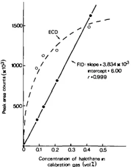
The standard curve which relates counts to partial pressure was obtained by injecting to the GC, 30 ulitre of four different calibration gases, using a gas-tight Hamilton syringe. To obtain the calibration gases, four glass bottles (550 ml) with a turnable stopcock with a Teflon septum were used. The septum was pierced with the syringe needle and 2, 3,5 and 10 pditre of liquid halothane was injected after flushing the bottles with oxygen for approximately 6 min. The calculated volume percents of halothane were plotted against the measured GC counts (fig. 1). (Partial pressure can be obtained by calculating volume percent/100 x barometric pressure.) The range of the partial pressures of the volatile anaesthetic was similar to that in the head space of the blood samples.
Tonometry and preparation of the standard blood curve
In order to link the measured gas concentration in the head space to partial pressure in the blood,
it was necessary to obtain standard blood of known partial pressure. An IL237 tonometer (Instrumen-tation Laboratory, Lexington, Ma., U.S.A.) was used to equilibrate blood with various concentra-tions of volatile anaesthetic gases. A clinical range of concentrations of the volatile anaesthetic obtained from a vaporizer was added to the calibration gas (5% carbon dioxide in oxygen). The gas mixture was warmed to 37 °C and humidified in the tonometer. The volatile anaesthetic concentration was verified at the tonometer outlet with a Beckman infra-red analyser. In the equilibration chamber of the tonometer, up to 5 ml of the patient's heparinized arterial blood can be equilibrated at 37 °C for 10 min and at a gas flow of 500 ml min"1.
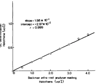
Equilibrated blood 0.5 ml was transferred with a 1-ml tuberculin syringe from the tonometer to a 5-ml glass vial, which was then sealed with a Teflon-neoprene septum (Supelco) held in place with a screw-cap. These vials were equilibrated by swirling in a 37 °C waterbath for 30 min. After piercing the septum of the vial, a 100-pJitre gastight syringe (Hamilton) was flushed with head space gas by moving the plunger rapidly up and down at least 10 times. Thirty microlitre of gas was then injected to the GC. This resulted in a standard blood curve which related counts in the head space (volume percent) to the partial pressure of the volatile anaesthetic in the gas phase during tonometry (fig. 2). Partial pressure in the arterial test blood can be obtained from the standard curve, if the sample is treated in the same manner as the standard blood sample.
Infra-red analysis
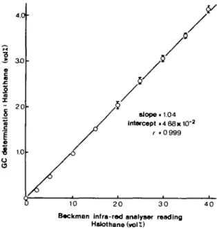
To link the partial pressure in the inspired/ex-pired gas to partial pressure in the blood, the same analyser can be used at the patient's mouthpiece and at the outlet of the tonometer. In this study, a Beckman LB-2 infra-red analyser was used. Its accuracy was verified by comparing its digital reading with the gas chromatographic measure-ment when gas mixtures from a vaporizer with halothane 0.2-4 vol% were used (fig. 3). These were the same partial pressures which were used to derive the standard blood curve using the tonometer.
Calculation of the blood/gas partition coefficient
The blood/gas partition coefficient (A) can be calculated if the concentration of the volatile anaesthetic in the head space of the equilibrated
sample is determined using the calibration curve presented in figure 1, which relates counts to concentration:
The quantity of volatile anaesthetic brought into the vial equals
CB T x VB
This amount equals the amount after equilibration in the waterbath:
CB T x VB = CB E x VB + CQE x Va
The blood/gas partition coefficient A is denned as:
A = CBT/CQ^ or A = CB E/ CG E
therefore
CB T = A x CG T and CB E = A x CG E
Inserting this to the above equation, we obtain: A x CG T x VB = A x CG E x VB + CG E x VG
Solving for A results in:
A =
in which equation all the variables can be measured directly.
(CG T = concentration of volatile anaesthetic in gas
phase in the tonometer; CB T = concentration of
volatile anaesthetic in blood after equilibration with the gas in the tonometer; CB E =
concen-tration in blood in vial after equilibration in the waterbath; CG E = concentration in the head space
after equilibration in the waterbath; VB = volume
of blood in the vial; Vo = volume of gas in the
vial.)
RESULTS
Gas chromatography
Figure 1 relates GC counts to concentrations of halothane in the calibration gas. With an ECD, a non-linear curve with a correlation coefficient r = 0.8152 was achieved. However, since the ECD was too sensitive for our needs, we used an FID (which is 1000 times less sensitive). This had a slope of 3.834 10s, an intercept of 6.00 and a
correlation coefficient of 0.999. The curve was linear over a range of 0.084-0.424 vol % which are the head space concentrations at clinically relevant concentrations, and the counts in this range were from 329 to 1629. Each point consisted of the mean of 20 measurements made from individually prepared calibration gases of halothane on three
1500 1000 5 0 0 ECD \ . /o
<v
/
°/ 1
/ /
/
.
J Q1 0.2 y s / / Q 3 / - ' / / FID' slope.3.834K103 ntercept • 6.00 r-0.999 0.4 03 Concentration of halothane n calibration gas (volZ)FIG. 1. Area of the chromatogram curve expressed as counts plotted against volume percent of halothane in calibration gases. The curve of the ECD is non-linear, its coefficient of variation is 8.6 %; each point consists of seven measurements. The curve of the FID is linear; each point is the mean of 20 measurements made on three different days. The coefficient of variation for halothane is 2.16% for all four concentrations. Partial pressure can be calculated using volume percent and
barometric pressure.
different days. The curve for isoflurane is not shown, but was equally linear in a range of 0.072-0.91 vol %. It had a slope of 2.568-103, an
intercept of 17.00 and a correlation coefficient of 0.999. The counts ranged from 204 to 2349.
The retention time (zero to peak) for halothane was 1.32 min and for isoflurane 1.13 min. The influence of water vapour on GC-analysis was tested by injecting 30 ulitre of the head space to the GC after equilibrating 0.5 ml of water instead of blood at 37 °C. Whether we injected the (humid) head space or room air to the GC, the counts were zero.
Tonometry and the standard blood curve
The tonometer proved to be adequate for equilibrating the gas phase with the blood phase, as was shown in various tests. The adequate equilibration time for 5 ml of blood was found to be 10 min, as shown in two sets of samples which had been equilibrated for up to 15 min at 0.6 vol % of halothane. Adequacy of equilibration was verified for temperature changes, water uptake
DETERMINATION OF HALOTHANE IN BLOOD §1 0.5 stope • 1.98 x 10'1 tntercspt • - 2 . 9 7 K 10"2 r .0.999 1.0 2.0 3.0 4.0
Beckman mfra-red analyser reading Hakrthane ( v o l Z )
FIG. 2. Blood samples are tonometered with various gas concentrations of halothane. The x-axis shows the Beckman infra-red analyser reading at the outlet of tonometer. The concentration in the head space of blood samples determined with gas chromatography is on the .y-axis. Each point is given as the mean of four measurements. The coefficient of variation is 4.77 %. If counts, instead of concentration, are plotted on the >-axis then the curve can be used directly as a standard curve relating counts of the head space to partial pressure in the
blood.
from the (humidified) calibration gas and desicca-tionofthebloodsample. The temperature remained stable at 37 °C during tonometry. There was no water uptake by the blood sample as judged by. haematocrit and the weight of the sample before and after tonometry (eight samples). The true-gas concentration, with which the blood sample had been equilibrated, was considered to be equal to the concentration measured at the outlet of the tonometer; the outlet concentration, as measured with the Beckman infra-red analyser, was 16% less than the inlet concentration over a range of 0.5-1.5 vol % halothane, during an infinite time and without the presence of water in the tonometer. This measurement was performed while maintaining back pressure to the vaporizer, to rule out back pressure effects on the vaporizer output, and was obtained by using a peep-valve before the tonometer, and concentrations were measured after that valve. Figure 2 relates concentration of halothane at the outlet of the tonometer as determined with the infra-red analyser to the concentration in the head space of equilibrated blood samples as determined by gas chromatography.
The gas concentrations in the head space were
much lower than at the tonometer outlet, as part of the halothane remained dissolved in the blood phase. The curve had a slope of 1.98 10"1, an
intercept of — 2.97 10~* and a correlation co-efficient of 0.999. It was linear in the tested range. Each point consisted of four measurements. If counts are plotted on the .y-axis instead of con-centration, then the curve can be used directly as a standard curve relating counts of the head space to partial pressure in the blood.
Tuberculin syringes and vials were checked for constancy of volume and for halothane loss by absorption and leakage. The volume of 0.5 ml of blood, as measured with 10 tuberculin syringes, was 0.503 ml (SD 0.00675) with a coefficient of variation (CV) 1.34 %. During 5 min there was no loss of halothane from syringes containing 0.5 ml of blood which had been tonometered with 1 vol % halothane and during 32 h there was no loss of halothane from vials containing 0.5 ml of blood which had been tonometered with 1 vol % halothane when the Teflon side of the septum had been turned towards the blood sample; the septum, which consists of neoprene, itself absorbs volatile anaesthetics. The volume of the content of six vials was 5.0012 ml (SD 0.0329), CV 0.66%.
Measurements of GC-counts in the head space of blood which had been tonometered with 0.5 vol % of halothane and which had been equilibrated in the water bath at 37 °C during varying periods of time showed no increase in counts after 20 min. Therefore, an equilibration time of 30 min was considered safe.
Infra-red analysis
Figure 3 relates concentration of halothane at the outlet of the tonometer, controlled and adjusted by the Beckman infra-red analyser (%-axis), to concentration of halothane as calculated with the counts of GC analysis (y-axis). The curve has a slope of 1.04, an intercept of — 4.68 • 10~* and a correlation coefficient of 0.999. The concentration measurements with the Beckman analyser corres-pond well to the measurements of GC. Each point consists of four measurements.
Calculation of the blood/gas partition coefficient
Data of blood/gas partition coefficients were determined in six dogs for halothane and in a further six dogs for isoflurane. Comparing uptake and distribution of the two anaesthetics, A amounted to 2.53±0.3 for halothane and to 1.30 ±0.10 for isoflurane.
4.0 •3 ~s ao
I
i
a X1
2 01
to u sJopo. 1.04 intercept 14 68K10'2 f - 0 9 9 9 10 2 0 3 0 4 0B«ckman infra-rod analyser reading HalothanetvolZ)
FIG. 3. Relation between the Beckman infra-red analyser readings of calibration gases and the determination of gas chromatography. Each point is given as the mean of four measurements and the standard deviation. The coefficient of
variation is 3.99 %.
DISCUSSION
The head space technique enabled us to measure the concentrations of volatile anaesthetics easily and accurately in body fluid or body tissue compartments. Approximately 2 h was required to analyse 40 samples—about twice as fast as with the extraction technique described by Toner and colleagues (1977). Besides being easy to perform, this method has several other advantages.
Most methods measure the content, although partial pressure is physiologically more relevant. To calculate partial pressure from the content, the determination of the partition coefficient is necessary. The partition coefficient is not constant, varying from patient to patient and even within one patient (Munson et al., 1978). It may depend on haemoglobin concentration (Laasberg and Hedley-Whyte, 1970; Cowles, Borgstedt and Gillies, 1971), albumin:globulin ratio (Laasberg and Hedley-Whyte, 1970), osmolarity (Lerman et al.,1983) or temperature (Stoelting and Longshore, 1972; Knill et al., 1983), the decisive factor being the serum txiglyceride concentration (Saraiva et al., 1977). Various analytical methods have been described for the determination of the partition coefficient (Butler and Freeman, 1962; Larson,
Eger and Severinghaus, 1962; Saidman et al., 1966; Laasberg and Hedley-Whyte, 1970; Cowles, Borgstedt and Gillies, 1971; Cromwell et al., 1971; Stoelting and Longshore, 1972; Saraiva et al., 1977). Determining both content and partition coefficient is not only time consuming, but gives imprecise results as the errors of both methods will be included in the result. Knill and co-workers (1983) have described a technique for the simultaneous measurement of the partition coefficient and the original blood content using a multiple gas-phase equilibration technique. Our method, which uses a tonometer to equilibrate the blood, is easy to perform and allows for direct determination of the partial pressure of the volatile anaesthetic in the blood simultaneously with the partition coefficient. Furthermore, partial pressure in inspired air can be compared with that in the blood, using the same measuring device at the patient's mouth piece and at the outlet of the tonometer. In this study we used an infra-red Beckman analyser, which is an established device for measuring the concentration of volatile anaesthetic in the gaseous phase.
To make standard curves with earlier techniques, either very small amounts of liquid volatile anaesthetic have to be added to the native blood, which may be technically difficult and inaccurate, or liquid volatile anaesthetic dissolved in saline has to be added (Yamamura et al., 1966; Jones, 1978). This changes the partition coefficient of the test blood. Standard curves using a tonometer tech-nique are more accurate and simpler.
Sampling syringes and equilibration vials have to be tested for loss of volatile anaesthetic (absorbance or leakage) or quantitative volume errors. According to Mapleson, Eynon and Jones (1968), even Hamilton syringes may show a retention of volatile anaesthetics in the Teflon tip of the plunger. The sample site and the spread of measurement is critical. While other techniques use large vials for equilibration (Cowles, Borgstedt and Gillies, 1971) and a sampling loop (Yokota et al., 1967) for the injection of large gas samples of comparatively low volatile anaesthetic concentra-tions washed-out from a tissue homogenization chamber into the GC, our technique uses small vials and requires small amounts of test blood. The equilibration of blood with the gaseous phase in the vials was found to be critically dependent on continuous vigorous shaking of the vials.
Cowles, Borgstedt and Gillies (1971) mention that the pressure in the head space increases
DETERMINATION OF HALOTHANE IN BLOOD 801
during equilibration as a result of loss of anaesthetic from the blood into the gas space, and warming and humidification of the gas in the head space of the tube. In our technique, the vials of the standard curve are treated in the same manner as the vials of test blood; therefore, changes in head space volume do not affect the measurement. However, the head space pressure changes, not only because of the above mentioned factors, but also as a result of shifting of oxygen and carbon dioxide between blood and head space. As these changes may be different for venous and arterial blood, counts from venous test blood cannot be compared with a standard curve which has been made with arterial blood. It is not known how important this fact is.
A few critical points have to be mentioned in the tonometer method: the adequacy of humidification of the tonometer gases is important to avoid dilution or desiccation of the blood sample. This is guaranteed in the described system. Absorbance of volatile anaesthetics through the walls of the tonometer container occurs as is shown by gas concentration measurements before and after the (dry) tonometer. At this point, measurements of concentration mirror the true gas concentration within the glass container in which blood and gas are equilibrated. Blood was equilibrated with gas in the tonometer for time periods of different duration and it could be shown that 5 ml of blood could be equilibrated sufficiently at a flow of 500 ml min"1 in 10 min.
Determination of the partition coefficient using this method is submitted to the same errors as the method described by Butler, Kelly and Zapp (1967): namely, the unknown change in the volume of the head space during equilibration in the vial. One advantage of this method is that, for the determination of the partial pressure, the partition coefficient is not used and, therefore, this error is not transmitted. The values mentioned were obtained before and after animal experiment, and the discrepancy betwen values presumably mirrors haemodilution as a result of frequent determinations of cardiac output. Comparable values from other authors for the halothane blood/gas partition coefficient in dog blood were: A = 2.38 + 0.042 x haematocrit for unanaes-thetized dogs and A = 1.69 + 0.049 x haematocrit for the anaesthetized dog (Steward, Allott and Mapleson, 1975). Steward also shows that mean A for halothane measured by different authors varies from 2.00 (Okuda, 1968) up to 3.51 (Steward,
Allott and Mapleson, 1975). These variabilities may result from the inter-individual physiological variability or the methods applied.
The volume of the head space contained in vials of arterial and venous blood is not the same; in the venous sample, a greater amount of oxygen is absorbed by the blood phase and slightly more carbon dioxide is released than in the arterial sample. This statement is supported by our pressure measurements in arterial and venous vials, after equilibration at 37 °C, which showed a significantly smaller pressure in the venous sample. This will result in a seemingly too great concentration in the venous sample. Whatever method is applied, blood must be equilibrated to a gas mixture in the head space of a defined concentration of oxygen, carbon dioxide and other gases, otherwise there will be a gas transfer and the volume of gas in the head space to which the volatile anaesthetic distributes will not be equal to the one obtained when standards are made using a tonometer. The partition coefficient for venous blood would then not be known. If nitrous oxide is used, its concentration must be known accurately for two reasons. First, nitrous oxide interferes strongly with the Beckman infra-red analyser and, therefore, the reference chamber of the analyser must be filled with exactly the same carrier gas as in the test gas. Second, the standard blood would have to be tonometered with the same gas, as slight changes of the volume of the head space might occur. If, for example, blood was equilibrated with 50 % nitrous oxide and 50 % oxygen and if the blood/gas partition coefficient for nitrous oxide is 0.5, then only 0.25 ml of nitrous oxide would be dissolved in the blood and could be expelled at best, therefore expanding the volume of the head space by 0.25/4.5 x 100 = 5.5%. In reality, the figure would be slightly smaller, as not all nitrous oxide would be expelled from the blood.
Volatile anaesthetic concentrations in various gaseous or liquid compartments have to be determined for clinical and research purposes. It is suggested by this investigation, that factors such as the importance of measuring partial pressure instead of content, absorbance of volatile anaes-thetic by vials and the difference in the head space gas volume in arterial and venous blood sample should be considered.
ACKNOWLEDGEMENTS
We wish to thank Eh- Rudolf Fluckiger for his advice on gas chromatography and Sarah NeviU for her assistance with the preparation of the manuscript.
802
REFERENCES
Allott, P. R., Steward, A., and Mapleson, W. W. (1971). Determination of halothane in gas, blood and tissues by chemical extraction and gas chromatography. Br. J.
Anaesth., 43, 913.
Atallah, M. M., and Geddes, I. C. (1972). The gas chromato-graphic estimation of halothane in blood using electron capture detector unit. Br. J. Anaesth., 44, 1035.
Borgstedt, H. H., and Gillies, A. J. (1965). Determination of the solubility of nitrous oxide in water by gas chromatography.
Anesthesiology, 26, 675.
Butler, R. A., and Freeman, J. (1962). Gas chromatography as a method for estimating concentrations of volatile anaesthetics in blood. Br. J. Anaesth., 34, 440.
Kelly, A. B., and Zapp, J. (1967). The determination of hydrocarbon anesthetics in blood by gas chromatography.
Anesthesiology, 28, 760.
Cousins.M. J.,andMazze,R. I.(1972).Arapiddirect-injectian method for measuring volatile anesthetics in whole blood.
Anesthesiology, 36, 293.
Cowles, A. L., Borgstedt, H. H., and Gillies, A. J. (1971). Solubilities of ethylene, cyclopropane, halothane and diethyl ether in human and dog blood at low concentrations.
Anesthesiology, 35, 203.
Cromwell, T. H., Eger, E. I. II, Stevens, W. C , and Dolan, W. M. (1971). Forane uptake, excretion, and blood solubility in man. Anesthesiology, 35, 401.
Davies, D. D. (1978). A method of gas chromatography using electron-capture detection for the determination of blood concentrations of halothane, chloroform and
trichloroethyl-ene. Br. J. Anaesth., 50, 147.
Douglas, R., Hill, D. W., and Wood, D. G. L. (1970). Methods for the estimation of blood halothane concentrations by gas chromatography. Br. J. Anaesth., 42, 119.
Fink, B. R., and Morikawa, K. (1970). A simplified method for the measurement of volatile anesthetics in blood by gas chromatography. Anesthesiology, 32, 451.
Hill, D. W., and Newell, H. A. (1965). Effect of water vapour on the sensitivity of a flame ionization detector for gas chromatography. Nature (Lond.), 206, 708.
Jacobs, E. S. (1964). The gas chromatographic determination of halopropane in blood. Anesth. Analg., 43, 177.
Jones, D. J. (1978). Rapid gas chromatographic assay for volatile anesthetics in blood. J. Pharmacol. Methods, 1, 155. Knill, R. L., Lok, P. Y. K., Strupat, J. P., and Lam, A. M.
(1983). Blood solubility of isoflurane measured by a multiple gas phase equilibration technique. Can. Anaesth. Soc. J., 30,
155.
Laasberg,L. H.,andEtsten,B. E.(1965).Gaschromatographic analysis of cyclopropane in whole blood. Anesthesiology, 26, 216.
Hedley-Whyte, J. (1970). Halothane solubility in blood and solutions of plasma proteins: effects of temperature, protein composition and hemoglobin concentration.
Anes-thesiology, 32, 351.
Larson, C. P. jr, Eger, E. I. II, and Severinghaus, J. W. (1962). The solubility of halothane in blood and tissue homogenates. Anesthesiology, 23, 349.
Lauven, P.M., Hack, G., and Stoeckel, H. (1979). Gas-chromatographische Bestimmung des Verteilungskoefn-zienten fur volatile Anaesthetika. Anaesthesist, 28, 104. Lerman, J., Willis, M. M., Gregory, G. A., and Eger, E. I. n
(1983). Osmolarity determines the solubility of anesthetics in aqueous solutions at 37 °C. Anesthesiology, 59, 554. Lowe, H. J. (1964). Flame ionization detection of volatile
organic anesthetics in blood, gases and tissues. Anesthesiology, 25,808.
Mapleson, W. W., Eynon, A. L., and Jones, P. L. (1968). The retention of halothane and other volatile anaesthetics in syringes. Br. J. Anaesth., 40, 805.
Miller, M. S., and Gandolfi, A. J. (1979). A rapid, sensitive method for quantifying enflurane in whole blood.
Anesthesi-ology, 51, 542.
Munson, E. S., Eger, E. I. u, Tham, M. K., and Embro, W. J. (1978). Increase in anesthetics uptake, excretion, and blood solubility in man after eating. Anesth. Analg., 57, 224. Okuda, I. (1968). A study on passage of inhalation anesthetics
from blood into cerebrospinal fluid with gas chromatography.
Arch. Jap. Chir., 37, 700.
Parker, K. D., Fontan, C. R., Yee, J. L., and Kirk, P. L. (1962). Gas chromatographic determination of ethyl alcohol in blood for medicolegal purposes. Anal. Chem., 34, 1234. Saidman,L. J.,Eger,E. I. n,Munson,E. S.,and Severinghaus, J. W. (1966). A method for determining solubility of anesthetics utilizing the Scholander apparatus.
Anesthesio-logy, 27, 180.
Saraiva, R. A., Willis, B. A., Steward, A., Lunn, J. N., and Mapleson, W. W. (1977). Halothane solubility in human
blood. Br.J. Anaesth., 49, 115.
Steward, A., Allott, R. P., and Mapleson, W. W. (1975). The solubility of halothane in canine blood and tissues. Br. J.
Anaesth., 47, 423.
Stocking, R. K., and Longshore, R. E. (1972). The effects of temperature on fluroxene, halothane, and methoxyflurane blood/gas and cerebrospinal fluid/gas partition coefficients.
Anesthesiology, 36, 503.
Toner, W., Howard, P. J., Scott, M. G., Black, G. W., and Dundee, J. W. (1977). Estimation of blood enflurane concentrations by gas—liquid chromatography. Br. J.
Anaesth., 49, 871.
Yamamura, H., Wakasugi, B., Sato, S., and Takebe, Y. (1966). Gas chromatographic analysis of inhalation anesthetics in whole blood by an equilibration method. Anesthesiology, 27, 311.
Yokota, T., Hitomi, Y., Ohta, K., and Kosaka, F. (1967). Direct injection method for gas chromatographic measure-ment of inhalation anesthetics in whole blood and tissues.