THE JOURNAL OF INFECTIOUS DISEASES • VOL. 149, NO.4. APRIL 1984 © 1984 by The University of Chicago. All rights reserved. 0022-1899/84/4904-0015$01.00
The Postantibiotic Effect in the Treatment of Experimental Meningitis
Caused by
Streptococcus pneumoniae in Rabbits
Martin G. Tauber, Oto Zak,
W. Michael Scheid, Bruno Hengstler, and Merle A. Sande
From the Department of Medicine, University of California, and the Medical Service, San Francisco General Hospital Medical Center, San Francisco, California; Ciba-Geigy, Basel, SWitzerland; and the Department of Internal Medicine, University of Virginia, Charlottesville, Virginia
The relevance of a postantibiotic effect in the treatment of pneumococcal meningitis was evaluated in a rabbit model. After administration of a single intravenous bolus of ampicillin at various dosages, such an effect was observed in all animals. The duration of this effect in vivo (2.5-18 hr) was consistently longer than that in vitro (1-4.3 hr); however, in rabbits the postantibiotic effect was eliminated by the administration of intravenous plus intracisternal fJ-Iactamase. In an assessment of the potential therapeu-tic benefit of the postantibiotherapeu-tic effect, the efficacy of two regimens of treatment with different intervals between doses was compared. One group of animals received ampicil-lin every 4 hr and another every 12 hr. With sufficiently high doses, drug concentrations in cerebrospinal fluid exceeded the minimal bactericidal concentration for most of the 4-hr interval but for only about one-third of the 12-hr interval. The rate of cure was sim-ilar for the two regimens and approximated 100010 when peak drug concentrations in cerebrospinal fluid exceeded the minimal bactericidal concentration by at least IO-fold.
The factors that influence efficacy of the treatment of infectious diseases with antibiotics are not well defined[1].Complex interactions between host de-fense mechanisms, bacteria, and antibiotics and difficulties associated with the accurate measure-ment of drug concentrations at the site of infection make it almost impossible to determine the exact relation between treatment and effect. Hence, the relative importance of such pharmacokinetic vari-ables as peak drug concentration, total dose, pe-riod during which drug concentrations exceed the MIC, and interval between doses has not been studied extensively with regard to most infections. Bacterial meningitis has several distinguishing features that make its treatment particularly com-plex. The blood-CSF barrier limits the penetration of antibiotics into the CSF [2]; active transport systems rapidly clear most fJ-lactam drugs from the CNS [3, 4]; and impaired host-defense
mecha-Received for publication [editor will add date in galleys]. We thank Sami Kunz and Julian Vaxelaire for technical assis-tance and Rebecca Brooks-Fournier for help with preparation of the manuscript.
Please address requests for reprints to Dr. Merle A. Sande, The Medical Service, Room 5H22, San Francisco General Hospital, 1001 Potrero Avenue, San Francisco, California 94110.
575
nisms in the CSF make bactericidal activity impor-tant [5, 6]. Since bacterial meningitis is such a dev-astating disease, the regimens currently recom-mended consist of large doses of antibiotics given either at frequent intervals or by constant infusion. However, with respect to the persistently high mor-bidity and mortality associated with the disease [7,
8] and the potential toxicity of the antibiotics used to treat it, a more detailed understanding of the principles operative in therapy for meningitis seems important.
We have shown [9] that two intermittent doses of penicillin (with an interval of 4 hr) are as effective as a constant infusion of the same amount of drug over 8 hr in reducing bacterial counts in rabbits with pneumococcal meningitis. The fact that trough drug levels below the MBC for the infecting organisms in 64% of CSF samples did not cause any apparent loss of bactericidal activity (com-pared with the activity of the drug at levels that were constantly above the MBC) suggested that a postantibiotic effect (PAE) exists in CSF in vivo. The occurrence of a PAE, defined as delayed re-growth of bacteria after limited exposure to anti-biotics, was recognized early in the antibiotic era
[10-12]. Eagle and colleagues documented this phenomenon in vivo and evaluated its potential role in the treatment of bacterial infections [13].
However, the relevance of the PAE for the treat-ment of meningitis has not been established.
We designed the present study to characterize the PAE in an animal model of pneumococcal meningitis and to determine its potential implica-tions for therapy. We examined the relation be-tween drug concentration in the CSF and bacterial growth kinetics at this site over time after a single iv injection of ampicillin. To evaluate the possibil-ity that the observed PAE was due to undetectable drug in the CSF, we characterized the bacterial growth curves in animals receiving a f3-lactamase during the PAE. In order to determine the possible impact of the PAE on therapy, we examined the relative influence of several pharmacokinetic variables (dose, peak CSF concentration, interval between doses) on the outcome of treatment.
Materials and Methods
Organism. A strain ofStreptococcus pneumo-niae type 3 originally isolated from a human adult
with meningitis and characterized earlier [14] was used in all experiments. The strain was grown in trypticase soy broth (TSB; Difco Laboratories, De-troit) supplemented with 50070 inactivated calf se-rum in a moist atmosphere of 5% CO2-95OJo air at
37 C for 18 hr. The culture was then centrifuged for 10 min at 7,000g, resuspended in 0.9070 NaCI, and stored in l-ml portions containing "-'107
cfu in the vapor phase of a liquid-nitrogen vessel.
Viable counts. Viable bacterial counts were de-termined in trypticase soy agar (TSA; Difco) sup-plemented with either 5% fresh sheep blood or 5% human blood. Plates were examined after incuba-tion at 37 C in a moist atmosphere of 5070 CO2 -95070 air for 18-24 hr. The limit of detectability was 10 cfu/ml for 0.1 ml of the culture in broth (in vitro tests) or CSF.
Antibiotics. A stock solution of 10 mg of am-picillin/ml (Penbritin's; Beecham, Bristol, Tenn) in 0.9070 NaCI was prepared daily and further diluted in 0.9% NaCI if required.
Inactivation of ampicillin. Penicillin-amido-f3-lactamhydrolase (E.C. 3.5.2.6.; Whatman Bio-chemicals, Maidenhead, Berkshire, England) was used for hydrolysis of ampicillin in the in vitro ex-periments. The final concentration of f3-lactamase was 0.8 unit/ml of agar or 1.6 units/ml of culture broth [15, 16]. In rabbits, ampicillin was inacti-vated by iv and intracisternal injection of
penicil-lin-amido-f3-lactamhydrolase (Riker Laboratories, Northridge, Calif); each animal simultaneously re-ceived 300,000 IV by the iv route and 100,000 IV intracisternally. The activity of f3-lactamase in CSF was confirmed by the demonstration that the CSF of control animals given f3-lactamase, as already described, completely inactivated ampicillin in vitro.
Antibiotic assay. Concentrations of ampicillin were determined by an agar well-diffusion tech-nique [17], with Micrococcus luteus ATCC 9341 (Sarcina lutea) as the test organism. Plates
(diam-eter, 85 mm) were filled with 15 ml of antibiotic medium no. 1 (Oxoid, London) containing 107
vi-able cells/ml of agar. Wells (diameter, 8 mm) were cut and filled with 0.05 ml of CSF sample or standard dilutions, and zones of inhibition were. measured after incubation overnight at 37 C. CSF standards were prepared in 0.9% NaCI, which has been shown to produce zones identical to those in infected or uninfected CSF [9].
In vitro studies. The MICs and MBCs of ampi-cillin were determined by a microtiter method in heart infusion broth with bacterial inocula of 104 and 106 cfu [14, 18]. The MIC was defined as the
lowest drug concentration that inhibited visible bacterial growth and the MBC as the lowest con-centration that killed 99.9% of the organisms over 24 hr. MICs and MBCs were also determined in pooled rabbit CSF and in broth with a pH of 6.8. The effects of in vitro exposure of the organisms to ampicillin were examined by a time-kill re-growth method [19]. All experiments were per-formed in TSB plus 50% calf serum by shaking at 200 cycles/min (Mini-Shakers; A. Kuhner, Basel, Switzerland). A stock culture of S pneumoniae
type 3 was grown for 4 hr in order to obtain orga-nisms in the logarithmic phase of growth. Portions (10 ml) of this culture were transferred into 25-ml flasks containing either ampicillin solutions in 0.9% NaCI or pure 0.9070 NaCI (control flasks). The time of the transfer was defined as time zero. Ampicillin was removed at intervals by centrifuga-tion three times for 10 min at 1,500g [20] or by inactivation with .f3-lactamase. Fresh, prewarmed TSB supplemented with 50% serum was used for resuspension of bacteria after centrifugation. Vi-able cell counts were performed on the original in-oculum before exposure, at the end of the period of exposure to the drug, and thereafter at required intervals during the regrowth phase. The PAE was
PAE in Treatment of Experimental Meningitis
calculated as the time necessary for the cultured in-oculum to increase by 1 10glO cfu/ml after removal of the antibiotic minus the time required by a con-trol, unexposed culture to increase by 1log., cfu/ ml. The same experiment was performed with pooled CSF (instead of supplemented TSB) and
f3-lactamase for inactivation of ampicillin.
Rabbit model of meningitis. New Zealand white and Chinchilla rabbits weighing 2-3 kg were used for the induction of pneumococcal meningi-tis, as described previously [21]. The rabbits were anesthetized with pentobarbital (30 mg/kg), and a dental acrylic helmet was attached to their skulls by four screws. At least 24 hr later, the animals were again anesthetized and attached by the helmet to a stereotactic frame. A Quincke spinal needle (25 gauge, 3Y2 inches (Becton, Dickinson, Rutherford, NJ) was introduced atraumatically into the cis-terna magna with a geared electrode inducer, and the animals were infected with an inoculum ofrv2 x 105cfu ofSpneumoniaein 0.2 m!. The needle
was removed, and the animals were returned to their cages. Eighteen hours later all animals demonstrated signs of meningitis (temperature of
~39.6C, lethargy, CSF pleocytosis, and positive
CSF culture results) [6, 9, 21].
PAE in vivo. At 18 hr the animals were anes-thetized with 1.5 g of iv-administered urethane/ kg (Merck Sharp and Dohme, Rahway, NJ). The spinal needle was again introduced into the cister-na magcister-na, and CSF was withdrawn serially every 2-3 hr for 24 hr for determination of bacterial ti-ters and drug concentration in CSF. At time zero the animals received an iv injection of ampicillin at one of the following doses (in mg/kg): 2, 3, 4, 6, 12.5, 20, or 62.5. In six animals the ampicillin was inactivated by the iv injection of fJ-lactamase (300,000 IV by the iv route and 100,000 IV by the intracisternal route) 2 or 4.5 hr after drug adminis-tration.
Treatment with different regimens. Animals were prepared as already described, and CSF for cultures was obtained by spinal taps performed 18 hr after infection. Animals were then assigned to one of 10 treatment groups or to a control group that received no treatment. Treatment consisted of either two iv injections of ampicillin 12 hr apart or four iv injections every 4 hr; thus, both regimens were completed after 12 hr. The dose in each injec-tion was one of the following (in mg/kg): 4.17, 6.25, 12.5, 25, or 37.5. Peak levels of drug in CSF
577
were determined 30 min after the first injection. Animals were observed every 4 hr during the exper-iment (with temperatures determined) and under-went final evaluation (including sampling of CSF for culture) 36 hr after the last dose. After each spinal tap the needle was removed.
Results
Susceptibility. The MIC and MBC of ampi-cillin for the test strain were 0.06-0.08 JAg/ml with an inoculum of 104 cfu/ml and 0.1-0.125 ug/ml
with an inoculum of 106cfu/ml, MICs and MBCs
were identical in broth at pH 7.4, in broth at pH 6.8, and in CSF ex vivo.
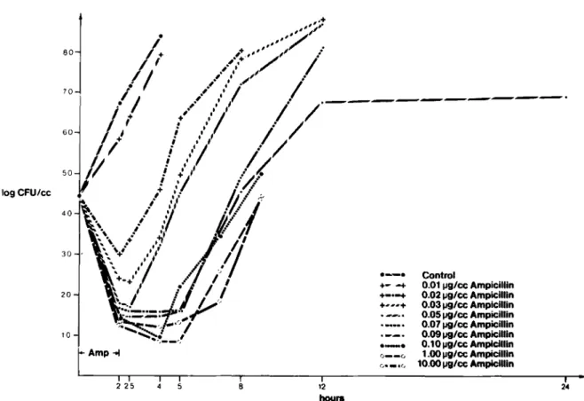
Characteristics of the PAE in vitro. Figure 1 shows the principal characteristics of growth of
Spneumoniae in the presence and absence of ampicillin. In the absence of antibiotics, the bac-teria grew promptly (in 4 hr) from an initial count of 6.2 loglo cfu/ml to 8log., cfu/rnl. The presence of ampicillin at concentrations at or above the MBC led to a uniform decline in bacterial titers of 1.5 10glO cfu/ml during the first 2 hr. The rate of killing was constant over a wide range of ampicil-lin concentrations (1-100 times the MBC). Re-moval of the antibiotic after exposure of the or-ganisms for 2 hr resulted in delayed regrowth (i.e., a PAE) in all experiments. Whether ampicillin was inactivated by fJ-lactamase or removed by centri-fugation and washing, the duration of the PAE in vitro was the same. Higher drug levels resulted in a longer PAE (figure 1). The lowest drug concen-tration that resulted in a measurable PAE was 0.03 JAg of ampicillin/ml (one-half the MIC), with a PAE duration of 1 hr. At a concentration of 10 JAg of ampicillin/ml, the PAE lasted for 4.3 hr. When the same experiments were performed in pooled rabbit CSF, the level of initial killing during the 2-hr period of ampicillin exposure was markedly re-duced (i.e., to 0.5 log., cfu/ml), and the average generation (or doubling) time of bacterial growth was increased (from 21 min in broth to 37 min in CSF). The duration of the PAE, however, was the same in broth and CSF.
Characteristics of the PAE in vivo. The dy-namic relation between drug concentrations and bacterial titers in CSF was examined in 17 animals that had been infected intracisternally 18-20 hr prior to the iv administration of ampicillin. The administration of different drug doses (1-62.5
logCFU/cc 80 70 60 20 10 Amp~
....
~. +- -+ +_0-+ +...+ e - e 0 - - 00--.0
Control 0.011J9/cc Ampicillin 0.021J9/cc Ampicillin 0.031J9/cc Ampicillin 0.051J9/cc Ampicillin 0.071J9/cc Ampicillin 0.091J9/cc Ampicillin 0.10 1J9/ccAmpicillin 1.00 1J9/ccAmpicillin 10.oop9/ccAmpicillin 24 8 2 25 12 hoursFigure 1. Effect of exposure for 2 hr to different concentrations of ampicillin on growth curves of Spneumoniae
in broth. Bacterial titers are plotted as log.,cfu/mlagainst time (hr). The control culture contained no ampicillin. After exposure for 2 hr to the indicated concentrations of ampicillin, the drug was removed from the culture by centrifu-gation and three washes.
mg/kg) resulted in a wide range of CSF concentra-tions (0.04-6.4 ug/rnl); the dose administered and the resulting peak CSF concentrations correlated. well (r = .921;P
<
.001). The dose and the peak CSF concentrations also correlated (r = .616; P<
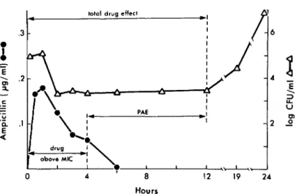
.02) with the period during which CSF concen-trations were above the MIC (median, 4hr; range, 1-7.5 hr).The mean bacterial titer(± SD) in CSF was 6.0 (± 1.0) 10glO cfu/ml at the time of drug adminis-tration. The injection of ampicillin was followed by a median drop in bacterial titers of 4.4 log., cfu/ml (range, 1.9-6.5 10glO cfu/ml). This decline in bacterial titers correlated with the dose of ampi-cillin administered (r = .619; P
<
.01).The total drug effect, defined as the period from the injection of ampicillin to the beginning of bac-terial regrowth (figure 2), lasted for a median of 11 hr (range, 3.5-20 hr) and correlated with the ampi-cillin dose(rs = .488; P
<
.05). In animals receiv-ing doses of ampicillin that produced peak CSFconcentrations of ~10 times the MBC, the total duration of the drug's effect consistently exceeded 12 hr. The PAE, defined as the total duration of the drug's effect minus the period during which drug concentrations were above the MIC (figure 2), ranged from 2.5 hr to 18 hr (median, 6.5 hr) and did not correlate with either the dose of ampicillin or the peak concentration of ampicillin in CSF(r
= .050; P = not significant).
Effect of [3-lactamase on the PAE in vivo. In six additional animals that were prepared identi-cally, the administration of an iv bolus of 12.5 mg of ampicillin/kg was followed by the injection of 400,000 IV of [3-lactamase (300,000 IV by the iv route and 100,000 IV into the cisterna magna) after 2 hr or 4.5 hr. In all animals the injection of [3-lactamase reversed the PAE. In both treatment groups, bacterial titers in CSF had increased by an average of 0.5log., cfu/ml 0.5 hr after administra-tion of the enzyme and by 0.9 10glO cfu/ml 1 hr after the injection (figure 3).
PAE in Treatment of Experimental Meningitis 579
Figure 2. Relation of ampicillin con-centrations and bacterial titers in CSF of a representative rabbit that received 12.5 mg of ampicillin/kg iv at time zero. Drug concentrations and bac-terial titers were determined in serial samples of CSF.
totol drug effect
.. I
;16
I I,
J
..
~
E < ; ~ LL. U PAE..
Ol 2 ...2 HoursFigure 3. Effect of inactivation of ampicillin in CSF by injection of fJ-Iactamase(t)in two representative rab-bits. Each animal received 12.5 mg of ampicillin/kg iv 18 hr after being infected. Bacterial titers of S pneumoniae were determined in CSF over time. The animals received 400,000 IV of fJ-Iactamase (300,000 IV by the iv route and 100,000 IV intracisternally) 2 hr and 4.5 hr after the injection of ampicillin.
large doses, with peak CSF concentrations of >10 times the MBC, sterilized the CSF of>90010 of the
animals, regardless of other variables in the treat-ment regimen (table 1).When regimens consisting of the same single dose but different total doses and intervals between doses (four doses at 4-hr in-tervals vs. two doses at 12-hr inin-tervals) were com-pared, the more frequent dosing regimen appeared to be preferable but differences did not reach sta-tistical significance (P ~ .2). On the other hand,
10 8 .. 6 Hours 2
o
2..
v v ... :::> LL. U m ..QInfluence of different regimens on therapeutic efficacy. A total of 235 rabbits with meningitis were used in studies of the influence of different doses of ampicillin and different intervals between doses on sterilization of CSF cultures 36 hr after the last injection. The 28 untreated control rabbits all died within 66 hr of bacterial inoculation or had positive CSF culture results (mean, 105cfu/ml)
and fever (~39.6 C); no spontaneous cures were observed. The remaining 207 animals were as-signed to the various treatment groups. Therapy was initiated 18 hr after infection, when 100% of the animals had fever and positive CSF culture re-sults (3.9 ± 1.2 log., cfu/ml). Forty-nine rabbits had to be excluded from the evaluation either because they died before the completion of the 66-hr experiment(n
=
15) or because no CSF could be obtained for the final evaluation(n = 34). The majority of the early deaths resulted from experi-mental procedures. Since evaluation of the results depended on the result of culture of the final CSF sample, data on all animals that could not be clas-sified with certainty for one of the reasons men-tioned were excluded. Even if all deaths had been counted as treatment failures, the outcome would not have been significantly changed.The two most important factors determining outcome in our experiments were the dose in a single injection of ampicillin and the peak drug concentrations in CSF. As previously stated, these two variables are closely related. Small single doses, which produced peak CSF concentrations of 0.2-0.5 ug/ml (two to five times the MBC), in-consistently resulted in sterile CSF; in contrast,
Table 1. Results of ampicillin treatment of meningitis caused by Spneumoniae in rabbits. -Single dose (mg/kg)
o
4.17 4.17 6.25 6.25 12.5 12.5 25.0 25.0 37.5 37.5 No. of doseso
2 4 2 4 2 4 2 4 2 4 Total dose (mg/kg)o
8.34 16.68 12.5 25 25 50 50 100 75 150Mean peak drug level in CSF (± SO)
o
0.20 ± 0.09 0.20 ± 0.14 0.20 ± 0.08 0.28 ± 0.16 0.52 ± 0.37 0.54 ± 0.35 1.14 ± 0.54 1.44 ± 0.46 1.44± 1.26 1.55 ± 0.89No. of animals with sterile CSF / no. evaluated(010 with sterile CSF)*
0/28 ( ) 0/10 ( ) 1/9 (11) 4/19 (21) 9/21 (43) 13/23 (57) 19/24 (79) 21/22 (95) 8/8 (100) 11/12 (92) 10/10 (100)
* Each pair of results listed consecutively in the table was compared by theX2test or the Fisher's exact test. In all cases the
differ-ences between values were insignificant(P>.05).
when regimens with the same total dose were com-pared, those involving drug administration every 12 hr (which resulted in higher individual peak CSF concentrations) were found to be slightly ad-vantageous but not significantly so (P ~ .2).
Another variable that may influence the bacteri-ologic response is the proportion of the interval be-tween doses during which CSF concentrations exceed the MIC. Our preliminary experiments showed that CSF concentrations are above the MIC for ""4 hr after an injection of 12.5 mg of am-picillin/kg and for ""6 hr after a dose of 37.5 mg/kg. Thus, regimens with single doses of 12.5 mg/kg administered every 4 hr result in CSF con-centrations above the MIC for 100070 of the in-terval between doses (i.e., the whole period of treatment), while those with single doses of 37.5 mg/kg administered every 12 hr produce CSF con-centrations that exceed the MIC for (at most) 50070 of the time. Our results did not show a significant influence of the interval between doses on the results of treatment. However, when the single dose administered every 12 hr was increased from 12.5 mg/kg to 25 mg/kg, the outcome of treatment was significantly better(P
<
.01), even though the period during which CSF concentrations were above the MIC increased only from 30% to 40% of the interval between doses. Thus, our results indicate that the period during which CSF concen-trations exceed the MIC is not a major determi-nant of outcome within the limits examined (i.e., with CSF concentrations above the MIC for >30070 of the interval between doses).Thus, the size of the single dose of ampicillin
and the peak concentration of the drug in CSF de-termine the bacteriologic result in our animal model of meningitis. Doses that result in peak CSF concentrations of >10 times the MBC are highly effective, with sterile CSF in >90070 of cases. The total amount of drug given during the period of treatment, the interval between doses, and the period during which the drug concentration in the CSF is above the MIC appear to be of minor importance.
Discussion
Early in the antibiotic era, Parker and colleagues [10, 11] observed that the inhibitory effect of peni-cillin on staphylococci in vitro persisted for some time after removal of the drug. This PAE has since been reproduced in vitro with many different or-ganisms and antibiotics [12, 19, 22]. A few years after Parker's initial observations, Eagle and col-leagues conducted a series of experiments on the importance of several variables of antibiotic treat-ment, including the PAE, in mice and rabbits [13, 23, 24]. These investigators found that a PAE simi-lar to that found in vitro also existed in vivo, that an increase of doses above a critical value did not enhance the bactericidal action of penicillin, and that the most important variable determining out-come was the period during which the concentra-tion of drug was maintained above the MIC. Inter-est in these quInter-estions has persisted. Most recently, studies conducted with a thigh model of pneumo-coccal infection in neutropenic mice demonstrated that penicillin produced a PAE only after the
ad-PAE in Treatment oj Experimental Meningitis
ministration of large doses and prolonged expo-sure, whereas erythromycin and tetracycline pro-duced a PAE similar to that observed in vitro [25]. Furthermore, the period during which drug con-centrations were above the MIC was found to be the most important factor determining the results of treatment in these experiments [25, 26]; these observations were similar to those of Eagle et al. [23,24].In another study Bakker-Woudenberg and associates examined the effectiveness of penicillin against pneumococcal pneumonia in decomple-mented rats and confirmed the importance of pro-longed periods during which drug concentrations exceeded the MIC for maximal therapeutic bene-fit [27].
The understanding of pharmacokinetic factors that influence the efficacy of therapy for bacterial meningitis is incomplete. Preliminary studies in a rabbit model of pneumococcal meningitis have suggested that a PAE exists in CSF [9], but the role of this PAE and its relation to other variables, such as dose and duration of drug exposure, have not been investigated.
The PAE documented in our studies in vivo of CSF differed in several respects from the PAE ob-served in vitro. The former lasted considerably longer than the latter (2.5-18 hr vs. 1-4.3 hr) and was considerably more variable. The duration of the in vivo PAE did not correlate with the dose or the peak concentration of ampicillin in CSF and could be reversed by the administration of fJ-Iacta-rnase, which inactivated residual drug in the sub-arachnoid space.
These characteristics suggest that the PAE ob-served in our animal model of meningitis may be a different phenomenon from the PAE observed in vitro. The strongest evidence for this hypothesis derives from the striking effect of fJ-Iactamase administration on the PAE in vivo. Our findings indicate that the in vivo effect is the consequence of the presence of small residual amounts of drug in CSF. When this drug is inactivated, no further PAE can be documented. This result is in contrast to that in vitro, where the PAE was found (in both this study and that of McDonald et al. [19]) to be unaffected by fJ-Iactamase.
Ifthe PAE in CSF is indeed attributable to the presence of small residual amounts of drug, its duration should reflect the pharmacokinetic char-acteristics of drug elimination from CSF. In earlier studies we found considerable variation of CSF
581
pharmacokinetics in infected rabbits [14, 28], and the slow gamma phase of drug elimination is prob-ably subject to similar variability. Furthermore, the duration of the gamma phase is probably a func-tion not only of the initial CSF concentrafunc-tions but also of other factors that can influence CSF pharmacokinetics (e.g., degree of inflammation, rate of CSF turnover). Thus, the observed variabil-ity of the PAE and its lack of correlation with the initial CSF peak in vivo may simply be the expres-sions of pharmacokinetic differences that affect the duration of the gamma phase of drug elimina-tion in individual rabbits.
In the absence of active drug in CSF (i.e., after fJ-Iactamase administration), we did not find a PAE in our model. This result is in agreement with the findings of other researchers who, examining the occurrence of a PAE in neutropenic mice in-fected with Spneumoniae, found either no PAE
[26] or only a short PAE after prolonged exposure to large doses of penicillin [25].Conversely, Eagle et al. documented a sustained PAE in a mouse model in the apparent absence of any residual drug [13]. The reasons for these discrepancies are not clear. Eagle and Musselman showed that only actively growing organisms exhibited a PAE in vitro [12].Pneumococci in CSF in vivo grow con-siderably more slowly than in broth, with genera-tion times of 60 min and 20 min, respectively [29]. Itis questionable, though, whether this difference in growth rate is the main reason for the absence of a drug-independent PAE in vivo since our in vitro studies showed no influence of the prolonged generation time in ex vivo CSF on the duration of the PAE.
Another interesting fact is that a PAE was ob-served in immunocompetent animals by Eagle et al. [13], whereas no drug-independent PAE could be documented in immunodeficient animals (neutropenic mice) [26] or in animals infected at a site of impaired host defenses (meningitis). It is conceivable that the occurrence of a PAE in vivo withSpneumoniae after exposure to penicillin is
dependent on either the presence of residual amounts of drug 'or functioning host-defense mechanisms.
The relevance of a PAE to therapy is difficult to assess unequivocally. The pharmacologic param-eter best suited to reflect the influence of the PAE is the interval between doses. However, when this interval is varied so that its influence on outcome
can be assessed, at least one other variable (the number of doses, the dose administered in a single injection, the total dose, or the duration of treat-ment) must also be changed. In our study we com-pared two different intervals (4 hr and 12 hr), varying the single and total doses of ampicillin while keeping the total duration of therapy con-stant (12 hr).
The most important factor determining efficacy of treatment in our experiments was the peak con-centration of ampicillin in CSF. The outcome of treatment improved as drug concentrations in CSF increased, and peak concentrations that were >10 times the MBC consistently resulted in sterile CSF in >90% of animals. On the other hand, within the limits of the dosing schedules examined, the inter-val between doses and the duration of CSF ampi-cillin concentrations above the MIC did not ap-pear to be of major importance with regard to outcome. If anything, prolonging of the interval between doses (with a constant amount of total drug) resulted in a favorable trend throughout all treatment groups; this effect was probably due to the resulting higher peak concentrations in CSF. Thus, even with small doses of drug, the benefit of the enhanced killing rate by the higher peak con-centrations of ampicillin in CSF [30] seems to out-weigh the hazard of eventual bacterial regrowth toward the end of the dosing interval. With large doses the total antibiotic effect observed in our preliminary experiments is apparently of sufficient duration to prevent bacterial regrowth during the 12-hr interval between doses.
Our findings are in contrast to those of Eagle et al. [13, 23, 24] and Gudmundsson et al. [26], which suggested that the duration of drug concentrations above the MIC was the most important determi-nant of outcome and that peak drug concentra-tions were of only minor import.Itis possible that the unique characteristics of meningitis (i.e., bac-terial growth conditions in CSF, pharmacokinetics in CSF, impaired host defenses in the subarach-noid space) are responsible for these discrepancies. Since the observed PAE in meningitis appears to be the result of a prolonged gamma phase of am-picillin elimination, direct extrapolation to infec-tions at sites with different pharmacokinetics is not possible. Also, since the PAE may vary con-siderably from one antibiotic to another and from one organism to another [22], our results apply only to the drug and organism tested.
Our findings emphasize points of potential im-portance in the treatment of pneumococcal menin-gitis.Ifit is to effect maximal bacterial killing, the dose of drug administered must result in peak CSF concentrations far above the MBC for the patho-gen. This notion has been confirmed in a recent study of the relation between the concentrations of severalf3-lactam antibiotics in CSF and the bacteri-cidal effect of these drugs [30] and has also been supported by clinical experiences [5, 31, 32]. In addition, the drug-dependent PAE in CSF prevents immediate bacterial regrowth after drug concen-trations drop below the MIC. The complexity of our model dictated the inclusion of only relatively small numbers of animals in each treatment group. The large inherent f3-error that results from these small numbers precludes a definite statement about equality of the two intervals between doses. Nevertheless, our study demonstrates that the pro-longed prevention of bacterial regrowth observed in vivo in CSF is associated with therapeutic effi-cacy. Furthermore, contrary to findings by Schmidt and Walley in experiments with a rat model of pneumococcal pneumonia [33], our studies failed to demonstrate a clear-cut advantage of long inter-vals between doses of drug. Thus, while our data suggest that an increase in these intervals can result in rates of cure similar to those obtained with sus-tained, high concentrations of drug in CSF as long as the peak concentration reaches a critical value
(~10times the MBC), they do not support the con-cept that a prolonged therapeutic interval is superior.
References
1. Kunin CM. Dosage schedules of antimicrobial agents: a historical review. Rev Infect Dis 1981;3:4-11
2. Fishman RA. Cerebrospinal fluid in diseases of the nervous system. Philadelphia: WB Saunders, 1980:49-54 3. Fishman RA. Blood-brain and CSF barriers to penicillin
and related organic acids. Arch Neurol 1966;15:113-24 4. Spector R, Lorenzo AV. Inhibition of penicillin transport
from the cerebrospinal fluid after intracisternal inocula-tion of bacteria. JClin Invest1974;54:316-25
5. Sande MA. Antibiotic therapy of bacterial meningitis: les-sons we've learned [editorial]. AmJMed1981;71:507-10 6. ScheId WM, Sande MA. Bactericidal versus bacteriostatic antibiotic therapy of experimental pneumococcal menin-gitis in rabbits. JClin Invest1983;71:411-9
7. Hodges GR, Perkins RL. Acute bacterial meningitis: an analysis of factors influencing prognosis. AmJMed Sci 1975;270:427-40
PAE in Treatment of Experimental Meningitis
meningococcemia- United States, 1978. MMWR 1979; 28:277-9
9. Sande MA, Korzeniowski OM, Allegro GM, Brennan RO, Zak0,Scheid WM. Intermittent or continuous therapy of experimental meningitis due to Streptococcus pneu-moniaein rabbits: preliminary observations on the post-antibiotic effect in vivo. Rev Infect Dis 1981;3:98-109 10. Parker RF, March He. Action of penicillin on
Staphylo-coccus.J Bacteriol 1946:51:181-6
ll. Parker RF, Luse S. The action of penicillin on Staphylococ-cus:further observations on the effect of a short ex-posure. J Bacteriol 1948;56:75-81
12. Eagle H, Musselman AD. The slow recovery of bacteria from the toxic effect of penicillin. J Bacteriol 1949;58: 475-90
13. Eagle H, Fleischman R, Musselman AD. The bactericidal action of penicillin in vivo: the participation of the host and the slow recovery of the organisms. Ann Intern Med 1950;33:544- 71
14. Sherertz RJ, Dacey R, Sande MA. Cefamandole in the therapy of experimental pneumococcal meningitis. J Antimicrob Chemother 1976;2:159-65
15. Waterworth PM. An enzyme preparation inactivating all penicillins andcephalosporins, J Clin Pathol 1973;26: 596-8
16. Sykes RB, Nordstrom K. Microiodometric determination of (J-Iactamase activity. Antimicrob Agents Chemother 1972;1:94-9
17. Zak 0,KradolferF.Effects of subminimal inhibitory con-centrations of antibiotics in experimental infections. Rev Infect Dis 1979;1:862-79
18. Thrupp LD. Susceptibility testing of antibiotics in liquid media. In: Lorian V, ed. Antibiotics in laboratory medi-cine. Baltimore: Williams and Wilkins, 1980:73-113 19. McDonald PJ, Craig WA, Kunin CM. Persistent effect of
antibiotics onStaphylococcus aureusafter exposure for limited periods of time. J Infect Dis 1977;135:217-23 20. Murray PR, Hampton CM. Recovery of pathogenic
bac-teria from cerebrospinal fluid. J Clin Microbiol 1980; 12:554-7
21. Dacey RG, Sande MA. Effects of probenecid on cerebro-spinal fluid concentrations of penicillin and cephalo-sporin derivatives. Antimicrob Agents Chemother 1974; 6:437-41
22. Bundtzen RW, Gerber AU, Cohn DL, Craig WA. Postanti-biotic suppression of bacterial growth. Rev Infect Dis 1981;3:28-37
23. Eagle H, Fleischman R, Musselman AD. Effect of schedule of administration on the therapeutic efficacy of penicil-lin: importance of the aggregate time penicillin remains
583
at effectively bactericidal levels. Am J Med 1950;9:280-99
24. Eagle H, Fleischman R, Levy M. "Continuous" vs. "discon-tinuous" therapy with penicillin; the effect of the interval between injections on therapeutic efficacy. N Engl J Med 1953;248:481-8
25. Craig WA, Turnidge JD, Gudmundsson S. In vivo demon-stration of postantibiotic effect (PAE) [abstract 634]. In: Program and abstracts of the 22nd Interscience Confer-ence on Antimicrobial Agents and Chemotherapy. Washington, DC: American Society for Microbiology, 1982
26. Gudmundsson S, Turnidge JD, Vogelman B, Craig WA. Correlation of pharmacokinetic parameters of penicillin and erythromycin with efficacy against S.pneumoniaein an animal model [abstract 288]. In: Program and ab-stracts of the 23rd Interscience Conference on Anti-microbial Agents and Chemotherapy. Washington, DC: American Society for Microbiology, 1983
27. Bakker-Woudenberg IAJM, Van den Berg JC, Fontijne P, Michel ME Therapeutic efficacy of continuous vs. inter-mittent administration of penicillin in pneumococcal pneumonia in normal rats and rats with impaired phago-cytosis [abstract 289]. In: Program and abstracts of the 23rd Interscience Conference on Antimicrobial Agents and Chemotherapy. Washington, DC: American Society
for Microbiology, 1983
28. Sande MA, Sherertz RJ, Zak 0,Strausbaugh LJ. Cepha-losporin antibiotics in therapy of experimental Strep-tococcus pneumoniae and Haemophilus influenzae meningitis in rabbits. J Infect Dis 1978;137:S161-8 29. Ernst JD, Decazes JM, Sande MA. Experimental
pneu-mococcal meningitis: role of leucocytes in pathogenesis. Infect Immun 1983;41:275-9
30. Tauber MG, Doroshow CA, Hackbarth CJ, Rusnak MG, Drake TA, Sande MA. Antibacterial activity of (J-Iactam antibiotics in experimental pneumococcal meningitis. J Infect Dis 1984;149:568-74
31. McCracken GH Jr. The rate of bacteriological response to antimicrobial therapy in neonatal meningitis.Am J Dis Child 1972;123:547-53
32. Landesman SH, Corrado ML, Shah PM, Armengaud M, Barza M, Cherubin CEo Past and current role for cepha-losporin antibiotics in treatment of meningitis. Am J Med 1981;71:693-703
33. Schmidt LH, Walley A. The influence of the dosage regi-men on the therapeutic effectiveness of penicillin G in experimental lobar pneumonia. J Pharmacol Exp Ther 1951;103:479-88