Advance Access publication 24 July 2012
Impacted maxillary canines and root resorptions of neighbouring
teeth: a radiographic analysis using cone-beam computed
tomography
Caroline S. Lai
*
,†, Michael M. Bornstein
**
,†, Lothar Mock
*
, Benjamin M. Heuberger
***
,
Thomas Dietrich
****
and Christos Katsaros
*
*Department of Orthodontics and Dentofacial Orthopedics, **Department of Oral Surgery and Stomatology, School of Dental Medicine, University of Bern, Switzerland, ***Clinic for Oral and Maxillofacial Surgery, Hospital of Luzern, Switzerland and ****Department of Oral Surgery, The School of Dentistry, University of Birmingham, UK
†These authors have contributed equally to this study
Correspondence to: Michael M. Bornstein, Department of Oral Surgery and Stomatology, School of Dental
Medicine, University of Bern, Freiburgstrasse 7, CH-3010 Bern, Switzerland. E-mail: [email protected]
SUMMARY The study analyses the location of impacted maxillary canines and factors influencing root
resorptions of adjacent teeth using cone-beam computed tomography (CBCT). In addition, the interrater reliability between observers of two different dental specialties for radiographic parameters will be evaluated. CBCT images of patients who were referred for radiographic localization of impacted maxillary canines and/or suspicion of root resorptions of adjacent teeth were included. The study analysed the exact three-dimensional location of the impacted canines in the anterior maxilla, frequency and extent of root resorptions, and potential influencing factors. To assess interrater agreement, Cohen’s correlation parameters were calculated.
This study comprises 113 patients with CBCT scans, and 134 impacted canines were analysed retrospectively. In the patients evaluated, 69 impacted canines were located palatally (51.49 per cent), 41 labially (30.60 per cent), and 24 (17.91 per cent) in the middle of the alveolar process. Root resorptions were found in 34 lateral incisors (25.37 per cent), 7 central incisors (5.22 per cent), 6 first premolars (4.48 per cent), and 1 second premolar (0.75 per cent). There was a significant correlation between root resorptions on adjacent teeth and localization of the impacted canine in relation to the bone, as well as vertical localization of the canine. Interrater agreement showed values of 0.546–0.877. CBCT provides accurate information about location of the impacted canine and prevalence and degree of root resorption of neighbouring teeth with high interrater correlation. This information is of great importance for surgeons and orthodontists for accurate diagnostics and interdisciplinary treatment planning.
IMPACTED MAXILLARY CANINES AND ROOT RESORPTION OF NEIGHBOURING TEETH
Introduction
Impaction is defined by the lack of eruption of a tooth into the appropriate position in the dental arch within the time and physiological limits of the normal eruption
pro-cess (Maverna and Gracco, 2007). Maxillary permanent
canines are the second most frequently impacted teeth after third molars (Walker et al., 2005; Liu et al., 2008), with a prevalence of approximately 1–3 per cent (Preda
et al., 1997; Chaushu et al., 1999; Mason et al., 2001; Walker
et al., 2005). Most studies report that palatal displacement (80– 90 per cent) of impacted maxillary canines is more common than labial displacement (10–20 per cent) (Ericson and Kurol, 2000; Walker et al., 2005; Bjerklin and Ericson, 2006). How-ever, using computed tomography (CT), Bjerklin and Ericson (2006) found the canines to be localized labially in 40 per cent of the cases, palatally in 42 per cent, and in a mid-alveolar position in 18 per cent. In a study using cone-beam computed
tomography (CBCT), Liu and coworkers (2008) found an even more frequent labial displacement of the canines, with percent-ages of 45 (labial), 40 (palatal), and 18 (mid-alveolar).
Complications due to ectopic eruption of maxillary canines—such as root resorption of adjacent teeth, ankylosis of the canine, and (follicular) cyst formation—have been reported (Ericson and Kurol, 1987a, 1988, 2000; Liu
et al., 2008). Many studies have shown that root resorption in combination with ectopic maxillary canines occurs more often in female patients (Sasakura et al., 1984; Ericson and Kurol, 1987b; Peck et al., 1994; Rimes et al., 1997). An accurate diagnosis of exact canine position and potential root resorptions may influence which of various orthodontic-surgical treatment options is chosen, such as: 1. orthodontic alignment of the impacted tooth after surgical exposure; 2. extraction of the resorbed lateral incisor, orthodontic alignment of the impacted tooth after surgical exposure in the position of the extracted tooth, space closure, and reshaping
of the canine to serve as lateral incisor; 3. orthodontic alignment of the impacted tooth after surgical exposure, extraction of the resorbed lateral incisor, and replacement of the lateral incisor by autotransplantation or implantation (Bjerklin and Ericson 2006; Alqerban et al., 2009a,b).
CBCT studies analysing the location of impacted maxil-lary canines and related root resorption of adjacent incisors are still rare in the literature (Walker et al., 2005; Liu et al., 2008; Oberoi and Knueppel, 2012). Some of these studies have a small sample size and report only limited correla-tions between canine impaction and root resorption. Fur-thermore, the data lacks information regarding interrater reliability of the radiographic image analysis.
Sound justification of radiographic examinations in children and young adults is important due to the higher risks associated with exposure (Horner et al., 2011). Tra-ditional radiological examination of children undergoing orthodontic assessment relies on a panoramic radiograph, supplemented by a lateral cephalometric and intraoral radi-ographs. In recent years, the availability of CBCT has led to this technique being used by a growing number of clinicians as a means of radiological examination. A recent review of
Kapila and coworkers (2011) provides a useful summary of the current status of CBCT in orthodontics.
In this study, the primary outcome variables were to evaluate the three-dimensional location of impacted maxil-lary canines, the frequency, extent, and determinants of root resorptions of neighbouring teeth using CBCT imaging. The secondary outcome variables analyse the interrater reli-ability between observers of two different dental specialties for radiographic parameters.
Materials and methods
Patients
CBCT images from patients who were referred for radio-graphic localization of impacted/ectopically erupting max-illary canines (uni- or bilateral) with or without suspicion of resorption of neighbouring teeth were consecutively admit-ted to the present study. The database between January 2009 and December 2010 of the Section of Dental Radiology and Stomatology, Department of Oral Surgery and Stomatology, University of Bern, was used. Patients with a known cleft palate were excluded from further analysis.
Methods
To keep the radiation dose to a minimum, all CBCT images were taken using a limited or dentoalveolar field of view (FOV: 4 × 4, 6 × 6, or 8 × 8 cm; 3D Accuitomo XYZ Slice View Tomograph, Morita Corp., Kyoto, Japan) so that only the area of clinical interest was irradiated. A basic voxel size of 0.08 mm was used for evaluation of all FOVs included. The operating parameters were set at 5.0 mA and 80 kV, and the exposure time was 17.5 seconds. The data were recon-structed in slices, and examined slice by slice in all three
dimensions (sagittal, coronal, and axial) on 1:1 scaled images using a specialized software program (i-Dixel, Morita Corp., Kyoto, Japan). Initially, all images were reformatted to place the palate/floor of the nose in a horizontal position in the sagittal views, and the central incisors in a vertical position in the coronal views. When needed, the magnifying tool and the ruler of the viewer were used. The following analysis and measurements were performed for every included subject: 1. Three-dimensional localization of the impacted canine:
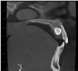
(a) Sagittal: location of the crown of the impacted canine in relation to the neighbouring teeth (mostly lateral incisors) classified in labial, median, or pala-tal position (Figure 1) was assessed using sagittal and/or coronal CBCT scans.
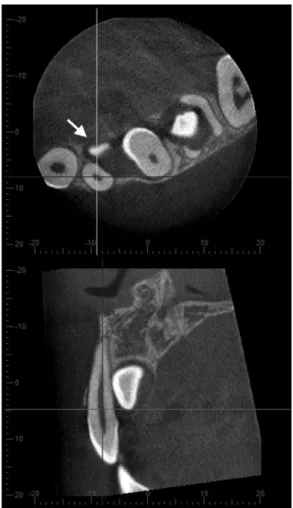
(b) Vertical: location of the cusp tip in relation to the long axis of the neighbouring incisor, subdivided into coronal, cervical third of the root, middle third of the root, apical third of the root, or apical to the root tip (Figure 2).
(c) Transversal: measurement of the cusp tip in relation to the midline based on the linear measurement meth-ods proposed by Walker et al. (2005). The shortest distance between the tip of the impacted canine and the mid-palatal sutures was measured perpendicularly in mm on respective axial CBCT scans (Figure 3). 2. Type of impaction in relation to the bone, grouped into
full bony impaction, retention with soft tissue coverage, or retention without soft tissue coverage.
3. Development of the root of the impacted canine, classified as complete root development with closed apex, almost complete root development with open apex, ¾ of the root length developed, or ½ of the root length developed. 4. Follicle size measurement at the widest area of the
folli-cle perpendicular to the crown of the impacted canine on
Figure 1 Representative example of a sagittal CBCT scan exhibiting a
coronal and axial CBCT slices. Distances greater than 3 mm were considered to be an enlarged follicle (Ericson
et al., 2001).
5. Presence of deciduous canines and possible root resorp-tion of deciduous canines, subdivided into missing canine, canine with intact root, or resorbed root.
6. Morphology of the lateral incisor: missing, peg shaped, and normal.
7. Proximity to and/or direct contact of the impacted canine with incisors or premolars (yes/no). Proximity was defined by ≤0.5 mm distance between the two teeth (Walker et al., 2005). If yes: location of the contact in relation to the long axis of the involved tooth, classified as the cervical, middle, or apical third of the root. 8. Root resorption of incisors or other teeth (premolars and
deciduous canines). Location of the root resorption in relation to the long axis of the involved tooth, classi-fied as the cervical, middle, or apical third of the root. Resorption was graded based on the system suggested by Ericson and Kurol (2000):
(a) No resorption: intact root surface, the cementum layer may have been lost.
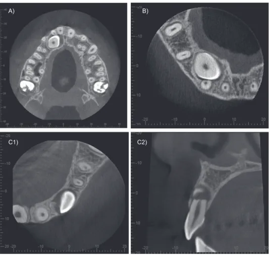
(b) Slight resorption: resorption up to half of the den-tine thickness (Figure 4A).
(c) Moderate resorption: resorption of the dentine midway to the pulp or more, the pulp lining being unbroken (Figure 4B).
(d) Severe resorption: resorption reaches the pulp ( Figure 4C).
All CBCT scans were reviewed by a single experienced orthodontist (C.S.L.). Additionally, the sagittal location of the impacted canine, and the presence, location, and severity of root resorption of neighbouring teeth, were also independently evaluated by another orthodontist (L.M.) and an oral surgeon (B.M.H.) to evaluate interrater agreement. To quantify the correlation between impaction and resorption (primary outcome variables), any disagreement between the observers was resolved by discussion for further analysis.
Statistical analysis
Summary statistics were calculated for all assessed param-eters. To assess interrater agreement, unweighted Cohen’s kappas were calculated. Logistic regression was used to cal-culate crude as well as age- and gender-adjusted odds ratios, and two-sided 95% confidence intervals (CI) were calcu-lated to evaluate the association between various factors and the prevalence of root resorption of at least one adjacent tooth. To account for clustering of bilateral teeth, general-ized estimating equations with an exchangeable correlation matrix were used. All statistical tests were two-sided at α = 0.05 and were performed using STATA 11.2 (Stata Corp., College Station, TX, USA).
Results
In this study, a total of 113 patients with CBCT scans were enrolled, and 134 impacted canines were analysed retrospectively. The mean age of the patients was 19.35 years
Figure 2 Vertical location of the cusp tip (arrow) in relation to the long
axis of the neighbouring lateral incisor on sagittal and axial CBCT scans, representative example.
Figure 3 Example of transversal measurement of the distance between the
(range: 8.7–77.2 years, SD ± 13.65 years). Of the 113 included patients, 39 (34.51 per cent) were male and 74 (65.49 per cent) were female (Table 1). Unilateral impaction was present in 92 patients (81.4 per cent), and 21 patients (18.6 per cent)
presented with bilateral impaction. Among these, 64 impacted canines were located on the right side (47.76 per cent).
The analysis of the three-dimensional location revealed that most of the impacted canines were located in a palatal
A B
Figure 5 Representative example of an impacted canine crossing the maxillary midline (A: volume rendered image and B: axial CBCT scan). A)
C1) C2)
B)
Figure 4 Representative example of different types root resorption: (A) slight root resorption on a right lateral incisor; (B) moderate root resorption on a
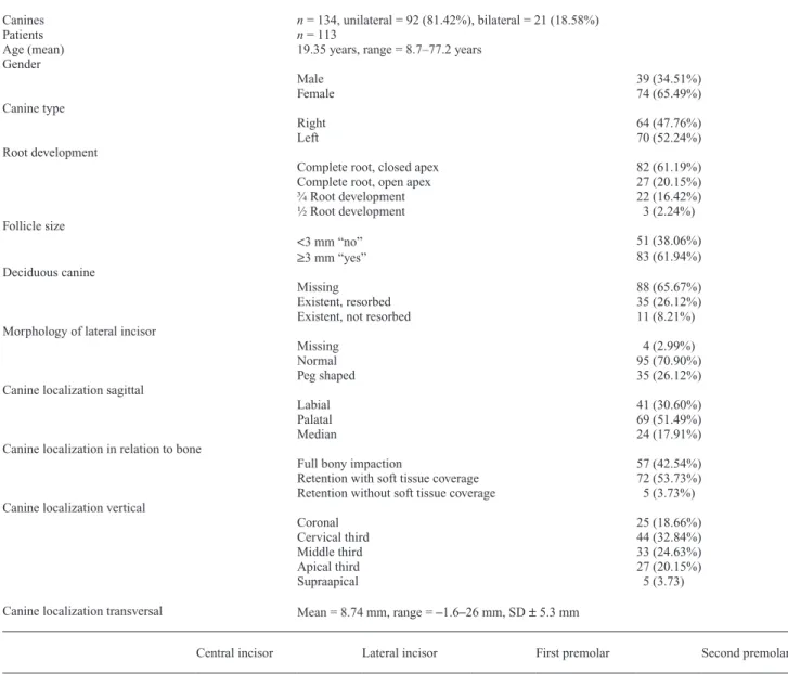
Table 1 Descriptive data regarding morphology and location of impacted canines.
Canines Patients
n = 134, unilateral = 92 (81.42%), bilateral = 21 (18.58%) n = 113
Age (mean) 19.35 years, range = 8.7–77.2 years Gender Male 39 (34.51%) Female 74 (65.49%) Canine type Right 64 (47.76%) Left 70 (52.24%) Root development
Complete root, closed apex 82 (61.19%) Complete root, open apex 27 (20.15%) ¾ Root development 22 (16.42%) ½ Root development 3 (2.24%) Follicle size <3 mm “no” 51 (38.06%) ≥3 mm “yes” 83 (61.94%) Deciduous canine Missing 88 (65.67%) Existent, resorbed 35 (26.12%) Existent, not resorbed 11 (8.21%) Morphology of lateral incisor
Missing 4 (2.99%)
Normal 95 (70.90%)
Peg shaped 35 (26.12%)
Canine localization sagittal
Labial 41 (30.60%)
Palatal 69 (51.49%)
Median 24 (17.91%)
Canine localization in relation to bone
Full bony impaction 57 (42.54%) Retention with soft tissue coverage 72 (53.73%) Retention without soft tissue coverage 5 (3.73%) Canine localization vertical
Coronal 25 (18.66%)
Cervical third 44 (32.84%)
Middle third 33 (24.63%)
Apical third 27 (20.15%)
Supraapical 5 (3.73)
Canine localization transversal Mean = 8.74 mm, range = –1.6–26 mm, SD ± 5.3 mm
Central incisor Lateral incisor First premolar Second premolar
Proximity/direct contact
No = 99 (73.88%) No = 29 (21.64%) No = 112 (83.58%) No = 131 Yes = 35 (26.12%) Yes = 101 (75.37%) Yes = 22 (16.42%) Yes = 3 (2.24%)
Missing = 4 (2.99%)
Location of proximity/direct contact
Cervical third 7 (5.22%) 14 (10.45%) 4 (2.99%) 1 (0.75%) Middle third 19 (14.18%) 58 (43.28%) 7 (5.22%) 0 Apical third 9 (6.72%) 29 (21.64%) 11 (8.21%) 2 (1.49%)
Resorption
No = 127 (94.78%) No = 96 (71.64%) No = 128 (95.52%) No = 133 (99.25%) Yes = 7 (5.22%) Yes = 34 (25.37%) Yes = 6 (4.48%) Yes = 1 (0.75%)
Location of resorption Cervical third 0 2 (1.49%) 0 0 Middle third 4 (2.99%) 16 (11.49%) 2 (1.49%) 0 Apical third 3 (2.24%) 16 (11.49%) 4 (2.99%) 1 (0.75%) Severeness of resorption Mild 3 (2.24%) 12 (8.96%) 3 (2.24%) 1 (0.75%) Moderate 1 (0.75%) 5 (3.73%) 0 0 Severe 3 (2.24%) 17 (12.69%) 3 (2.24%) 0
position (51.49 per cent, 69 canines), and only 30.6 per cent (41 canines) were located buccally. In most cases, the verti-cal location of the canine cusp tip in relation to the long axis of the adjacent tooth was in the cervical third of the root (32.84 per cent, 44 teeth), followed by a location in the middle third (24.63 per cent, 33 canines), and in the apical third (20.15 per cent, 27 canines). The mean distance of the cusp tip of the impacted canine to the midline of the upper jaw was 8.74 mm (range: 1.6–26 mm). Only two impacted canines crossed the transversal midline (Figure 5). Transpo-sition of the impacted canine was found in only two cases. For further details, see Table 1.
In relation to the bone, most canines exhibited retention with soft tissue coverage (53.73 per cent, 72 canines), fol-lowed by canines with complete bone coverage (42.54 per cent, 57 canines). In 61.94 per cent (83 cases) of the cases, the follicle was enlarged. In the population analysed, 35 peg-shaped laterals (26.12 per cent) and 4 missing laterals (2.99 per cent) were found. In 82 cases the impacted canines showed complete root development (81.34 per cent), and in 27 cases the root development was almost complete. For further details, see Table 1.
For the 134 ectopic canines, 41 adjacent incisors exhib-ited signs of resorption: 34 lateral incisors (25.37 per cent) and 7 central incisors (5.22 per cent). In six cases with root resorption on central incisors, the lateral incisors exhibited signs of resorption as well. Only six adjacent first premolars (4.48 per cent) and one second premolar (0.75 per cent) with root resorption were found. Among the 21 bilateral cases, there were 4 cases with bilateral root resorption on adja-cent incisors and 2 cases with only one side affected. Root resorption on permanent incisors was located primarily in the middle third of the root (48.78 per cent), followed by the apical third of the root (46.34 per cent) and the cervical third of the root (4.88 per cent). In 36 cases there was a vis-ible contact between the impacted canine and the resorbed incisor. For further details, see Table 1.
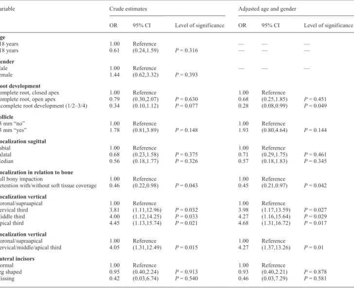
There was no correlation between age of the patient and prevalence of root resorption (P = 0.49), but a slightly higher prevalence of resorption was found for older patients when the patients were divided by age into two groups: ≥18 years and <18 years, although this difference was not sta-tistically significant (P = 0.316). A stasta-tistically significant higher prevalence of root resorption was present when there was complete root development of the impacted canine with a closed apex compared with when there was incomplete root development of the canine (P = 0.049). Although not statistically significant, female subjects seemed to be more affected than male subjects (P = 0.39). For further details, see Table 2.
Visible contact between the impacted canines and the neighbouring teeth with and without root resorption was frequently found, mostly for lateral incisors (101 cases), for central incisors (35 cases), for first premolars (22 cases), and for second premolars (3 cases). The location of contact
was found to be in the middle third of the root in 84 of the cases (52.17 per cent), in the apical third in 51 cases (31.68 per cent), and in the cervical third in 26 cases (16.15 per cent). For further details, see Table 1.
With regard to root resorption and three-dimensional localization of the canines in the sagittal plane, there was no difference in prevalence of root resorption on adjacent teeth between labially and palatally impacted canines (P = 0.375). Regarding retention with or without soft tissue or bone cov-erage, there was a higher prevalence of root resorption in adjacent teeth when the canine was fully covered by bone (P = 0.043). Furthermore, there was a significant correla-tion between prevalence of root resorpcorrela-tion and locacorrela-tion of the cusp tip in the vertical plane in relation to the long axis of the adjacent teeth. Compared to a coronal or supraapical location of the cusp tip, there was a higher risk of resorp-tion when the cusp tip was located in the cervical, middle, or apical third of the root of the adjacent tooth (P = 0.01). Furthermore, no correlation could be found between root resorption and peg-shaped (P = 0.878) or missing laterals (P = 0.581). For further details, see Table 2.
In 46 of the cases, deciduous canines were still present, and 34 of those exhibited resorbed roots. In 20 cases with root resorptions, the follicle of the impacted canine was enlarged. Direct contact between the impacted canine and the resorbed deciduous tooth was found in only 2 out of 34 (5.9 per cent) deciduous canines, compared with 41 out of 49 (83.7 per cent) resorbed permanent teeth (OR 88.3, 95% CI 17.1, 457).
Interrater agreement evaluated for the sagittal location of the impacted canine, and the presence, location, and sever-ity of root resorption of neighbouring teeth for the three different observers exhibited values ranging from 0.546 to 0.877 (Cohen’s kappa; Table 3).
Discussion
The prevalence of disturbances in eruption of the maxillary canines seems to vary within a range of 1–3 per cent (Preda
et al., 1997; Chaushu et al., 1999; Mason et al., 2001;
Walker et al., 2005), and females seem to be more affected (Becker et al., 1981; Preda et al., 1997; Ericson and Kurol 2000). In our study, there were more female subjects than males resulting in a ratio of almost 2:1. Walker and cowork-ers (2005) speculate that the difference in overall craniofa-cial growth and development between the sexes, as well as genetics, could be possible reasons for that finding. Another reason could be that girls and women seek orthodontic treatment more frequently than males. Furthermore, Zilber-man and coworkers postulate that an experimental group represents a biased sample and may show higher or differ-ent gender ratios than in a true epidemiological (general) population (Zilberman et al., 1990).
In our study sample, we found a high prevalence of palatally impacted canines (51.49 per cent). In European and North American samples, impacted maxillary canines
were also more often found to be located palatally (85–92.6 per cent) (Preda et al., 1997; Ericson and Kurol 2000). In Asian samples, however, impacted canines were more often located buccally (45.2 per cent) than palatally (40.5
per cent) (Liu et al., 2008). Earlier studies have shown that labial and palatal displacement of maxillary canines are very different phenomena (Jacoby, 1983; Peck et al., 1994). Labial displacement is usually due to an inadequate dental arch space, whereas palatal displacement often occurs despite adequate arch space. The etiology of palatally displaced canines can be divided into local or genetic factors. Local factors, such as persistent deciduous canines, delayed eruptive pathways, and missing or anomalous lateral incisors, have been described (Bass, 1967; Becker
et al., 1981). Differences between various studies with
regard to prevalence and location of impacted canines may also be due to differences in patient selection.
The etiology of root resorption is still unclear. It has been postulated that enlarged dental follicles, as well as the pressure caused by an erupting tooth, may be responsible for root resorption of adjacent teeth (Marks et al., 1997). However, Ericson and coworkers (2001) have concluded, based on a CT examination, that the dental follicle does not Table 2 Statistical analysis regarding root resorptions in correlation with various clinical/radiographic parameters.
Variable Crude estimates Adjusted age and gender
OR 95% CI Level of significance OR 95% CI Level of significance
Age ≥18 years 1.00 Reference — — — <18 years 0.61 (0.24,1.59) P = 0.316 — — — Gender Male 1.00 Reference — — — Female 1.44 (0.62,3.32) P = 0.393 Root development
Complete root, closed apex 1.00 Reference 1.00 Reference
Complete root, open apex 0.79 (0.30,2.07) P = 0.630 0.68 (0.25,1.85) P = 0.451
Incomplete root development (1/2–3/4) 0.34 (0.10,1.12) P = 0.077 0.28 (0.08,0.99) P = 0.049
Follicle
<3 mm “no” 1.00 Reference 1.00 Reference
≥3 mm “yes” 1.78 (0.81,3.89) P = 0.148 1.93 (0.80,4.64) P = 0.144
Localization sagittal
Labial 1.00 Reference 1.00 Reference
Palatal 0.68 (0.23,1.58) P = 0.375 0.71 (0.29,1.75) P = 0.461
Median 0.56 (0.18,1.77) P = 0.326 0.57 (0.18,1.83) P = 0.345
Localization in relation to bone
Full bony impaction 1.00 Reference 1.00 Reference
Retention with/without soft tissue coverage 0.46 (0.22,0.98) P = 0.043 0.45 (0.21,0.97) P = 0.042
Localization vertical
Coronal/supraapical 1.00 Reference 1.00 Reference
Cervical third 3.81 (1.11,12.96) P = 0.032 3.98 (1.17,13.59) P = 0.027
Middle third 4.00 (1.12,14.25) P = 0.033 4.27 (1.16,15.64) P = 0.029
Apical third 4.45 (1.13,15.74) P = 0.021 4.68 (1.31,16.72) P = 0.017
Localization vertical
Coronal/supraapical 1.00 Reference 1.00 Reference
Cervical/middle/apical third 4.05 (1.31,12.49) P = 0.015 4.27 (1.37,13.26) P = 0.01
Lateral incisors
Normal 1.00 Reference 1.00 Reference
Peg shaped 0.95 (0.40,2.24) P = 0.913 0.93 (0.40,2.21) P = 0.878
Missing 0.42 (0.03,6.74) P = 0.540 0.46 (0.03,7.29) P = 0.581
Table 3 Interrater agreement between two experienced
orthodontists (C.L. and L. M.) and an oral surgeon (B.H.) using Cohen’s kappa values.
Variable Kappa values
Labiopalatal location of the impacted canine 0.877 Prevalence of proximity/direct contact 0.858 Location of proximity/direct contact 0.546 Prevalence of root resorption 0.649 Severity of root resorption 0.783 Kappa values: no agreement, <0 ; slight, 0–0.2 ; fair, 0.21–0.40 ; moderate, 0.41–0.60 ; substantial, 0.61–0.80; almost perfect, 0.81–1 (Landis and Koch, 1977)
cause root resorption of permanent teeth. They concluded that resorption of the permanent maxillary incisor is caused by the physical contact between the incisor and the canine, and by direct pressure from the canine as a part of the erup-tion process (Ericson et al., 2001). An ectopic canine with a well-developed root, erupting medially to the long axis of the adjacent incisor and inclined ≥25° to the midline of the jaw, presented the greatest risk for root resorption of the lateral incisor (Ericson and Kurol, 1987b, 2000).
This study supports previous findings that there is a cor-relation between prevalence of root resorption of permanent teeth and proximity/contact of the impacted canine. In the present sample, 30 resorbed lateral incisors showed prox-imity/contact with the impacted canine, as did 6 resorbed central incisors, 5 resorbed first premolars, and 1 resorbed second premolar. Only four lateral incisors, one central incisor, and one first premolar were resorbed without direct canine contact. In this study, there was an overall but not statistically significant tendency for enlarged canine fol-licles to occur together with root resorption. Therefore, our study supports the findings of Ericson and coworkers (2001), but due to the limited sample size some caution is still necessary. In addition, there seems to be a borderline statistical risk of root resorption if the root development of the impacted canine was complete and the apex closed com-pared with incomplete root development.
The position of the impacted canine may have an influ-ence on root resorption. There was a higher but not sta-tistically significant prevalence of root resorption when the canine was located labially. A significant correlation between root resorption and full bony impaction of the canine was found (P = 0.043). Furthermore, there may be a higher risk of root resorption of neighbouring teeth when the cusp tip is located along the long axis of the root of the adjacent tooth, and there seems to be a significantly lower risk of root resorption when the canine cusp tip is located supraapically or coronally. There was no correlation between root resorption and gender in our study. Nonethe-less, females seem to have a tendency toward higher risk of root resorption, which was also documented in previous reports (Ericson and Kurol, 1987b, 2000).
The reported incidence of root resorption also depends on the radiographic imaging method used. Conventional periapical X-rays have been shown to be an inaccurate method for diagnosing root resorption (Ericson and Kurol, 1987a,b). The prevalence of root resorption on maxillary incisors using intraoral X-rays was reported to be 12 per cent (Ericson and Kurol, 1987b). Even when using a step-wise (Ericson and Kurol, 1986) or the tube shift method (Clark, 1909), as well as in combination with panoramic views and lateral cephalographs, root resorption may be overlooked in 50 per cent of the cases (Chaushu et al., 1999;
Ericson and Kurol, 2000; Mason et al., 2001; Heimisdottir
et al., 2005). By using three-dimensional visualization, the diagnostic accuracy is significantly increased, since beam
projection is always orthogonal and provides information in all three planes of the skull (Becker et al., 2010; Haney
et al., 2010; Pazera et al., 2011). CBCT for three-dimen-sional visualization was introduced in dentistry more than a decade ago (Mah et al., 2003). CBCT scans have become established for orthodontic diagnostic and treatment plan-ning procedures, such as the localization of ectopic teeth, evaluation before orthognathic surgery, visualization of the temporomandibular joint, airway analysis, and the assess-ment of cleft palate patients (Nakajima et al., 2005; Bjerklin and Ericson, 2006; Bornstein et al., 2010). In vitro stud-ies on human skulls have shown no significant differences between different CBCT systems for assessing the severity of root resorptions (Alqerban et al., 2009a, 2011b). Nev-ertheless, different voxel sizes of different CBCT devices could influence the detectability of initial or slight root resorptions. There is definitely a need to evaluate the influ-ence of different CBCT operating parameters on the diag-nosis and classification of severity of root resorptions.
In a CT study analysing 12 patients with 17 impacted canines, Ericson et al. (2000) found that the ipsilateral lateral incisor was the tooth most commonly affected by root resorp-tion (38 per cent), followed by the ipsilateral central incisor (9 per cent). In addition, there was a high correlation between the CT diagnosis and direct visual observation of the roots of extracted teeth. Liu et al. (2008) found root resorption in 27.2 per cent of lateral and 23.4 per cent of central incisors. Root resorption on premolars appears to be rare (Postlethwaite, 1989; Cooke and Nute, 2005). In our study, we found root resorption in 25.37 per cent of the lateral incisors, 5.22 per cent of the central incisors, 4.48 per cent of the first premolars, and 0.75 per cent of the second premolars. A lower percent-age (17.7 per cent) of root resorption on permanent teeth was reported in a recent study evaluating CT scans (Cernochova
et al., 2011), with a prevalence of 12.6 per cent for lateral incisors, 4.8 per cent for first premolars, and 2.1 per cent for central incisors. In contrast to our study, only ‘severe’ root resorption was recorded, explaining the lower percentages.
Root resorption as result of impacted canines seems to be a rapid, progressive process that almost always ceases once the impacted canine has been removed from the affected root area (Becker and Chaushu, 2005). Even with pulpal involvement, lateral incisors with root resorption may not exhibit clinical symptoms and may show good long-term healing and prognosis (Milberg, 2006; Falahat et al., 2008). Previous studies have shown that the amount of information obtained from three-dimensional analysis is significantly greater than from conventional periapical and panoramic radiography (Ericson and Kurol, 2000; Alqerban et al., 2011a), and consequently this may have an influence on the treatment plan (Bjerklin and Ericson, 2006; Botticelli et al., 2010;
Haney et al., 2010; Wriedt et al., 2012). Bjerklin and Ericson (2006) have shown that almost 44 per cent of the treatment plans were modified after CT investigations brought further information about the presence of root resorption.
CBCT provides accurate information about location of the canine, which is of diagnostic importance for planning potential surgical procedures (Becker et al., 2010). Despite the expected advantage of CBCT imaging in tooth localiza-tion, it is important to consider also the impact on manage-ment of patients, the increased radiation dose and the likely higher cost of CBCT examinations in comparison with con-ventional radiography that has served dentists and specialist orthodontists well over many years. In a recent systematic review, the SEDENTEXCT consortium suggests (Horner
et al., 2011) that where it was practice to use multi-slice CT
scans for localization of unerupted teeth (Alqerban et al., 2009b), CBCT is likely to be preferred today.
CBCT has clear advantages over CT, the most important being less radiation administered to the patient (Cohenca
et al., 2007; Hirsch et al., 2008). Nevertheless, it is of importance to use smaller FOV for CBCT imaging when possible, thus adhering to the ALARA (as low as reasona-bly achievable) principle in medical radiology (McCollough
et al., 2009). To the best of our knowledge, this study is the
first in the dental literature to assess interrater agreement for the severity and location of root resorption between three observers from different dental specialties. In previ-ous studies, CBCT or CT scans were analysed by one single observer (Cernochova et al. 2011), or twice at two different time points by the same observer (Liu et al., 2008), with assessment of intrarater reliability.
This study resulted in high interrater agreement as to the location of the impacted canine in the sagittal plane and the prevalence of proximity/direct contact of the impacted canine and the adjacent roots. Furthermore, there was sub-stantial agreement between the two orthodontists and the oral surgeon in assessing the prevalence and the severity of root resorption on adjacent teeth – a finding that cer-tainly has a major impact on treatment planning. To assess potential differences between orthodontists and oral sur-geons in evaluating CBCT images, more observers would be needed. However, there was only a moderate agreement for the location of proximity. Although a ruler was used, the predefined range of ≤0.5 mm for defining a contact between teeth may be a factor influencing interrater reli-ability. Future studies are needed to evaluate if and how diagnostic parameters can be refined to result in higher interrater reliability scores.
Conclusions
Precise localization of an impacted canine in the sagittal plane, as well as assessment of the presence and degree of root resorption of neighbouring teeth, is mandatory in order for surgeons and orthodontists to be able to make an accur-ate diagnosis and interdisciplinary treatment plan. When used to supplement clinical examination and conventional radiographic imaging, CBCT provides additional accur-ate information about location of the impacted canine and
prevalence and degree of root resorption of neighbouring teeth, with high interrater correlation. This study found a statistically significant correlation between root resorption on adjacent teeth and localization of the impacted canine in relation to bone or soft tissue coverage as well as vertical localization of the impacted canine in relation to the long axis of the neighbouring incisor.
Acknowledgements
The authors are grateful to Walter B. Bürgin, School of Dental Medicine, University of Bern, for his assistance in the statistical analysis.
References
Alqerban A, Jacobs R, Souza P C, Willems G 2009a In-vitro comparison of 2 cone-beam computed tomography systems and panoramic imaging for detecting simulated canine impaction-induced external root resorp-tion in maxillary lateral incisors. American Journal of Orthodontics and Dentofacial Orthopedics 136: 764.e1–11
Alqerban A, Jacobs R, Lambrechts P, Loozen G, Willems G 2009b Root resorption of the maxillary lateral incisor caused by impacted canine: a literature review. Clinical Oral Investigations 13: 247–255
Alqerban A, Jacobs R, Fieuws S, Willems G 2011a Comparison of two cone beam computed tomographic systems versus panoramic imaging for localisation of impacted maxillary canines and detection of root resorption. European Journal of Orthodontics 33: 93–102
Alqerban A, Jacobs R, Fieuws S, Nackaerts O, The SEDENTEXCT Pro-ject Consortium, Willems G 2011b Comparison of 6 cone-beam com-puted tomography systems for image quality and detection of simulated canine impaction-induced external root resorption in maxillary lateral incisors. American Journal of Orthodontics and Dentofacial Orthope-dics 140: e129–139
Bass T B 1967 Observations on the misplaced upper canine tooth. The Dental Practitioner 18: 25–33
Becker A, Chaushu S 2005 Long-term follow-up of severely resorbed max-illary incisors after resolution of an etiologically associated impacted canine. American Journal of Orthodontics and Dentofacial Orthopedics 127: 650–654
Becker A, Chaushu S, Casap-Caspi N 2010 Cone-beam computed tomog-raphy and the orthosurgical management of impacted teeth. Journal of the American Dental Association 141: 14S–18S
Becker A, Smith P, Behar R 1981 The incidence of anomalous maxillary lateral incisors in relation to palatally-displaced cuspids. Angle Ortho-dontist 51: 24–29
Bornstein M M, Pazera P, Katsaros C 2010 The use of three-dimensional reconstructions in the diagnosis of impacted teeth. In: Daskalaki A (ed.). Informatics in oral medicine: advanced techniques in clinical and diagnostic technologies. Hershey: Medical Information Science Reference, pp. 171–181
Botticelli S, Verna C, Cattaneo P M, Heidmann J, Melsen B 2010 Two- versus three-dimensional imaging in subjects with unerupted maxillary canines. European Journal of Orthodontics 33: 344–349
Bjerklin K, Ericson S 2006 How a computerized tomography examination changed the treatment plans of 80 children with retained and ectopically positioned maxillary canines. Angle Orthodontist 76: 43–51
Cernochova P, Krupa P, Izakovicova-Holla L 2011 Root resorption associ-ated with ectopically erupting maxillary permanent canines: a computed tomography study. European Journal of Orthodontics 33: 483–491 Chaushu S, Chaushu G, Becker A 1999 The use of panoramic radiographs
to localize displaced maxillary canines. Oral Surgery, Oral Medicine, Oral Patholology, Oral Radiology, and Endodontology 85: 511–516
Clark C A 1909 Radiographs of the teeth and associated parts. Proceedings of the Royal Society of Medicine 2: 39–46
Cohenca N, Simon J H, Roges R, Morag Y, Malfaz J M 2007 Clinical indi-cations for digital imaging in dento-alveolar trauma. Part 1: Traumatic injuries. Dental Traumatology 23: 95–104
Cooke M E, Nute S J 2005 Maxillary premolar resorption by canines: three case reports. International Journal of Paediatric Dentistry 15: 210–212 Ericson S, Bjerklin K, Falahat B 2001 Does the canine dental follicle cause
resorption of permanent incisor roots? A computed tomographic study of erupting maxillary canines. Angle Orthodontist 72: 95–104
Ericson S, Kurol J 1986 Radiographic assessment of maxillary canine eruption in children with clinical signs of eruption disturbance. Euro-pean Journal of Orthodontics 8: 133–140
Ericson S, Kurol J 1987a Radiographic examination of ectopically erupt-ing maxillary canines. American Journal of Orthodontics and Dentofa-cial Orthopedics 91: 483–492
Ericson S, Kurol J 1987b Incisor resorption caused by maxillary cuspids. A radiographic study. Angle Orthodontist 57: 332–346
Ericson S, Kurol J 1988 Resorption of maxillary lateral incisors caused by ectopic eruption of the canines: A clinical and radiographic analysis of predisposing factors. American Journal of Orthodontics and Dentofacial Orthopedics 94: 503–513
Ericson S, Kurol J 2000 Incisor resorptions due to ectopic maxillary canines imaged by computerized tomography: a comparative study in extracted teeth. Angle Orthodontist 70: 276–283
Falahat B, Ericson S, Mak D’Amico R, Bjerklin K 2008 Incisor root resorption due to ectopic maxillary canines: a long-term radiographic follow-up. Angle Orthodontist 78: 778–785
Haney E, Gansky S A, Lee J S, Johnson E, Maki K, Miller A J, Huang J C 2010 Comparative analysis of traditional radiographs and cone-beam computed tomography volumetric images in the diagnosis and treatment planning of maxillary impacted canines. American Journal of Orthodon-tics and Dentofacial Orthopedics 137: 590–597
Heimisdottir K, Bosshardt D, Ruf S 2005 Can the severity of root resorp-tion be accurately judged by means of radiographs? A case report with histology. American Journal of Orthodontics and Dentofacial Orthope-dics 128: 106–109
Hirsch E, Wolf U, Heinicke F, Silva M A 2008 Dosimetry of the cone beam computed tomography Veraviewepocs 3D compared with the 3D Accuitomo in different fields of view. Dentomaxillofacial Radiology 37: 268–273
Horner K, Armitt G, 2011 SEDENTEXCT project members 2011 Radia-tion protecRadia-tion: cone beam CT for dental and maxillofacial radiology. Evidence based guidelines. v2.0 final. (http://www.sedentexct.eu/files/ guidelines_final.pdf)
Jacoby H 1983 The etiology of maxillary canine impactions. American Journal of Orthodontics 84: 125–132
Kapila S, Conley R S, Harrell Jr W E 2011 The current status of cone beam computed tomography imaging in orthodontics. Dentomaxillofa-cial Radiology 40: 24–34
Landis J R, Koch G G 1977 The measurement of observer agreement for categorical data. Biometrics 33: 159–174
Liu D G, Zhang W L, Zhang Z Y, Wu Y T, Ma X C 2008 Localization of impacted maxillary canines and observation of adjacent incisor resorp-tion with cone-beam computed tomography. Oral Surgery, Oral Medi-cine, Oral Patholology, Oral Radiology, and Endodontology 105: 91–98
Mah J K, Danforth R A, Bumann A, Hatcher D 2003 Radiation absorbed in maxillofacial imaging with a new dental computed tomography device. Oral Surgery, Oral Medicine, Oral Patholology, Oral Radiology, and Endodontology 96: 508–513
Marks S C, Schroeder H E, Andreasen J O 1997 Theories and mechanism of tooth eruption. In: Andreasen J O, Kölsen-Pedersen J, Laskin D M (eds). Textbook and color Atlas of tooth impactions. Mosby, St Louis, pp. 20–65
Mason C, Papadakou P, Roberts G J 2001 The radiographic localization of impacted canines: a comparison of methods. European Journal of Ortho-dontics 23: 25–34
Maverna R, Gracco A 2007 Different diagnostic tools for the localiza-tion of impacted maxillary canines: clinical consideralocaliza-tions. Progress in Orthodontics 8: 28–44
McCollough C H, Primak A N, Braun N, Kofler J, Yu L, Christner J 2009 Strategies for reducing radiation dose in CT. Radiologic Clinics of North America 47: 27–40
Milberg D J 2006 Labially impacted maxillary canines causing severe root resorption of maxillary central incisors. Angle Orthodontist 76: 173–176
Nakajima A, Sameshima G T, Arai Y, Homme Y, Shimizu N, Dougherty SrH 2005 Two- and three-dimensional orthodontic imaging using lim-ited cone beam-computed tomography. Angle Orthodontist 75: 895–903 Oberoi S, Knueppel S 2012 Three-dimensional assessment of impacted
canines and root resorption using cone beam computed tomography. Oral Surgery, Oral Medicine, Oral Patholology, Oral Radiology 113: 260–267
Pazera P, Bornstein M M, Pazera A, Sendi P, Katsaros C 2011 Incidental maxillary sinus findings in orthodontic patients: a radiographic analysis using cone beam computed tomography (CBCT). Orthodontics and Cran-iofacial Research 14: 17–24
Peck S, Peck L, Kataja M 1994 The palatally displaced canine as a dental anomaly of genetic origin. Angle Orthodontist 64: 249–256
Peck S, Peck L, Kataja M 1996 Prevalence of tooth agenesis and peg-shaped max-illary lateral incisor associated with palatally displaced canine (PDC) anomaly. American Journal of Orthodontics and Dentofacial Orthopedics 110: 441–443 Postlethwaite K M 1989 Resorption of premolar roots by ectopic canines.
British Dental Journal 9: 23–24
Preda L, La Fianza A, Di Maggio E M, Dore R, Schifino M R, Campani R
et al. 1997 The use of spiral computed tomography in the localization of
impacted maxillary canines. Dentomaxillofacial Radiology 26: 236–241 Rimes J R, Mitchell C N T, Wilmott D R 1997 Maxillary incisor root
resorption in relation to the ectopic canine: a review of 26 patients. European Journal of Orthodontics 19: 79–84
Sasakura H, Yoshida T, Murayama S, Hanada K, Nakajima T 1984 Root resorption of upper permanent incisor caused by impacted canine. Inter-national Journal of Oral Surgery 13: 299–306
Walker L, Enciso R, Mah J 2005 Three-dimensional localization of maxil-lary canines with cone-beam computed tomography. American Journal of Orthodontics and Dentofacial Orthopedics 128: 418–423
Wriedt S, Jaklin J, Al-Nawas B, Wehrbein H 2012 Impacted upper canines: examination and treatment proposal based on 3D versus 2D diagnosis. Journal of Orofacial Orthopedics 73: 28–40
Zilberman Y, Cohen B, Becker A 1990 Familial trends in palatal canines, anomalous lateral incisors, and related phenomena. European Journal of Orthodontics 12: 135–139