M A J O R A R T I C L E
Species-Specific Recognition of Aspergillus
fumigatus by Toll-like Receptor 1 and Toll-like
Receptor 6
Ivana Rubino,1Alix Coste,2Didier Le Roy,1Thierry Roger,1Katia Jaton,2Michael Boeckh,4Michel Monod,3
Jean-Paul Latge´,5Thierry Calandra,1and Pierre-Yves Bochud1
1Department of Medicine, Infectious Diseases Service,2Institute of Microbiology, and3Department of Dermatology, University Hospital and
University of Lausanne, Switzerland;4Fred Hutchinson Cancer Research Center, Seattle, Washington; and5Department of Parasitology and
Mycology, Pasteur Institute Paris, France
Background. Aspergillus fumigatus causes invasive aspergillosis, a potentially fatal infection in oncohematological patients. Innate immune detection of A. fumigatus involves Toll-like receptor (TLR) 4 and TLR2, which forms a heterodimer with either TLR1 or TLR6. The role of those coreceptors in Aspergillus sensing is unknown.
Methods. Cytokine production was measured in bone marrow–derived macrophages (BMDMs) from wild-type (WT) and TLR-deficient mice after incubation with a WT and an immunogenic RodA-deficient (DrodA-47) strain of A. fumigatus and in lungs from these mice after intranasal mold inoculation. Aspergillus fumigatus–mediated NF-jB activation was measured in HEK293T cells transfected with plasmids expressing mouse or human TLRs.
Results. Bone marrow–derived macrophages from TLR1- and TLR6-deficient mice produced lower amounts of interleukin 12p40, CXCL2, interleukin 6, and tumor necrosis factor a than BMDMs from WT mice after stimulation with A. fumigatus. Lungs from TLR1- and TLR6-deficient mice had diminished CXCL1 and CXCL2 production and increased fungal burden after intranasal inoculation of DrodA A. fumigatus compared with lungs from WT mice. DrodA strain-mediated NF-jB activation was observed in HEK293T cells expressing mouse TLR2/1, mouse TLR2/6, and human TLR2/1 but not human TLR2/6.
Conclusions. Innate immune detection of A. fumigatus is mediated by TLR4 and TLR2 together with TLR1 or TLR6 in mice and TLR1 but not TLR6 in humans.
Aspergillus fumigatus is a ubiquitous mold that can cause invasive aspergillosis (IA), a potentially lethal infection in oncohematological patients. With an incidence rate ranging 5%–15%, IA is one of the most frequent in-fections in patients undergoing intensive myeloablative chemotherapy for acute leukemia or allogenic hemato-poietic stem cell transplantation [1]. Despite the avail-ability of newer antifungal drugs, IA is still associated with a high mortality (30%–60%) [2]. Understanding the pathogenic mechanisms of IA may have important
consequences for the prevention and management of this infection in patients at risk.
Strategically located at the host-pathogen interface, Toll-like receptors (TLRs) are essential components of the innate immune system. They constitute a family of at least 12 transmembrane proteins localized either on the cell surface (TLR1, 2, 4, 5, and 6) or within endocytic vesicles (TLR3, 7, 8, and 9) of mammalian cells. The extracellular domain of TLRs is characterized by the presence of leucine-rich repeats that detect specific microbe-associated molecular patterns [3]. Their in-tracellular domain interacts with signaling molecules, leading to the activation of transcription factors and the release of soluble mediators such as cytokines and chemokines that initiate and shape adaptative immune response [4,5].
The involvement of TLR2 and TLR4 in the innate immune detection of A. fumigatus was suggested by several studies measuring cytokine production from bone marrow–derived macrophages (BMDMs) from
Received 11 March 2011; accepted 18 October 2011; electronically published 7 February 2012.
Presented in part: IDSA 2010 Annual Meeting. IDSA 48th Annual Meeting, Vancouver BC, 21-24 October 2010. Abstract 1508.
Correspondence: Pierre-Yves Bochud, MD, Department of Medicine, Infectious Diseases Service, Centre Hospitalier Universitaire Vaudois (CHUV), Rue du Bugnon 48, 1011 Lausanne (Suisse), Switzerland (pierre-yves.bochud@chuv.ch). The Journal of Infectious Diseases 2012;205:944–54
Ó The Author 2012. Published by Oxford University Press on behalf of the Infectious Diseases Society of America. All rights reserved. For Permissions, please e-mail: journals.permissions@oup.com
wild-type (WT) and TLR-deficient mice [6–8], a study mea-suring transfection of TLR plasmids into different cell-lines [9], and in vivo models of IA [10, 11]. However, these ex-periments were not univocally confirmed [9,12,13] and did not assess the possible role of TLR1 and TLR6, known as TLR2 coreceptors [14–16].
Recent studies show that A. fumigatus RodA protein, which constitutes interwoven rodlet fascicles on the outermost cell wall of conidia, prevents innate immune recognition and could in part explain incongruity so far reported in literature [17,18]. Aimanianda et al showed that removal of this protein from A. fumigatus either chemically, genetically (by using a mutant deficient in RodA, DrodA), or biologically (by germination) en-hanced immune activation [17]. Human dendritic cells and murine alveolar macrophages stimulated by dormant conidia (DOC) from DrodA A. fumigatus produced large amounts of tumor necrosis factor a (TNF-a), interleukin 6 (IL-6), interleukin 10 (IL-10), and interleukin 1b (IL-1b), whereas the same cells stimulated by DOC from WT A. fumigatus produced almost undetectable amounts of these cytokines [17].
In this study, we used the immunogenic rodletless mutant strain of A. fumigatus (DrodA-47) to investigate the role of TLR1, TLR2, TLR4, and TLR6 in the immune responses to A. fumigatus. This strain was used together with WT A. fumigatus in cells from knockout mice as well as HEK293T cells transfected with TLR plasmids. We confirmed the role of TLR2 and TLR4 in the innate immune detection of this pathogen in both mice and humans and showed that this detection involves both TLR1 and TLR6 in mice and TLR1 but not TLR6 in humans. Moreover, we analyzed the role of TLR1 domains in the detection of this pathogen in humans and also the functional effects of poly-morphisms located in the coding region of TLR1 and previously associated with an increased risk of developing IA in immuno-compromised patients [19].
MATERIALS AND METHODS
Mice
Mice deficient in TLR1, TLR2, TLR3, TLR4, and TLR6 (all from C57BL/6 background) were described elsewhere [20–25]. Wild-type female C57BL/6 mice purchased from Charles River Laboratories were used as controls. All mice were bred and housed with filtered air under standard conditions. All animal procedures were approved by the Office Ve´te´rinaire du Canton de Vaud (authorizations number 876.6 and 877.6) and performed according to the institution’s guidelines for animal experiments.
Cells and Reagents
Human HEK293T cells (American Type Culture Collection No. CRL-1573) were cultured in cellular grade Petri dishes in Dulbecco’s modified Eagle medium (Invitrogen). Bone
marrow–derived macrophages and bone marrow–derived dendritic cells (BMDCs) were obtained from femoral and tibial bone marrow cells and cultured in Iscove’s modified Dulbecco’s medium (Invitrogen) supplemented with monocyte and granulocyte-monocyte colony-stimulating factor, res-pectively [26]. All media were supplemented with 2 mmol/L
L-glutamine, 50 lmol/L 2-mercaptoethanol, 100 IU/mL of
penicillin, 100 lg/mL of streptomycin (all from Invitrogen) and 10% heat-inactivated fetal calf serum (Sigma–Aldrich), and cells were maintained in a humidified incubator at 37°C and 5% carbon dioxide. Adherent cells were harvested on day 5 (HEK293) and day 7 (BMDMs and BMDCs), enu-merated, and used for the experiments.
Stimulations were performed using 100 ng/mL of Salmonella minnesota ultra pure lipopolysaccaride (LPS) (List Biological Laboratories), 1–10 lg/mL palmitoyl-cys((RS)-2,3-di(palmi-toyloxy)-propyl)-Ser-Lys-Lys-Lys-Lys-OHtrifluoro-acetate salt (Pam3CSK4; EMC microcollections), 100 ng/mL of
macro-phage-activating lipopeptide-2 (MALP-2) (Invivogen), and 10 lg/mL polyinosine-polycytidylic acid (Invivogen) DOC and germinated conidia (GC) from A. fumigatus Centraalbureau voor Schimmelcultures, Utrecht, The Netherlands (CBS) 144-89 (WT) and DOC from A. fumigatus DrodA CBS 144-89 at multiplicity of infection of 1. Dormant conidia were obtained from 6-day cultures grown on Sabouraud agar plate at 37°C. Germinated conidia were obtained by incubating DOC for up to 8 hours in Sabouraud liquid medium (2% glucose and 1% mycopeptone) at 37°C [17]. Conidia were killed by exposure to 2% paraformaldehyde (PFA) (4°C, overnight).
Cytokine and Chemokine Quantification
Bone marrow–derived macrophages from WT and TLR-deficient mice were incubated with WT and DrodA strains of A. fumigatus and subsequently analyzed for cytokine expres-sion and secretion after 4 hours and 24 hours, respectively. This timing resulted in near maximal stimulation of cellular responses. Total RNA was isolated from BMDMs using the RNeasy kit (Qiagen). Reverse transcription of 1 lg of RNA was performed using the ImProm II Reverse Transcription System kit (Promega). Quantitative polymerase chain reac-tion (PCR) was performed with a 7500 Fast Real-Time PCR System using the Power SYBR Green PCR Master Mix (PE Applied Biosystems) and primer pairs listed inTable 1. All samples were tested in triplicate as described elsewhere [27]. Gene-specific expression was expressed relative to the expression of HPRT in arbitrary units (AU) (Table 1). Col-lected cell-culture supernatants were processed for enzyme-linked immunosorbent assay (ELISA) quantification of mouse interleukin 12p40 (IL-12p40) and CXCL2 (R&D System).
In Vivo Infection Model
For infection assays, female mice aged 8–12 weeks (18–22 g) were inoculated intranasally twice (6 hours apart) with 20 lL
phosphate-buffered saline plus 0.05% of Tween 80 (Sigma-Aldrich), either alone or mixed with 108colony-forming units (CFUs) of DOC. One day after inoculation, mice were eu-thanized by carbon dioxide. Collected lungs were homoge-nized in 2 mL of phosphate-buffered saline with a Medic Tools tissue homogenizer. Chemokine expression and fungal burden were quantified by CFU counts and quantitative real time PCR (RT-PCR). Colony-forming units adjusted to the weight of tissue were calculated by plating several dilutions of tissue homogenates onto Sabouraud agar plates. The remaining homogenate was used for quantitative PCR of cytochrome B measurement as described above and using primer pairs listed in
Table 1.
DNA Expression Vectors
Wild-type and mutant human TLR1 (hTLR1) constructs were kindly provided by T. R. Hawn (University of Washington, Seattle [28]); human TLR1ext/TLR6int and TLR6ext/TLR1int
plasmids were provided by R. I. Tapping (University of Illinois, Urbana [29]). Other plasmids were published else-where (human TLR 2 [hTLR2], human TLR6 [hTLR6] and human CD14 [hCD14] [30, 31]). Murine plasmids were purchased from Invivogen (mouse TLR1 [mTLR1], mouse TLR6 [mTLR6], and mouse CD14 [mCD14]) or described elsewhere (mouse TLR 2 [mTLR2] [32]). Endothelial cell-leukocyte adhesion molecule (ELAM) luciferase reporter plasmid and transfection control pRL-TK plasmid were pur-chased (Promega). Plasmids were amplified in Escherichia coli, purified using the EndoFree Plasmid kit (Qiagen).
Luciferase Assay
The day before transfection, HEK293T cells were seeded at 2 3 104 cells per well in 96-well plates (Costar). Transient transfection with 50 ng of ELAM plasmid, 5 ng of pRL-TK plasmid, and 10 ng of each plasmid present in the mix was
performed using Effectene transfection reagent (Qiagen). Equal DNA quantity was obtained by adding appropriate amounts of empty plasmid. Toll-like receptor plasmid ex-pression was confirmed by RT-PCR. Twenty-four hours after transfection, cells were stimulated for 4 hours and lysed in passive lysis buffer. Luciferase activity was measured using the Dual-Luciferase Reporter Assay System (Promega) on a FLUOstar Omega luminometer (BMG Labtech).
Statistical Analysis
The unpaired Student t test was used for statistical analysis of experiments. Differences were considered statistically sig-nificant at P , .05.
RESULTS
Murine TLR1 and TLR6 Mediate Immune Responses to A. fumigatus
To determine the role of mTLR1 and mTLR6 in the innate immune detection of A. fumigatus, we analyzed the pro-duction of IL-12p40, CXCL2, IL-6, and TNF-a by BMDMs from WT, TLR1-, TLR2-, TLR3-, TLR4-, and TLR6-deficient mice in response to A. fumigatus. Bone marrow–derived macrophages were incubated with a WT strain of A. fumigatus (CBS 144-89) and a strain deficient in the RodA coating protein (DrodA CBS 144-89), known for its strong immu-nogenic properties. The production of IL-12p40, CXCL2, IL-6, and TNF-a was measured by RT-PCR and ELISA. Wild-type BMDMs stimulated with DOC from the DrodA strain of A. fumigatus produced increased IL-12p40, CXCL2, IL-6, and TNF- a protein (Figure 1) and messenger RNA (mRNA) (Supplementary Figure 1 A–D) levels compared with those stimulated with DOC from the WT strain. Interleukin 12p40, CXCL2, IL-6, and TNF-a production induced by both WT and DrodA strains of A. fumigatus
Table 1. Oligonucleotides Used in Reverse-Transcription Polymerase Chain Reaction Analyses
Forward (5’/3’) Reverse (5’/3’) Human HPRT GAACGTCTTGCTCGAGATGTG CCAGCAGGTCAGCAAAGAATT TLR2 GCCTCTCCAAGGAAGAATCC TCCTGTTGTTGGACAGGTCA TLR1 GGGTCAGCTGGACTTCAGAG AAAATCCAAATGCAGGAACG TLR4 AAGCCGAAAGGTGATTGTTG CTGAGCAGGGTCTTCTCCAC TLR6 GAACATGATTCTGCCTGGGT GCTGTTCTGTGGAATGGGTT Mouse Hprt GTTGGATACAGGCCAGACTTTGTTG GATTCAACTTGCGCTCATCTTAGGC
Il12b GGAAGCACGGCAGCAGAATA AACTTGAGGGAGAAGTAGGAATGG
Cxcl1 CCGCTCGCTTCTCTGTGC CTCTGGATGTTCTTGAGGGAATC Cxcl2 CCAACCACCAGGCTACAG CTTCAGGGTCAAGGCAAAC Aspergillus fumigatus cytochrome B TTGTATTCTTCATGCCTAACGCAa CGGAACAATAGCAGGTGGAGTT a probe: AGGTGATAGTGAAAATTATGTTATGGCTAATCCAATGC.
was decreased in BMDMs deficient in TLR1 and almost aboli-shed in BMDMs deficient in TLR2, TLR4, and TLR6, but not TLR3, compared with WT BMDMs (Figure 1). As a control, the ability of WT and TLR-deficient cells to produce these cytokines was assessed by stimulation with specific TLR agonists: LPS (TLR4 ligand), Pam3CSK4 (TLR1/2 ligand),
and MALP-2 (TLR6/TLR2 ligand) (data not shown). Similar results were obtained in another experiment measuring IL-6 production by BMDCs, instead of BMDMs, from TLR-deficient mice after A. fumigatus stimulation (Supplementary Figure 1E–F).
To assess the role of TLR1 and TLR6 during the germi-nating phase of the mold, which is mimicked by deletion of the rodlet layer, we compared the immune responses after stimulation with GC and DOC from the WT strain of A. fumigatus as well as DOC from the DrodA strain. Bone
marrow–derived macrophages stimulated with both GC from the WT strain and DOC from the DrodA strain pro-duced increased levels of IL-12p40 and CXCL2 compa-red with those stimulated with DOC from the WT strain (Figure 2). Furthermore, BMDMs deficient in TLR1 and TLR6 showed impaired production of IL-12p40 and CXCL2 after stimulation with all 3 different forms of A. fumigatus (Figure 2). Altogether, these data suggest that the innate im-mune detection of A. fumigatus in mice involves TLR1, TLR2, TLR4, and TLR6 and that the DRodA strain of A. fumigatus has an activation pattern similar to that of WT GC.
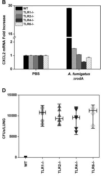
To further assess the role of TLR1 and TLR6 in the innate immune detection of A. fumigatus, we used an in vivo infection model of A. fumigatus pneumonia. CXCL1 and CXCL2 mRNA expression, as well as A. fumigatus burden, was measured in lungs from WT, TLR1-, TLR2-, TLR4-, and TLR6-deficient mice
Figure 1. Murine Toll-like receptor (TLR) 1, TLR2, TLR4, and TLR6 are required to produce interleukin 12p40 (IL-12p40), CXCL2, interleukin 6 (IL-6), and tumor necrosis factor a (TNF-a) in response to wild-type (WT) and DrodA Aspergillus fumigatus. Bone marrow–derived macrophages from WT, TLR12/2, TLR22/ 2
,TLR32/2, TLR42/2, and TLR62/2mice were incubated for 24 hours with 2% PFA DrodA A. fumigatus (multiplicity of infection [MOI], 1) and WT A. fumigatus (MOI, 1). IL-12p40 (A ), CXCL2 (B ), IL-6 (C ), and TNF-a (D ) secretion was quantified by enzyme-linked immunosorbent assay. Data are means 6 standard deviations of triplicates from 1 experiment representative of 3 experiments. Abbreviations: PBS, phosphate-buffered saline; PFA, paraformaldehyde.
24 hours after intranasal inoculation of DrodA A. fumigatus. In lungs from WT mice, the infection strongly increased CXCL1 and CXCL2 mRNA levels (Figure 3A and 3B), whereas the mold was nearly undetectable as revealed by CFU counts and RT-PCR detecting A. fumigatus cytochrome B gene (Figure 3C and 3D). In lungs from TLR1-, TLR2-, TLR4-, and TLR6-deficient mice, the expression of CXCL1 and CXCL2 mRNA was dramatically reduced (Figure 3A and 3B), whereas large amounts of the mould were found in lung homogenates (Figure 3C and 3D). Similarly, lungs from TLR1- and TLR6-deficient mice infected with the WT (instead of the DrodA) strain of A. fumigatus showed an increased fungal bur-den compared with lungs from WT mice (Figure 3E). The presence of conidia in lungs was confirmed by staining of frozen tissue sections with Blankophor (not shown) [33].
To determine the influence of TLRs on mortality, TLR1- and TLR6-deficient mice as well as WT mice (4 each) were infected with WT and DrodA A. fumigatus as described above and fol-lowed up for 30 days. Survival rate was 100% in TLR1- and TLR6-deficient mice as well as in WT mice. Altogether, these results suggest that the absence of TLR1, TLR2, TLR4, and TLR6 causes impaired production of inflammatory mediators in response to the mold and delayed fungal clearance. However, the absence of TLR1 or TLR6 does not induce mortality in this model.
Mediation of Immune Responses to A. fumigatus by Human TLR1 but Not TLR6
To compare the role of human and mouse TLR1 and TLR6 in the innate immune detection of A. fumigatus, HEK293T cells were transiently transfected with vectors expressing human
Figure 2. Mouse Toll-like receptor (TLR) 1 and TLR6 are required to express and secrete interleukin 12p40 (IL-12p40) and CXCL2 in response to wild-type (WT) and DrodA Aspergillus fumigatus dormant (DOC) conidia and WT A. fumigatus germinated conidia. Bone marrow–derived macrophages from WT and TLR1- and TLR6-deficient mice were incubated for 4 hours (A, B ) or 24 hours (C, D ) with 2% PFA DrodA A. fumigatus DOC, WT A. fumigatus DOC, and germinated conidia (GC) at a multiplicity of infection of 1. Expression of IL-12p40 and CXCL2 was measured by reverse-transcription polymerase chain reaction (A, B ), and secretion was quantified by enzyme-linked immunosorbent assay (C, D ). Data are means 6 standard deviations of triplicates from 1 experiment representative of 3 experiments. Abbreviations: AU, arbitrary units; mRNA, messenger RNA; PBS, phosphate-buffered saline; PFA, paraformaldehyde.
Figure 3. Toll-like receptor (TLR) 1-, TLR2-, TLR4-, and TLR6-deficient mice show impaired chemokines expression in response to Aspergillus fumigatus and increased fungal burden in lungs compared with wild-type (WT) mice. Wild-type, TLR12/2, TLR22/2, TLR42/2, and TLR62/2mice were infected
twice, 6 hours apart, with 108colony-forming units (CFUs) of DrodA (A–E ) or WT (E ) A. fumigatus. Twenty-four hours after infection lungs were collected and tested for CXCL1 and CXCL2 messenger RNA (mRNA) expression by reverse-transcription polymerase chain reaction (RT-PCR) (A, B ). Lung homogenates were plated onto Sabouraud agar plates for CFU counts (C, E ) and analyzed for quantification of A. fumigatus cytochrome B copy number by RT-PCR (D ). Abbreviation: PBS, phosphate-buffered saline.
and mouse TLR1 and TLR6 (each in combination with TLR2 and CD14 from the corresponding species), together with a NF-jB luciferase reporter (ELAM) and a transfection control (pRL-TK). The day after transfection, cells were incubated for 4 hours with WT or DrodA A. fumigatus or appropriate controls, and luciferase activity was measured. Both strains of A. fumigatus induced NF-jB activation in cells expressing the mTLR2/mTLR1) (Figure 4A) and the hTLR2/hTLR1 (Figure 4B) combination, as well as the mTLR2/mTLR6 com-bination (Figure 4C) but not the hTLR2/hTLR6 combination (Figure 4D). Cells expressing either the human or mouse TLR2/
TLR1 combination were able to respond to Pam3CSK4(TLR2/
TLR1 ligand) but not to MALP-2 (TLR2/TLR6 ligand) (Figure 4A and 4B). Cells expressing the human or mouse TLR2/TLR6 combination showed NF-jB activation in response to MALP-2 but not to Pam3CSK4 stimulation (Figure 4C and 4D). These
results suggest that detection of A. fumigatus in mice requires the presence of TLR2 together with TLR1 or TLR6, whereas detection in humans relies on TLR2 along with TLR1 but not TLR6.
To further ascertain the role of TLR1 and TLR6 in the detection of A. fumigatus in human, HEK293T cells were
Figure 4. Innate immune response to wild-type (WT) and DrodA Aspergillus fumigatus is mediated by murine Toll-like receptor (TLR) 2/TLR1 (mTLR2/mTLR1) or TLR2/TLR6 (mTLR2/mTLR6) heterodimers and human TLR2/TLR1 (hTLR2/hTLR1). HEK293T cells were transfected with murine and human TLR1 and TLR2 (A, B ) or murine and human TLR2 and TLR6 (C, D ) alone or together with murine (mCD14) and human CD14 (hCD14). Cells were cotransfected with the ELAM luciferase and pRL-TK plasmids. The next day, cells were incubated for 4 hours with phosphate-buffered saline (PBS), Pam3CSK4(1 lg/mL) (A, B ), MALP-2 (100 ng/mL) (C, D). Wild-type or DrodA A. fumigatus (multiplicity of infection, 1) results are expressed as the ratio of
luciferase to Renilla luciferase activity (relative luciferase units [RLU]). Data are means 6 standard deviations of triplicates from 1 experiment representative of 3 experiments. Abbreviations: ELAM, endothelial cell-leukocyte adhesion molecule; MALP-2, macrophage-activating lipopeptide-2.
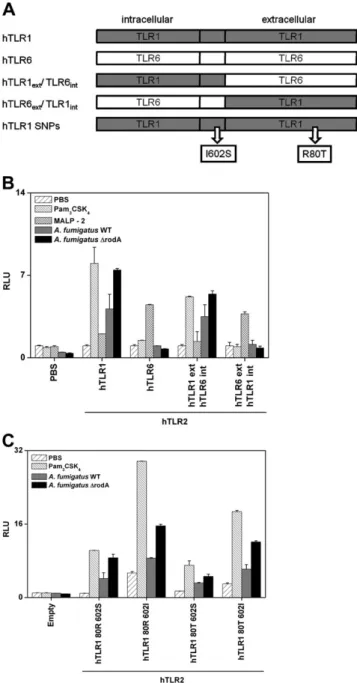
transfected with hTLR2 in combination with chimeric con-structs carrying the extracellular domain from hTLR1 with the intracellular domain from hTLR6 (hTLR1ext/TLR6int) or
the extracellular domain from hTLR6 with the intracellular domain of hTLR1 (hTLR6ext/hTLR1int) (Figure 5A and 5B).
Wild-type and DrodA A. fumigatus induced NF-jB activation in cells expressing chimeric TLRs carrying the extracellular domain from TLR1 (hTLR1ext/TLR6int) but not in those
carrying the extracellular domain from TLR6 (hTLR6ext/
hTLR1int) (Figure5A and 5B). As a control, Pam3CSK4
in-duced NF-jB activation in cells expressing chimeric TLRs carrying the extracellular domain from TLR1 (hTLR1ext/
hTLR6int), and MALP-2 induced NF-jB activation in those
carrying the extracellular domain from TLR6 (hTLR6ext/
hTLR1int), as described elsewhere [29]. Altogether, these data
show that TLR1, but not TLR6, mediates the innate immune detection of A. fumigatus in humans and that the extracellular domain of human TLR1 is required for this detection.
Because TLR1 is involved in the innate immune response to A. fumigatus in humans, we investigated the functional role of 2 common genetic polymorphisms located in the coding region of TLR1, G1805T (S602I; minor allele frequency [MAF], 0.25 in whites) and G239C (R80T; MAF, 0.06 in whites), which have been previously associated with suscepti-bility to IA [19] and/or with impaired TLR1 function [28,34].
Figure 5. Extracellular domain of human Toll-like receptor (TLR) 1 (hTLR1) and G1805T (S602I) and G239C (R80T) polymorphism can modulate NF-jB activation on stimulation with wild-type (WT) and DrodA Aspergillus fumigatus. A, Wild-type hTLR1 and human TLR6 (hTLR6) plasmids carry human complementary DNA (cDNA) cloned into a construct with V5 epitope TAG (pEF6-V5His-TOPO, Invivogen). Chimeric plasmids carry hTLR1 extracellular and transmembrane domains merged with TLR6 intracellular domain (hTLR1ext/hTLR6int) or hTLR6 extracellular and
transmembrane domains merged with TLR1 intracellular domain (hTLR6ext/hTLR1int). Human TLR1 haplotype plasmids carry a WT version
of TLR1 from human cDNA cloned into a construct with V5 epitope TAG (pEF6-V5His-TOPO; Invivogen) that has been genetically modified using site-directed mutagenesis (QuikChange Site-Directed Mutagenesis; Stratagene) to reproduce all possible haplotypic combinations of G1805T (S602I), located in the transmembrane junction, and G239C (R80T), located in the ectodomain. B, C, HEK293T cells were transfected with different mixes of plasmids, as indicated. B, In all conditions, cells were transfected with endothelial cell-leukocyte adhesion molecule
Figure 5 continued. (ELAM) and pRL-TK together with empty vector or human TLF1 (hTLR2) and human CD14 (hCD14). As reported, cells have been complemented also with chimeric plasmids hTLR1ext/hTLR6int or
hTLR6ext/hTLR1intor WT hTLR1 or hTLR6, as control. C, In all conditions,
cells were transfected with ELAM and pRL-TK together with empty vector or hTLR2 and hCD14. As reported, cells have been also transfected with plasmids carrying WT hTLR1 (80R 602S) and all hTLR1 haplotypes (80R 602I, 80T 602S; 80T 602I). One day after transfection, cells were incubated for 4 hours with phosphate-buffered saline (PBS), Pam3CSK4
(1 lg/mL), MALP-2 (100 ng/mL), or DrodA A. fumigatus (multiplicity of infection 1), as indicated. NF-jB activation was measured by the luminescent activity of a NF-jB firefly luciferase reporter plasmid (relative luciferase units [RLU'). Data are means 6 standard deviations of triplicates from 1 experiment representative of 3 experiments.
Supplementary Figure 1.Mouse Toll-like receptor (TLR) 1, TLR2, TLR4, and TLR6 are required to express interleukin 12p40 (IL-12p40), CXCL2, interleukin 6 (IL-6), and tumor necrosis factor a (TNF-a) in response to wild-type (WT) and DrodA Aspergillus fumigatus. Bone marrow–derived macrophages (BMDMs) (A–D ) and bone marrow–derived dendritic cells (BMDCs) (E–F ) from WT, TLR12/2, TLR22/2, TLR32/2, TLR42/2, and TLR62/2 mice were incubated for 4 hours with 2% PFA DrodA
A. fumigatus (multiplicity of infection [MOI], 1) and WT A. fumigatus (MOI, 1). For BMDMs, expression of IL-12p40 (A ), CXCL2 (B ), IL-6 (C ), and TNF-a (D ) was measured by reverse-transcription polymerase chain reaction (RT-PCR). For BMDCs, expression (E ) and secretion (F ) of IL-6 was measured by RT-PCR and enzyme-linked immunosorbent assay, res-pectively. Data are means 6 standard deviations of triplicates from 1 experiment representative of 3 experiments. Abbreviations: AU, arbitrary units; mRNA, messenger RNA; PBS, phosphate-buffered saline.
HEK293T cells were transfected with hTLR1 plasmids con-taining the 4 haplotypic combinations of G1805T (S602I) and G239C (R80T) (Figure 5A and 5C). HEK293T trans-fected with plasmids carrying the 602I allele showed higher levels of NF-jB activation at baseline and after stimulation with WT and DrodA A. fumigatus compared with those carrying the 602S allele. Cells transfected with plasmids carrying the 80T allele revealed lower levels of NF-jB acti-vation after stimulation with WT and DrodA A. fumigatus compared with those carrying the 80R allele. These results suggest that hTLR1 variants modulate innate immune res-ponses to A. fumigatus.
DISCUSSION
In this study, we investigated the role of TLRs in the innate immune detection of A. fumigatus in mice and humans. Previous studies suggested a role for TLR2 and TLR4 in the detection of this pathogen [6,8,12], but these observations were not univocally confirmed [9, 12]. Moreover, limited information was available on the role of TLR1 and TLR6, which both dimerize with TLR2 for signaling [14–16]. We showed that the detection of A. fumigatus is mediated by TLR1 and TLR6 in mice and TLR1 but not TLR6 in humans, together with TLR2 in both species. Our data also confirm the role of TLR4 in both mice and humans.
Immune studies of A. fumigatus are complicated by the evolving composition of its cell wall during its growth, thereby conferring different immunological properties to the pathogen [35]. Investigators used different growth stages of the fungus (ie, resting conidia [8,12], swollen conidia [12], or hyphae [6,8,12]). Resting conidia produce a rodlet layer, which constitutes a mechanical protection from immune cells, leading to weak, almost undetectable, innate immune responses [17]. The weak immunogenicity of the fungus, at least during this early step, may explain why some studies failed to detect the involvement of TLR2 [9], TLR4 [9,12], and MyD88 [9,13]. In the present study, we used a mutant form of A. fumigatus (DrodA), which does not produce this masking layer and mimics the more immunogenic, germinated form of the fungus [17]. Dormant conidia from this immuno-genic strain produced similar, but stronger, immune responses than the WT strain.
Species-specific differences in TLR ligand detection have been described elsewhere. Mice but not humans can detect taxol through TLR4 [36], trilauroylated peptides through TLR2 [37], and LPS from Leptospira through TLR4 [38]. Phylogenetic studies suggest that the different TLRs arose from $1 common ancestor by gene duplications events and subsequent deletions and substitutions [39, 40]. It was proposed that the TLR1 family (including TLR1, TLR2, TLR6, and TLR10) underwent more species-specific changes
during evolution compared with other families [41]. Human TLR1 and TLR6 are closely located on chromosome 4 [39], suggesting a gene duplication event. As for the other TLRs, the TIR signaling domains of TLR1 and TLR6 have more sequence homology (96% in paralogs and 80% in orthologs) than the leucine-rich repeat (LRR) ligand discriminating do-mains (71% and 46%, respectively) [42]. This suggests that the adaptive pressure shaped the ligand binding site to im-prove the recognition and the defense against host-specific pathogens. This is consistent with our transfection experiments using TLR1/TLR6 chimeric plasmids, showing that TLR1, but not TLR6, was able to detect A. fumigatus in humans, and that this specificity was conferred by the TLR1 external LRR-con-taining, but not internal TIR-conLRR-con-taining, domain.
The multiplicity of pattern-recognition receptors (PRRs) targeting the mold would suggest that the innate immune recognition is redundant and that deficiency in a single PRR would only account for a moderate effect. However, con-sistent with previous observations [43], our data show that the absence of a single PRR or adaptor protein leads to a relatively strong impairment of cytokine production on A. fumigatus stimulation and/or an important increase in the fungal burden after infection. In fact, the activation of several PRRs (either simultaneously or at different times of the in-fection) may be necessary for appropriate immune responses. This strategy may be adopted by the innate immune system to avoid continuous and deleterious activation of inflammatory responses to a pathogen to which we are exposed on a daily basis, such as A. fumigatus.
It is not known whether this innate immune signal requires a single ligand or multiples ligands from A. fumigatus nor whether different TLRs are activated by the same or by dis-tinct ligands. Some ligands were shown to be detected by both TLR1/2 and TLR2/6 heterodimers (such as Ureaplasma parvum lipoprotein and Chlamydia trachomatis elementary bodies [44,45]), but others are restricted to TLR1/2 (triacylated lipopeptides and lipoarabinomannan [23, 46]) or TLR2/6 (diacylated lipopeptides [22]) heterodimers. However, identifi-cation of A. fumigatus exact ligand(s) may be limited by the ability to effectively isolate components of the pathogen’s cell wall and intracellular compartment.
Identification of the PRRs involved in the innate immune detection of A. fumigatus may have important consequences for patient management. Single nucleotide polymorphisms in the TLR4 gene from hematopoietic stem cell trans-plantation donors have been associated with an increased susceptibility to IA in the recipient [47]. We examined the functional role of 2 TLR1 single nucleotide polymorphisms located in the extracellular domain (G239C, R80T) and in the junction of the transmembrane and intracellular domain (G1805T, S602I) that were described in a report of a genetic study on susceptibility to IA [34]. Our work shows that TLR1
80T is associated with a lower NF-jB activation than TLR1 80R. This is consistent with the findings of the genetic study, showing that 80T is associated with an increased risk of IA [19]. Our experiments showed that TLR1 602I leads to higher NF-jB activation in response to A. fumigatus than TLR1 602S. This is consistent with other studies showing that TLR1 602I confers a better transmembrane expression of the receptor than TLR1 602S and is protective against leprosy [28, 34]. R80T is located in the highly conserved extracellular domain of TLR1 shown to be necessary for TLR1 ligand recognition. One can speculate that this poly-morphism has an important impact on the folding of the LRR domain, leading to an impairment of ligand binding and/or TLR1 function, as already described for other TLRs [48, 49]. Our results may contribute to explain the genetic variability in susceptibility to IA and develop new preventing strategies in high-risk patients [19,47].
Supplementary Data
Supplementary materialsare available at The Journal of Infectious Dis-eases online (http://www.oxfordjournals.org/our_journals/jid/). Supple-mentary materials consist of data provided by the author that are published to benefit the reader. The posted materials are not copyedited. The contents of allsupplementary dataare the sole responsibility of the authors. Questions or messages regarding errors should be addressed to the author.
Notes
Financial support. This work was supported by research funding from the Leenaards Foundation; Santos-Suarez Foundation; the Swiss National Science Foundation (grants 32003B-127613 to P. Y. B. and 310030-118266 to T. C.); and the European Community’s Seventh Framework Program (FP7-2007–2013 under grant agreement HEALTH-F2-2010-260338–ALLFUN).
Potential conflicts of interest. All authors: No reported conflicts. All authors have submitted the ICMJE Form for Disclosure of Potential Conflicts of Interest. Conflicts that the editors consider relevant to the content of the manuscript have been disclosed.
References
1. Ben-Ami R, Lewis RE, Kontoyiannis DP. Invasive mould infections in the setting of hematopoietic cell transplantation: current trends and new challenges. Curr Opin Infect Dis 2009; 22:376–84.
2. Trifilio S, Singhal S, Williams S, et al. Breakthrough fungal infections after allogeneic hematopoietic stem cell transplantation in patients on prophylactic voriconazole. Bone Marrow Transplant 2007; 40:451–6. 3. Kumagai Y, Akira S. Identification and functions of pattern-recognition
receptors. J Allergy Clin Immunol 2010; 125:985–92.
4. O’Neill LA, Bowie AG. The family of five: TIR-domain-containing adaptors in Toll-like receptor signalling. Nat Rev Immunol 2007; 7: 353–64.
5. Lawrence T. The nuclear factor NF-kappaB pathway in inflam-mation. Cold Spring Harb Perspect Biol 2009; 1:a001651. 6. Bellocchio S, Moretti S, Perruccio K, et al. TLRs govern neutrophil
activity in aspergillosis. J Immunol 2004; 173:7406–15.
7. Braedel S, Radsak M, Einsele H, et al. Aspergillus fumigatus anti-gens activate innate immune cells via Toll-like receptors 2 and 4. Br J Haematol 2004; 125:392–9.
8. Meier A, Kirschning CJ, Nikolaus T, Wagner H, Heesemann J, Ebel F. Toll-like receptor (TLR) 2 and TLR4 are essential for Aspergillus-induced activation of murine macrophages. Cell Microbiol 2003; 5:561–70. 9. Dubourdeau M, Athman R, Balloy V, et al. Aspergillus fumigatus
induces innate immune responses in alveolar macrophages through the MAPK pathway independently of TLR2 and TLR4. J Immunol 2006; 177:3994–4001.
10. Bellocchio S, Montagnoli C, Bozza S, et al. The contribution of the Toll-like/IL-1 receptor superfamily to innate and adaptive immunity to fungal pathogens in vivo. J Immunol 2004; 172:3059–69.
11. Bretz C, Gersuk G, Knoblaugh S, et al. MyD88 signaling contributes to early pulmonary responses to Aspergillus fumigatus. Infect Immun 2008; 76:952–8.
12. Mambula SS, Sau K, Henneke P, Golenbock DT, Levitz SM. Toll-like receptor (TLR) signaling in response to Aspergillus fumigatus. J Biol Chem 2002; 277:39320–6.
13. Marr KA, Balajee SA, Hawn TR, et al. Differential role of MyD88 in macrophage-mediated responses to opportunistic fungal pathogens. Infect Immun 2003; 71:5280–6.
14. Jin MS, Kim SE, Heo JY, et al. Crystal structure of the TLR1-TLR2 heterodimer induced by binding of a tri-acylated lipopeptide. Cell 2007; 130:1071–82.
15. Kang JY, Nan X, Jin MS, et al. Recognition of lipopeptide patterns by Toll-like receptor 2–Toll-like receptor 6 heterodimer. Immunity 2009; 31:873–84.
16. Farhat K, Riekenberg S, Heine H, et al. Heterodimerization of TLR2 with TLR1 or TLR6 expands the ligand spectrum but does not lead to differential signaling. J Leukoc Biol 2008; 83:692–701.
17. Aimanianda V, Bayry J, Bozza S, et al. Surface hydrophobin prevents immune recognition of airborne fungal spores. Nature 2009; 460:1117–21. 18. Thau N, Monod M, Crestani B, et al. Rodletless mutants of Aspergillus
fumigatus. Infect Immun 1994; 62:4380–8.
19. Kesh S, Mensah NY, Peterlongo P, et al. TLR1 and TLR6 poly-morphisms are associated with susceptibility to invasive aspergillosis after allogeneic stem cell transplantation. Ann N Y Acad Sci 2005; 1062:95–103.
20. Adachi O, Kawai T, Takeda K, et al. Targeted disruption of the MyD88 gene results in loss of IL-1– and IL-18–mediated function. Immunity 1998; 9:143–50.
21. Hoshino K, Takeuchi O, Kawai T, et al. Cutting edge: Toll-like receptor 4 (TLR4)–deficient mice are hyporesponsive to lipopoly-saccharide: evidence for TLR4 as the Lps gene product. J Immunol 1999; 162:3749–52.
22. Takeuchi O, Kawai T, Muhlradt PF, et al. Discrimination of bacterial lipoproteins by Toll-like receptor 6. Int Immunol 2001; 13:933–40. 23. Takeuchi O, Sato S, Horiuchi T, et al. Cutting edge: role of Toll-like
receptor 1 in mediating immune response to microbial lipoproteins. J Immunol 2002; 169:10–14.
24. Yamamoto M, Sato S, Hemmi H, et al. Role of adaptor TRIF in the MyD88-independent Toll-like receptor signaling pathway. Science 2003; 301:640–3.
25. Alexopoulou L, Holt AC, Medzhitov R, Flavell RA. Recognition of double-stranded RNA and activation of NF-kappaB by Toll-like receptor 3. Nature 2001; 413:732–8.
26. Roger T, Froidevaux C, Le Roy D, et al. Protection from lethal gram-negative bacterial sepsis by targeting Toll-like receptor 4. Proc Natl Acad Sci U S A 2009; 106:2348–52.
27. Delaloye J, Roger T, Steiner-Tardivel QG, et al. Innate immune sensing of modified vaccinia virus Ankara (MVA) is mediated by TLR2-TLR6, MDA-5 and the NALP3 inflammasome. PLoS Pathog 2009; 5:e1000480.
28. Hawn TR, Misch EA, Dunstan SJ, et al. A common human TLR1 polymorphism regulates the innate immune response to lipopep-tides. Eur J Immunol 2007; 37:2280–9.
29. Omueti KO, Beyer JM, Johnson CM, Lyle EA, Tapping RI. Domain exchange between human Toll-like receptors 1 and 6 reveals a region required for lipopeptide discrimination. J Biol Chem 2005; 280:36616–25.
30. Bochud PY, Hawn TR, Aderem A. Cutting edge: a Toll-like recep-tor 2 polymorphism that is associated with lepromatous leprosy is unable to mediate mycobacterial signaling. J Immunol 2003; 170: 3451–4.
31. Into T, Kiura K, Yasuda M, et al. Stimulation of human Toll-like receptor (TLR) 2 and TLR6 with membrane lipoproteins of Myco-plasma fermentans induces apoptotic cell death after NF-kappa B activation. Cell Microbiol 2004; 6:187–99.
32. Roger T, David J, Glauser MP, Calandra T. MIF regulates innate immune responses through modulation of Toll-like receptor 4. Nature 2001; 414:920–4.
33. Monod M, Jaccoud S, Stirnimann R, et al. Economical microscope configuration for direct mycological examination with fluorescence in dermatology. Dermatology 2000; 201:246–8.
34. Johnson CM, Lyle EA, Omueti KO, et al. Cutting edge: a common polymorphism impairs cell surface trafficking and functional responses of TLR1 but protects against leprosy. J Immunol 2007; 178:7520–4. 35. Bozza S, Perruccio K, Montagnoli C, et al. A dendritic cell vaccine
against invasive aspergillosis in allogeneic hematopoietic trans-plantation. Blood 2003; 102:3807–14.
36. Lien E, Means TK, Heine H, et al. Toll-like receptor 4 imparts ligand-specific recognition of bacterial lipopolysaccharide. J Clin Invest 2000; 105:497–504.
37. Grabiec A, Meng G, Fichte S, Bessler W, Wagner H, Kirschning CJ. Human but not murine Toll-like receptor 2 discriminates between tri-palmitoylated and tri-lauroylated peptides. J Biol Chem 2004; 279:48004–12.
38. Nahori MA, Fournie-Amazouz E, Que-Gewirth NS, et al. Differential TLR recognition of leptospiral lipid A and lipopolysaccharide in murine and human cells. J Immunol 2005; 175:6022–31.
39. Kruithof EK, Satta N, Liu JW, Dunoyer-Geindre S, Fish RJ. Gene conversion limits divergence of mammalian TLR1 and TLR6. BMC Evol Biol 2007; 7:148.
40. Zhou H, Gu J, Lamont SJ, Gu X. Evolutionary analysis for functional divergence of the Toll-like receptor gene family and altered functional constraints. J Mol Evol 2007; 65:119–23.
41. Roach JC, Glusman G, Rowen L, et al. The evolution of vertebrate Toll-like receptors. Proc Natl Acad Sci U S A 2005; 102:9577–82. 42. Kubarenko A, Frank M, Weber AN. Structure-function relationships of
Toll-like receptor domains through homology modelling and molec-ular dynamics. Biochem Soc Trans 2007; 35:1515–18.
43. Garlanda C, Hirsch E, Bozza S, et al. Non-redundant role of the long pentraxin PTX3 in anti-fungal innate immune response. Nature 2002; 420:182–6.
44. Shimizu T, Kida Y, Kuwano K. Ureaplasma parvum lipoproteins, including MB antigen, activate NF-{kappa}B through TLR1, TLR2 and TLR6. Microbiology 2008; 154:1318–25.
45. Bas S, Neff L, Vuillet M, et al. The proinflammatory cytokine response to Chlamydia trachomatis elementary bodies in human macrophages is partly mediated by a lipoprotein, the macrophage infectivity potenti-ator, through TLR2/TLR1/TLR6 and CD14. J Immunol 2008; 180: 1158–68.
46. Tapping RI, Tobias PS. Mycobacterial lipoarabinomannan mediates physical interactions between TLR1 and TLR2 to induce signaling. J Endotoxin Res 2003; 9:264–8.
47. Bochud PY, Chien JW, Marr KA, et al. Toll-like receptor 4 poly-morphisms and aspergillosis in stem-cell transplantation. N Engl J Med 2008; 359:1766–77.
48. Hawn TR, Verbon A, Lettinga KD, et al. A common dominant TLR5 stop codon polymorphism abolishes flagellin signaling and is associ-ated with susceptibility to Legionnaires’ disease. J Exp Med 2003; 198: 1563–72.
49. Hidaka F, Matsuo S, Muta T, Takeshige K, Mizukami T, Nunoi H. A missense mutation of the Toll-like receptor 3 gene in a patient with influenza-associated encephalopathy. Clin Immunol 2006; 119: 188–94.