Molecular characterization of the genes encoding DNA gyrase and topoisomerase IV of Listeria monocytogenes
8
0
0
Texte intégral
(2) R. Lampidis et al. of the gyrase and topoisomerase genes (source of DNA), as well as mutagenesis of gyrA (donator of DNA) and the complementation experiments (recipient strain). E. coli DH5α was from Gibco-BRL, Life Technologies, Inc., Gaithersburg, MD, USA. E. coli TOP10 was from Invitrogen BV, Groningen, The Netherlands.. Culture media and antibiotics L. monocytogenes was grown in brain-heart infusion (BHI) broth (BD/Difco, Franklin Lakes, NJ, USA), E. coli strains were grown in Luria–Bertani (LB) broth, both at 37°C. Antibiotics for selection of transformants were used at the following concentrations (Sigma-Aldrich Corp., St Louis, MO, USA): ampicillin, 100 mg/L (E. coli); erythromycin, 50 mg/L (E. coli) or 5 mg/L (L. monocytogenes); chloramphenicol, 10 mg/L (E. coli and L. monocytogenes).. Determination of MICs MICs were determined in duplicate using Etest (AB Biodisk, Solna, Sweden; purchased from Viva Diagnostika GmbH, Hürth/Köln, Germany) on Mueller–Hinton agar plates according to the manufacturer’s instructions. Test strips covered the following MIC ranges: 0.002–32 mg/L for ciprofloxacin and sparfloxacin, 0.016–256 mg/L for nalidixic acid.. Isolation of DNA E. coli plasmid DNA was isolated with a QIAspin Miniprep Kit (Qiagen N.V., Venlo, The Netherlands). L. monocytogenes EGD chromosomal DNA was isolated with QIAamp and DNeasy Tissue Kits (Qiagen) according to the manufacturer’s protocols for bacteria. For inverse PCR, chromosomal DNA was isolated by proteinase K and SDS lysis as described by Leimeister-Wächter & Chakraborty,10 and cleaned up with phenol/chloroform extraction; the total amount of DNA isolated from 25 mL of overnight culture was resuspended in 600 µL of sterile distilled water. For both chromosomal DNA isolation procedures, the cell walls were initially disrupted with mutanolysin (Sigma) (cell pellet resuspended in 1 mL of 100 mM sodium phosphate buffer pH 7.0, containing 250 U of mutanolysin) for 1–3 h.. Recombinant DNA techniques Recombinant DNA techniques were applied according to standard protocols.11 For cloning, amplicons were ligated blunt-ended into vector pUC18, pre-digested with SmaI (pUC18 Ready-To-Go; Amersham Pharmacia Biotech AB, Uppsala, Sweden). For complementation and mutagenesis, the gyrBA genes were PCR amplified with primers GC-3 and GC-4R (Table 1) and ligated into the multiple cloning site (internal of cat68::lacZα, constitutively transcribed from promoter P59) of vector pHPS9 (cmR, erR).12 The vector was cut. with NheI and SmaI (gyrBA divergently orientated to promoter P59, pNT) or with BamHI and SmaI (gyrBA in the same orientation as P59, pW1-18).. PCR Goldstar DNA polymerase (Eurogentec Bel SA, Seraing, Belgium) was used applying standard parameters for concentrations: 1× reaction buffer as supplied by the manufacturer, 1.5 mM MgCl2, 200 µM of each dNTP, 12.5 pmol of each primer (Table 1) and 1 U of DNA polymerase per kb of amplicon in 50 µL total reaction volume. The concentration of degenerate primers in the PCR mixture was increased to match the degree of degeneration (e.g. two-fold increased concentration for an oligonucleotide with two degenerate nucleotide positions). The final MgCl2 concentration was raised to 3 mM for amplification of parC QRDR with primers PAC-1 and PAC-2R and to 2 mM for amplification of gyrB (primers GYB-1X and GYB-2R) and parE (primers PAE-1 and PAE-2RX). Cycling parameters were: 2.5 min at 95°C as an initial denaturation step; 30 cycles of 30 s at 95°C, 30 s at the appropriate annealing temperature (5°C below Tm of oligo), and 1 min of extension per kb of amplicon at 72°C; followed by a last extension step of 2 min at 72°C. For inverse PCR,13 20 µL of chromosomal DNA were digested overnight with an appropriate restriction enzyme (EcoRI, HindIII; 150 U in 100 µL reaction volume), extracted with phenol/chloroform, religated (5 U ligase in 400 µL reaction volume) and, after precipitation, resuspended in 60 µL of sterile distilled water. Aliquots of 2.5–10 µL were used as a template in the PCR. For cloning of the full-length genes, mutagenesis and re-sequencing PCRs, proofreading DNA polymerases Pfu turbo (Stratagene Inc., La Jolla, CA, USA) or Pwo (Roche/Hoffmann-La Roche Ltd, Basel, Switzerland) were used. Amplification products were purified with the QIAquick PCR purification kit (Qiagen).. Site-directed mutagenesis Site-directed mutagenesis was achieved by applying recombination PCR (RPCR)14 to the gyrA-QRDR (PCR-amplified with primers QRD-1 and QRD-2R) of L. monocytogenes EGD, cloned into pUC18. Primers for mutagenesis were AMP-2 and AMP-1R from the pUC18 ampR gene in combination with FE-1 and FE-2R, TS-1 and TS-2R, TF-1 and TF-2R, or MS-1 and MS-2R (Table 1). Following mutagenesis in pUC18, QRDR DNA fragments were exchanged between plasmids after restriction with SmaI and BstXI (unique restriction sites in gyrBA, flanking the QRDR).. DNA sequencing and analysis DNA was sequenced at MWG (MWG-Biotech AG, Ebersberg, Germany). DNA and protein sequences were analysed with DNASIS (Hitachi Software Engineering Co., Ltd, Yokohama, Japan) and the HUSAR analysis package via the inter-. 918.
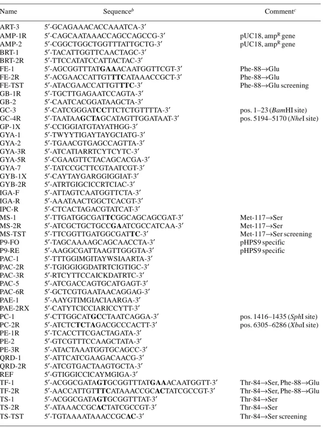
(3) DNA gyrase and topoisomerase IV of L. monocytogenes Table 1. Primers used in this studya Name ART-3 AMP-1R AMP-2 BRT-1 BRT-2R FE-1 FE-2R FE-TST GB-1R GB-2 GC-3 GC-4R GP-1X GYA-1 GYA-2 GYA-3R GYA-5R GYA-7 GYB-1X GYB-2R IGA-F IGA-R IPC-R MS-1 MS-2R MS-TST P9-FO P9-RE PAC-1 PAC-2R PAC-3R PAC-5 PAC-6R PAE-1 PAE-2RX PC-1 PC-2R PE-1R PE-2 PE-3R QRD-1 QRD-2R REF TF-1 TF-2R TS-1 TS-2R TS-TST. Sequenceb. Commentc. 5′-GCAGAAACACCAAATCA-3′ 5′-CAGCAATAAACCAGCCAGCCG-3′ 5′-CGGCTGGCTGGTTTATTGCTG-3′ 5′-TACATTGGTTCAACTAGC-3′ 5′-TTCCATATCCATTACTAC-3′ 5′-AGCGGTTTATGAAACAATGGTTCGT-3′ 5′-ACGAACCATTGTTTCATAAACCGCT-3′ 5′-ATACGAACCATTGTTTC-3′ 5′-TGCTTGAGAATCCAGTA-3′ 5′-CAATCACGGATAAGCTA-3′ 5′-CATCGGGATCCTTCTCTGTTTTA-3′ 5′-TAATAAGCTAGCATAGTTGGATAAT-3′ 5′-CCIGGIATGTAYATHGG-3′ 5′-TWYYTIGAYTAYGCIATG-3′ 5′-TGAACGTGAGCCAGTTA-3′ 5′-ATCATIARRTCYTCYTC-3′ 5′-CGAAGTTCTACAGCACGA-3′ 5′-TATCCGCTTCGTAATCGT-3′ 5′-CAYTAYGARGGIGGIAT-3′ 5′-ATRTGIGCICCRTCIAC-3′ 5′-ATTAGTCAATGGTTCTA-3′ 5′-AAATAACTGGCTCACGT-3′ 5′-CTCACTAGACGTATCAT-3′ 5′-TTGATGGCGATTCGGCAGCAGCGAT-3′ 5′-ATCGCTGCTGCCGAATCGCCATCAA-3′ 5′-TTCGGTTGATGGCGATTC-3′ 5′-TAGCAAAAGCAGCAACCTA-3′ 5′-AAGGCGATTAAGTTGGGTA-3′ 5′-TTTGGIMGITAYWSIAARTA-3′ 5′-TGIGGIGGDATRTCIGTIGC-3′ 5′-RTCYTTCCAICKDATRTC-3′ 5′-ATCGACCAGTGCATGAGT-3′ 5′-GCTCGTGAATAACAGGAG-3′ 5′-AAYGTIMGIACIAARGA-3′ 5′-CATYTCICCIARICCYTT-3′ 5′-CTTGGCATGCCTAATCAGGA-3′ 5′-ATCTCTCTAGACGCCCACTT-3′ 5′-TCACCTTCGACTAGATA-3′ 5′-GTCGTTTCCAAGCTATA-3′ 5′-ATACTAAATGGTGCAGCC-3′ 5′-ATTCATCGAAGACAACG-3′ 5′-ATCGTGACTAAGTGCTA-3′ 5′-GTIGGICCICAYMGIGA-3′ 5′-ACGGCGATAGTGCGGTTTATGAAACAATGGTT-3′ 5′-AACCATTGTTTCATAAACCGCACTATCGCCGT-3′ 5′-ACGGCGATAGTGCGGTTTAT-3′ 5′-ATAAACCGCACTATCGCCGT-3′ 5′-TGTAAAATAAACCGCAC-3′. aIUPAC. pUC18, ampR gene pUC18, ampR gene. Phe-88→Glu Phe-88→Glu Phe-88→Glu screening. pos. 1–23 (BamHI site) pos. 5194–5170 (NheI site). Met-117→Ser Met-117→Ser Met-117→Ser screening pHPS9 specific pHPS9 specific. pos. 1416–1435 (SphI site) pos. 6305–6286 (XbaI site). Thr-84→Ser, Phe-88→Glu Thr-84→Ser, Phe-88→Glu Thr-84→Ser Thr-84→Ser Thr-84→Ser screening. abbreviations; I, inosine. introduced to create mutational or restriction sites, are in bold. cNucleotide positions according to the published sequence. Introduced restriction sites are in parentheses. bMismatches,. 919.
(4) R. Lampidis et al. net (German Cancer Research Center, Heidelberg, Germany; HUSAR is based on the GCG Package, Genetics Computer Group, Inc.). FASTA searches were carried out at the EMBL European Bioinformatics Institute (Cambridge, UK).. Nucleotide sequence accession number. at 37°C before streaking them on selective BHI plates. Transformants were obtained after 2–5 days of incubation at 37°C. Transformants were PCR-screened with plasmid/insertspecific and L. monocytogenes iap-specific17 primer pairs.. RNA extraction and RT–PCR. The nucleotide sequence data reported here will appear in the GenBank nucleotide sequence databases with the following accession numbers: AF084042 for the gyrBA genes and AF084044 for the parEC genes.. Gyrase homology modelling The sequences of E. coli and L. monocytogenes EGD GyrA were aligned with the program XSAE (C. Broger, personal communication). The program MOLOC15 was used for onscreen modelling based on the sequence alignment and on the published X-ray structure of the 59 kDa N-terminal fragment of E. coli GyrA.16. Total RNA was isolated from E. coli using an RNeasy Kit (Qiagen) and on-column DNase (Qiagen) treatment according to the manufacturer’s instructions. Total RNA (1 µg) was reverse transcribed with primer IGA-R (gyrA) or BRT-2R (gyrB) using a ‘First Strand cDNA Synthesis Kit for RT–PCR’ (Roche) according to the manufacturer’s protocol. cDNA (5 µL) was used in the subsequent PCRs (50 µL reaction volume, 1× reaction buffer, 1.5 mM MgCl2, 12.5 pmol each primer, 1.5 U Goldstar DNA polymerase; 45°C annealing temperature) with ART-3/IGA-R (gyrA) and BRT-1/BRT-2R (gyrB) (Table 1).. Transforming plasmids into L. monocytogenes. Results. L. monocytogenes EGD from mid-log growth phase (OD550 = 0.6–0.7) was pelleted at 3000g, washed once in 1/10 vol. 3.5 SMEM (952 mM sucrose/3.5 mM MgCl2) and resuspended in 1/100 vol. 3.5 SMEM (cells were kept at 4°C during all steps). Using a Gene Pulser II unit with Pulse Controller Plus (BioRad Laboratories Inc., Hercules, CA, USA), 100–200 µL cell aliquots were electroporated with 10 µg of DNA in 0.2 cm cuvettes (Gene Pulser settings: 2.5 kV, 25 µF, 100 Ω). After pulsing, the bacteria were pre-cultured in 1 mL of BHI for 3 h. PCR amplificaton of the QRDRs The GyrA and ParC protein sequences of Bacillus subtilis, Streptococcus pneumoniae, Staphylococcus aureus and E. coli were aligned using DNASIS. Pairs of degenerated PCR primers were selected from conserved regions, GYA-1 and PAC-2R from GyrA, PAC-1 and PAC-2R from ParC (Figure 1). These primers were designed so that they would not amplify gyrA and parC of L. monocytogenes EGD. Figure 1. L. monocytogenes SLCC 5853 genomic DNA regions containing the gyrBA (a) and parEC (b) operons. PCR products and primers used for cloning are shown above the genes.. 920.
(5) DNA gyrase and topoisomerase IV of L. monocytogenes simultaneously (although PAC-2R has binding sites in both genes). Internal parts of gyrB (primers GYB-1X and GYB2R) and parE (primers PAE-1 and PAE-2RX) were PCR amplified in the same way.. PCR amplification of the full-length genes The missing DNA regions between gyrB and gyrA as well as between parE and parC were amplified with primers derived from the sequences already obtained (primers GB-2 and IGA-R for the gyrBA genes, PE-2 and IPC-R for parEC genes), demonstrating that gyrB and gyrA as well as parE and parC are contiguous in L. monocytogenes EGD. The missing 5′ and 3′ regions were amplified either by inverse PCR or by using degenerate primers derived from conserved amino acid stretches again (Figure 1 and Table 1). In the case of gyrB, a degenerate primer (REF) for recF was chosen, assuming that recF precedes gyrB in L. monocytogenes, as it does in B. subtilis. The gyrA 3′ region was PCR amplified, first using primer GYA-2 in combination with the degenerate primer GYA-3R and subsequently by an inverse PCR with primers GYA-5R and GYA-7 (EcoRI cut DNA). The parE 5′ region was PCR amplified, first with primers PE-1R and GP-1X (degenerate), followed by inverse PCR with primers PE-3R and IGA-F (HindIII cut DNA). The parC 3′ region was PCR amplified using primers IGA-F and primer PAC-3R (degenerate) and completed with inverse PCR using primers PAC-5 and PAC-6R (HindIII cut DNA). The obtained sequences span 8419 bp (gyrBA plus flanking regions) and 7563 bp (parEC plus flanking regions). The organization of both chromosomal regions is shown in Figure 1. Each locus was PCR amplified again for cloning and resequencing with proofreading DNA polymerases in order to minimize misincorporations. gyrBA was PCR amplified with primers GC-3 and GC-4R (Table 1) and cloned into vector pHPS9 for sequencing and complementation studies. As parEC (PCR-amplified with primers PC-1 and PC-2R; Table 1) from L. monocytogenes could not be cloned in E. coli, the amplicon was resequenced directly.. Sequence analysis The DNA gyrase subunits GyrA (842 aa) and GyrB (646 aa) show the highest similarity to GyrA (71.4% identity) and GyrB (71.2% identity) from B. subtilis. Preceding gyrB, a gene similar to recF can be found. The deduced amino acid sequence (only partial, 106 aa, C-terminus) shows 77.1% identity to RecF from B. subtilis. Downstream of gyrA, four open reading frames (ORFs) are located. ORF1 (504 aa) codes for a protein with similarity to cardiolipin synthase (cls) from Bacillus firmus (37.5% identity). The deduced amino acid sequence of ORF2 (172 aa) is similar to spermidine N1-acetyltransferase (speG) from E. coli (70.6% identity).. ORF3 codes for a protein (327 aa) showing similarity to mevalonate kinase (mvaK1) from Enterococcus faecium (40.8% identity), and ORF4 codes for a protein (only partial, 79 aa, N-terminus) with similarity to mevalonate diphosphate decarboxylase (mvaD) from Staphylococcus haemolyticus (59.5% identity). Interestingly, ORF3 overlaps ORF4, the start codon of ORF4 being located 41 bp upstream of the stop codon of ORF3. The topoisomerase IV subunits ParC (819 aa) and ParE (655 aa) show highest similarities to their counterparts ParC (64.0% identity) and ParE (76.0% identity) from B. subtilis. Three ORFs are located upstream of parE. The first (ORFA, only partial, 103 aa, C-terminus) codes for a protein with similarity to LacX from Lactococcus lactis subsp. lactis (32.0% identity). ORFB (198 aa) and ORFC (135 aa) code for proteins with similarity to B. subtilis YneS (58.6% identity) and YneT (61.8% identity), respectively. Downstream of parC ORFD (155 aa) codes for a protein with similarity to autoinducer-2 production protein LuxS from Helicobacter pylori (68.5% identity). No significant similarity to proteins in the databases was found for ORFE (only partial, 187 aa, N-terminus). Sequence analysis hints at a putative but perfect –10 box preceding gyrB (TATAAT, nt 332–337 in the published sequence). However, no matching –35 consensus sequence is found here (consensus sequences without –35 boxes have been described for gyrase genes). No promoter consensus sequences and no terminator-like structures seem to be present in the intergenic space between gyrB and gyrA. In contrast, a terminator-like structure is located downstream of gyrA (nt 4933–4989). The situation is similar for parE and parC: a terminator-like structure is situated downstream of parC (nt 6127–6166) and a rather weak putative promoter is located upstream of parE [TTGTCA-(N)15-ATTAAT, nt 1582–1608 in the published sequence]. A QRDR alignment of gyrA of L. monocytogenes and of other quinolone-susceptible bacterial species (Figure 2) revealed two altered residues, commonly associated with resistance to fluoroquinolones5 (for an overview see Piddock 18). At position 88 (87 in E. coli numbering) the aromatic amino acid phenylalanine is found instead of the negatively charged aspartate/glutamate residue. At position 84 (83 in E. coli) threonine replaces serine. A third possible variation of importance was discovered in the N-terminus of GyrA of L. monocytogenes by superimposing three-dimensional models of the N-terminal portions of GyrA of E. coli and L. monocytogenes using Swiss-PdbViewer (Glaxo Wellcome/GlaxoSmithKline, UK). The side-chain of Met-117 (Ser-116 in E. coli) is bulkier than usually found at this position and points towards the two other altered side-chains. All three side-chains are located in close proximity to each other. In 1999, Pestova et al.19 reported on S. pneumoniae gyrase mutants with decreased susceptibility to fluoroquinolones. Beside alterations in ParC. 921.
(6) R. Lampidis et al.. Figure 2. Comparison of the QRDRs of L. monocytogenes (L. mo), B. subtilis (B. su), S. pneumoniae (S. pn), S. aureus (S. au) and E. coli (E. co). Numbering of residues according to E. coli GyrA.. alone or in ParC and ParE, a Ser-114→Gly (116 in E. coli) amino acid substitution in GyrA was found in these mutants (although no direct role in the contribution to resistance could be assigned to this substitution in complementation experiments).. Mutagenesis and complementation In order to investigate the role of the mentioned amino acid substitutions of gyrase of L. monocytogenes, gyrA of L. monocytogenes EGD was mutagenized on plasmid pW1-18 (where gyrBA can be transcribed from plasmidic promoter P59). Phe-88 was replaced with Glu on pW1-A, Thr-84 with Ser on pW1-B, and both positions were changed on pW1-C. Met-117 alone was replaced with Ser on pW1-D and Phe-88→Glu was introduced additionally on pW1-E. Mutations were verified by sequencing the exchanged DNA fragments; the insert of pW1-C was sequenced completely. On the basis of reports that sensitive, plasmid-borne alleles of gyrA are dominant over chromosomal alleles,20–22 the plasmids were transformed into L. monocytogenes EGD (Table 2). The presence of transformed mutants was verified with PCR. Gyrase-specific primers (GC-3 and GC-4R) were used in combination with plasmid-specific primers (P9-FO and P9-RE, forward and reverse primers with binding sites in the multiple cloning site of vector pHPS9) to check for the presence of the plasmids carrying the mutagenized alleles. A set of primers with 3′ bases complementary to the codons of the newly introduced amino acids in gyrA was used to verify the presence of the amino acid substitutions: primer FE-TST for Phe-88→Glu, TS-TST for Thr-84→Ser and MS-TST for Met-117→Ser (Table 1). L. monocytogenes EGD wild type served as a negative control in both screening PCRs. To confirm that indeed L. monocytogenes was transformed, mutants were screened with L. monocytogenes-specific primers (MONO-A and MONO-B).17. Susceptibility testing No differences in the susceptibility patterns between L. monocytogenes wild type and the complemented strains were detected for nalidixic acid. The strains SLCC 5853-A, -B and. -C showed slightly decreased MICs of ciprofloxacin and sparfloxacin. The double mutation in SLCC 5853-C conferred the highest degree of sensitivity to ciprofloxacin (MIC 0.38 mg/L), followed by the intermediate phenotype of SLCC 5853-A (MIC 0.5 mg/L), whereas SLCC 5853-B showed the lowest reduction of the MIC (0.75 compared with 1 mg/L for the control strain SLCC 5853-18, complemented with wild-type gyrBA). The MIC of sparfloxacin was reduced to 1 mg/L compared with 1.5 mg/L for the control strain SLCC 5853-18 in all three cases. No differences in the MICs for all tested antibiotics were observed for SLCC 5853-D and SLCC 5853-E, again compared with SLCC 5853-18 (Table 2). No differences in the pattern of susceptibility to nalidixic acid, either between E. coli DH5α (nalR, gyrA96) wild type or complemented with all plasmids, or between E. coli TOP10 (nalS) wild type or complemented with pW1-18 or pW1-C, were observed (data not shown).. Expression of gyrase RT–PCR with primers specific for gyrA or gyrB of L. monocytogenes revealed transcription of both genes in complemented E. coli DH5α, showing that the constructed plasmids were functional. Transcripts of both genes were also detected in E. coli complemented with pNT, where gyrBA is orientated divergently to plasmidic promoter P59 (data not shown). Transcription of gyrA and gyrB was tested in E. coli, as two alleles of these genes were present in the complemented Listeria strains (plasmidic on vector pHPS9 and chromosomal), which might have resulted in false positive results. The vector pHPS9 had already been used sucessfully for cloning and expression of listerial genes in Listeria spp.23. Discussion The complete gyrBA and parEC genes were cloned and sequenced (Figure 1) and could subsequently be structurally characterized. Assignments were based on close sequence similarities to DNA gyrase and topoisomerase IV subunits of B. subtilis, S. pneumoniae, S. aureus and E. coli.. 922.
(7) DNA gyrase and topoisomerase IV of L. monocytogenes Table 2. Plasmids and determination of MICs MIC (mg/L)b Plasmid/inserta. CIP. SPAR. NA. No plasmid (L. monocytogenes EGD wild type) pHPS9 (cloning vector, no insert) pW1-18 (gyrA wild type)c pW1-A (Phe-88→Glu)c pW1-B (Thr-84→Ser)c pW1-C (Phe-88→Glu, Thr-84→Ser)c pW1-D (Met-117→Ser)c pW1-E (Met-117→Ser, Phe-88→Glu)c. 2 1 1 0.5 0.75 0.38 1 1. 2 1.5 1.5 1 1 1 1.5 1.5. >256 >256 >256 >256 >256 >256 >256 >256. aL.. monocytogenes EGD SLCC 5853 complemented with the plasmid. ciprofloxacin; SPAR, sparfloxacin; NA, nalidixic acid. cInsert of the plasmid is gyrBA (can be transcribed from P59), mutation of gyrA is indicated in parentheses. bCIP,. Sequence analysis of both loci indicates that gyrBA and parEC might constitute transcriptional units, i.e. operons. Terminator-like sequences are only found downstream of gyrA and parC, and promoter-like sequences seem to be located only upstream of gyrB and parE. Further analysis of the gyrase genes confirmed the presence of Phe and Thr residues in GyrA at positions 88 and 84, respectively, in place of Asp/Glu and Ser residues conserved in many quinolone-susceptible bacteria. After site-directed mutagenesis of these residues, the recombinant gyrBA alleles were transformed into L. monocytogenes, producing heterodiploid strains for this locus. Introducing these plasmidborne, presumably sensitive alleles of DNA gyrase had no effect on the sensitivity pattern of L. monocytogenes transformants to nalidixic acid. The plasmidic mutations Phe-88→Glu or Thr-84→Ser produced a slightly elevated sensitivity for fluoroquinolones (Table 2). As this effect was very modest, no definitive conclusion could be drawn about the phenotype of the gyrA gene, or the basis of the inherent resistance of L. monocytogenes to nalidixic acid and fluoroquinolones. This raises the following possibilities regarding the nalidixic acid resistance of L. monocytogenes: (i) the revealed alterations of the amino acid sequence in the QRDR of GyrA are not exclusively responsible for the nalidixic acid-resistant phenotype; (ii) sensitive, plasmid-borne alleles of gyrA are not dominant over chromosomal alleles in L. monocytogenes (as described for other bacterial species20–22); and (iii) other mutations or factors such as efflux pumps dominate over the presumed DNA gyrase mutations. Preliminary experiments with reserpine, a known inhibitor of Gram-positive efflux pumps,24 added to the susceptibility test agar plates25 indicate that efflux does not contribute to the resistance to nalidixic acid, as the phenotypes of the strains tested were not changed.. Also, addition of chloramphenicol or erythromycin to the agar plates, in order to prevent plasmid loss, finally had no effect (data not shown). The quinolone class of antimicrobial drugs is not yet applicable in the treatment of listeriosis, although newer derivatives of the quinolones exhibit improved activity against Listeriae (and Gram-positive bacteria in general). However, quinolones would be of particular interest in the treatment of listeriosis, as they are accumulated intracellularly in the eukaryotic cell, thereby having the potential to reach the intracellular Listeriae in high concentrations, where other antibiotics like β-lactams (the drug of choice against listeriosis) perform inadequately.4 But, as L. monocytogenes is intrinsically resistant to nalidixic acid, the elucidation of the resistance mechanism is more complicated in this case compared with naturally sensitive microorganisms, where the development of resistance (i.e. mutations) can be monitored in relation to the degree of resistance.. Acknowledgements This work was supported by the Forschungsfonds grants of the Klinikum Mannheim, project No. 098200/99-203.. References 1. Gellin, B. G. & Broome, C. V. (1989). Listeriosis. Journal of the American Medical Association 261, 1313–20. 2. Low, J. C. & Donachie, W. A. (1997). A review of Listeria monocytogenes and listeriosis. Veterinary Journal 153, 9–29. 3. Beerens, H. & Tahon-Castel, M. M. (1966). Medium with nalidixic acid for isolation of streptococci, S. pneumoniae, Listeria and Erysipelothrix. Annales de l’Institut Pasteur 111, 90–3. 4. Hof, H., Nichterlein, T. & Kretschmar, M. (1997). Management of listeriosis. Clinical Microbiology Reviews 10, 345–57.. 923.
(8) R. Lampidis et al. 5. Yoshida, H., Kojima, T., Yamagishi, J. & Nakamura, S. ( 1988). Quinolone-resistant mutations of the gyrA gene of Escherichia coli. Molecular and General Genetics 211, 1–7.. 16. Cabral, M. J. H., Jackson, A. P., Smith, C. V., Shikotra, N., Maxwell, A. & Liddington, R. C. (1997). Crystal structure of the breakage–reunion domain of DNA gyrase. Nature 388, 903.. 6. Vila, J., Ruiz, J., Goñi, P. & De Anta, M. T. (1996). Detection of mutations in parC in quinolone-resistant clinical isolates of Escherichia coli. Antimicrobial Agents and Chemotherapy 40, 491–3.. 17. Bubert, A., Köhler, S. & Goebel, W. (1992). Homologous and heterologous regions within the iap gene allow genus- and speciesspecific identification of Listeria spp. by polymerase chain reaction. Applied and Environmental Microbiology 58, 2625–32.. 7. Yamagishi, J., Yoshida, H., Yamayoshi, M. & Nakamura, S. (1986). Nalidixic acid-resistant mutations of the gyrB gene of Escherichia coli. Molecular and General Genetics 204, 367–73. 8. Perichon, B., Tankovic, J. & Courvalin, P. (1997). Characterization of a mutation in the parE gene that confers fluoroquinolone resistance in Streptococcus pneumoniae. Antimicrobial Agents and Chemotherapy 41, 1166–7. 9. Drlica, K. & Zhao, X. (1997). DNA gyrase, topoisomerase IV, and the 4-quinolones. Microbiology and Molecular Biology Reviews 61, 377–92. 10. Leimeister-Wächter, M. & Chakraborty, T. (1989). Detection of listeriolysin, the thiol-dependent hemolysin in Listeria monocytogenes, Listeria ivanovii and Listeria seeligeri. Infection and Immunity 57, 2350–7. 11. Sambrook, J., Fritsch, E. F. & Maniatis, T. (1989). Molecular Cloning: A Laboratory Manual, 2nd edn. Cold Spring Harbor Laboratory Press, Cold Spring Harbor, NY, USA. 12. Haima, P., van Sinderen, D., Schotting, H., Bron, S. & Venema, G. (1990). Development of a β-galactosidase α-complementation system for molecular cloning in Bacillus subtilis. Gene 86, 63–9. 13. Ochman, H., Medhora, M. M., Garza, D. & Hartl, D. L. (1990). Amplification of flanking sequences by inverse PCR. In PCR Protocols: A Guide to Methods and Applications (Innis, M. A., Gelfand, D. H., Sninsky, J. J. & White T. J., Eds), pp. 219–27. Academic Press, San Diego, CA, USA. 14. Jones, D. H. & Winistorfer, S. C. (1990). Simultaneous sitespecific mutagenesis of two distal sites by in vivo recombination of PCR products. Biotechniques 2, 273–8. 15. Gerber, P. R. & Müller, K. (1995). MAB: a generally applicable molecular force field for structure modelling in medicinal chemistry. Journal of Computer-Aided Molecular Design 9, 251–68.. 18. Piddock, L. J. V. (1999). Mechanisms of fluoroquinolone resistance: an update 1994–1998. Drugs 58, Suppl. 2, 11–8. 19. Pestova, E., Beyer, R., Cianciotto, N. P., Noskin, G. A. & Peterson, L. R. (1999). Contribution of topoisomerase IV and DNA gyrase mutations in Streptococcus pneumoniae to resistance to novel fluoroquinolones. Antimicrobial Agents and Chemotherapy 43, 2000–4. 20. Hane, M. W. & Wood, T. H. (1969). Escherichia coli K-12 mutants resistant to nalidixic acid: genetic mapping and dominance studies. Journal of Bacteriology 99, 238–41. 21. Heisig, P. & Wiedemann, B. (1991). Use of a broad-hostrange gyrA plasmid for genetic characterization of fluoroquinoloneresistant Gram-negative bacteria. Antimicrobial Agents and Chemotherapy 35, 2031–6. 22. Nakamura, S., Nakamura, M., Kojima, T. & Yoshida, H. (1989). gyrA and gyrB mutations in quinolone-resistant strains of Escherichia coli. Antimicrobial Agents and Chemotherapy 33, 254–5. 23. Engelbrecht, F., Dominguez-Bernal, G., Hess, J., Dickneite, C., Greiffenberg, L., Lampidis, R. et al. (1998). A novel PrfA-regulated chromosomal locus, which is specific for Listeria ivanovii, encodes two small, secreted internalins and contributes to virulence in mice. Molecular Microbiology 30, 405–17. 24. Neyfakh, A. A., Borsch, C. M. & Kaatz, G. W. (1993). Fluoroquinolone resistance protein NorA of Staphylococcus aureus is a multidrug efflux transporter. Antimicrobial Agents and Chemotherapy 37, 128–9. 25. Brenwald, N. P., Gill, M. J. & Wise, R. ( 1998). Prevalence of a putative efflux mechanism among fluoroquinolone-resistant clinical isolates of Streptococcus pneumoniae. Antimicrobial Agents and Chemotherapy 42, 2032–5.. 924.
(9)
Figure


Documents relatifs
We will retain universal prokaryotic primers and archaeal specific primers within the 16S rRNA gene, two housekeeping genes, gyrB(encoding the ß subunit of the DNA
DNA barcoding and molecular systematics of the benthic and demersal organisms of the CEAMARC survey.
Results from multiple studies including or based on CEAMARC specimens point out that for many groups, barcoding is a valuable tool for the identification of marine specimens from
Besides, the dual observation of the loss of TRAIL expression (a cytokine-promoting cell death [31]) and the overexpression of ILT7 (whose inter- action with BST2 at the surface
Title: Specific identification of Gallibacterium by a PCR using primers targeting the 16S rRNA and 23S rRNA genes.. Authors: Anders Miki Bojesen, Maria
Stéphane Cociancich, UMR Cirad-Inra-Montpellier SupAgro BGPI, Montpellier, France; Kathrin Schneider, Technische Universität, Berlin, Germany; Sandrine Duplan, Isabelle Pieretti,
*Jeu "Il était une fois...": les joueurs s'assoient en cercle et désignent un meneur de jeu/ Celui-ci commence à raconter une histoire qu'il invente puis s'arrête
Calculez la somme ou la différence de chaque
The 2 copies of the a-amylase gene from a strain of D melanogaster have already been partly sequenced (Boer and Hickey, 1986), and the 2 coding regions showed a
