HAL Id: hal-02094590
https://hal-amu.archives-ouvertes.fr/hal-02094590
Submitted on 26 May 2020
HAL is a multi-disciplinary open access
archive for the deposit and dissemination of
sci-entific research documents, whether they are
pub-lished or not. The documents may come from
teaching and research institutions in France or
abroad, or from public or private research centers.
L’archive ouverte pluridisciplinaire HAL, est
destinée au dépôt et à la diffusion de documents
scientifiques de niveau recherche, publiés ou non,
émanant des établissements d’enseignement et de
recherche français ou étrangers, des laboratoires
publics ou privés.
Distributed under a Creative Commons Attribution| 4.0 International License
human gut microbiota
Sophie Mathieu, Mélanie Touvrey-Loiodice, Laurent Poulet, Sophie
Drouillard, Renaud Vincentelli, Bernard Henrissat, Gudmund Skjåk-Bræk,
William Helbert
To cite this version:
Sophie Mathieu, Mélanie Touvrey-Loiodice, Laurent Poulet, Sophie Drouillard, Renaud Vincentelli,
et al.. Ancient acquisition of “alginate utilization loci” by human gut microbiota. Scientific Reports,
Nature Publishing Group, 2018, 8 (1), pp.8075. �10.1038/s41598-018-26104-1�. �hal-02094590�
www.nature.com/scientificreports
Ancient acquisition of “alginate
utilization loci” by human gut
microbiota
Sophie Mathieu
1, Mélanie Touvrey-Loiodice
1, Laurent Poulet
1, Sophie Drouillard
1,
Renaud Vincentelli
2,3, Bernard Henrissat
2,3,4, Gudmund Skjåk-Bræk
5& William Helbert
1In bacteria from the phylum Bacteroidetes, the genes coding for enzymes involved in polysaccharide degradation are often colocalized and coregulated in so-called “polysaccharide utilization loci” (PULs). PULs dedicated to the degradation of marine polysaccharides (e.g. laminaran, ulvan, alginate and porphyran) have been characterized in marine bacteria. Interestingly, the gut microbiome of Japanese individuals acquired, by lateral transfer from marine bacteria, the genes involved in the breakdown of porphyran, the cell wall polysaccharide of the red seaweed used in maki. Sequence similarity analyses predict that the human gut microbiome also encodes enzymes for the degradation of alginate, the main cell wall polysaccharide of brown algae. We undertook the functional characterization of diverse polysaccharide lyases from family PL17, frequently found in marine bacteria as well as those of human gut bacteria. We demonstrate here that this family is polyspecific. Our phylogenetic analysis of family PL17 reveals that all alginate lyases, which have all the same specificity and mode of action, cluster together in a very distinct subfamily. The alginate lyases found in human gut bacteria group together in a single clade which is rooted deeply in the PL17 tree. These enzymes were found in PULs containing PL6 enzymes, which also clustered together in the phylogenetic tree of PL6. Together, biochemical and bioinformatics analyses suggest that acquisition of this system appears ancient and, because only traces of two successful transfers were detected upon inspection of PL6 and PL17 families, the pace of acquisition of marine polysaccharide degradation system is probably very slow.
The high complexity of polysaccharides arises from the stereochemical diversity of their constitutive monosac-charide residues and from the many ways to assemble these residues through glycosidic bonds. Primarily pro-duced by photosynthesis, these macromolecules represent the most abundant biomass and carbon source for land and marine heterotrophic organisms. The biodegradation of polysaccharides requires large arrays of enzymes that can specifically cleave each of the constitutive glycosidic bonds, either by hydrolysis (glycoside hydrolases, GH), β-elimination (polysaccharide lyases, PL) or oxidation (lytic polysaccharide mono-oxygenases, LPMO). Thus, the complexity of the polysaccharide substrates is accompanied by a wide diversity of polysaccharide-degrading enzymes. The International Union of Biochemistry and Molecular Biology lists more than 200 EC numbers that correspond to different GH substrate specificities (http://www.enzyme-database.org)1 and the carbohydrate-active enzymes database (CAZy database, http://www.cazy.org/)2 features 145 GH, 27 PL and 13 LPMO sequence-based families, and these numbers are still growing, e.g. the recent addition of seven new GH families3.
Members of the individual sequence-based families of carbohydrate-active enzymes listed in the CAZy data-base share the same folds and the same catalytic amino acids2. In the case of GHs, the orientation of the cleaved glycosidic bond and the stereochemical outcome of the reaction (e.g. retaining or inverting the anomeric config-uration at the site of cleavage) are essentially conserved in each family4,5. However, many sequence-based families are polyspecific, i.e. group enzymes of different substrate specificity. For several large families of GHs, subfamilies have been shown to improve correlation with substrate specificity and also to identify clades that contain enzymes without any experimentally demonstrated function6–9. Similarly, several families of PLs have been discovered
1CERMAV, CNRS and Grenoble Alpes Université, BP53, 38000, Grenoble Cedex 9, France. 2Centre National de la
Recherche Scientifique (CNRS), UMR7257, Université Aix-Marseille, Marseille, 13288, France. 3INRA, USC 1408
AFMB, 13288, Marseille, France. 4Department of Biological Sciences, King Abdulaziz University, Jeddah, Saudi
Arabia. 5Department of Biotechnology, Norwegian University of Science and Technology, NTNU Sem Sælands vei
6-8, 7491, Trondheim, Norway. Correspondence and requests for materials should be addressed to W.H. (email:
william.helbert@cermav.cnrs.fr) Received: 10 January 2018
Accepted: 4 May 2018 Published: xx xx xxxx
to be polyspecific and most of them have also been divided into subfamilies10. In contrast to the GH families however, PL families contain fewer members and the conservation of function within subfamilies is probably less robust11, making functional predictions from sequence data alone less reliable.
In bacteria belonging to phylum Bacteroidetes, genes involved in the degradation of a given polysaccharide are often clustered on the genome and coregulated. These gene clusters have been termed “polysaccharide utili-zation loci” (PULs)12,13. This colocalization offers an additional lever for the prediction of the function of other polysaccharide-degrading enzymes when one or several members of a PUL have been biochemically character-ized. Over 26,000 predicted PULs are listed in the CAZy database (http://www.cazy.org/PULDB/) based on the analysis of 820 bacteria from the human gut microbiote and from the environment14,15. Analysis of the marine bacterial genomes or metagenomes suggests a large diversity of PULs for the breakdown of marine polysaccha-rides16–22. However, the precise function (specificity) of the PULs can only be hypothesized based on sequence similarity of protein sequences found in the PUL with characterized, but distantly related, carbohydrate-active enzymes. Hypotheses on the specificity of marine PULs are usually formulated by confronting the putative func-tion of genes with the available literature on the composifunc-tion or structure of marine polysaccharides. The funcfunc-tion of several PULs involved in the degradation of marine polysaccharides has been demonstrated by transcriptom-ics, proteomics or enzymology methods. There are marine PULs that target laminaran and alginate18 (the storage and cell wall polysaccharides of brown algae, respectively), ulvan22 (the cell wall polysaccharide of the green algae
Ulva and Enteromorpha sp.), and porphyran23 (the cell-wall polysaccharide of the red alga Porphyra sp.). Porphyran-degrading enzymes (porphyranases) have reportedly been horizontally transferred from a marine
Bacteroidetes to Bacteroidetes harbored in the Japanese gut microbiota23. Acquisition of the porphyran-degrading enzymes has been attributed to the algae-rich diet common in Japan, suggesting that contaminated raw food may have been the vector of the transferred genes. Porphyranases are glycoside hydrolases that belong to the polyspe-cific GH16 family, which also contains agarases and carrageenases and other enzymes for the degradation of red algal polysaccharides. Phylogenetic analyses of family GH16 show that the porphyranase clade groups with both marine and gut Bacteroidetes and shares a common ancestor with agarases and carrageenases23. These results highlight the plasticity of the gut microbiome and its ability to adapt to diet. However, the frequency of acquisi-tion of a novel gene introduced by diet and the mechanisms of adaptaacquisi-tion to diet remain obscure.
The possible occurrence of alginate lyases and carrageenases in the human gut microbiome has been suggested based on sequence similarity of proteins encoded by gut bacteria to alginate lyases24 and on the fact that fecal bac-teria can degrade carrageenan25. However, the functional characterization of the corresponding proteins has not been performed, leaving porphyran as the only experimentally demonstrated example of a marine polysaccharide degraded by enzymes found in the human gut microbiome. Here, we conducted a functional characterization of alginate lyases from family PL17 to establish that human gut bacteria do encode an alginate-degradation system and, combined with phylogenetic analyses, we discuss the putative evolutionary steps that led to the transfer of marine polysaccharide degradation systems to the human gut microbiome.
Results
Selection and production of recombinant PL17 enzymes.
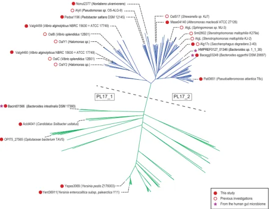
The phylogenetic tree computed from the alignment of the catalytic modules of 272 PL17 sequences shows a clear division into two main clades (Fig. 1). One clade contains all the biochemically characterized enzymes already described in the family, all being alginate lyases (shown in blue in Fig. 1). This clade corresponds to subfamily PL17_210. The other clade (shown in green in Fig. 1) forms a less homogenous group with lineages that root deeply, reflecting high sequence divergence. This clade groups the members of subfamily PL17_1 along with sequences not assigned to any subfamily. To the best of our knowledge, no enzymes from this highly divergent clade have been characterized experimentally and thus the specificity of the corresponding proteins cannot be reliably predicted. Subfamily classification improves sub-strate specificity predictions when subfamilies contain many characterized and isofunctional members; however, predictive power decreases with the size of dataset and, as illustrated in the PL6 family, when several substrate specificities and modes of action (endo- or exo-lyases) are found in the same subfamily11. The shape of the phy-logenetic tree of family PL17, the small number of published protein sequences and the absence of any charac-terized members in a distantly related clade suggest that there may be other specificities or modes of action. To explore the functional diversity in family PL17, we selected 13 target proteins spread across the phylogenetic tree for production and enzymatic characterization.All the targeted proteins fused to a His6-tag were successfully overexpressed in a soluble manner in E. coli
BL21(DE3) grown overnight at 20 °C. The proteins were purified by affinity and size-exclusion chromatogra-phy. The proteins were analyzed by SDS-PAGE and displayed molecular weights ranging from 71.2 to 84.7 kDa, as expected. As reported for the Saccharophagus degradans alginate lyase26, size-exclusion chromatography showed that several proteins such as Valg4460, Pedsa1196 and Mase04140 form dimers, but dimerization was not detected consistently in our experimental conditions.
Functional screening of PL17 family members.
Alginates extracted from brown algae are composed of d-mannuronic acid (M) and l-guluronic acid (G) residues distributed in blocks or alternately along the polysac-charide chain. Composition and distribution of the uronic residues vary greatly according to the algal species27. Isolation of the blockwise fractions of poly-M and poly-G substrates can be obtained by acid hydrolysis followed by selective precipitation experiments28, but highly pure material is difficult to prepare. Therefore, to obtain an accurate description of substrate specificities, we used a poly-M substrate with a regular structure biosynthesized by genetically engineered Pseudomonas fluorescens to avoid the presence of l-guluronic acid residues in the com-position29. Regular poly-MG chains were obtained after enzymatic epimerization, which led to a substrate devoid of G-block contamination30. Substrates with G-blocks were obtained by acid hydrolysis of alginate, but some traces of M-residues were still present28.www.nature.com/scientificreports/
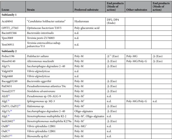
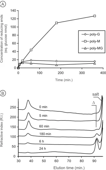
The eight proteins selected from the PL17_2 clade all displayed alginate lyase activity (Table 1), as reported for other members of this clade based on digestion experiments conducted with either raw alginate substrates or on isolated poly-M or poly-G block fractions prepared by chemical treatment26,31–35. The substrate specificity of the enzymes was determined using at least two independent production of enzymes by measuring the veloc-ity of degradation using poly-M, poly-G and poly-MG substrates (Fig. 2A). The degradation kinetics revealed that the preferred substrate was always poly-M chains. For Pedsa1196 (Pedobacter saltans) and Mase04140 (Alteromonas macleodii), the enzymes also showed weak activity on the poly-MG substrate (Table 1). The degra-dation of poly-M chains monitored by size-exclusion chromatography revealed that the alginate lyases produced a single unsaturated monosaccharide end product (4-deoxy-L-erythro-5-hexoseulose uronic acid) (Fig. 2B). No product of intermediate size was produced, demonstrating that the alginate lyases of the clade PL17_2 are exo-polymannuronan lyases.
The five proteins selected from the other clade were all inactive on alginates extracted from algae or on algi-nates with regular structures (poly-M, poly-G and poly-MG). They were thus assayed with a collection of oligo- and polysaccharides containing uronic residues in their structure. No degradation of oligo- or polysaccharides used in this work was observed for the proteins Bacint01566 (Bacteroides intestinalis, ZP_03014006), Ypes3069 (Yersinia pestis, Z176003) and Yent36911 (Yersinia enterocolitica subsp. palearctica Y11, CBY28839). However, Acid4041 (“Candidatus Solibacter usitatus”, ABJ85006) degraded hyaluronic acid according to an endolytic mode of action giving di- and tetrasaccharide end products. Other substrates with β-linked glucuronic acid residues in their structures (e.g. poly-glucuronic acid, xanthan) were not degraded (Fig. S1A). OPIT5_27565 (Opitutaceae
bacterium TAV5, WP_009508481) was found to be active on poly-β(1,4)-D-glucuronic acid. This enzyme had
an endo- mode of action and the smallest end products were di- and trisaccharides (Fig. S1B). All the screening results, substrate specificities and modes of action are summarized in Table 1.
Discussion
Polyspecificity of family PL17.
Alginate lyases were all found in the same clade corresponding to the PL17_2 subfamily and they all shared the same exo-M-lyase mode of action. Most of these enzymes are of marine origin, consistent with the degradation of alginate from marine brown algae. The specificity and the mode of action of the alginate lyases were carefully determined using alginate substrates with known, controlled structures (poly-M, poly-G or poly-MG chains). The results were in agreement with the previous enzymology and crystallography studies carried out on other recombinant enzymes26,31–35. One report indicates that the expression of the marine strain Gramella forsetti PL17 enzyme is upregulated when alginate is added to the culture medium18, supporting substrate specificity towards alginate. Similarly, the transcription of Zobellia galactinovorans PL17 has been induced in the presence of alginate24. Therefore, the clade defining the PL17_2 subfamily appears monospecific in light of all data recorded to date.Figure 1. Phylogenetic tree of the PL17 family. The tree can be divided into two main clades, separated here
by a dashed line. In the clade above the dashed line, shown in blue, all the characterized enzymes are exo-mannuronan lyases. The characterized enzymes below the dashed line, shown in green, include hyaluronic and poly-glucuronic acid lyases.
The results obtained with the PL17 members outside of the first clade were more contrasted. We were not able to identify the substrate for three enzymes (Bacint01566, Ypes3069, Yent36911), but we identified the function of two enzymes: namely, hyaluronan lyase for Acid 4041 and glucuronan lyase for OPIT5_27565. These two activities have never been reported before in family PL17. The hyaluronan and β-1,4-glucuronan substrates share structural simi-larities, especially the β-linked glucuronic acid residue which undergoes enzymatic cleavage by β-elimination. Both enzymes had an endo-acting mode of action, in contrast to the exo-M lyases. Thus, differences in substrate specific-ity and mode of action accompany the segregation of the sequences into two clades on the PL17 phylogenetic tree. Our characterization of hyaluronan and glucuronan lyases in family PL17 also indicates that the previously reported functional data were not sufficient to accurately predict the function of PL17 members based on sequence similarity, and that the poly-specificity of this family has clearly been underestimated. In addition, because several enzymes in our study did not show activity on any of the substrates we used, it is also likely that there are other specificities. In any case, caution should be taken when attempting to assign a function to uncharacterized members that are only distantly related to alginate, glucuronan and hyaluronan lyases.
Alginate utilization loci in human gut microbiota.
The genes coding for enzymes involved in the rec-ognition, internalization and degradation of oligomers and alginate polymers as well as enzymes associated with the metabolic pathways of the end products are colocalized in PULs in the genome of several marine Bacteroidetes strains18,24. In the PULDB database (www.cazy.org/PULDB/), which lists the PULs predicted in a large number ofBacteroidetes, we observed that all PULs of the human gut microbiote containing PL17 alginate lyases also contain
alginate lyases from the PL6_1 subfamily (Fig. 3). Recent functional investigations have shown that the members of the PL6_1 subfamily are endo-alginate lyases11. Therefore, the exo-acting PL17_2 enzymes likely cleave the oligo-alginates produced by the PL6_1 endo-acting lyases. The conservation of colocalized gene pairs coding for PL17_2 and PL6_1 alginate lyases probably reflects a conserved alginate-degradation system.
In addition to the conserved pairs of PL6_1 and PL17_2 genes, the physical organization of the genes in several PULs appears well conserved in several bacteria regardless of their origin – be it marine or human gut (Fig. 3). Using a homology-based approach, Thomas and co-workers24 hypothesized that the alginate degradation system has been laterally transferred from marine to human gut Bacteroidetes. The detailed biochemical characteriza-tion of families PL6 and PL17 now paves the way to a more robust funccharacteriza-tional prediccharacteriza-tion for members of these
Locus Strain Preferred substrate
End products (Mode of
action) Other substrates
End products (Mode of action) Subfamily 1
Acid4041 “Candidatus Solibacter usitatus” Hyaluronan DP2, DP4 (Endo)
OPIT5_27565 Opitutaceae bacterium TAV5 Poly-glucuronic acid
Bacint01566 Bacteroides intestinalis n.d. Ypes3069 Yersinia pestis Z176003 n.d. Yent36911 Yersinia enterocolitica subsp. palearctica Y11 n.d.
Subfamily 2
Pedsa1196 Pedobacter saltans Poly-M Δ** (Exo) Poly-MG Δ (Exo)
Mase04140 Alteromonas macleodii Poly-M Δ (Exo) Poly-MG/Poly-G Δ (Exo) Alg17c Saccharophagus degradans 2–40 Poly-M Δ (Exo)
Valg4459 Vibrio alginolyticus n.d. Valg4460 Vibrio alginolyticus n.d.
Bacegg03249 Bacteroides eggerthii Poly-M Δ (Exo) Patl3651 Pseudoalteromonas atlantica T6c Poly-M Δ (Exo) Nonul2377 Nonlabens ulvanivorans Poly-M Δ (Exo) AlyII31 Pseudomonas sp. OS-ALG-9 Poly-M* n.d.
AlgL32 Sphingomonas sp. MJ-3 Poly-M* n.d. Poly-MG/Poly-G n.d.
OalY1, OalY233 Halomonas sp. Poly-M* Δ (Exo)
Alg17c26 Saccharophagus degradans 2–40 Oligo-alginates Δ (Exo)
AlgL34 Stenotrophomas maltophilia KJ-2 Poly-M*, Oligo-alginates n.d.
Smlt260235 Stenotrophomonas maltophilia K279a Poly-M* Δ (Exo)
OalB51 Vibrio splendidus 12B01 Poly-MG* n.d.
OalC51 Vibrio splendidus 12B01 Poly-M n.d.
OalS1752 Shewanella sp.Kz7 Poly-M n.d.
Table 1. Substrate specificity and mode of action. *The substrates used to characterize the enzyme specificities
were alginate or enriched poly-M or poly-G fractions obtained by acid hydrolysis. **Δ:
4-deoxy-L-erythro-5-hexoseulose uronic acid The table summarizes the substrate specificities, end products and mode of action (endo-, exo-) of the polysaccharide lyases investigated. The alginate lyases described in the literature are also reported in the table (shaded in gray).
www.nature.com/scientificreports/
Figure 2. Characterization of the exo-acting mannuronan (M) lyase Nonul2377. (A) Representative
degradation kinetic revealing the poly-M substrate specificity. Incubation on standard alginate substrates was conducted, at least, with two independent production of enzymes. (B) The exolytic mode of action of the enzymes was visualized using size-exclusion chromatography. Δ: 4-deoxy-L-erythro-5-hexoseulose uronic acid.
Figure 3. Alginate utilization loci in the human gut microbiome containing PL17 alginate lyase. The
families. In particular, our experimental data can discriminate true “alginate utilization loci” from other PULs and especially exclude those containing PL17 proteins that do not cluster with alginate lyases in the phylogenetic tree. Interestingly, the exo-M-lyase activity that we found for Bacteroides eggerthii (Baceg_03249) demonstrates the occurrence of an active alginate lyase in the human gut microbiome.
Ancient acquisition of an alginate utilization locus by the human gut microbiome.
The phyloge-netic tree of family PL17 shows that human gut alginate lyases root deeply in the tree (Fig. 1). The mode of action B.eggerthii (Baceg_03249) is similar to all other alginate lyases and its genomic environment (i.e. PUL organization)
is well preserved, suggesting that the divergence of protein sequences cannot be attributed to a major functional shift. The deep root has one of two explanations. Either it reflects the recent transfer of a PL17 gene belonging to an environmental strain, not present in sequence databanks, that has evolved independently from the other lineages, or it is the ancient acquisition of the PL17 gene by the human gut microbiome followed by independent evolution.
To test these hypotheses, we compared the G + C ratio of the alginate lyases in the PULs with that in the cor-responding full genome. The G + C ratios of the genomes of the human gut reference strains (Bacteroidetes sp. 1_1_30, Bacteroides sp. D2, Bacteroides sp. 2_2_4, Bacteroides sp. 3_1_23, Bacteroides clarus YIT 12056) were very similar to that of the alginate utilization loci, suggesting old acquisition of the alginate lyases by these organisms. In contrast with these five reference strains, the alginate utilizing loci of B. eggerthii DSM 20697 has a different gene organization and had a G + C ratio very different from that of the full genome suggesting it has been the subject of lateral gene transfer36. Because all the PL17 alginate lyases of the human gut clustered in the same clade of the phylogenetic tree, it is thus likely that – after an ancient horizontal transfer – the human microbiome algi-nate lyase genes evolved under selection pressures that differ from those experienced by the original donor. The transfer of PL17 alginate lyase genes between bacteria of the human gut microbiota probably occurred more than once, explaining the distinctive distribution of genes from several strains and the probable recent acquisition of
B. eggerthii DSM 20697 alginate lyase from a different donor.
Due to the colocalization of the PL6 and PL17 alginate lyases in PULs, the acquisition of PL6 alginate lyases is likely to have followed a very similar scenario. We built a phylogenetic tree of family PL6 including all PL6 algi-nate lyases predicted in the known strains of the human gut microbiota (Fig. S2). The gut algialgi-nate lyases grouped into two distinct branches that rooted deep in the tree. One branch groups all the PL6_1 genes that colocalize with PL17_2 alginate lyases, indicating a common ancestor for these PL6_1 enzymes. The second clade contains PL6 alginate lyase genes that were localized in a PUL composed of only PL6 alginate lyases. Altogether, this sug-gests that PL6 alginate lyases were probably acquired by the gut microbiota twice, from two donor strains that possessed PL6 alginate lyases in different genomic environments.
The phylogenetic analyses of the PL17_2 and PL6_1 alginate lyases colocalized in the same PUL suggest a single ancient acquisition of this PUL by a member of the human gut microbiota. Comparison of the phyloge-netic trees is not a direct proof of the co-acquisition of PL17_2/PL6_1 alginate lyases; however, due to their complementary mode of action and their co-occurrence in many marine strains, their co-transfer from marine to gut Bacteroidetes appears highly likely. The retention of the PL17/PL6 gene pairs in the genomes of human gut bacteria for a long period of time supposes that the functional enzymes were expressed and conferred an advan-tage to the host strain. Therefore, it is reasonable to assume that all the elements of the alginate utilization locus, including the machinery for the detection of the alginate substrate and the activation of alginate lyase expression, were all transferred en bloc from a marine to a human gut Bacteroidetes.
Food: a vehicle of genes and a selective pressure on transferred genes.
Lateral transfer between a marine strain and human gut Bacteroidetes was first demonstrated in a gene coding for the porphyranase involved in the degradation of the red algal cell wall polysaccharides found in sushi roll (maki) ingredients23. It was then suggested that genes were transferred to the human gut from marine Bacteroidetes associated with algae eaten as food. Analyses of the phylogenetic trees of PL17, PL6 and GH16 (which includes agarases and porphyranases) suggest that, despite the frequent consumption of algae over many generations, the transfer of genes coding for marine polysaccharide-degrading enzymes is quite rare and ancient. Genomic data available to date are a snapshot of the genetic capital of the microbiome. Possible temporary acquisition of other alginate degradation systems – now lost – may have occurred. The bioinformatics analyses conducted here highlight the most endur-ing transfer events from marine to gut Bacteroidetes. Phylogenetic trees do not provide any information on the dynamics of gene transfer, particularly on the duration of residence of a newly acquired gene by the host strain.The newly transferred genes can persist in the microbiome only if there are selection pressures on the host bacterium to keep the new genes. The regular presence of algal food sources in the digestive tract is therefore a likely factor that gives a competitive advantage to the gene-receiving bacterium. Obviously, food and its associ-ated bacteria are potential sources of genetic material, but the algal diet represents the main selection pressure that promoted the retention of genes coding for algal polysaccharide-degrading enzymes.
What are the substrates of the unknown PL17 enzymes.
The function of several members of family PL17 could not be identified using the collection of polysaccharides that were available to us. To infer what the substrates of these enzymes may be, we examined the nature of the other enzymes encoded in the correspond-ing PULs. In addition to alginate-utilization loci, PULs of the human gut microbiome containcorrespond-ing PL17 genes can be divided into two broad sets according to the composition of the PUL. The first set associates PL17 genes with genes encoding glycoside hydrolases from families GH30_4, GH105 and GH2 (Fig. 4). Family GH105 is composed of α- or β-glucuronyl hydrolases that cleave the Δ-4,5 unsaturated residue at the non-reducing end of the lyase product. Subfamily GH30_4 contains two characterized β-D-fucosidases and family GH2 contains β-galactosidases, β-mannosidases and β-glucuronidases. A literature survey of oligo- and polysaccharides composed of uronic res-idues, preferably β-D-glucuronic resres-idues, β-D-fucose and β-linked glycosides, identifies, for example, the glycanwww.nature.com/scientificreports/
part of triterpenoid saponins extracted from several plants37–39. The corresponding PUL of the strain Tannerella sp. 6_1_58FAA_CT B contains a gene coding for a sulfatase in addition to glycoside hydrolases (Fig. 4). Sulfated saponins extracted from marine organisms such as starfish and sea cucumber may also be potential substrates40,41.
The PULs in the second set comprise a PL17-encoding gene and several glycoside hydrolases active on α-linked glycosides (GH31, GH63, GH92) and β-linked glycosides (GH2, GH3, GH43_2). This PUL, which con-tains CAZymes from seven different families, suggests that it targets a very complex polysaccharide whose struc-ture cannot be easily deduced. However, this polysaccharide plausibly has a strucstruc-ture very different from alginate, hyaluronic acid, poly-glucuronic acid and the substrate of the first set of PULs discussed above. The complexity of this putative substrate is probably what prevented us from detecting any activity of several PL17 members using the set of simple substrates at our disposal. Thus, the widely differing genomic environments of PL17-containing PULs and the demonstration that this family is polyspecific indicate that at least two polysaccharide substrates other than those used here are the specific substrates for members of this family.
The clade of the PL17 phylogenetic tree (depicted in green in Fig. 1) that groups together hyaluronan lyase and glucuronan lyase contains a lineage of enzymes found in the human gut microbiome that cluster inde-pendently of other enzymes. The deep root of the branch suggests ancient acquisition by the human microbiome. These enzymes did not share the glucuronan lyase activity of the closest characterized enzymes and the pro-tein sequence divergence probably reflects functional adaptation to a substrate yet to be identified. Importantly, although we were not able to attribute a function to these proteins, a BLAST search of their sequences against metagenomics data sets derived from Japanese, Danish, Spanish and American stool samples (GenomeNet Database Resources)42,43 revealed the occurrence of these enzymes in populations living on three continents, suggesting a widespread role of these enzymes in the human gut microbiome.
Conclusion
The diversity of substrate specificity in polysaccharide lyase family PL17 was investigated using a rational bioin-formatics selection of enzymes and individual biochemical characterization of recombinant proteins. Two new enzyme activities (hyaluronan and glucuronan lyases) were characterized in the family. The lack of activity on tested substrates for several PL17 members and the analysis of the composition and organization of correspond-ing PULs suggests at least two other lyase activities in this family.
The phylogenetic analysis of family PL17 suggests the ancient acquisition of alginate lyase genes by the human gut microbiome. These genes probably spread to other bacteria of the microbiome by horizontal transfer between strains and was retained and evolved under selection pressures exerted by diet. This scenario is also likely for the PL6 alginate lyase genes that co-occur with PL17 genes in the alginate utilization loci of Bacteroidetes species in the human gut microbiome. The alginate lyase genes were possibly transferred horizontally from a marine
Bacteroidetes, following a scenario similar to the gene in “porphyran utilization loci”, i.e. via consumption of
marine algae. Our analyses suggest that regular consumption of algae acts as a selection pressure on Bacteroidetes strains hosting algal polysaccharide-degrading enzymes.
Methods
Bioinformatics analysis of polysaccharide lyase family 17.
GenBank protein accessions of family PL17 members were extracted from the CAZy database website (www.cazy.org) and used to retrieve the corre-sponding amino acid sequences from NCBI (www.ncbi.nlm.nih.gov). The amino acid sequences were trimmed to isolate the PL17 catalytic domain based on the three-dimensional structure of Saccharophagus degradans (PDB code 4NEI). The resulting sequences were aligned using MUSCLE44. The alignment was used to compute aFigure 4. Two types of arrangements in PULs containing PL17 proteins inactive on alginate. These two sets
of PULs differed in their organization and, more importantly, in the composition of glycoside hydrolase (GH) family representatives.
distance matrix using maximum likelihood distances45 and BLOSUM62 substitution parameters46. The resulting distance matrix was then used to build a phylogenetic tree that was divided into subfamilies using Secator47.
Cloning of alginate lyase genes from family PL17.
Thirteen selected genes covering the sequence diversity of PL17 family were cloned as previously described for the PL6 family genes11. Genomic DNA and bac-terial strains were obtained from the DSMZ-German Collection of Microorganisms and Cell Cultures. The gene names, their accession numbers and the templates (purified genomic DNA or bacterial clones) used for clon-ing are listed in Table S1. Occurrence of signal peptides and their cleavage sites were identified usclon-ing SignalP48. Primers designed for amplification of the targeted genes without the signal peptide are listed in Table S2. The primers contain sequences of NcoI/XhoI, BamHI/XhoI or EcoRI/HindIII pairs of restriction enzymes, all of which are compatible for cloning in the expression pET28a plasmid (Novagen, USA).Given that the genomic DNA of Y. pestis Z176003, “Ca. Solibacter usitatus” and O. bacterium TAV5 were not avail-able, the genes yent36911, acid4041, opit524565 were synthesized and optimized for expression in E. coli. yent36911 and acid4041 were synthesized by MWG Operons (Germany) and cloned in pET28a using NcoI and XhoI restriction sites at the 5′ and 3′ ends, respectively. opit524565 was synthesized and cloned in pHTP1 by NZYtech Lda (Portugal).
Heterologous expression and purification of recombinant PL17 lyases.
Protein expression was performed in E. coli BL21 (DE3) strains induced at OD600 = 0.6 with 0.2 mM IPTG for 16 h according to Mathieuet al.11. Opit_524565 expression was conducted using Rosetta (DE3) pLysS E. coli strains using auto-inducible media (NZY auto-inducible LB medium – NZTech, Portugal) at 25 °C for 24 h.
Recombinant proteins were purified by affinity chromatography using a nickel agarose affinity resin (Ni-NTA resin, Qiagen) loaded on poly-prep
®
chromatography columns (Bio-Rad 731-1550) and eluted with buffer 20 mM Tris-HCl pH 8, 500 mM NaCl containing increasing amounts of imidazole (20 mM, 50 mM, 150 mM, 300 mM and 500 mM). After purification, the proteins were desalted on 20 mM Tris-HCl pH 8.3, 150 mM NaCl, 2 mM CaCl2, using a NAP-5 column (GE-Healthcare) as recommended by the manufacturer.For Acid4041 and Opit527565 proteins, soluble proteins obtained after cell lysis were purified by affinity chro-matography using a 1 mL HisTrap
™
HP column (GE Healthcare) connected to a NGC chromatography system (Bio-Rad). After a wash step of 5 volumes of buffer A, the protein of interest was eluted with a 20–500 mM gra-dient of imidazole for 20 min and injected on a ENRich650 column (Bio-Rad), pre-equilibrated with buffer B, 20 mM Tris-HCl pH 7.5, 100 mM NaCl. The purity of the fractions was verified by 10% SDS-PAGE analysis.Polysaccharide substrates and activity screening.
High molecular weight mannuronan (poly-M) (fraction of guluronic acid FG = 0, [η] = 940 mL.g−1) was produced by an algG− mutant strain of Pseudomonasfluorescens29. Alginate with a strictly alternating poly-MG sequence (F
G = 0.46 and FGG = 0) was prepared by
incu-bating poly-M with a recombinant mannuronan-C-5 epimerase AlgE4 from Azotobacter vinelandii expressed in
Hansenula polymorpha30. Poly-G (F
G = 0.95, DPn = 20) was prepared from Laminaria hyperborea28.
Xanthan and heparin sodium salt were purchased from Dextra Laboratories Ltd. (Reading, United Kingdom). Hyaluronan, dermatan sulfate and gellan were purchased from Glycomix Ltd (Berkshire, United Kingdom). Citrus pectins with 30% and 70% methylation were kindly provided by Cargill (Baupt, France). Sulfated chon-droitin was purchased from TCI Europe N.V. (Zwijndrecht, Belgium). Heparan sulfate was a kind gift from Dr. Romain Vivès (IBS, Grenoble, France). Ulvan and alginate from Ascophyllum sp. were prepared by CEVA (Pleubian, France). Heparosan and unsulfated chondroitin were prepared Dr. Bernard Priem (CERMAV, Grenoble, France) using genetically modified E. coli strains49.
Screening was conducted according to a protocol adapted from Fer et al.50. The polysaccharide solutions (125 µL, 0.4% w/v in water) were dispensed into the wells of 96-well filter microplates (10 kDa, PES, Pall) with an equal volume of buffer and 10 µL of desalted enzymes (concentrations ranging from 0.5 µM to 10.6 µM containing 2 mM CaCl2). Three pH levels were tested: pH 5 (0.2 M sodium acetate buffer), pH 7 (0.2 M MOPS buffer) and
pH 9.2 (0.2 M glycine buffer). A negative control was performed using the buffer without enzyme. Microplates were incubated for 20 h at 25 °C with shaking. After filtration, 80 µL of filtrate was transferred to a microplate (Dutscher) and 160 µL of ferricyanide solution (1.5 g K3[Fe(CN)6], 24 g Na2CO3, 1 mL NaOH 5 M, qsp 1 L) were
added. The microplate was then heated at 95 °C for 15 min in a thermocycler and cooled to room temperature. The absorbance of 200 µL of sample was read at 415 nm with a microplate reader (Bio-Rad, model 680).
Degradation kinetics.
Enzymatic assays were carried out by incubating 1.4 mL of poly-M, poly-MG and poly-G (0.2% w/v in 50 mM Tris-HCl pH 8, 1 mM CaCl2) with a few microliters, ranging from 15 to 50 µL, ofthe purified enzymes at 25 °C. The protein concentration of the purified enzyme ranged from 5 to 70 µM. The production of reducing ends was measured using the ferricyanide method. Aliquots (100 µL) were transferred to a 900 µL ferricyanide solution (0.3 g.L−1 K
3[Fe(CN)6], 24 g Na2CO3, 1 mL 5 M NaOH, qsp 1 L) which stopped
the enzymatic reaction. The solution was heated to 100 °C for 10 min and, after cooling to room temperature, the absorbance of sample was measured at 420 nm with a CARY50 Bio UV/Vis spectrophotometer (Varian).
Biochemical characterization of Acid4041 and Opit_527565.
Salt concentration, pH and tempera-ture optima of Acid4041 were measured using 0.1% of hyaluronic acid and 1 mM CaCl2. Initial velocity ofpoly-saccharide degradation was measured at pH levels ranging from 5 to 9 using four 100 mM MES, MOPS and Tris buffers. Optimal temperature was determined at the optimum pH (100 mM MES pH 6.5) at temperatures ranging from 10 °C to 70 °C, the enzyme being pre-incubated for 15 min at the tested temperature. Nine salt concentra-tions were also tested, from 0 to 400 mM NaCl, at the optimum pH. The biochemical parameters of Opit_527565 were determined according similar protocol. Salt concentration and optimum temperature were measured at in 100 mM Tris buffer pH 7.5 corresponding to the optimum pH (Figs S3 and S4).
www.nature.com/scientificreports/
Size-exclusion chromatography.
Enzymatic reactions were stopped by heating the samples to 100 °C for 10 min. After filtration (0.2 µm), the samples were eluted on 0.1 M NaCl pre-equilibrated Superdex S200 10/300 and Superdex peptide 10/300 (GE Healthcare) columns mounted in series and connected on an HPLC Ultimate 3000 chromatography system (Thermofischer). The injection volume was 20 µL and the elution was performed at 0.4 mL.min−1 in the same eluent. Oligosaccharides were detected by differential refractometry (detector iota 2 –precision instruments) and UV spectrometry at 235 nm (Thermofischer). Retention time of the monosaccharide end-product (4-deoxy-Lerythro-5-hexoseulose uronic acid) was previously determined in Mathieu et al.11.
Purification of Acid4041 end products.
First, 60 mg of hyaluronic acid was dissolved in 50 mM MES pH 6.5, 20 mM NaCl at 0.4% and degraded with 20 µL of Acid4041 at 3.2 µM, for 20 h at 25 °C with shaking. Then, after enzyme inactivation by boiling and sample filtration on 0.2 µm, the hyaluronan oligosaccharides were puri-fied on three HiLoad®
26/600 Superdex®
30 pg (GE Healthcare) columns mounted in series and connected to a semi-preparative size-exclusion chromatography system which consisted of a Knauer pump (pump model 100), a refractive detector (iota2 Precision instrument) and a fraction collector (Foxy R1) mounted in series. The elution was conducted at a flow rate of 1.2 mL.min−1 at room temperature using 100 mM (NH4)2CO3 as eluent. The
frac-tion containing pure oligosaccharides (DP2 and DP4) was collected and freeze-dried.
1
H NMR.
Samples were exchanged twice with D2O and were transferred into a 5 mm NMR tube. 1H NMR
spectra were recorded at 323 K using an Advance III 400 MHz spectrometer (Bruker). Chemical shifts are expressed in ppm in reference to water. No suppression of the HOD signal was performed.
References
1. McDonald, A. G. & Tipton, K. F. Fifty-five years of Enzyme classification: advances and difficulties. FEBS J. 281, 583–592 (2014). 2. Lombard, V., Golaconda Ramulu, H., Drula, E., Coutinho, P. M. & Henrissat, B. The Carbohydrate-active enzymes database (CAZy)
in 2013. Nucleic Acids Res 42, D490–D495 (2014).
3. Ndeh, D. et al. Complex pectin metabolism by gut bacteria reveals novel catalytic functions. Nature 544, 65–70 (2017). 4. Davies, G. & Henrissat, B. Structures and mechanisms of glycosyl hydrolases. Structure 3, 853–859 (1995).
5. Henrissat, B. & Davies, G. J. Structural and sequence-based classification of glycoside hydrolases. Curr. Op. Struct. Biol. 7, 637–644 (1997). 6. Stam, M. R., Danchin, E. G., Rancurel, C., Coutinho, P. M. & Henrissat, B. Dividing the large glycoside hydrolase family 13 into
subfamilies: towards improved functional annotations of α-amylase-related proteins. Protein Eng Des Sel 19, 555–562 (2006). 7. St John, F. J., González, J. M. & Pozharski, E. Consolidation of glycosyl hydrolase family 30: a dual domain 4/7 hydrolase family
consisting of two structurally distinct groups. FEBS Lett 584, 4435–4441 (2010).
8. Aspeborg, H., Coutinho, P. M., Wang, Y., Brumer, H. & Henrissat, B. Evolution, substrate specificity and subfamily classification of glycoside hydrolase family 5 (GH5). BMC Evol Biol 12, 186, https://doi.org/10.1186/1471-2148-12-186 (2012).
9. Mewis, K., Lenfant, N., Lombard, V. & Henrissat, B. Dividing the large glycoside hydrolase family 43 into subfamilies: a motivation for detailed enzyme characterization. Appl. Environ. Microbiol. 82, 1686–92 (2016).
10. Lombard, V. et al. A hierarchical classification of polysaccharide lyases for glycogenomics. Biochem J. 432, 437–44 (2010). 11. Mathieu, S., Henrissat, B., Labre, F., Skjåk-Bræk, G. & Helbert, W. Functional exploration of the polysaccharide lyase family PL6.
PLoS One 11(7), e0159415 (2016).
12. Martens, E. C., Koropatkin, N. M., Smith, T. J. & Gordon, J. I. Complex glycan catabolism by the human gut microbiota: the Bacteroidetes sus-like paradigm. J. Biol. Chem. 284, 673–24,677 (2009).
13. Cockburn, D. W. & Koropatkin, N. M. Polysaccharide degradation by the intestinal microbiota and its influence on human health and disease. J Mol Biol 428, 3230–3252 (2016).
14. Terrapon, N., Lombard, V., Gilbert, H. J. & Henrissat, B. Automatic prediction of polysaccharide utilization loci in Bacteroidetes species from the human gut microbiota. Bioinformatics 31, 647–655 (2015).
15. Terrapon, N. et al. PULDB: the expanded database of Polysaccharide Utilization Loci. Nucleic Acids Res, https://doi.org/10.1093/nar/ gkx1022 (2017).
16. Gómez-Pereira, P. R. et al. Genomic content of uncultured Bacteroidetes from contrasting oceanic provinces in the North Atlantic Ocean. Environ Microbiol 14, 52–66 (2012).
17. Mann, A. J. et al. The genome of the alga-associated marine Flavobacterium Formosa agariphila KMM 3901T reveals a broad potential for degradation of algal polysaccharides. Appl Environ Microbiol 79, 6813–6822 (2013).
18. Kabisch, A. et al. Functional characterization of polysaccharide utilization loci in the marine Bacteroidetes ‘Gramella forsetii’ KT0803. ISME J. 8, 1492–1502 (2014).
19. Hahnke, R. L. et al. High quality draft genome sequence of Flavobacterium rivuli type strain WB 3.3-2T (DSM 21788T), a valuable source of polysaccharide decomposing enzymes. Stand. Genomic Sci. 10, 46, https://doi.org/10.1186/s40793-015-0032-y (2015). 20. Xing, P. et al. Niches of two polysaccharide-degrading Polaribacter isolates from the North Sea during a spring diatom bloom. ISME
J 9, 1410–1422 (2015).
21. Sun, C. et al. Isolation and complete genome sequence of Algibacter alginolytica sp. nov., a novel seaweed-degrading Bacteroidetes bacterium with diverse putative polysaccharide utilization loci. Appl Environ Microbiol 82, 2975–2987 (2016).
22. Foran, E. et al. Functional characterization of a novel “ulvan utilization loci” found in Alteromonas sp. LOR genome. Algal Res 25, 39–46 (2017).
23. Hehemann, J. H. et al. Seaweed diet enabled transfer of CAZymes from marine bacteria to Japanese gut bacteria. Nature 464, 908–912 (2010).
24. Thomas, F. et al. Characterization of the first alginolytic operons in a marine bacterium: from their emergence in marine Flavobacteriia to their independent transfers to marine Proteobacteria and human gut Bacteroides. Environ Microbiol 14, 2379–2394 (2012). 25. Li, M., Shang, Q., Li, G., Wang, X. & Yu, G. Degradation of Marine Algae-Derived Carbohydrates by Bacteroidetes Isolated from
Human Gut Microbiota. Mar. Drugs 15, 92, https://doi.org/10.3390/md15040092 (2017).
26. Park, D., Jagtap, S. & Nair, S. K. Structure of a PL17 family alginate lyase demonstrates functional similarities among exotype depolymerases. J Biol Chem 289, 8645–8655 (2014).
27. Draget, K.I., Smidsrød, O. & Skjåk-Bræk, G. Alginates from Algae. Biopolymers, 6, https://doi.org/10.1002/3527600035.bpol6008 (2005). 28. Haug, A., Larsen, B. & Smidsrød, O. Studies of the sequence of uronic acid residues I alginic acid. Acta Chem Scand 21, 691–704B (1967). 29. Gimmestad, M. et al. The Pseudomonas fluorescens AlgG protein, but not its mannuronan C-5-epimerase activity, is needed for
alginate polymer formation. J Bacteriol 185, 3515–3523 (2003).
30. Aarstad, O. A., Tøndervik, A., Sletta, H. & Skjåk-Bræk, G. Alginate sequencing. An analysis of block distribution in alginates using specific alginate degrading enzymes. Biomacromol 13, 106–116 (2012).
31. Kraiwattanapong, J., Ooi, T. & Kinoshita, S. Cloning and sequence analysis of the gene (alyII) coding for an alginate lyase of
32. Park, H. H., Kam, N., Lee, E. Y. & Kim, H. S. Cloning and characterization of a novel oligoalginate lyase from a newly isolated bacterium Sphingomonas sp. MJ-3. Mar Biotechnol 14, 189–202 (2012).
33. Yang, X. M., Li, S. Y., Wu, Y., Yu, W. G. & Han, F. Cloning and characterization of two thermo- and salt-tolerant oligoalginate lyases from marine bacterium Halomonas sp. FEMS Microbiol Lett 363, fnw079, https://doi.org/10.1093/femsle/fnw079 (2016). 34. Shin, J. W., Lee, O. K., Park, H. H., Kim, H. S. & Lee, E. Y. Molecular characterization of a novel oligoalginate lyase consisting of
AlgL- and heparinase II/III-like domains from Stenotrophomonas maltophilia KJ-2 and its application to alginate saccharification.
Korean J Chem Eng 32, 917–924 (2015).
35. MacDonald, L. C., Weiler, E. B. & Berger, B. W. Engineering broad-spectrum digestion of polyuronides from an exolytic polysaccharide lyase. Biotechnol Biofuels 9, 43, https://doi.org/10.1186/s13068-016-0455-8 (2016).
36. Liu, L. et al. The human microbiome: A hot spot of microbial horizontal gene transfer. Genomics 100, 265–270 (2012).
37. Voutquenne-Nazabadioko, L. et al. Triterpenoid saponins from the roots of Gypsophila trichotoma Wender. Phytochemistry 90, 114–127 (2013).
38. Guo, S., Falk, E., Kenne, L., Rönnberg, B. & Sundquist, B. G. Triterpenoid saponins containing an acetylated branched D-fucosyl residue from Quillaja saponaria Molina. Phytochemistry 53, 861–868 (2000).
39. Xua, W. et al. Pentacyclic Triterpenoid Saponins from Silene viscidula. Helv Chim Acta 93, 2017–2014 (2010).
40. De Marino, S. et al. Bioactive Asterosaponins from the starfish Luidia quinaria and Psilaster cassiope. Isolation and structure characterization by two-dimensional NMR spectroscopy. J. Nat. Prod. 66, 515–519 (2003).
41. Khan, S. S. et al. Urease inhibitory activity of ursane type sulfated saponins from the aerial parts of Zygophyllum fabago Linn.
Phytomedicine 21, 379–382 (2014).
42. Kanehisa, M., Furumichi, M., Tanabe, M., Sato, Y. & Morishima, K. KEGG: new perspectives on genomes, pathways, diseases and drugs. Nucleic Acids Res. 45, D353–D361 (2017).
43. Kanehisa, M., Sato, Y., Kawashima, M., Furumichi, M. & Tanabe, M. KEGG as a reference resource for gene and protein annotation.
Nucleic Acids Res 44, D457–D462 (2016).
44. Edgar, R. C. MUSCLE: multiple sequence alignment with high accuracy and high throughput. Nucleic Acids Res 32, 1792–1797 (2004). 45. Guindon, S. & Gascuel, O. A. Simple, fast, and accurate algorithm to estimate large phylogenies by maximum likelihood. Systematic
Biol 52, 696–704 (2003).
46. Henikoff, J. G. Amino acid substitution matrices from protein blocks. Proc. Natl. Acad. Sci. USA 89, 10915–10919 (1992). 47. Wicker, N., Perrin, G. R., Thierry, J. C. & Poch, O. Secator: A program for inferring protein subfamilies from phylogenetic trees. Mol
Biol Evol 18, 1435–1441 (2001).
48. Petersen, T. N., Brunak, S., von Heijne, G. & Nielsen, H. SignalP 4.0: discriminating signal peptides from transmembrane regions.
Nature Meth 8, 785–786 (2011).
49. Barreteau, H., Richard, E., Drouillard, S., Samain, E. & Priem, B. Production of intracellular heparosan and derived oligosaccharides by lyase expression in metabolically engineered E. coli K-12. Carbohydr Res 360, 19–24 (2012).
50. Fer, M. et al. Medium-throughput profiling method for screening polysaccharide-degrading enzymes in complex bacterial extracts.
J. Microbiol. Meth. 89, 222–229 (2012).
51. Jagtap, S. S., Hehemann, J. H., Polz, M. F., Lee, J. K. & Zhao, H. M. Comparative biochemical characterization of three exolytic oligoalginate lyases from Vibrio splendidus reveals complementary substrate scope, temperature, and pH adaptations. Appl Environ
Microbiol 80, 4207–4214 (2014).
52. Wang, L., Li, S., Yu, W. & Gong, Q. Cloning, overexpression and characterization of a new oligoalginate lyase from a marine bacterium. Shewanella sp. Biotechnol Lett 37, 665–671 (2015).
Acknowledgements
The research leading to these results received funding from Brittany Regional Council and OSEO as part of the FUI-AAP14 “Poly-mer” project and from the French National Research agency under the grant ANR-14-CE06-0017-01. WH acknowledges the Cross Disciplinary Program Glyco@Alps program (ANR-15-IDEX-02). S.M. was funded by the Brittany Regional Council and OSEO as part of the FUI-AAP14 “Poly-mer” project. L.P. was funded the French National Research agency under the grant ANR-14-CE06-0017-01.
Author Contributions
S.M., M.T.-L. and L.P. performed enzymology experiments on the PL_17 enzymes, S.D. characterized the end-products by NMR, R.V. participated in the production of recombinant proteins, B.H. preformed phylogenetic analyses of the PL6 and PL17 families, G.S.-B. prepared the standard alginate substrates (Poly-M, poly-MG and Poly-G), W.H. designed the experiments and wrote the article.
Additional Information
Supplementary information accompanies this paper at https://doi.org/10.1038/s41598-018-26104-1.
Competing Interests: The authors declare no competing interests.
Publisher's note: Springer Nature remains neutral with regard to jurisdictional claims in published maps and
institutional affiliations.
Open Access This article is licensed under a Creative Commons Attribution 4.0 International
License, which permits use, sharing, adaptation, distribution and reproduction in any medium or format, as long as you give appropriate credit to the original author(s) and the source, provide a link to the Cre-ative Commons license, and indicate if changes were made. The images or other third party material in this article are included in the article’s Creative Commons license, unless indicated otherwise in a credit line to the material. If material is not included in the article’s Creative Commons license and your intended use is not per-mitted by statutory regulation or exceeds the perper-mitted use, you will need to obtain permission directly from the copyright holder. To view a copy of this license, visit http://creativecommons.org/licenses/by/4.0/.


