PFC/JA-86-3
Design and Performance of Compact Vacuum-Compatible Submillimeter Viewing Dumps
K. Kato and I. H. Hutchinson Plasma Fusion Center
Massachusetts Institute of Technology Cambridge, MA 02139
January 1986
This work was supported by the U.S. Department of Energy Contract
No. DE-AC02-78ET51013. Reproduction, translation, publication, use
and disposal, in whole or in part by or for the United States
govern-ment is permitted.
By acceptance of this article, the publisher and/or recipient ac-knowledges the U.S. Government's right to retain a non-exclusive, royalty-free license in and to any copyright covering this paper.
Design and Performance of Compact Vacuum-Compatible
Submillimeter Viewing Dumps
K. Kato and I. H. Hutchinson
Plasma Fusion Center, Massachusetts Institute of Technology
Cambridge, Massachusetts, 02139, U.S.A.
ABSTRACT
Compact, vacuum-compatible submillimeter viewing dumps, whose design
pa-rameters were based on the results of simple ray calculations, have been fabricated
from Pyrex, Macor, and alumina. The measured performance of the dumps agrees
qualitatively with calculated results. The absorption is somewhat lower than
ex-pected, but still acceptable(> 99% absorption for Pyrex and Macor). In an
elec-tron cycloelec-tron plasma emission experiment on Alcator C tokamak, comparison of
the emission spectra with and without the dump shows that the dump in this
con-figuration is effective in supressing the unwanted radiation by approximately 90%.
The additional reflection is believed to be due to a portion of the antenna pattern
missing the dump.
INTRODUCTION
Vacuum compatible viewing dumps that absorb electromagnetic radiation in
the far infrared and millimeter wavelengths are required in plasma experiments such
as electron density profile measurements using ECE(electron cyclotron emission)[1],
and ion temperature measurements using FIR Thomson scattering[2]. Without
viewing dumps, these measurements are severely contaminated by reflections off the
metallic walls. In this paper, the design and performance of submillimeter viewing
dumps constructed for an ECE experiment on Alcator C tokamak[3] is discussed.
A simple approach of using an eccosorb screen, for example, is not satisfactory for
this purpose since the dump must be compatible with the high vacuum and the
high temperature environment of the plasma confinement vessel.
In the past, dumps made of graphite have been used at 385pm(780GHz) for
Thomson scattering in- Alcator C[4], but with little success owing to graphite's
relatively high reflectivity. Eccosorb filled glass capsules have been used at
fre-quencies below 30GHz, providing increase in measured polarization of ECE from
the Tokapole device[5]. The requirements for dumps discussed here include high
absorptivity in the wide frequency range of 100GHz to 1000GHz and plasma
com-patibility.
I. DESIGN AND FABRICATION
An absorptivity of greater than 99% is used as the design goal of the dumps
for the ECE diagnostic. A spatial constraint, arising from the small access and
limited space in Alcator C[6] limits the size of the dump(especially the thickness)
to 7cm x 7cm x 1cm. For design purposes, two assumptions are made. They are;
(1) the dump material is chosen to have a large absorption coefficient although
still with the real part of the refractive index much greater than the imaginary part, and (2) geometrical optics is valid. The first assumption allows us to consider the fraction of the ray which is transmitted into the material to be completely absorbed. The second assumption allows us to execute simple ray-tracing to plot the ray trajectory. The ray's angle of incidence obtained in this way enables the calculation of the transmitted and the reflected electric field components for a given wave polarization and the refractive index of the material. These relations are given
by[7],
(E,
(N
2/N1)
2 cos0 -(N
2/N
1)2- sin
2 0Ei
(N
2IN 1)2 cos 0 + (N2/N, ) 2 - sin2 E Cos 0 -(N
2/N,)
2 - sin2 0. E cos 0+
(N2/N,)2
- sin20 =N,
(1
(Er
E
N2Ei
Ei 1- Ewhere E is the electric field, N is the refractive index(here assumed to be purely real), r, t, and i refer to reflected, transmitted, and incident rays, respectively, and 1 and 2 refer to the different dielectric media. Polarizations, parallel(j) and perpendicular(.L), are defined with respect to the plane of incidence. The symbols and geometries used in the above equations are defined in Fig.1. In general, parallel polarization is reflected less, throughout most of the range of angles of incidence.
The above two assumptions are at best crude. -The assumption of large
reciprocal of the absorption coefficient. In this case, a fraction of the transmitted
ray can reemerge, contributing to the reflected power. The second assumption of
geometrical optics breaks down when the dimensions of the viewing dump
struc-ture becomes comparable to the wavelength. Hence, both of these assumptions
tend to underestimate the reflectivity for dumps of size comparable to the inverse
absorption coefficient, or the wavelength, respectively.
Shapes often used for viewing dumps include conic structure, pyramid arrays,
an array of horns, or straight grooves. These all have the effect of increasing
absorp-tion by inducing multiple reflecabsorp-tions. The first three shapes are difficult to fabricate,
but the reflectivity is independent of the relative orientation of the dump and the
polarization vector. On the other hand, a groove structure is somewhat easier to
make, but is sensitive to the polarization. The shape of the dumps discussed here
is the straight groove structure, selected primarily for its ease in fabrication.
In general, the relationship between k, the number of front surface reflections
before the ray reemerges from the groove, and
g,,the groove angle can be written,
k =
9(2)
,
for a ray normally incident on the dump. A fractional k indicates that the ray
eventually emerges non-normal to the dump. This relationship arises as a simple
consequence of ray tracing, and is illustrated in Fig.2, using real space(a), and
semi-circle representation(b). The groove angle shown here is
45*.
Three candidate materials are selected, which have both good vacuum
proper-ties and absorption properproper-ties. They are Pyrex, Macor[8], and alumina. All three
materials are compatible with the Alcator vacuum(10Torr), with low
reflectiv-ity and high absorption coefficient. Table I shows the refractive indicies for these
materials. The absorption coefficient, a(cm-
1), at 400GHz for Pyrex, Macor, and
alumina are 6.2[9, 5.1. and 0.41[10], respectively. From this data, Pyrex is expected
to provide the best performance, followed by Macor. then alumina. Both Macor and alumina can withstand temperatures in excess of 1000*C. Pyrex has a softening point temperature of approximately 800'C.
Also shown in Table I are the front surface reflectivities computed using Eq.(1) for Pyrex, Macor, and alumina for normally incident rays and for various groove angles. The values indicate that for Pyrex and Macor, the design goal of 99% absorption can be achieved with a groove angle of 450 or with four reflections. The
requirement becomes more critical for alumina, which requires an angle of 300. In all cases, it is seen that parallel polarized waves have much lower front surface reflectivity.
In spite of the calculated results of Table I, all three dumps were made with the 450 groove angle, so the alumina dump is not expected to fulfill the design goal.
The size of the dump structure was determined such that the groove spacing is 5mm and the depth is 6mm. There is also a 4mm thick substrate of the same material.
The schematic of the dump with dimensions is shown in Fig.3(a). While it was possible to make this out of one piece of Macor by machining, Pyrex and alumina, which had to be ground, were made of many identical groove pieces as shown in
Fig.3(b)[11}. The pieces were then placed together to form the entire dump. The lip along the perimeter is used to hold the dump in place.
II. PERFORMANCE
The performance of the dumps was evaluated in the frequency range 100GHz to 1000GHz. The measurement configuration is shown in Fig.4. A mercury arc
lamp[121 with a chopper is used as the broad band source, and the reflected signal is detected by the InSb detector connected to a lock-in amplifier. The spectral response of the mercury arc lamp-InSb detector system is ideally matched to the frequency range of interest, with the peak of the spectrum at 550GHz and FWHM of 700GHz[13]. To minimize the angle subtended by the source and the detector, a copper waveguide extension. was attached to the detector, placing it behind the source. In this way, a minimum angle of 20' between the source and the detector is achieved while still ensuring that the detector only see the dump surface. For polarization measurements, fine wire grid polarizers were placed in front of the source. The sample dump was held in place by an aluminum frame covered with eccosorb microwave absorber. The angular spread of the reflected radiation is obtained by scanning the detector in a circular path about the dump.
Both polarized and unpolarized radiation were used as the source. In addition, since the detector can only be scanned in the plane of the table, measurements were taken for the two orientations of the groove, vertical and horizontal(Fig.4 shows the dump in vertical orientation). A measurement with a stainless steel slab placed in the dump frame is used for reflected power normalization.
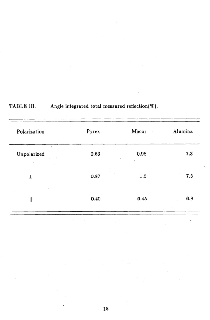
Table II shows the results of specular reflection measurement for the three polarizations; unpolarized, electric field vector parallel to the plane of incidence, and perpendicular to the plane of incidence(See Fig.1). (H) and (V) after the dump material indicate horizontal and vertical orientation, respectively. All measurements
are normalized to the specularly reflected power of the stainless steel slab(= 100). Measured values indicate higher absorption for the parallel polarization in general, in qualitative agreement with results of Table I. The ordering of the effectiveness also agrees with the calculations. Pyrex is found to be the most effective, with Macor slightly worse, and alumina being the worst of the three. However, the quantitative
agreement is only approximate for the perpendicular polarization, and is off by orders of magnitude for the parallel polarization. We believe these discrepancies are due to incomplete absorption of the transmitted power in the body of the dump, and may therefore be reduced in a thicker dump. Note that the specular reflectivity value for a given dump depends on the groove orientation. The horizontal orientation always gives larger values. In subsequent text, the angular measurements obtained with the grooves horizontal is referred to as the H-scan while the spectra with grooves oriented vertical is referred to as the V-scan.
Fig.5 shows the reflected power as a function of the detector angle measured from the specular position for unpolarized radiation. The V-scan shows a relatively low peak and a wide tail, extending to large angles. The cause of this wide tail is believed to be low frequency(f <250GHz) waves diffracting off the dump grooves, and internal reflections within the dump giving rise to waves reemerging in random directions. The H-scan shows a relatively high peak with narrow width, extending approximately to 150. The shapes of the profiles for a given dump orientation are
quite similar for all materials and polarizations. Measurements with the polarized source produce similar spectra, shifted up or down by the relative value of reflection at the specular angle.
The difference in the specularly reflected power for different groove orientations is accounted for mainly by the retrospecular nature of the front surface reflected rays. As an example, the ray shown by a dashed line in Fig.2(a) enters the groove at 100 to the normal and exits it parallel to the original ray. Thus, in the V-scan,
these retro-specular reflections do not get detected, and a lower value for reflected power results.
Therefore, to obtain the aggregate reflection spectra over the hemisphere facing the detector, a rather simple approach is taken, of adding the retrospecular power
component to the V-scan so as to make the peak value at normal reflection equal, as shown schematically in Fig.6. Insofar as the angular distribution of reflected power can be approximated as a retrospecular peak plus other broader features, this will provide a reasonable estimate, regardless of the exact cause of the broader scatter-ing. The total angular distribution is then taken to be the product of separate angu-lar functions in the horizontal and vertical directions, I(OH,OV) =
fH(OH)fV(OV)-Thus the total reflected power is given by
flr/2f i/2
PR =
II
I(OH, OV)dOH cos OVdOV
J -i/2 J -,r/2
(3)
=
I
fH(OH)dOHI
fv(Ov)cosOvd6v,_-7/2 -7r 2
where PR is the total reflected power. For the purpose of this integration, orthog-onal detector angles from the normal, OH and OV, are assumed similar to spherical
coordinate variables, (the polar angle) and 4(the azimuthal angle), with OH =
and OV = 7r/2 -0. Negligible error is introduced since post of the reflected power is
concentrated near OH ~ Oy ~ 0. Nevertheless, By, with its greater angular spread,
is associated with the polar angle to bring out the spherical dependence. By com-paring this with the total reflected power from a stainless steel slab, the absorption
by the dump is obtained. Results are summarized in Table III. It shows that the
design goal of less than 1% reflection is achieved with the Pyrex dump regardless
of the source polarization. The goal is also achieved for parallel polarization with the Macor dump, but alumina dump is noticeably worse.
The spectral response of the dump was measured using the mercury arc lamp source and a rapidly-scanning Michelson interferometer[14] in place of the broad .band detector. This is a particularly difficult and time consuming measurement since the level of the mercury arc radiation reflected off the dump is equivalent to
that of a T
<30Kblackbody. A single measurement was made for the Macor dump,
in the H-scan orientation for the parallel polarization. The normalized reflected
power curve, measured by co-addition of more than 40,000 interferograms, is shown
by a broken line in Fig.7. Further attempt to reduce the noise appears impractical.
The smoothed approximation indicated by the solid line shows less absorption at
the low frequency end where geometrical optics breaks down and the absorption
coefficient is lower. Small amplitude features of the smoothed curve at higher
fre-quencies are probably not statistically significant. This inferior performance at low
frequency is also supported by a separate measurement using a low pass filter at
the broad band detector aperture. The frequency integrated response,
appropri-ately weighted by the source spectrum, is in fair agreement with the value shown
in Table II.
III. APPLICATION
These dumps were fabricated as a part of the vertical viewing ECE experiment,
the purpose of which is to isolate the region of ECE to a narrow chord to measure the
electron velocity distribution[15]. The measurement requires a specialized optical
arrangement, as well as the viewing dump at the termination point.
The configuration of the experiment is shown schematically in Fig.8. The Pyrex
or Macor dump was placed at the bottom of the tokamak torus, surrounded by a .
stainless steel frame to provide additional protectign from the plasma particles.
The distance between the tip of the grooves and the limiter-defined plasma edge is
approximately 1.5cm. Three front surface mirrors were used to image the plasma
onto the Michelson interferometer aperture.
Second harmonic ECE profiles from thermal plasmas at a magnetic field of
8Tesla were used for the performance analysis in the range 380GHz < f S 560GHz.
These profiles, selected for their reproducibility, were taken with and without the
Macor dump, and for ordinary and extraordinary modes of propagation(E 11 BT
and E I BT, respectively). They are shown in Fig.9. The dump grooves were
oriented parallel to the toroidal field, so that attenuation of extraordinary mode
emission is favored.
Without the dump, emission from essentially the entire plasma diameter(33cm)
is observed because the intensity is dominated by the multiple passes of reflected
rays through the plasma. The emission with the dumps has dramatically decreased
width, corresponding to about 3cm in the major radius direction for the FWHM.
This is consistent with the calculations of beam width at the plasma center, so that
most of the radiation is coming from within the line of sight.
The comparison of the emission levels from outside the line of sight shows that
the optical arrangement is effective in reducing the reflected emission by ;> 90%.
This discrepancy in performance is believed to be due to a portion of the antenna
pattern missing the dump and accepting reflections off the frame, even though the
antenna pattern width at the dump was minimized consistent with the other optical
requirements.
The Pyrex dump was found to have cracked at the groove tips (apparently
because of thermal shocks) after three weeks(~ 1000 plasma discharges, t
=
0.5sec/
discharge). There was no structural damage to the Macor dump(also after three
weeks). No obvioi's performance degradation from either dump was observed
dur-ing this period, despite metal particle deposits on the surface. Furthermore, no
additional plasma impurity problems caused by the dump were observed, despite
its close proximity to the plasma.
In summary, we have fabricated and tested submillimeter viewing dumps made of Pyrex, Macor, and alumina. A simple front surface reflectivity calculation pro-vides a fair estimate of their performance, which, for Pyrex and Macor, is > 99%
absorption of the incident radiation. Use of these dumps in a plasma ECE experi-ment has resulted in a dramatic reduction of multiple reflections.
ACKNOWLEDGEMENTS
The authors gratefully acknowledge Dr. Woskoboinikow for many helpful dis-cussions and advice, especially with respect to the initial selection of the candidate dump materials. Many thanks go to Drs. Gandy and Gomez for helpful suggestions on the performance tests, and to the rest of the Alcator Group for their support. This work was supported by Department of Energy contract DE-AC02-78ET51013.
REFERENCES
1] F. Engelmann and M. Curatolo, Nucl. Fusion, 13(1973)497.
2] P. Woskoboinikow, H.C. Praddaude, W.J. Mulligan, D.R. Cohn. B. Lax, and
H.R. Fetterman, 6th Int. Conf. Infrared and Millimeter Waves, Conf. Digest,
IEEE Cat. No. 81 CH1645-1 MTT(1981) M-3-1.
[3] K. Kato and I.H. Hutchinson, Bull. Am. Phys. Soc. Vol.29, No.8, p.1223 (Oct.
1984).
[4] P. Woskoboinikow, R. Erickson and W.J. Mulligan, Int. J. Infrared and
Mil-limeter Waves, Vol.4, p.1
045(1983).
[5] M.A. Sengstacke, R.N. Dexter, and S.C. Prager, Phys. Fluids, 28(1985)403.
[6] C. Weggel, W. Hamburger, B. Montgomery, and N. Pierce, Proc. 7th Symposium
on Engineering Problems of Fusion Research, p.54, Knoxville, Tennessee, 1977.
[7] G. Bekefi and A. Barrett, Electromagnetic 'Vibrations, Wates, and Radiation,
MIT Press, Cambridge, Mass. 1977.
[8]
V.0. Altemose and A.R. Kacyon, "Vacuum Compatibility of Machinable Glass
Ceramics," Corning Tech. Paper, Corning Glass Works, Corning, N.Y.
[9]
K.H. Breeden and A.P. Sheppard, Radio Science, 3 (1968) 205.
[10] M.N. Afsar and K.J. Button, IEEE Transactions on Microwave Theory and
Techniques, Vol.MTT-31, No.2, Feb. 1983.
[11] Grinding of dump pieces were done at Ceramic Grinding Company, Waltham,
MA.
[12] M.F. Kimmit, Far Infrared Techniques, Pion (1970).
[13] I.H. Hutchinson and S.E. Kissel, Proc. 4th Int. Conf. Infrared and Millimeter
Waves and their Applications, p.76, Miami Beach, 1979.
|15
1.H. Hutchinson and K. Kato, "Diagnosis of Mildly Relativistic Electron
Distri-butions by Cyclotron Emission," PFC/JA-85-15, to be published in Nucl. Fusion.
FIGURE CAPTIONS
Fig.1.
Notation of field vectors at a dielectric interface, used in Eq.(1).
Fig.2.
(a) Path of a normally incident ray through a
45'
groove in real
space(solid line). The path of a ray incident at 100 to normal is shown by the
dashed line.
(b) Path of a normally incident ray through a
450
groove in semi-circle
representation. The groove is duplicated and spread into a semi-circle so that the
path of the ray is shown by a straight line.
Fig.3.
(a) Dimensions of the viewing dumps. (b) Modular construction of the
Pyrex and the Alumina dumps.
Fig.4.
Measurement configuration as viewed from above(not to scale).
Fig.5.
Reflected power from different dumps as a function of the detector
angle. (Noise level ce 5 x 10⁻³ A.U.)Fig.6.
Schematic picture of the normal incidence approximation. The angular
distribution of the reflected power, I, is plotted as a function of the two orthobonal
orientations,
OHfor the H-scan, and
Byfor the V-scan.
Fig.7. Frequency response of the Macor dump at 10' specular orientation, with grooves oriented in the H-scan direction and for parallel polarization. Broken
line shows the measured spectrum. Solid line is the smoothed approximation.
Fig.8. Experiment configuration of the Vertical Viewing ECE diagnostic.
Fig.9. Second harmonic ECE spectrum from Alcator C plasma ne : 1.7 x
102 0m- 3, 350kA < Ip < 400kA, BT = 8Tesla. Trace A. Extraordinary mode,
with-out the dump; Trace B. ordinary mode, withwith-out the dump; Trace C. extraordinary
TABLE I.
Refractive indicies and computed reflectivities for different
mate-rials. Reflectivities are computed for the parallel and perpendicular electric field
orientation. (Values are normalized to incident power of unity.)
-
Polarization
Pyrex
Macor
Alumina
N
09
= 7r/3?r/3
7r/4
7r/4
2.1i 3 x 10-60.02
1 x 10⁻⁷ 0.004 1 x 10- 73 x
10~4 2.4ii 4 x 10⁻⁵ 0.03 8 x 10-130.01
2 x 10⁻⁷ 0.001 3.111 8 x 10⁻⁴ 0.07 8 x 10~6 0.037 x 10⁻⁸
0.006-I
I
7r/6
7r/6
1
From Ref.9.
From Ref.10.
Measured specular reflectivity(%) at 100 incidence angle.
Dump
Unpolarized
S.S.
Pyrex(H)
Pyrex(V)
Macor(H)
100
0.32 0.074 0.58Macor(V)
Alumina(H)
Alumina(V)
0.13
4.2 1.3100
0.460.17
0.079
0.060
0.91
0.230.12
0.12
4.31.2
100
3.8 1.4TABLE II.
Angle integrated total measured reflection(%).
Polarization
Pyrex
Macor
Alumina
Unpolarized
0.63
0.98
7.3
0.87
1.5
7.3
0.40
0.45
6.8
Er±
Erl,
Ei.
Ei
Figure 1.7
N
1
N
2Etllt
pp0
0
0
z- - 0 - -n - -lov - - - - - - - -Figure 2.0
(u.s0
z
kk l4*
.... .. ...
~
yip~ ~
.;y pr .45O--
\-rAW
/
5m
~
t4-J6mm
1I-4mm
80mm
.I
'
t
Figure 3.80 mm
a)
InSb
Detector
Angular Scan
Mercury
Arc Lamp
1" Waveguide
Polarizer-
2
-200 .
30 cm
18
cm
Eccosorb
Al. Frame
AI.Back Plate
106
Figure 4.100
*Ses.
o
Alumina (H)
*
Alumina
(v)
10
->
Macor(H)
V
Pyrex (H)
A
Macor(V)
Pyrex
(v)
.01-0
10
20
30
40
Detector Angle (Degrees) from Specular Position
V-scan Profile
H-scan Profile
OH
Ov
H-scan Profile
Added to V-profile
NINI
Figure 6.II I I I I I I