HAL Id: inserm-02449474
https://www.hal.inserm.fr/inserm-02449474
Submitted on 22 Jan 2020
HAL is a multi-disciplinary open access
archive for the deposit and dissemination of
sci-entific research documents, whether they are
pub-lished or not. The documents may come from
teaching and research institutions in France or
abroad, or from public or private research centers.
L’archive ouverte pluridisciplinaire HAL, est
destinée au dépôt et à la diffusion de documents
scientifiques de niveau recherche, publiés ou non,
émanant des établissements d’enseignement et de
recherche français ou étrangers, des laboratoires
publics ou privés.
In vitro response of rat pleural mesothelial cells to talc
samples in genotoxicity assays (sister chromatid
exchanges and DNA repair)
S Endo-Capron, A. Renier, X Janson, L Kheuang, M Jaurand
To cite this version:
S Endo-Capron, A. Renier, X Janson, L Kheuang, M Jaurand. In vitro response of rat pleural
mesothelial cells to talc samples in genotoxicity assays (sister chromatid exchanges and DNA repair).
Toxicology in Vitro, Elsevier, 1993, 7, pp.7 - 14. �10.1016/0887-2333(93)90107-G�. �inserm-02449474�
Toxic. in Vitro Vol. 7, No. 1, pp. 7-14, 1993 0887-2333/93 $6.00+0.00 Printed in Great Britain. All rights reserved Copyright © 1993 Pergamon Press Ltd
IN VITRO
RESPONSE OF RAT PLEURAL MESOTHELIAL
CELLS TO TALC SAMPLES IN GENOTOXICITY ASSAYS
(SISTER CHROMATID EXCHANGES A N D D N A REPAIR)
S. ENDO-CAPRON*, A. RENIER*, X. JANSON~', L. KHEUANG* and M. C. JAURAND*~ *INSERM-U139, Laboratoire de Toxicologie Cellulaire et Mol6culaire de l'Environnement, CHU Henri Mondor, 94010 Cr&eil and tLaboratoire d'Etude des Particules Inhal6es, DASS, 11 rue Georges Eastmann,
75013 Paris, France
(Received 16 April 1992; revisions received 11 August 1992)
Abstract--The genotoxicity of three samples of talc has been determined using in vitro cell systems previously developed for testing asbestos fibres. The talc samples used consisted of particles of respirable size in order to test the effect of particles likely to be deposited in the lung. Genotoxicity was tested in cultures of rat pleural mesothelial cells (RPMC) using genotoxicity assays for unscheduled DNA synthesis (UDS) and sister chromatid exchanges (SCEs). The effects were compared with those obtained with negative controls (attapulgite and anatase) and positive controls (chrysotile and crocidolite asbestos). In contrast to asbestos, none of the talc samples, nor the negative controls, induced enhancement of UDS or SCEs in treated cultures in comparison with the untreated cultures.
INTRODUCTION
Talc is a mineral commonly used in various industries including the ceramics, paper, plastics, paints, pharmaceutical and cosmetics industries. It is a magnesium silicate of similar chemical composition to chrysotile asbestos fibres but with a different structure. IARC has evaluated the biological effects of talc (IARC Working Group, 1987); according to their findings the results obtained in previous exper- iments in vivo and in vitro were inadequate to evaluate the carcinogenicity or genotoxicity of talc because of the limited number of studies. Data from animal studies did not show an excess of pleural sarcomas or mesotheliomas after the intrapleural administration of talc (Endo-Capron et al., 1990; Stanton et al., 1977; Wagner et al., 1977). From data from epidemio- logical studies, the IARC Working Group (1987) concluded that it was possible that carcinogenicity could result from exposure to some specific samples found to be associated with fibrous tremolite. How- ever, epidemiological studies have been updated recently and no evidence of increased risk of lung cancer has been found (Weill et al., 1990). Some authors have examined the association between genital talcum powder exposure and ovarian cancer (Harlow and Weiss, 1989); no appreciable altered risk was ~:To whom correspondence should be addressed.
Abbreviations: FCS = foetal calf serum; HU = hydroxy- urea; RPMC = (rat pleural mesothelial ceils); SCE = sister chromatid exchange; TEM = transmission electron microscopy; UDS = unscheduled DNA synthesis.
observed following exposure to baby powders, which are reported to contain only talc, but an increased risk was associated with the use of talc-containing powders, that is, also containing deodorizing sub- stances or a variety of other free and bound silica (Harlow and Weiss, 1989).
The present experiments were designed to deter- mine whether talc particles of respirable dimensions exerted a genotoxic effect on cultures of rat pleural mesothelial cells (RPMC). Pleural mesothelial cells are an important target for fibrous particles inhaled from our environment and can be used as test models to determine the in vitro effects of particle matter. In addition, talc has been used to overcome pleural effusion (IARC Working Group, 1987). It is therefore of interest to determine the effects of pure talc on RPMC. In previous experiments, we have used RPMC to study the genotoxicity of asbestos fibres. Enhance- ment of unscheduled DNA synthesis (UDS; Renier
et al., 1990) and sister chromatid exchanges (SCE; Achard et al., 1987) have been observed in cultured RPMC after exposure to chrysotile or crocidolite fibres, but not after exposure to a non-carcinogenic sample of attapulgite. Identical tests were applied in this study in order to determine the effects of pure talc.
MATERIALS AND METHODS
Particles and test compounds. Three samples of European talc provided by Eurotalc (Brussels, Belgium) were studied. One sample each of French talc (no. 7841). Italian talc (no. 5726) and Spanish
8 S. ErqDO-CAPRON et al.
talc (no. 5725) have been tested. Samples contained 90-95% of talc, the other compounds being chlorite and dolomite. Anatase (a gift from P. Sebastien, Cerchar, France) and attapulgite (from Mormoiron, France) were tested as negative reference particles; Rhodesian chrysotile and crocidolite from the Union Internationale Contre le Cancer (UICC) as positive reference particles. The particles were dispersed in culture medium at a concentration of 560/zg/ml by sonication for 5 min (20 KHz, 3 W). Chemicals used as controls, mitomycin C (Choay, Paris, France) and K2CrO 4 (Aldrich Chemical Co., Milwaukee, MO, USA), were solubilized in water and in culture medium, respectively.
Transmission electron microscopy ( TEM). Particles
at a concentration of 100#g/ml were dispersed in culture medium. An aliquot of the suspension was filtered through a 0.40-/./M pore size Nuclepore filter. The filters were transferred to electron microscopic grids and dissolved according to the method routinely used in the laboratory (Srbastien et al., 1978). The
size of the particles was determined following a systematic scanning of the grid at two magnifications ( × 33,000 and x 26,000).
Cell culture. Rat pleural mesothelial cells (RPMC)
were obtained as described elsewhere (Jaurand et al.,
1981). Briefly, primary RPMC cultures were obtained by scraping the parietal pleura and allowing the cells to grow in multiwell tissue culture plates. The cultures were maintained in complete medium (i.e. Ham's F 10 medium; Flow Laboratories, Irvine, Ayrshire, Scot- land) supplemented with 2mM-L-glutamine (Flow Laboratories), 1 mM-vitamin C (Sigma Chemical Co., St Louis, MO, USA), 10mM-HEPES (Seromed, Berlin, Germany), 10% foetal calf serum (FCS; from Boehringer, Mielan, France), 100 U penicillin ml and 50/~g streptomycin ml (both antibiotica from Flow Laboratories). When the cells reached confluence they were subcultured. From passage 5, R P M C were subcultured approximately every week by standard trypsinization and used between passages 5 and 15.
Ultrastructural analysis. 24 hr after plating, talc
was added to the RPMC in the tissue culture dishes at a concentration of 10#g/cm 2. Electron micro- scopic studies were carried out according to standard methods previously described (Jaurand et al., 1979).
The solid compound concentration was expressed as /zg/cm 2 to take into consideration the particle settling; in these culture conditions, 1/~g/cm 2 is equivalent to 5 # g/ml.
Unscheduled DNA synthesis (UDS). RPMC were
cultured in 24-well cluster dishes (Falcon, France); 8 × 10 4 cells were plated per well in complete medium.
Cells reached confluence after 4 days of incubation. The medium of the confluent culture was replaced with R P M I (Flow Laboratories) containing 1% FCS (Boehringer), 5mM-hydroxyurea (HU; Sigma) to arrest cells in G1, 100U penicillin/ml and 5 0 # g streptomycin/ml (both from Flow Laboratories). The cells were incubated for 24 hr in a humidified atmos-
phere of 5% CO2 in air at 37°C. The cells were then treated for 24 hr with the indicated dose of particles (1/~g/cm 2 is equivalent to 5/~g/ml) in 1% FCS medium containing 5mM-HU and [methyl-3H]
thymidine (Amersham, les Ulis, France) at 4 # Ci/ml. The amount of radioactivity incorporated into D N A was determined as described elsewhere (Renier et al.,
1990). Six wells were used per treatment. After treat- ment, cells were washed three times with phosphate buffered saline. Acid-soluble material was removed by rinsing with 10% cold trichloracetic acid for 10 min and incubated in a mixture of 0.2 M-NaOH and 1% sodium dodecyl sulphate. Aliquots of 200/z 1 were mixed with scintillation fluid (Pico-fluor, Pack- ard) and radioactivity was measured with a Beckman LS 6000SC scintillation counter. Cell D N A content was determined according to West et al. (1985) in
separate wells treated with the minerals in the same conditions as described above. Results are expressed as dpm//tg DNA. All studies were carried out with coded samples.
Sister chromatid exchanges (SCEs). RPMC were
plated at a density of 2 x 10⁶ cells per 75-cm 2 flask in R P M I medium supplemented with 10% FCS. Cells were treated either with test chemicals or with several concentrations of particles plus 3 # g bromodeoxy- uridine/ml 24 hr after the plating of the culture. In these culture conditions, 1/zg/cm 2 is equivalent to 7.5/zg/ml. The cultures were incubated with the test compound at 37°C for 48 hr in the dark. 2 hr before harvesting cells, colchicine (Sigma) at a final con- centration of 0.2/~g/ml was added to each culture. Metaphase cells were then detached with 0.25% trypsin (Eurobio, Paris, France), collected in 15-ml corex tubes and centrifuged at 1500 rpm for 7 min. The supernatant was removed. Cells were treated with 0.075 M-KCI at 37°C for 30 min before fixation in methanol-glacial acetic acid (3 : 1, v/v). The fixative was changed three times and the last fixation step lasted for one night. The cell suspension was dropped onto an ice-cold slide. Cells were stained by the fluorescence plus Giemsa technique (Perry and Wolff, 1974). 30 metaphases exhibiting 37-42 chromosomes were counted per assay. All studies were carried out with coded samples.
Statistical analysis. The significance of UDS data
was evaluated using Student's t-test. The number of SCEs observed in treated cell cultures was com- pared with that in the untreated cultures using the Mann-Whitney test.
R E S U L T S
T E M study o f particles
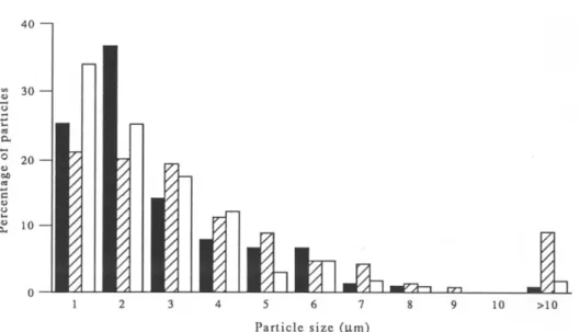
The size distribution of the talc samples is reported in Fig. 1. The characteristics of the talc, anatase, crocidolite and chrysotile particles are reported in Table 1. The mean size of the three talc samples was in the ranking order 5725 = 7841 < 5726. The number
401
3 0 e~ 2 0 ~ to ~ m o 1 2 3 4 5 6 7 8 9 10 P a r t i c l e s i z e (].tin)Fig. 1. Size distribution o f talc samples no. 5725 (m), no. 5726 ([~) and no. 7841 ( R ) .
L
>IOPlate 2. Rat pleural mesothelial cells, (a) untreated or (b) treated with talc particles (arrows) at 50/~g/cm-" for 48 hr. Phase contrast microscopy ( x 115).
Plate 3. Rat pleural mesothelial cells treated with talc particles (arrows) at 50 u g / c m 2 for 48 hr. Electron microscopy ( x 6400).
Genotoxicity o f a tale in mesothelial cells Table I. Characteristics of the particle samples
Sample
Mean No. of particles length No. of of length
(#m) particles/gg >4#m/#g Talc 5725 2.6 13.0 x 10⁴ 2.1 × 10⁴ Talc 5726 4.0 9.8 x 10⁴ 2.8 x 10⁴ Talc 7841 2.6 3.3 x 10⁴ 0.4 x 10⁴ Anatase 0.7 2.2 X 10 9 0 Crocidolite UICC 3.1 3.0 x 10⁶ 5.1 × 10 5* Chrysotile UICC 3.2 1.1 x l0 T 2.8 x 10⁶. *Fibres having a diameter ~< 1.5 #m.
o f p a r t i c l e s p e r u n i t w e i g h t w a s in t h e r a n k i n g o r d e r 5725 > 5726 > 7841. T h e r e f o r e , t h e n u m b e r o f p a r - ticles h a v i n g a size g r e a t e r t h a n 4 # m is a p p r o x i m a t e l y t h e s a m e in t w o s a m p l e s a n d s m a l l e s t in s a m p l e 'no. 7841. T E M s t u d y s h o w e d t h a t n o n e o f t h e t h r e e s a m p l e s o f talc c o n t a i n e d a s b e s t o s fibres ( P l a t e 1). T h e m e a n l e n g t h o f c r o c i d o l i t e a n d c h r y s o t i l e fibres is b e t w e e n t h o s e o f talc s a m p l e s 5725 a n d 7841 a n d t h a t o f s a m p l e 5726. T h e n u m b e r o f c r o c i d o l i t e o r c h r y s o t i l e p a r t i c l e s h a v i n g a l e n g t h g r e a t e r t h a n 4 # m is 2 0 - 1 0 0 t i m e s m o r e t h a n t h a t o f talc. A n a t a s e is a v e r y s m a l l p a r t i c l e h a v i n g a n a v e r a g e size o f less t h a n 1 # m w i t h n o p a r t i c l e l a r g e r t h a n 4 # m .
Structural and ultrastructural studies
N o s t r u c t u r a l c h a n g e h a s b e e n o b s e r v e d f o l l o w i n g t r e a t m e n t o f R P M C w i t h talc ( P l a t e 2). It a p p e a r e d t h a t t h e n u m b e r o f cells w a s r e d u c e d c o m p a r e d w i t h t h a t o f u n t r e a t e d cells b u t n o sign o f c y t o l y s i s w a s d e t e c t e d . T E M s t u d i e s h a v e i n d i c a t e d a c a p a c i t y o f R P M C t o i n g e s t talc a n d a n a t a s e particles. P l a t e 3 s h o w s t h a t talc p a r t i c l e s w e r e l o c a t e d in t h e peri- n u c l e a r r e g i o n a n d o r g a n e l l e s d i d n o t s e e m c h a n g e d in c o m p a r i s o n w i t h u n t r e a t e d cells.
Unscheduled DNA synthesis (UDS)
T a b l e s 2 a n d 3 s h o w t h e effect o f t r e a t m e n t o f R P M C w i t h r e f e r e n c e p a r t i c l e s o r talc s a m p l e s .
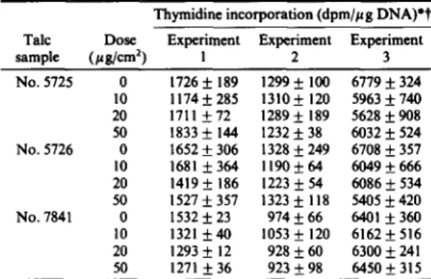
11 Table 3. Unscheduled DNA synthesis in pleural mesothelial cells
treated with different talc samples at several doses Thymidine incorporation (dpm//~g DNA)*t Talc Dose Experiment Experiment Experiment
sample (#g/cm 2) I 2 3 No. 5725 0 1726 __ 189 1299 +_ 100 6779 -+ 324 10 1174_+285 1310_+ 120 5963_+740 20 1711 +72 1289+ 189 5628__.908 50 1833 -+ 144 1232 -+ 38 6032 __ 524 No. 5726 0 1652 _+ 306 1328 _+ 249 6708 _+ 357 l0 1681 ___364 ll90 -+ 64 6049_+666 20 1419 _+ 186 1223 _+ 54 6086 _+ 534 50 1527 +_ 357 1323 _+ 118 5405 _+ 420 No. 7841 0 1532 _+ 23 974 _+ 66 6401 _+ 360 10 1321 _+ 40 1053 _+ 120 6162 _+ 516 20 1293 _+ 12 928 _+ 60 6300 + 241 50 1 2 7 1 _ + 3 6 9 2 3 _ + 9 8 6450+315 *Experiments 1 and 2 were carried out with a specific activity of
methyl-3H
of 20-30 Ci/mmol; experiment 3 was carried out with a specific activity of 40-60 Ci/mmol.?Values are means + SD for six replicates.
A n a t a s e d i d n o t e n h a n c e U D S in R P M C . Cells t r e a t e d w i t h c r o c i d o l i t e at 10 # g / c m 2 o r c h r y s o t i l e at 4 o r 10/~g/cm 2 a l w a y s s h o w e d a significant e n h a n c e m e n t o f U D S c o m p a r e d w i t h u n t r e a t e d cells. N o n e o f t h e talc s a m p l e s t e s t e d h e r e e n h a n c e d U D S .
Sister chromatid exchanges
T h e n u m b e r s o f S C E s f o r r e f e r e n c e particles, c h e m i c a l s a n d talc s a m p l e s a r e s h o w n in T a b l e 4, T h e c o n t r o l particles, a t t a p u l g i t e a n d a n a t a s e , d i d n o t i n d u c e a significant m o d i f i c a t i o n in t h e n u m b e r o f S C E s . I n c o n t r a s t , i n c r e a s e d n u m b e r s o f S C E s w e r e o b s e r v e d w h e n R P M C w e r e t r e a t e d w i t h t h e g e n o t o x i c c h e m i c a l s m i t o m y c i n C a n d K2CrO4. A statistically significant e n h a n c e m e n t o f S C E s w a s o b t a i n e d in cells t r e a t e d w i t h 2 n g m i t o m y c i n C / m l ( P < 0.005) o r 0.5 # g K2CrO4/ml ( P < 0.005). T h e m e a n n u m b e r o f S C E s w a s significantly i n c r e a s e d b y c h r y s o t i l e at 1 # g / c m : ( P < 0.005) o r c r o c i d o l i t e a t 2 # g / m 2 ( P < 0 . 0 5 ) , w i t h significant i n c r e a s e s o c c u r r i n g in t w o o u t o f f o u r a n d t h r e e o u t o f e i g h t e x p e r i m e n t s w i t h c h r y s o t i l e a n d c r o c i d o l i t e ,
Table 2. Unscheduled DNA synthesis in pleural mesothelial cells treated with different reference particles at several doses
Thymidine incorporation (dpm/#g DNA)? Dose
Particle (#g/cm 2) Experiment 1 Experiment 2 Experiment 3 Crocidolite~ 0 1299 _ I O0 1327 __. 57 6086 __ 299 4 1625 + 191"* 1425 + 926 9572 _ 463** I0 1668 + 53*** 1489 + 203* 8323 + 308*** Chrysotile:~ 0 1495 + 106 1362 _ 117 5632 + 326 4 1744 + 188"** 1498 _+ 116" 7590 + 649*** 10 1646 _+ 124"* 1598 + 64*** 8157 + 341"** Anatase~ 0 1316 -I- 153 6169 + 760 6579 + 413 2 1271 + 61 6096 +_ 705 6785 _+ 650 4 1380 _+ 276 6535 _+ 565 7214 +_ 301 10 1318 + 264 6405 -+ 480 7764 _+ 456** tValues are means _+ SD of six replicates and those marked with asterisks differ significantly
(Student's t-test) from the corresponding value for untreated cells (*P <0.05; **P <0.01; ***P < 0.001).
~:Experiments 1 and 2 were carried out with a specific activity of
methyl-3H
of 20-30 Ci/mmol; experiment 3 was carried out with a specific activity of 40--60 Ci/mmol.§Experiment I was carried out with a specific activity of
methyl-3H
of 20-30 Ci/mmol; experiments 2 and 3 were carried out with a specific activity of 40-60 Ci/mmol.12 S. ENDo-CAPRON et al. Table 4. SCE induction in R P M C treated with reference particles,
chemicals and talc samples
No. of significant
No. of experiments/
experi- Dose No. of SCEs/ no. of Treatment ments (# g/cm2)? metaphase$ experiments Attapulgite 3 0 17.6 ± 2.4 0/3 20 19.7 ± 1.4 Anatase 9 0 14.6 ± 2.9 0/4 2 12.9 ± 3.0 5 13.9 ± 2.8 Chrysotile 4 0 15.2 ± 1.6 2/4 1 20.2 ± 3.7** Crocidolite 8 0 14.9 ± 4.6 3/8 2 16.8 __+ 5.0* Mitomycin C 8 0 12.6 ± 1.5 4/4 2 47.0 ± 12.7"* K2CrO4 8 0 15.4 ± 3.4 4/4 0.5 38.6 ± 4.2** Talc no. 5725 3 0 12.2 ± 1.8 0/3 2 12.2 ± 1.0 5 11.8 ± 2.8 10 11.3 ± 0.4 15 12.6 ± 2.4 Talc no. 5726 3 0 12.2 ± 1.8 0/3 2 12.2 ± 1.8 5 9.8 ± 1.0 10 12.2 ± 1.8 15 12.2 ± 2.3 Talc no. 7841 3 0 12.1 ± 1.1 0/3 2 1 1 . 9 ± 1.1 5 1 1 . 0 ± 0 . 8 10 11.9 ± 0.7 15 11.1 ± 1.3 ?Except mitomycin C (ng/ml) and K2CrO 4 (/~g/ml).
SValues are means ± SD for the number of experiments shown, and those marked with asterisks differ significantly (Mann-Whitney test) from the corresponding values for untreated cells (*P < 0.05; **P < 0.005).
respectively. The number of chromosomes per metaphase and SCE frequencies in R P M C exposed to talc samples 5725, 5726 and 7841 are shown in detail in Tables 5 and 6. No difference in the number of chromosomes per metaphase in treated cells was observed in comparison with untreated cells. More- over, treatment with several concentrations, from 2 to 15 #g/cm 2, did not increase SCE frequency.
Table 5. N u m b e r of chromosomes per metaphase in R P M C treated with three talc samples
No. o f chromosomes/metaphase* Dose Experiment Experiment Experiment Talc sample ( # g / c m 2) 1 2 3 No. 5725 0 40.1 ± 2.6 41.0 + 1.6 41.2 __. 1.5 2 41.1 ± 1.6 41.2 ± 1.1 4 0 . 7 _ 1.5 5 40.3 ± 1.7 41.1 ± 1.4 41.0 __+ 1.5 10 40.8 ± 1.6 40.8 ± 1.7 40.7 ± 1.6 15 40.2 ± 1.7 40.9 ± 1.6 41.2 ± 1.8 No. 5726 0 40.4 ± 2.6 41.0 ± 1.6 41.2 ± 1.5 2 4 0 . 3 ± 2 . 0 4 1 . 1 ± 1 . 3 40.9 ± 1.5 5 40.7 ± 1.6 40.9 ± 1.4 40.5 ± 1.9 10 40.7 ± 1.7 40.6 ± 1.6 40.6 ± 1.4 15 40.5 ± 1.7 N D 41.1 ± 1.7 No. 7841 0 41.1 ± 1.3 41.0 ± 1.6 41.1 ± 1.4 2 40.7 ± 2.1 41.1 + 1.2 41.2 ± 1.5 5 41.2 ± 1.4 41.0 ± 1.7 40.9 ± 1.7 10 40.7 ± 2.0 40.7 ± 1.8 40.7 ± 2.0 15 4 0 . 6 ± 2 . 0 40.7 ± 1.8 41.1_.+1.9 N D = not done *Mean ± SD of 30 metaphases.
Table 6. N u m b e r of SCEs in R P M C treated with three talc samples No. o f SCEs/metaphase* Dose Experiment Experiment Experiment
Talc sample (#g/cm 2) I 2 3 No. 5725 0 10.1 ± 3.5 2 11.4± 3.5 5 9.0 ± 3.6 10 11.4± 3.7 15 11.6± 3.5 No. 5726 0 10.1 ± 3.5 2 11.0 ± 2.9 5 9.3 ± 3.0 10 10.6 ± 3.4 15 1 0 . 5 ± 2 . 8 No. 7841 0 12.0 ± 4 . 3 2 10.6 ± 3.0 5 10.8 ± 4.9 10 1 2 . 1 ± 5 . 4 15 11.0± 3.8 N D = not done *Mean ± SD of 30 metaphases. DISCUSSION 1 3 . 3 ± 5 . 7 1 3 . 2 ± 5 . 8 11.8±4.1 1 3 . 3 ± 4 . 9 1 1 . 6 ± 3 . 9 1 4 . 7 ± 6 . 9 1 0 . 9 ± 3 . 5 1 1 . 6 ± 5 . 6 1 0 . 9 ± 3 . 2 1 5 . 3 ± 5 . 4 1 3 . 3 ± 5 . 7 1 3 . 2 ± 5 . 8 11.4±4.1 1 4 . 3 ± 5 . 0 1 1 . 0 ± 3 . 6 9 . 2 ± 6 . 2 1 1 . 9 ± 3 . 6 1 4 . 1 ± 5 . 0 N D 1 3 . 8 ± 5 . 4 1 3 . 3 ± 5 . 7 1 1 . 1 ± 4 . 4 1 2 . 2 ± 3 . 8 1 2 . 9 ± 4 . 9 1 0 . 3 ± 3 . 6 1 2 . 0 ± 4 . 0 1 1 . 2 ± 4 . 5 1 2 . 5 ± 6 . 2 9 . 9 ± 3 . 7 1 2 . 5 ± 3 . 9
In the in vitro studies reported here we investigated the effects of talc in genotoxic assays. We observed that the three talc samples did not increase UDS or SCEs, or produce aneuploidy in RPMC. In contrast, chrysotile and crocidolite fibres consistently enhanced UDS, as well as increasing SCEs in some of the experiments. This is in agreement with previous observations in our laboratory (Achard et al., 1987; Renier et al., 1990). SCE enhancement was also obtained after treatment of R P M C with mitomycin C and K2CrO4, agents previously known to induce SCE (Darroudi and Natarajan, 1989; Kato and Shimada, 1975; Levis and Bianchi, 1982; Littlefield et al., 1979; Perry, 1980). The negative reference particle, anatase, did not increase either UDS or the frequency of SCEs in comparison with untreated RPMC.
In spite of the fact that talc is a magnesium silicate, as are chrysotile fibres, the in vitro responses of the two particles are different. As far as the mechanisms of genotoxicity of particles are concerned, several factors might account for the different responses, in particular phagocytosis, granulometry and the shape of the particles. Several questions can be addressed. First, is the lack of genotoxic action of talc due to the absence of phagocytosis? Phagocytosis seems to play an important role in the genotoxic effect of particles, because fibres phagocytosed could interact with the mitotic spindle (Hesterberg and Barrett, 1985) or chromosomes (Wang et al., 1987). This may then induce aneuploidy by chromosomal missegrega- tion (Hesterberg and Barrett, 1985; Palekar et al.,
1987). Our TEM study showed that RPMC can ingest talc particles. This cellular process has been also observed with chrysotile and crocidolite asbestos fibres (Jaurand et al., 1979 and 1983). Despite phago- cytosis, talc did not induce aneuploidy since the number of chromosomes per metaphase in talc- treated cells was not different from that in untreated cells (Table 5). Therefore, the lack of chromosomal
Genotoxicity of a talc in mesothelial ceils 13 damage might be related to different mechanical or
physicochemical properties o f talc in comparison with mineral fibres.
Secondly, is the absence of genotoxic action due to the size o f the talc particles? F r o m the data reported in the literature, the carcinogenic potency of par- ticulate matter seems to be dependent on both shape and dimension. F o r example, Stanton et al. (1981) have reported that after intrapleural inoculation into the rat, the frequency o f pleural sarcomas was dependent on the n u m b e r of fibres less than 0.25/~m in diameter and more than 8 # m in length. Moreover, an in vitro assay has shown that thick glass fibres were more efficient than thin fibres, on a per n u m b e r basis, in transforming Syrian hamster embryo cells. In addition, no transformation was obtained when the fibre length was reduced to 0 . 9 5 p m (Hesterberg and Barrett, 1984). In contrast to asbestos fibres, talc does not have a fibrous shape, but rather a polygonal form. Fibre samples containing long fibres can be deposited in the airways because of their small diam- eter, whereas respirable talc particles with a diameter higher than 5 # m do n o t reach the deep lung. The absence o f an in vivo effect of talc might also be due to the small size of the particles. The size and number o f particles per unit weight are different in the three talc samples. G r a n u l o m e t r i c study o f the talc samples showed that the m e a n size was in the ranking order 5725 = 7841 < 5726 and o f the same order as that o f asbestos fibres. However, the number of long ( > 4/zm) particles is m u c h higher in asbestos samples than in the talc samples used here.
The three talc samples did not enhance U D S or induce SCEs in comparison with untreated R P M C . This is in contrast to the results with asbestos, es- pecially with regard to the U D S assay in which a significant response was observed with both types of asbestos fibres. The S C E results seem less convincing; in effect, no consistent positive enhancement o f SCEs was found with crocidolite, thus lessening the signifi- cance of the negative response obtained with talc. However, our observations are in agreement with
in vivo data reported by Stanton et al. (1981) and with our previous results obtained with sample no. 7841, which showed that talc did not produce tumours following intrapleural inoculation ( E n d o - C a p r o n
et al., 1990), as well as with in vitro results that showed that talc did not induce c h r o m o s o m a l effects in m a m m a l i a n cells in vivo and in vitro ( I A R C W o r k i n g Group, 1987).
Acknowledgements--This work has been supported by INSERM funds and Eurotalc subvention.
REFERENCES
Achard S., Perderiset M. and Jaurand M. C. (1987) Sister chromatid exchanges in rat pleural mesothelial ceils treated with crocidolite, attapulgite or benzo 3-7 pyrene.
British Journal o f Industrial Medicine 44, 281-283.
Darroudi F. and Natarajan A. T. (1989) Cyto- genetical characterization of chinese hamster ovary X-ray-sensitive mutant ceils Xrs5 and Xrs6. III. Induction of cell killing chromosomal aberrations and sister chromatid exchanges by bleomycin, mono- and bi-functional alkylating agents. Mutation Research
212, 123-125.
Endo-Capron S., Fleury-Feith J., Nebut M., De Neef R. and Jaurand M. C. (1990) Some in vivo and in vitro studies carried out with talc samples. In Health Related
Effects o f Phyllosilicates. Edited by J. Bignon. NATO ASI series, Serie G: Ecological sciences, Vol. G21. pp. 367-375. Springer-Verlag, Berlin.
Harlow B. L. and Weiss N. S. (1989) A case-control study of borderline ovarian tumors: the influence of perineal exposure to talc. American Journal of Epidemiology 130, 390-394.
Hesterberg T. W. and Barrett J. C. (1984) Dependence of asbestos and mineral dust-induced transformation of mammalian cells in culture of fiber dimension. Cancer
Research 44, 2170-2180.
Hesterberg T. W. and Barrett J. C. (1985) Induction by asbestos fibres of anaphase abnormalities: mechanism of aneuploidy induction and possibly carcinogenesis.
Carcinogenesis 6, 473-475.
IARC Working Group (1987) IARC Monographs on the
Evaluation o f the Carcinogenic Risk o f Chemicals to Humans. Vol. 42. Silica and some silicates, pp. 185-224. International Agency for Research on Cancer, Lyon. Jaurand M. C., Bastie-Sigeac I., Bignon J. and Stoebner P.
(1983) Effect of chrysotile and crocidolite on the morphology and growth of rat pleural mesothelial cells.
Environmental Research 30, 255-269.
Jaurand M. C., Bernaudin J. F., Renier A., Kaplan H. and Bignon J. (1981) Rat pleural mesothelial ceils in culture.
In Vitro 17, 98-105.
Jaurand M. C., Kaplan H., Thiollet J., Pinchon M. C., Bernaudin J. F. and Bignon J. (1979) Phagocytosis of chrysotile fibers by pleural mesothelial cells in culture.
American Journal o f Pathology 94, 529-538.
Kato H. and Shimada H. (1975) Sister chromatid exchanges induced by mitomycin C: a new method of detecting DNA damage at chromosome level. Mutation Research
28, 459-464.
Levis A. G. and Bianchi V. (1982) Mutagenic and cyto- genetic effects of chromium compounds. In Biological and
Environmental Aspects o f Chromium. Edited by Langard. pp. 171-209. Elsevier Biomedical Press.
Littlefield L. G., Colyer S. P., Sayer A. and Dufrain R. J. (1979) Sister-chromatid exchanges in human lymphocytes exposed during GO to four classes of DNA damaging chemicals. Mutation Research 67, 259-269.
Palekar L. D., Eyre J. F., Most B. M. and Coffin D. L. (1987) Metaphase and anaphase analysis of V79 cells ex- posed to erionite, UICC chrysotile and UICC crocidolite.
Carcinogenesis 8, 553-560.
Perry P. E. Chemical mutagens and sister chromatid exchange. (1980) In Chemical Mutagens, Principles and
Methods for their Detection. Vol. 6. Edited by F. J. de Serres and A. Hollaender. pp. 1-39. Plenum Press, New York.
Perry P. and Wolff S. (1974) New Giemsa method for the differential staining of sister chromatids. Nature, London 251, 156-158.
Renier A., I.rvy F., Pilli&e F. and Jaurand M. C. (1990) Unscheduled DNA synthesis in rat pleural mesothelial cells treated with mineral fibres. Mutation Research 241, 361-367.
Srbastien P., Billon M. A., Janson X., Bonnaud G. and Bignon J. (1978) Utilisation du microscope 61ectronique/t transmission (MET) pour la mesure des contaminations par l'amiante. Archives des Maladies Professionnelles 39, 229-248.
14 S. ENDO-CAPRON et al. Stanton M. F., Layard M., Tegeris A., Miller E., May M.
and Kent E. (1977) Carcinogenicity of fibrous glass: pleural response in the rat in relation to fiber dimension.
Journal o f the National Cancer Institute 58, 587-603.
Stanton M. F., Layard M., Tegeris A., Miller E., May M., Morgan E. and Smith A. (1981) Relation of particle dimension to carcinogenicity in amphibole asbestos and other fibrous minerals. Journal o f the National Cancer
Institute 67, 965-975.
Wagner J. C., Berry G., Cooke T. J., Hill R. J., Pooley F. D. and Skidmore J. W. (1977) Animal experiments with talc. In Inhaled Particles IV. Edited by W. H. Walton and B. McGovern. Part 2, pp. 647-654. Pergamon Press, Oxford.
Wang N. S., Jaurand M. C., Magne L., Kheuang L.,
Pinchon M. C. and Bignon J. (1987) The interaction between asbestos fibres and metaphase chromosomes of rat pleural mesothelial cells in culture. American Journal
o f Pathology 126, 343-349.
Weill H., Abraham J. L , Balmes J. R., Case B., Churg A. M., Hughes J., Schenker M. and S~bastien P. (1990) Health effects of tremolite. American Review o f
Respiratory Diseases 142, 1453-1458.
West D. C., Satter A. and Kumar S. (1985) A simplified in situ solubilization procedure for the determination of DNA and cell number in tissue cultured mammalian cells.