Deoxyribose Oxidation Chemistry and Endogenous DNA
Adducts
by Xinfeng Zhou
M.S. in Chemistry, University of Washington, 2001
Submitted to the Biological Engineering Division in Partial Fulfillment of the Requirements for the Degree of
PhD in Applied Biosciences At the
MASSACHUSETTS INSTITUTE OF TECHNOLOGY
June 2006
© 2006 Massachusetts Institute of Technology. All rights reserved.
Signature of Author ... . ... - . ...
Biological Engineering Division May 2006
Certified by...
Peter C. Dedon Professor of Biological Engineering and Toxicology Thesis Supervisor Accepted by ... US~-Iis-tr UEINSTITUTE OF TECHNOLOGY NOV
15 2006
ARCHIVES
LIBRARIES
Ram Sasisekharan Chairman, Committee on Graduate Students Professor of Biological Engineering---This doctoral thesis has been examined by a committee of the Biological Engineering Division as
follows:
Professor Bevin P. Engelward... ;~p ... ... Chairman
Professor Peter C. Dedon... ... ... Supervisor
Professor John M. Essigmann ... ... ...
Deoxyribose Oxidation Chemistry and Endogenous DNA Adducts
by Xinfeng Zhou
Submitted to the Biological Engineering Division on May 8th, 2006 in Partial Fulfillment of the Requirements for the Degree of PhD in Applied Biosciences
ABSTRACT
Endogenous and exogenous oxidants react with cellular macromolecules to generate a variety of electrophiles that react with DNA produce cytotoxic and mutagenic adducts. One source of such electrophiles is deoxyribose in DNA itself. Oxidation of each position in deoxyribose generates a unique spectrum of products, many of which are highly reactive with DNA bases and lead to formation of adducts. The objective of this thesis was to clarify the chemistry of deoxyribose oxidation, with a focus on C4'-oxidation that gives rise to 3'-phosphoglycolate residues on the DNA backbone and releases base propenal or malondialdehyde, and to investigate the role of base propenals in the formation of an important endogenous DNA adduct, M1 dG.
First, an index of total deoxyribose oxidation was developed, one that provides a means to compare different oxidizing agents. This method exploits the reaction of aldehyde- and ketone-containing deoxyribose oxidation products with 14C-methoxyamine to form stable oxime derivatives that are quantified by accelerator mass spectrometry. Sensitive GC/MS methods were developed to quantify 3'-phosphoglycolate residues from deoxyribose C4'-oxidation and HPLC/post-column derivatization methods were developed to quantify the corresponding base propenal or malondialdehyde. Combined with the quantification of total deoxyribose oxidation and the alternative product of C4'-oxidation, the 4'-ketoaldehyde abasic site, under the same conditions, these results offered direct insights into the partitioning of C4'-oxidation and the chemical mechanisms of deoxyribose oxidation in DNA.
With a foundation of deoxyribose oxidation chemistry and analytical methods, the in
vitro DNA oxidative damage induced by y-irradiation, Fe2+/EDTA, bleomycin and peroxynitrite
was explored. The results revealed that malondialdehyde was neither sufficient nor necessary for the formation of MlIdG, while base propenal was effective in generating MIdG. These observations were extended to an E. coli cell model in which the membrane content of
polyunsaturated fatty acids was controlled. The results revealed that lipid peroxidation caused by y-irradiation was insufficient to produce MIdG in cells and the level of MIdG adducts was inversely correlated with the quantity of membrane polyunsaturated fatty acids when cells were treated with peroxynitrite. Finally, MIdG showed a moderate (-50%) increase in tissues from a mouse model of inflammation, while etheno-adducts induced by lipid peroxidation increased
-3-fold. These results are again consistent with lipid peroxidation as a minor source of MIdG.
Thesis Supervisor: Peter C. Dedon
Title: Professor of Biological Engineering and Toxicology
-5-Acknowledgements
Many people have contributed to my thesis, to my education, and to my life, and it is now my great pleasure to take this opportunity to thank them.
First and foremost, I want to thank my thesis supervisor, Professor Peter Dedon, for his endless guidance and support in the last five years. Peter has given me enormous freedom to pursue my research while at the same time providing guidance to ensure that my efforts contribute to the mainstream of research. Peter has carefully read, over the years, many drafts of this thesis. I am grateful for his valuable comments, encouragement and thoughts for further development of my ideas. I will always be thankful for being given the opportunity to work in his laboratory. It gives me great pleasure to thank Professor Bevin Engelward, Professor John Essigmann, and Professor Steven Tannenbaum, the faculty members who have served on my thesis committee. Thank you all for having been so encouraging, giving so generously of your time and expertise, and contributing valuable insights to my thesis work.
I am deeply indebted to many current and former members of Dedon lab for their support and their contribution to my thesis. The group has been a source of friendships as well as good advice and collaboration. Many thanks to Dr. Min Dong and Dr. Mohammed Awada for getting me started in the lab. I was privileged to collaborate with Dr. Bo Pang, Dr. Bingzi Chen and Yelena Margolin for part of my thesis work. They have been a continuous source of inspiration and support. Special thanks to Dr. Eric Elmquist and Dr. Michael DeMott for the time and effort they devoted to proofreading my manuscripts and thesis. Thanks to Dr. Tao Jiang, Dr. Shivashnkar Kalinga, Ms. Marita Barth, Ms. Debra Dederich, and Mr. Vasileios Dendroulakis for all the stimulating discussions. My thanks also go to Ms. Olga Parkin and Jackie Goodluck for all their help.
I am also very grateful to colleagues from other research groups for significant contributions to this thesis. Dr. Koli Taghizadeh from the Center for Environmental Health Sciences (CEHS) at MIT helped me develop a variety of GC/MS, LC/MS and LC/MS-MS analysis methods. Dr. Rosa Liberman and Dr. Paul Skipper from the research group of Prof. Steven Tannenbaum developed the AMS instrument at MIT and performed all the analyses for the 14C-labeled samples. Dr. Hogbin Yu developed the LC-MS/MS method to quantify nucleobase oxidation. Dr. Pete Wishnok and Ms. Elaine Plummer-Turano provided generous help in using mass spectrometry and other equipments. Ms. Laura Trudel from the research group of Prof. Gerald Wogan provided the SJL/RcsX mice for our study. I thank Professor Lawrence Marnett (Vanderbilt University) for the gift of MIdG-containing DNA standard and Elaine Plummer for expert assistance with GC/MS analyses.
Lastly, I would like to thank my family for all their love and encouragement.
-6-Table of Contents
Chapter 1 Background and Literature Review
1.1. Introduction ... 21
1.2. Sources of reactive oxidants ... ... .24
1.2.1 Reactive oxygen species ... ... 25
1.2.2 Reactive nitrogen species... ... 27
1.2.3 Reactive halogen species ... ... 29
1.3. DN A base dam age ... 30
1.3.1 Hydroxyl radical induced base dam age ... 31
1.3.2 ONOO- and ONOOCO2 induced base damage... 34
1.3.3 N203-induced base dam age... 36
1.3.4 Sum m ary ... 37 1.4. DN A deoxyrobise oxidation ... 38 1.4.1 C l'-oxidation ... ... ... ... 41 1.4.2 C2'-oxidation ... ... 42 1.4.3 C3'-oxidation ... ... 43 1.4.4 C4'-oxidation ... 44 1.4.5 C5'-Oxidation ... 48
1.5. Com plex DN A lesions ... ... 49
1.6. Biological consequences of DN A deoxyribose oxidation ... 51
1.7. Lipid peroxidation products ... ... 54
1.7.1 M alondialdehyde... 57
1.7.2 4-Hydroxy-2-nonenal ... ... 58
1.7.3 Acrolein ... 59
1.7.4 4-oxo-2-nonenal ... 60
1.7.5 Other lipid peroxidation products ... 61
1.8. Protein oxidation ... ... 62
1.9. M aintenance of DN A integrity ... 63
1.9.1 Defense against ROS/RN S/RH S ... 63
1.9.2 Repair of DN A dam age ... ... 63
1.10. Sum m ary ... 68
-7-Chapter 2 Quantification of DNA strand breaks and abasic sites
A b stract ... ... ... 8 3
2.1. Introduction... 84
2.1.1 Formation of strand breaks and abasic sites ... ... 84
2.1.2 Quantification of strand breaks and abasic sites ... ... 85
2.1.3 Accelerator mass spectrometry ... 87
2.2. M aterials and m ethods ... ... 89
2 .2 .1 M aterials ... 89
2.2.2 D N A treatm ent... 90
2.2.3 Methoxyamine labeling & AMS analysis... ... 90
2.2.4 Plasm id nicking assay ... 92
2.2.5 Aldehyde reactive probe assay... 93
2.2.6 Method validation using uracil-enriched DNA... ... 94
2.2.7 C ell culture and exposure... ... 95
2.2.8 G enom ic D N A isolation ... ... 95
2.2.9 Genomic DNA purification...95
2.3. Results ... 97
2.3.1 Optimization and validation of [14C]-methoxyamine labeling ... 97
2.3.2 Quantification of strand breaks and abasic sites in y-irradiated DNA... 101
2.3.3 [14C]-Methoxyamine labeling of DNA damage induced by ONOO ... 103
2.3.4 H202-induced cellular DNA damage ... 105
2 .4 .D iscu ssion ... 109
R eferences... 114
-8-Chapter 3 Quantification of C4' deoxyribose lesions 3'-Phosphoglycolate and
4'-ketoaldehyde abasic sites
A bstract ... . ... 118
3.1. Introduction... 119
3.1.1 DNA deoxyrobise oxidation ... 119
3.1.2 C 4'-oxidation products ... 120
3.1.3 Quantification of deoxyribose oxidation products ... 121
3.1.4 Quantification of 3'-phosphoglycolate and 4'-ketoaldehyde abasic sites... 124
3.2. Materials and methods ... 126
3 .2 .1 M aterials ... 126
3.2.2 Instrumental analyses... 127
3.2.3 Synthesis of PGL standard... 127
3.2.4 DNA damage by y-irradiation and Fe(II)-bleomycin ... 128
3.2.5 PG L quantification ... 128
3.2.6 Total deoxyribose oxidation and keto-1 '-aldehyde quantification ... 129
3.3. R esults... 130
3.3.1 GC/MS analysis of PGL ... 130
3.3.2 PGL induced by Fe(II)-bleomycin and y-radiation ... 132
3.3.3 PGL vs. total deoxyribose oxidation ... 133
3.3.4 4'-ketoaldehyde induced by Fe(II)-bleomycin and y-radiation... 134
3 .4 . D iscu ssion ... ... ... 13 5 3 .5 . C on clu sion ... 138
R eferen ces ... ... 139
-9-Chapter 4 Chemical and biological evidence for base propenals as the major
source of M
1dG adducts in cellular DNA
A bstract ... .... 142
4.1.1 Sources of M IdG ... 143
4.1.2 Mutagenesis and repair of MldG ... 144
4.1.3 MIG/MldG Quantification... 146
4.2. Materials and methods ... 148
4 .2 .1 M aterials ... 14 8 4.2.2 Instrumental analyses... 148
4.2.3 Reaction of DNA with oxidizing agents ... 149
4.2.4 Quantification of MDA and base propenals ... 150
4.2.5 Quantification of M1dG ... 151
4.2.6 Modulation of the PUFA content of E. coli ... 152
4.2.7. y-Radiation and ONOO- treatment of E. coli ... 153
4.3. Results... 154
4.3.1. Correlation of Ml dG formation with MDA or base propenal in DNA ... 154
4.3.2 Control of the fatty acid composition of E. coli membranes ... 156
4.3.3 Correlation of M1dG formation with oxidant-induced lipid peroxidation in E. coli. 158 4.4. D iscussion ... . . ... ... 160
4.5. Conclusion ... 165 R eferen ces ... ... 16 6
-10-Chapter 5 A survey of DNA biomarkers from a SJL mouse model of
nitric oxide overproduction
A bstract ... . . . ... ... .... 172
5.1. Introduction... 173
5.1.1 Potential RNS induced DNA damage in inflammatory tissues ... 173
5.1.2 Mutagenesis and repair of etheno adducts ... 176
5.1.3 Etheno adduct quantification ... 178
5.1.4 SJL mouse model for NO- induced inflammation study ... 179
5.2. Materials and methods ... 181
5.2 .1 M aterials ... 18 1 5.2.2 Isotopic-labeled internal standards ... 181
5.2.3 Instrumental analyses... 182
5.2.4 RcsX cell line and animal experiments ... 182
5.2.5 DNA isolation from tissues... 183
5.2.6 Quantification of deamination products and etheno adducts ... 184
5.3. R esults ... 185
5.3.1 Isolation of genomic DNA from mouse tissues ... 187
5.3.4 Analysis of etheno adducts in SJL mice ... 192
5.3.5 Analysis of MldG adduct... 193
5.4. D iscussion ... 195
5.5. Conclusions ... 203
R eferen ce ... 204
-Chapter 6 Conclusions and Future Studies
6.1. Conclusions... ... ... 211 6.2. Future Studies ... ... ... 214 References ... ... ... . ... 219
-12-List of Abbreviations
BER base excision repair
bp base pair
B;STFA bisstrifluoroacetamide
EDTA ethelenediaminedetraacetic acid
El electron impact ionization
GC gas chromatography
HINE 4-hydroxy-2-nonenal
H1202 hydrogen peroxide
HPLC high pressure liquid chromatography
MDA malondialdehyde
NMIG pyrimido[ 1 ]purin- 10 Osone
M ldG pyrimidoppurin- 10(3H)one-2'-deoxyribose
MRM multiple reaction monitoring
MS mass spectrometry
NO- nitric oxide
nt deoxynucleotide
02 superoxide anion radical
*GOH hydroxyl radical
ONOO- peroxynitrite ONCOOCO2- nitrosoperoxycarbonate 8-oxodG 8-oxo-7,8-dihydro-2'-deoxyguanosine PGA phosphoglycoladehyde PGL pohsophoglycolate
-13-PUFA polyunsaturated fatty acids
RHS reactive halogen species
RNS reactive nitrogen species
ROS reactive oxygen species
SIM selective ion monitoring
TBA thiobarbituric acid
-14-List of Figures
Figure 1-1. Figure 1-2. Figure 1-3. Figure 1-4. Figure 1-5. Figure 1-6. Figure 1-7. Figure 1-8. Figure 1-9. Figure 1-10. Figure 1-11. Figure 1-12. Figure 1-13. Figure 1-14. Figure 1-15. Figure 1-16. Figure 1-17. Figure 1-18. Figure 1-19. Figure 1-20. Figure 1-21. Figure 1-22.Malondialdehyde and base propenals react with dG to form MldG
Formation of reactive oxygen species
Reaction of reactive nitrogen species
Reactive species produced during peroxynitrite decomposition
Formation of reactive halogen species
Product formation from guanine oxidation
Product formation from adenine oxidation
Product formation from cytosine oxidation
Product formation from thymine oxidation
Oxidation products from the reaction of dG with Secondary oxidation products of dG reaction with
ONOOCO2-Products of N-nitrosative nucleobase deamination
Overview of deoxyribose oxidation products
The seven abstractable hydrogen atoms in deoxyribose
Proposed H-l' abstraction pathway and products
Activation of neocrazinostatin, Calicheamicin and Esperamicin
Proposed H-2' abstraction pathway and products
Proposed H-3' abstraction pathway and products
Structure of bleomycin and iron-bleomycin complex
Proposed H-4' abstraction pathway and products by activated bleomycin
Proposed H-5' abstraction pathway and products
Free radical-induced double lesions
-15-Figure 1-23. Covalent trapping of DNA repair enzyme by 2'-deoxyribonolactone Figure 1-24. Formation of 1,N2-glyoxal adducts of dG from phosphoglycolaldehyde
Figure 1-25. Chemical transformations of the three phases of lipid peroxidation
Figure 1-26. The pathways and products of lipid peroxidation
Figure 1-27. MDA structure and formation of MDA-DNA adduct
Figure 1-28. Formation of propano and etheno adducts from 4-hydroxy-2-nonenal Figure 1-29. Formation of propano adducts from acrolein
Figure 1-30. Proposed mechanism for the formation of dC adducts by 4-oxo-2-alkenals
Figure 1-31. Pathways of Human base excision
Figure 1-32. Catalytic mechanisms of bifunctional and monofunctional glycosylases. Figure 2-1. Overview of deoxyribose oxidation products
Figure 2-2. Derivatization of aldehydes and ketones in native abasic sites and deoxyribose oxidation products as stable oximes of oxyamine
Figure 2-3. Outline of AMS system at MIT Biological Engineering AMS laboratory
Figure 2-4. Time course for the incorporation of 14CH
30NH2 into damaged DNA
Figure 2-5. Stability of oxime adduct
Figure 2-6. 14CH
30NH2 labeling of DNA containing defined quantities of abasic sites Figure 2-7. 14CH
30NH21abeling of y-radiation-induced DNA damage Figure 2-8. ONOO- dose-response for 1 4CH
30NH2 labeling
Figure 2-9. 14CH
30NH2 labeling of ONOO-induced DNA damage
Figure 2-10. H202 induced 14CH30NH2 reactive sites in TK6 cellular DNA
Figure 2-11. H202 induced ARP reactive sites in cellular DNA.
Figure 3-1. Overview of deoxyribose oxidation products
Figure 3-2. Reactition of 5'-(2-phosphoryl-1,4-dioxobutane) residues with O-benzylhydroxyl amine and hydrazine to form oxime and pyridazine derivative
-16-Figure 3-3. Figure 3-4. Figure 3-5. Figure 3-6. Figure 3-7. Figure 3-8. Figure 3-9. Figure 3-10. Figure 4-1. Figure 4-2. Figure 4-3. Figure 4-4. Figure 5-1. Figure Figure Figure 5-2. 5-3. 5-4. Figure 5-5.
derivitization of phosphoglycoaldehyde by pentafluorobenzylhydroxylamine and O-Bis-(trimethylsilyl)trifluoroacetamide (BSTFA)
Gneral strategy for the detection of phosphoglycolate and 4' -keto- l' -aldehyde abasic sites using GC/MS
Synthesis of PGL
Chromatogram and mass spectrum of PGL using positive El of GC/MS
Standard curve for the GC/EI-MS analysis of PGL
Phosphoglycolate dose response curves
Correlation between PGL and deoxyribose oxidation events in DNA treated with (a) y-radiation and (b) Fe(II) -bleomycin
4'-Ketoaldehyde abasic site dose response curves
Malondialdehyde and base propenals react with dG to form M1dG
HPLC resolution of MDA and base propenals
Correlation between MldG and the generation of either MDA or base propenals in purified DNA treated with different oxidants
Correlation between MIdG (B,D) and lipid peroxidation (A,C) in E. coli with controlled levels of PUFA and treated with either (A,B) y-radiation or (C,D) ONOO-.
DNA damage mechanism by NO- induced inflammation and potential DNA adducts
Proposed AlkB reaction mechanism for repair of etheno adducts
Macrophage-mediated cytoxicity by RcsX cells
Example of HPLC resolution of normal deoxynucleosides and various DNA damage adducts prior to LC/MS/MS analysis
Formation of nucleobase deamination products in spleen and liver DNA of SJL mice bearing the RcsX tumor
-17-Formation of 8-oxodG in spleen and liver DNA of SJL mice bearing the RcsX tumor
Figure 5-7.
Figure 6-1.
Formation of edA and EdG and MldG in spleen and liver DNA of SJL mice bearing the RcsX tumor
13C
3-MldG formed from peroxidation of uniform-['3C]-labeled lipids vs. unlabled M1dG formed from deoxyribose oxidation product base propenal.
E. coli DNA Glycosylases for Oxidative Base Damage
Human DNA Glycosylases for Oxidative Base Damage
Comparison of three genomic DNA isolation methods
Fatty acid composition (mole %) of E. coli cells grown in defined media
DNA adducts derived from direct damage and indirect damage mechanism in SJL mouse spleen.
-19-List of Tables
Table 1-1. Table 1-2. Table 2-1. Table 4-1. Table 5-1.Chapter
1
Background and Literature Review
-1.1. Introduction
There is now substantial evidence linking reactive oxygen and nitrogen species to aging
and chronic diseases [2], as illustrated by the epidemiological evidence associating chronic
inflammation and increased cancer risk [3-5]. Many of these effects have been associated with
reactions of endogenous and exogenous oxidants with DNA bases to produce mutagenic lesions
such as 8-oxo-d(G' and thymine glycol. The oxidants also react with lipids, carbohydrates and
proteins to generate electrophilic species capable of reacting with DNA bases to form secondary
adducts. This is illustrated by the reaction of a metabolite of hydroxynonenal, a product of
peroxidation of polyunsaturated fatty acids (PUFA), with dG, dA, and dC to form etheno adducts
[6]. A similar argument has been made for the PUFA peroxidation product, malondialdehyde
(MDA), which reacts in vitro with dG to form M1dG, the exocyclic
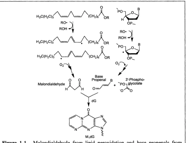
pyrimido[1,2-a]purin-10-(3H)-one adduct of dG (Figure 1-1).
In recent years, evidence has emerged that deoxyribose oxidation also plays a critical role
in the genetic toxicology of oxidative stress, including involvement in complex DNA lesions,
cross-linking with DNA repair proteins and the formation of endogenous DNA adducts. For
example, as part of a complex DNA lesion, closely opposed strand breaks and oxidized abasic
sites are resistant to repair by endonucleases and other enzymes [7, 8]. Studies also suggest that
deoxyribose oxidation may be an alternative to lipid peroxidation as a source of DNA-reactive
electrophiles. Oxidation of deoxyribose in DNA produces a variety of oxidized abasic sites and
strand breaks with different sugar residues, many of which are electrophilic and thus capable of
reacting with local nucleophiles to form adducts [9-13]. For example, the n-elimination product
of the 5'-(2-phosphoryl-1,4-dioxobutane) residue arising from 5'-oxidation of deoxyribose
(trans-1,4-dioxo-2-butene) reacts with dG, dA and dC to form stable bicyclic adducts [9, 10]. Research
-in Dedon laboratory has also shown the reaction of 3'-phosphoglycolaldehyde residues, which
are products of 3'-oxidation of deoxyribose in DNA to form the glyoxal adducts of guanine [11].
Similarly, we demonstrated that the base propenal products of deoxyribose 4'-oxidation,
structural analogs of the enol tautomer of MDA Figure 1-1), also react with DNA to form MjdG
[12], though with significantly greater efficiency than MDA [12, 13]. This may explain the
30-to 60-fold greater mutagenicity of base propenals than MDA [13].
H3C(H2C)3 • (CH2)6 OR RO* ROH H3C(H2C)3 (CH2)6 OR H3C(H2C)3 (CH2)6 OR Base o CM MalondialdehydeH Propenal B
H
O0
ý d G P B OP-RO, -ROH B OPR 02 7 3'-Phospho-+ PO glycolate 0o -o 0 N MldGFigure 1-1. Malondialdehyde from lipid peroxidation and base propenals from deoxyribose oxidation react with dG to form M1dG. B represents a base.
The goal of these studies undertaken in this thesis was to define chemical basis for the
formation of MldG as a product of either lipid peroxidation-derived MDA or deoxyribose
-oxidation-derived base propenals. This work entailed characterization of the chemistry of
4'-oxidation of deoxyribose in DNA, development of analytical methods for quantification of DNA
and lipid oxidation products, and application of analytical methods to quantify MldG in vitro, in
cells and in tissues from a mouse model of inflammation.
The thesis is organized as 6 chapters. In Chapter 1, we first present a comprehensive review of
DNA base and sugar damage with an emphasis on deoxyribose oxidation pathways and products,
especially 4'-oxidation products due to the relevance to our research. The chapter then discusses
lipid peroxidation and secondary DNA adduct from lipid peroxidation. Finally it covers the
mechanisms protecting cells/organisms against DNA damage. As a foundation for subsequent
studies, the studies presented in Chapter 2 address the development of analytical methods to
quantify strand breaks and abasic sites, which could serve as an index of total deoxyribose
oxidation for comparing different oxidizing agents. In Chapter 3, we present the results of efforts
to quantify 3'-phosphoglycolate residues and to define the spectrum of deoxyribose 4'-oxidation
products associated with different oxidizing agents. These methods are applied in Chapter 4 to
define the roles of base propenals and malondialdehyde in the formation of MldG in vitro and in
cultured cells. Chapter 5 extends these studies to DNA adducts in tissues from a mouse model of
inflammation, with development of analytical methods to quantify etheno adducts derived from
lipid peroxidation and to investigate the relationship between etheno adducts and M1dG
formation. Finally, in Chapter 6 we conclude this thesis by summarizing our contributions to
DNA damage study and offer directions for future research.
-23-1.2.
Sources of reactive oxidants
Cellular DNA is constantly subjected to reactions with a plethora of reactive oxidants.
These reactions are triggered by exposure of cells to exogenous chemicals (e.g., environmental
agents, food constituents, etc.) or they can result from endogenous metabolic processes, such as
oxidants generated by oxidative phosphorylation, P450 metabolism, peroxisomes, and
inflammatory cell activation. Oxidative damages can also be induced by exogenous sources, such
as radiation, metal ions, chlorinated compounds, barbiturates, phorbol esters and some
peroxisome proliferating compounds, all of which have been shown to induce oxidant formation
in vitro and in vivo [14, 15]. Endogenous and exogenous oxidants can either directly damage
DNA or they can induce DNA adduct formation indirectly by activation of otherwise inert
molecules by generation of reactive electrophiles by oxidation of lipids, proteins, carbohydrates
and even DNA itself. The former mechanism is illustrated by polycyclic aromatic hydrocarbon
benzo[a]-pyrene and aflatoxin B 1, both of which have been connected to bladder cancer and
liver cancer respectively due to an adduct formed between their epoxide metabolite and DNA
bases [16, 17]. This thesis focuses on the latter to study secondary DNA adducts formed by
electrophiles from oxidation of lipids and DNA deoxyribose.
While reactive oxidants are often important components of the immune defense system,
oxidative stress occurs when there is an imbalance between the oxidants and endogenous
antioxidant systems in favor of the former due to an excess of free radicals, a decrease in
antioxidant levels, or both [18]. For example, sustained production of reactive oxidants by
phagocytes during chronic inflammation induces collateral damage in adjacent normal tissues,
-which contributes to a range of diseases. In this section we will review the chemistry and biology
of the formation of these oxidative species.
1.2.1
Reactive oxygen species
Reactive oxygen species (ROS) is the term used to describe a variety of species, which
not only includes oxidizing radicals such as superoxide (02'), hydroxyl (.OH), peroxyl (ROO-),
and alkoxyl (RO*) radicals but also includes nonradicals such as hydrogen peroxide (H202), ozone (03), and singlet oxygen ('0 2)-that are oxidizing agents or are easily converted into
oxidizing radicals. During mitochondrial oxidative metabolism, a small fraction (0.2-0.6%) of
molecular oxygen is converted to reactive oxygen species, primarily superoxide anion radical 02, through one-electron reduction [19]. 02* is then converted to hydrogen peroxide (H202)
spontaneously or in reactions catalyzed by superoxide dimutase (SOD). The resulting H202 is further converted to hydroxyl radical (.OH) by Fenton chemistry involving Fe2 +, Cu+, or other
metal ions. The process is summarized in Figure 1-2. ROS are also released from neutrophils and
macrophages through a process known as respiratory burst [20]. During the respiratory burst, membrane-bound NAD(P)H oxidase is activated and reduces oxygen to 02', which is then
converted to other reactive oxygen species as described above.
-25 -NAD(P)H + 202 -~- NAD(P)+ + H+ + 202 SOD 202 + 2H - O- HOOH + 02 HOOH + Fe2+ -~ OH- + -OH + Fe3+
Molecular oxygen itself is a relatively weak univalent electron acceptor that cannot
efficiently oxidize biomolecules. In contrast, O2', H202, and *OH are much stronger univalent oxidants. However, the anionic charge of 02 inhibits its effectiveness as a direct oxidant of
electron-rich molecules, while the reactivity of H202 is diminished by the stability of its
oxygen-oxygen bond [19]. Of these species, *OH is considered to be the major reactive oxygen-oxygen species
contributing to endogenous oxidation of cellular molecules. Since the rate of reactions of *OH is
controlled by diffusion, the reactions are non-specific and occur close to the site of its formation.
A wide variety of -OH-induced damages, including oxidized bases and deoxyribose,
DNA-protein and DNA-DNA cross-links have been identified and will be discussed in detail in the
subsequent sections of Chapter 1.
Singlet oxygen '02 is an excited form of dioxygen in which the ;r antibonding electrons are spin-paired. 102 can be formed endogenously by energy transfer to oxygen by excited
chromophores in stimulated phagocytes during the respiratory burst. 102 also reacts with
biomolecules and exerts genotoxic, virucidal and cytotoxic effects [21, 22]. In contrast to *OH,
which reacts almost indifferently with all nucleobases and the sugar moiety of DNA, the reaction
of 102 with DNA is highly specific with guanine base as the exclusive target and 8-oxodG as the
main oxidation product [23].
Besides endogenous sources, ROS can also be generated by a variety of exogenous
sources. Ionizing radiation can induce the homolysis of H20 to form .OH and 'H. It can also
directly deposit energy into covalent bonds to cause damage to biolmolecules. The non-ionizing
UVA component of solar radiation has been shown to produce 102 in its lowest excited state [21]. Tobacco smoke contains a variety of free radicals, which generate free radicals in exposed
-tissues [24]. In addition, drugs such as Phenobarbital and carcinogens such as benzo(a)pyrene
have also been shown to induce the formation of ROS [25].
1.2.2 Reactive nitrogen species
The class of reactive nitrogen species (RNS) includes, among other species, nitric oxide
(NO.), nitrous anhydride N203, peroxynitrite (ONOO), and nitrosoperoxycarbonate
(ONOOCO2). NO- is produced from L-arginine by nitric oxide synthase, with NADPH as an
electron donor and HEM, FMN, FAD and tetrahydrobiopterin as cofactors. Three distinct
isoforms of NOS have been identified: endothelial NOS (eNOS), inducible NOS (iNOS) and
neuronal NOS (nNOS) [26]. Both eNOS and nNOS are constitutively expressed and are
activated by calcium and calmodulin, while iNOS is induced in macrophages, endothelium, hepatocytes and mast cells by bacterial endotoxin and cytokines IL-1 and tumor necrosis factor
(TNF) [27]. NO. can also be produced nonenzymatically from nitrite at low pH under reducing
conditions [28].
One important reaction of NO- is oxidation by molecular oxygen to form the powerful
nitrosating agent N203, as shown in Figure 1-3. Current evidence suggests that N203 is the
predominant species in aqueous media [29]. Based on these equations, the half-life of NO* is
inversely proportional to its concentration, with competition between hydrolysis to form nitrite
[30] and reactions with amines, thiols, or hydroxyl groups to form NO* adducts through
nitrosation.
-2NO + 02 -- 2NO2
NO + N ~2 N203
N203 + H20 ~ 2NO2- + 2H÷
Figure 1-3. Reaction of reactive nitrogen species
Macrophage activation induces both 02' and NO. production, both of which can react
together at a diffusion-controlled rate (k = 6.6-19 x 109 M'S -') to yield ONOO [31, 32]. While approximately equal rates of 02' and NO. generation produces the maximal amount of ONOO-
-induced damage, widely differing amounts of either 02' or NO* lead to side reactions with
ONOOU to form NO2, and thus inhibiting ONOO -induced oxidation [33]. Individually, 02" and
NO- are both relatively unreactive. However ONOO& is very reactive and rapidly decomposes in the absence of CO2 via a proton-catalyzed homolysis with k = 1.3 s"1, with -67% of caged
radicals reacting to form NO3-and the reaming -33% escape the cage and become free *OH and
*NO2 as shown in Figure 1-4. Under biological conditions, ONOO- reacts rapidly (tl/2 < 0.1 pS) with CO2 to form nitrosoperoxycarbonate ONOOCO2_. Homolysis of the 0-0 bond produces
carbonate (CO') and *NO3 2. The concentration of CO2 in vivo is relatively high (1-2 mM) due to the high levels (12 mM) of bicarbonate in intracellular fluids [34]. This suggests that the reaction
of ONOO- with CO2 is a major pathway for ONOO& consumption in vivo. Since the intrinsic life
time of ONOOCO2- is considerably shorter than ONOO (tl/2 z 0.7s) [35], the cellular targets for
ONOO- are likely restricted to the location of its formation.
-RNS contribute to direct genotoxicity through reaction with DNA and indirect
genotoxicity due to activation of nitrosamines, lipidoxidation-induced DNA damage and
inhibition of DNA repair enzymes. For example, RNS have been shown to inhibit
formamidopyrimidine-DNA glycosylase (Fpg) and 3-methyladenine-DNA glycoxylase (Alka)
by a nitrosation reaction at RNS concentrations 100-fold less than those required to deaminate
DNA [36].
1.2.3 Reactive halogen species
Reactive halogen species (RHS) include hypochlorous acid (HOC1), hypobromous acid
(HOBr), and hypoiodous acid (HOI). The lysosomal enzyme myeloperoxidase in the granules of
neutrophils catalyses the H202-mediated oxidation of halides to form RHS, as shown in Figure
1-5.
-29-slow
NO' + 02- - ONOO- <-+ ONOOH - [ONO..OH] - NO3- + H*
CO2
ONOOCO
2Proucts,
NO
2+.OH
67% 3
CO2+ NO-<--'- 3 [CO3. .NO2] -- CO3-+ NO2
Similar to ROS and RNS, RHS are strongly oxidizing and halogenating species that can
induce lipid peroxidation [37], halogenation of DNA bases (e.g., 5-chlorouracil) [38], and
cross-linking of DNA and protein [39], all of which may play a rose in tissue damage during
inflammation [40].
In summary, these reactive oxidants can alter cell function by directly damaging cellular
molecules as well as by generating electrophilic species capable of causing further damage. The
following sections address the reactions of ROS, RNS and RHS with biomolecules and the
resulting primary and secondary DNA adducts.
1.3.
DNA base damage
Free radicals can adversely alter DNA and have been implicated in aging and a number
of human diseases [41, 42]. In a given human cell, an estimated 104 oxidative DNA lesions per
day are formed [43]. DNA damage, if not repaired, can lead to mutations and cell death. The
magnitude of oxidative DNA damage has led many research groups to study the mechanisms of
DNA oxidative damage, to identify the damage products and to define their biological
significance.
-30-HOOH + H+ + CI- -- H20 + HOCI
HOOH + H+ + Br- - H20 + HOBr
HOOH + H÷ +
I-
H20 + HOIROS/RNS/RHS can oxidize both nucleobases and deoxyribose moieties by addition or H
atom abstraction. For example, *OH can add to double bonds of DNA bases and abstract H
atoms from the C-H bonds of deoxyribose. Given the prominence of nucleobase lesions and their
potential to cause DNA mutation, carcinogenesis and aging [44], this section of the background
and literature review will address the base damages in detail.
1.3.1 Hydroxyl radical induced base damage
OH is often considered to be the major ROS responsible for endogenous oxidation of
DNA and it serves as an illustration for the types of damage products generated by ROS and
RNS. As noted earlier, *OH arises from a variety of sources and rapidly reacts at its site of
formation either by H atom abstraction or addition to C=C. Given its low oxidation potential
relative to other DNA bases, guanine is the most reactive toward a variety of oxidants including
*OH. *OH can add to the C4 (-60%), C5 (-15%) and C8 (-25%) positions of guanine, with
subsequent dehydration of C4-OH- and C5-OH-adduct radicals yielding a guanine(-*H) radical.
The radical can either be reduced back to guanine or further oxidized to
2-amino-5-[(2-deoxy-3-D-erythro-pentofuranosyl)amino]-4H-imidazol-4-one and
2,2-diamino-4-[(2-deoxy-o-D-erythro-pentofuranosyl)amino]-5(21)-oxazolone as shown in Figure 1-6. The C8-OH-adduct radicals can
be oxidized to form final product 8-oxoG or reduced to form the formamidopyrimidine,
2,6-diamino-4-hydroxy-5-formamidopyrimidine (FapyGua) [45].
-Adenine undergoes reactions analogous to guanine, yielding C2-OH-, C4-OH- and
C8-OH-adduct radicals that further react to form 2-hydroxyadenine, 8-hydroxyadenine and 4,
6-diamino-5-formamidopyrimidine (FapyAde) [46], as shown in Figure 1-7.
NH2 NH2 NH2 NH2
N
N
N N N
N
N
NHCHO
N N H HO N N H N N'N H N INH 2
2-hydroxyadenine 8-hydroxyadenine 4,
6-diamino-5-formamidopyrimidine
Figure 1-7. Product formation from adenine oxidation
For cytosine, OH11 adds at the C5 position to the extent of -87%, whereas -10% of *OH
adds to the C6 [47] (Figure 1-8) .Molecular 02 then adds to the C5-OH-adduct radical
generating C5-OH-6-peroxyl radicals that subsequently eliminate 02' followed by reaction with
water to yield cytosine glycol. Deamination and dehydration of cytosine glycol yields uracil
glycol, 5-hydroxyuracil and 5-hydroxycytosine. In the absence of oxygen, the C5-OH-adduct
-32-radical can be reduced to hydroxy-6-hydrocytosine, which can be further converted to
5-hydroxy-6-hydrouracil through deamination [42]. Other reactions of C5-OH-6-peroxyl and COH-5-peroxyl radical result in the formation of the intermediates, 5-OH-hydroperoxide and
6-OH-5- hydroperoxide that then decompose to 4-amino-5-hydroxy-2, 6(1H, 5H)-pyrimidinedione
and 4-amino-6-hydroxy-2, 5(1H, 6H)-pyrimidinedione [48], respectively. The former deaminates
to give dialuric acid, which is readily oxidised to yield alloxan, while the latter deaminates to
isodialuric acid [49] (Figure 1-8).
*OH adds preferentially to the C-5 of thymine, whereas approximately 10-15% of .OH
adds to C-6 and 5 - 10% abstracts a H atom from the 5'-methyl group to form an allyl radical
(Figure 1-9). Similar to cytosine, the C5-OH-adduct radical can be oxidized to thymine glycol or
reduced to 5-hydroxy-6-hydrothymine and 6-hydroxy-5-hydrothymine, as shown in Figure 1-9.
Reaction with molecular oxygen yields peroxyl radicals that decompose to
-methylhydantoin [50]. The oxidation of the allyl radical of thymine yields
5-hydroxymethyluracil and 5-formyluracil.
C5-CH3 H HN OH S.OH O N H H O CH3 HN OH H
thymine glycol 5-hyd
hydro 0 . -OH n. T HI N H O- N H N OH H H H C6-OH-adduct radical
(-30%) 6-hydroxy-5- hydrothymine 5-hy(
0 0
HN 6H2 OHN CH20H
0 N H N H
H H
allyl radical 5-(hydroxymethyl)uracil
(-10%)
Figure 1-9. Product formation from thymine oxidation
roxy-6-thymine CH3 N OH H droxy-5-methyl hydantoin O HN CHO H 5-formyluracil
1.3.2
ONOO-
and ONOOCO
2-induced base damage
As we have discussed in Section 1.2, ONOO- is among the most reactive species of RNS.
ONOO- induced damage is influenced by CO2. In the absence of CO2, the majority of
ONOO-induced DNA damage is deoxyribose oxidation. The presence of CO2 results in the formation of
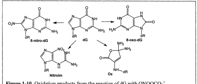
ONOOCO2 and cause a shift from deoxyribose oxidation to base oxidation and nitration with little change in the total number of lesions [51, 52]. ONOOC02z primarily reacts with dG to form 5-guanidino-4-nitroimidazole (Nitrolm), 2,2-diamino-4- {(2-deoxypentofuranosyl) amino}
-- -
34-HN
5(2H)-oxazolone (oxazolone; OZ), and 8-nitro-dG as shown in Figure 1-10 [53]. 8-NitrodG rapidly depurinates (tn/2 - 1-4h) at neutral pI1 and ambient temperature to yield the measurable 8-nitroGua [54]. More recent evidence from the Tannenbaum group also revealed that 8-nitrodG
can further react with ONOOCO2- to yield additional 8-oxodG [55].
As a result of the lower oxidation potential of 8-oxodG compared to dG (0.74V vs.
1.29V), it is not surprising that 8-oxodG has been shown to be at least 1000-times more
susceptible to further oxidation by ONOOCO2 than dG and forms a variety of secondary lesions
as shown in Figure 1-11 [53]. -35-frh 0
H
N N NH H I N NH H2N
dRa dR dK ui. 1' 8-nitro-dG dG N NH2N TNH2
H
Nitrolm OzONOOC02-1.3.3 N
20
3-induced base damage
Autooxidation of NO* yields N203, a potent nitrosating agent that induces the
deamination of DNA bases. N-Nitrosation of a primary amine initially produces a nitrosamine
that is quickly followed by the replacement of -NIH2 group bye H20. As shown in Figure 1-12,
nitrosation by N203 (and NO2" in acidified solutions) converts adenine to hypoxanthine
(2'-deoxyinonsine, dl), cytosine to uracil (dU in nucleoside form), 5-methylcytosine to thymine, and
guanine to both xanthine (2'-deoxyxanthosine, dX) and oxanosine (2-deoxyoxanine, dO:
-36-observed only at low pH), as well as abasic sites and inter- or intra-strand G-G cross-links [56,
57].
1.3.4
Summary
Aforementioned pyrimidine and purine adducts are far from complete. More than 100
base lesions have been identified using different oxidants. Many of these products have been
identified in mammalian cells and tissues as well, suggesting their importance in vivo.
-37-It is worth noting that the sites of initial oxidation and terminal damage sites are not
necessarily identical in DNA because of a phenomenon called long distance electron transfer. So
bases with lower redox potential, dG, is more likely to be oxidized than other bases. Electrons
were shown to transferred within the DNA duplex from guanine to the electron-deficient centers
created by radiation [58], chemical oxidants and laser-induced photoionization [47, 59]. Further
evidence showed that dG-repeats had lower redox potential than isolated dG, with 5'-dG having
the lowest potential, and as a result was a more likely target. The oxidation product of dG,
8-oxodG has even lower redox potential than dG. Considering the persistent existence of 8-8-oxodG
in vivo, it might be an important target for DNA damage as well.
1.4.
DNA deoxyrobise oxidation
Although significant attention has been paid to nucleobase lesions given their role in
toxicity and mutagenesis associated with oxidative species, there is growing evidence that
deoxyribose oxidation plays a major role in the biological response to oxidative stress. Oxidation
of each carbon in deoxyribose yields a unique spectrum of sugar residues or oxidized abasic sites,
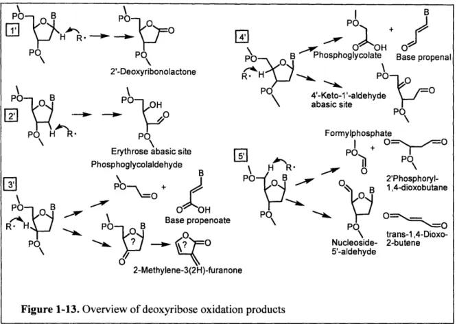
as shown in Figure 1-13 [60]. Among these products are several reactive electrophiles capable of
forming mutagenic adducts with DNA bases [11, 12].
-38-\ H R PO PO 2'-Deoxyribonolactone PO 0 B PO Po
PO
H R
PO
Erythrose abasic site Phosphoglycolaldehyde3PO-_+
\ i0I-penal o B O~OH Base propenoate PO B P0
H R
PO \ Formylphosphate I + PO+ PO POP S • O 2'Phosphoryl-B 1,4-dioxobutane PO 0 \ trans-1,4-Dioxo-Nucleoside- 2-butene 5'-aldehyde 2-Methylene-3(2H)-furancFigure 1-13. Overview of deoxyribose oxidation products
The deoxyribose in DNA has seven carbon-bound hydrogen atoms that are available for
abstraction by radicals and other one-electron oxidants that are designated as H-l', H-2', H-2",
H-3', H-4', H-5', and H'5", as shown in Figure 1-14 [61]. Mechanistic information on the
reaction of the *011 with nucleic acids has been extensively studied using ionizing radiation as a
source of .OH1. Because of the high reactivity of *OH, a wide spectrum of products has been
detected in irradiated DNA. For biologically relevant duplex DNA, besides the difference in
reactivity due to carbon-hydrogen bond energies, the shape of the double helix has been
proposed to influence the accessibility of the various C-H bonds to -OH. Using
Fe(II)/EDTA/H202/ ascorbate system as a source of putative hydroxyl radicals, Balasubramanian
et al. proposed that solvent accessibility dictates the relative reactivity of deoxyribose carbon
atoms in duplex DNA with *OH [62]. They exploited deuterium isotope effects to demonstrate
-39-4
F
that Fe-EDTA-induced oxidation exhibits a preference in the following order: 5' > 4' > 2' = 3' >
1'. An atomistic stochastic model of *OH radical reactions with DNA was developed to compute
relative -OH attack probabilities at individual deoxyribose hydrogen atoms [63]. Results from
this computational model show that *OH radicals exhibit preferential attack at different
deoxyribose hydrogens, as suggested by their corresponding percentage solvent-accessible
surface areas. The percentage OH attack probabilities for the deoxyribose hydrogens
[1H(5')+2H(5'), 11(4'), H(3'), 1H(2')+2H(2'), H(1')] were calculated as approximately 54.6%,
20.6%, 15.0%, 8.5% and 1.3%, respectively, averaged across the sequence. These results are in
good agreement with the latest experimental site-specific DNA strand break data of
Balasubramanian et al. [62]
Studies have shown the oxidation of each deoxyribose carbon position produces a unique
spectrum of products. The chemical mechanism, as well as the products of these oxidation
reactions, has been a major focus of the Dedon research group. So in this section, we will discuss
DNA deoxyribose oxidation (proposed) pathways and products in detail.
40 -OR' H-5' I I O O=P-O-C /0 B O H-5" 04 H H-2' C H C3 0C2
6
H-2"
I O=P-O I OR1.4.1
C1'-oxidation
The H-i' is buried in the minor groove of B-DNA and is believed to be relatively
inaccessible to solvent [64]. Hence, the importance of H-l' as a reactive site is thought to be
limited primarily to minor groove binding molecules, where the oxidant is generated in the
groove in close proximity to H-l'. One of the well-studied DNA cleavage reactions resulting
from H-l' is that promoted by bis(1,10-phenanthroline)copper(I) [65, 66]. Hence, reactive
copper-bound oxidants (e.g., [CuO]+, [CuOH]2+ and CuO2H) are believed to be the oxidizing
species rather than *OH. Two products, 2'-Deoxyribonolactone and 5-methylene-2(5H)-furanone, were identified as the major products species resulting from H-I' oxidation, as shown in Figure
1-15 [67].
Another class of DNA-cleaving agents that reacts at the C1' position is the enediyne
antibiotic family. These natural products from the eubacteria Actinomycetales include
neocarzinostatin (NCS), calicheamicin, esperamicin and dynemicin A, etc. As shown in Figure
1-16, enediynes produce oxidative DNA damage by a common mechanism involving reductive
activation to form a diradical species that, when positioned in the minor groove of one DNA
-41-strand, may abstract hydrogen atoms from deoxyribose on each DNA strand. For
neocarzinostatin, the lesions occur predominantly at AGC*GCT and AGT*ACT sequences and
involves mainly 1' chemistry at C of AGC, 4' chemistry at the T of AGT, and 5' chemistry at the
other T residues on the complementary strands [62, 68, 69].
1.4.2 C2'-oxidation
Due to the low solvent accessibility of H-2' and H-2", as well as the low reactivity of
these C-H bonds because of the instability of C2' radical [64], these positions are believed to
play a small role in deoxyribose oxidation. Dizdaroglu et al. proposed that a major product of
C2'-oxidation is erythrose abasic site (Figure 1-17) and characterized erythrose abasic site in
y 42 y
-RS- R SR R, SR R SR R
ROl RO ROH DNA ROIW H
RO RO RO RO H
Neocarzionstatin Enyne-[3]-cumulene Diradical intermediate
U
R
DNA RO 0S O
"'POR
DihvdrothioDhene Benzenoid diradical Calicheamicin
RS-SSSH3 H
HO S HO S
HO -HO SOR DNA OR
OR O O / Ot H
O RIO R10
R'O R0O
Esperamicin A'
Figure 1-16. Activation of neocarzinostatin, Calicheamicin and Esperamicin
A
irradiated DNA using GC/MS [70]. Since the hydroxyl group next to the phosphate ester causes
the site to be unstable, the erythrose abasic site has been shown to undergo retroaldol reaction to
yield 3'-phosphoglycolaldehyde residues under heating or alkaline conditions [71]. In addition to
ionizing radiation, photo-activation of 5-bromouracil has also been observed to induce C2'
oxidation [72].
1.4.3 C3'-oxidation
The C3'-hydrogen atom is located in the major groove of DNA. Since the majority of the
known oxidative cleavage agents bind in the minor groove rather than the major groove,
C3'-oxidation was not well studied. C3'-C3'-oxidation is considered to be minor in *OH-induced
deoxyribose oxidation due to limited accessibility of H-3' and the reduced stability of a
3'-deoxyribosyl radical. Major groove binding photoactive Rhodium(III) complexes were shown to
induce C3'-oxidation efficiently [73]. The proposed products of C3'-oxidation includes
phosphoglycoladehyde (PGA) residue and base propenoate in an oxygen-dependent mechanism
and 2-Methylene-3(2H)-furanone under anaerobic conditions as shown in Figure 1-18 [61].
-Using sequencing gel technique, Sitlani et al. tentatively identified PGA termini attached to the
3'-end of the strand breaks arising from deoxyribose oxidation by using Rhodium(III) complexes.
They further showed that the molar ratio between PGA and based propenoates was indeed 1:1,
which is consistent with the proposed pathway.
PO B PO% B
1
o- o
PO PO OH-2-Methylene 0 -3(2H)-furanone Base propenoateFigure 1-18. Proposed H-3' abstraction pathway and products
1.4.4
C4'-oxidation
The C4'-hydrogen atom is located on the outer edge of minor groove of DNA and is
relatively solvent accessible. This C4' carbon-hydrogen bond also has relatively low bond
dissociation energy, which makes the C4'-hydrogen atom a major target for *OH and other minor
groove-binding oxidants. DNA damage initiated from 4'-hydrogen abstraction has been observed
for ionizing radiation, Fe2+/EDTA, Fenton-generated -OH, bleomycin, and enediyne antibiotics.
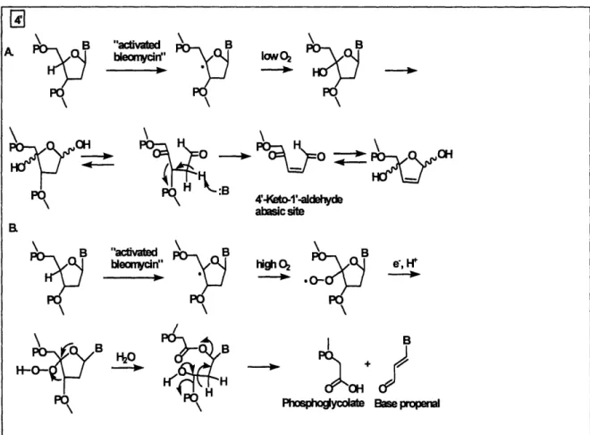
One of the best-characterized pathways of C4'-oxidation is the H-4' abstraction by
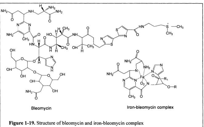
bleomycin*Fe(II) [74, 75]. The bleomycin antibiotics represent a family of glycopetide-derived
antitumor antibiotics widely used in clinical setting to treat cancer [76]. The attack of DNA by
44
-\e%
Bleoymcin*Fe(II) complex requires a sequence of preliminary steps, including "activation". The
drug binds to DNA in the minor groove and is "activated" either by molecular oxygen and two
electrons, or by hydrogen peroxide (Figure 1-19). Although the exact identity of the oxidizing
species in activated bleomycin is not settled, it is generally believed that a peroxide-Fe(III) *
bleomycin is likely to be the activated form [77].
Using deuterium-labeled deoxyribose, Stubbe et al. showed that the abstraction of H-4'
by activated belomycin was the rate-limiting step in bleomycin-induced DNA degradation [75,
78.]. Degradation of 4'-deoxyribosyl radical has two outcomes associated with two sets of
products (Figure 1-20). One set of products includes strand breaks terminated by 5'-phosphate
and 3'-phosphoglycolate moieties. The remainder of the cleaved nucleoside is released as base
propenal. The other set of degradation products includes free nucleobase and an abasic sugar,
2-deoxypentos-4-ulose (4'-keto-l'-aldehyde). Under neutral pH conditions and high oxygen
45 -.HN S\ -CH 3 CH3 >\ _-N NH2 ,N OO-R 0
Bleomycin Iron-bleomycin complex
tension, molecular oxygen reacts with the carbon radical to form a peroxy radical that may be
reduced to a peroxide. This intermediate further degrades to base propenal and
3'-phosphoglycolate. At low oxygen tension, the carbon radical is further oxidized to a carbocation
that adds water to yield a 4'-hydroxyl species. This product is proposed to give rise to a putative
hemiketal species that is expected to eliminate the nucleobase while rearranging to yield
4'-keto-I'-aldehyde abasic site [79]. It was shown that bleomycin causes the formation of cytosine-,
thymine-, and adenine-propenals accompanied by 3'-phosphoglycolate [80], while the
4'-keto-l'-aldehyde accounted for -40% of bleomycin-induced DNA oxidation products [79].
A
B "activated
B
A bleomydrn" PPp \P\ 4'A1Ko-1'-aldehyde abasic site \ B highQ
- o K>B "activated
B
bleomycidn" Ii.gPhosphoaycolate Base propenal
Figure 1-20. Proposed H-4' abstraction pathway and products by activated bleomycin H
PO
-46-B
Unlike bleomycin, y-radiation induces deoxyribose C4'-oxidation to form
malondialdehyde and a free base instead of base propenal [80], a difference that indicates
different reaction mechanisms. It is believed that bleomycin remains bound to DNA near the C4'
radical after hydrogen atom abstraction and influences the formation of final products. The
formation of malondialdehyde in irradiated DNA follows a linear dose-dependence and is
accompanied by the formation of 3'-phosphoglycolate residues [80, 81].
Enediynes such as NCS and calicheamicin were also shown to induce C4'-oxidation [82].
DNA deoxyribose oxidation induced by activated NCS has been extensively studied [83]. It has
been demonstrated that at least 80% of the DNA cleavage involves 5'-oxidation, which leads to
the 5'-aldehyde of dA and dT selectively [84], while less than 20% of the strand breaks resulted
from hydrogen atom abstraction at C4' and Cl' positions [85, 86]. The C6 radical of activated
NCS abstracts a H atom from the C5' of deoxyribose and the remaining NCS C2 radical reacts
across the minor groove with the opposite strand at the C4' (or Cl') position in the case of
suitable special DNA sequences that promote double-strand breaks. Although NCS yields the
same products from C4'-oxidation as bleomycin, the mechanism is believed to be different
[86-88]. Bleomycin-induced abasic site formation is oxygen-independent, while both abasic site
formation and 3'-phosphoglycolate formation are oxygen-dependent for NCS [89]. Similarly,
calicheamicin and esperamincins C-E also induce DNA cleavage by C4'-hydrogen abstraction
on one strand and C5'-hydrogen abstraction on the other strand. With calicheamicin, bistranded
lesions represent >95% of the DNA damage and the 4'-chemistry on one strand is accompanied
by products of 5'-hydrogen atom abstraction on the complementary strand [90].
There is also evidence for participation of thiols in reactions of the deoxyribose carbon
radicals after their formation. Neutral and positively charged thiols cause a reduction in the level
-of double-stranded DNA lesions induced by NCS and esperamincins relative to that -of
negatively charged thiols [91]. For calicheamicin, no such quenching effect was observed. Yet,
neutral and, to an even greater extent, positively charged thiols inhibit the formation of
3-phosphoglycolate residues with a proportional increases in the formation of the alternative abasic
sites, which may be due to concentration of the positive thiols on or around the negatively
charged backbone of DNA [82].
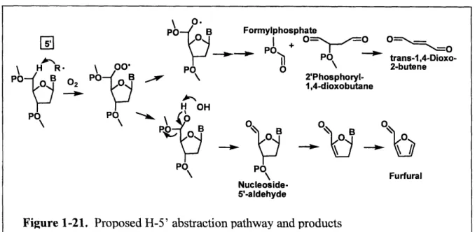
1.4.5 C5'-Oxidation
In the minor groove of the B-DNA helix, both H5' and H5" have been estimated to be
the most solvent accessible deoxyribose hydrogen atoms [61]. C5'-oxidation has been observed
by y-radiation, Fenton-generated *OH, enediyne antibiotics, cationic metal porphyrins, and the
perhydroxyl radical [92, 93]. As shown in Figure 1-21, the reaction partitions to form either a
nucleoside 5'-aldehyde residue attached to the 5'-end of the DNA strand, or a 5'-formylphosphate
residue attached to the 3'-end of the DNA strand that is accompanied by a four-carbon fragment
on the 5'-end, which was identified as 5'-(2-phosphoryl-1,4-dioxobutane) [94]. Formation of a
hydroxyl group at the C5' carbon radical gives rise to a C5' aldehyde at one strand terminus and
a 3'-phosphate at the other at ambient temperature [95]. Upon heating, consecutive 3- and
6-elimination result in the formation of furfural, free base, and a 5'-phosphate terminus. Pratviel et
al. developed a GC/MS method to quantify furfural and showed that furfural can be used as a
convenient marker of C5' hydroxylation of deoxyribose [95]. Alternatively, the deoxyribose
5'-peroxide may form an oxygen radical intermediate with subsequent p-scission of the 4'-5' bond
(Creigee-type rearrangement) to generate the 3'-formylphosphate and 5'-(2-phosphoryl-1,
-48-dioxobutane) residue. The Dedon group has shown that the trans-5'-(2-phosphoryl-
1,4-dioxobutane) residue has the potential to undergo 3-elimination to form 1,4-dioxo-2-butene, a
highly reactive a, P-unsaturated dicarbonyl species [94].
As discussed in Section 4.5, the activated enediynes abstract a H atom from the C5' of
deoxyribose. The resulting nucleoside 5'-aldhyde accounts for a majority of the damage
produced by C5'-hydrogen abstraction, while 3'-formylphosphate and
2'-phosphoryl-1,4-dioxobutane products are often the minor lesions [90, 96].
1.5. Complex DNA lesions
A special type of oxidative DNA damage, termed a complex lesion, has been associated
with ionizing radiation. This type of damage consists of two or more lesions-single-strand
breaks or modified bases-located within one to two helical turns on the same strand or on
opposite strands. Radiation, especially high LET radiation, produces dense ionization in the
-49-PO B Formylphosphate P POC! O 0 PO trans-i,4-Dioxo-H R \ 00 PO 2-butene PO B O Po- B \ 2'Phosphoryl-2 0 1,4-dioxobutane H OH P0 PO O
O
B
0
0
BO
PO PO \\ Furfural Nucleoside-5'-aldehydeFigure 1-21. Proposed H-5' abstraction pathway and products -
-vicinity of the DNA helix and induces multiple oxidation events. The best known example of
complex lesion is double strand break (DSB), which is generated when the two complementary
stands of the DNA double helix are broken simultaneously at sites that are sufficiently close to
one another to allow the dissociation of the helix [97]. As potent inducer of mutation and cell
death, a DSB is probably the most toxic DNA lesions with a single DSB capable of killing a
metazoan cell if it leads to the inactivation of an essential gene or, more commonly, triggers
apoptosis [98]. Evidence also showed a causal link between the generation of DSBs and the
induction of mutations and chromosomal translocations with tumorigenic potential [99, 100].
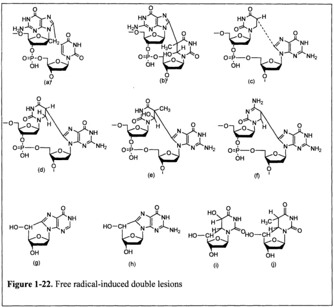
Another example of complex lesion, termed tandem lesion (Figure 1-22), involves two
vicinal modifications on the same DNA strand and is generated by one initial radical hit that
leads to cyclization [101]. These cyclo-lesions are formed through a covalent linkage between
the bases of adjacent nucleotides or linkage between the base and deoxyribose moeities of the
same nucleotide. A well-studied example, 5',8-Cycolpurine (g and h in Figure 1-22) nucleosides
were first identified more than 20 years ago [102] and have been observed in y-irradiated isolated
DNA [103] and in DNA from mammalian tissues [104]. 5', 8-cycolpurine nucleosides arise
from the attack of the C-5' centered radical of the sugar moiety at the C-8 of the purine within the
same nucleoside leading to intramolecular cyclization followed by oxidation of the thus formed
N-7 centered radical. These compounds represent a concomitant damage to both sugar and base
moieties of the same nucleoside and thus can be considered complex lesions. Because of the
presence of a covalent bond between the sugar and purine moieties, these tandem lesions are not
repaired by base excision repair but instead by nucleotide excision repair. Thus, they may play a
role in diseases with defective nucleotide excision repair.