HAL Id: inserm-02905134
https://www.hal.inserm.fr/inserm-02905134
Submitted on 23 Jul 2020
HAL is a multi-disciplinary open access archive for the deposit and dissemination of sci-entific research documents, whether they are pub-lished or not. The documents may come from teaching and research institutions in France or abroad, or from public or private research centers.
L’archive ouverte pluridisciplinaire HAL, est destinée au dépôt et à la diffusion de documents scientifiques de niveau recherche, publiés ou non, émanant des établissements d’enseignement et de recherche français ou étrangers, des laboratoires publics ou privés.
sleep apnea: Is there a link?
Céline Dard, Sébastien Bailly, Jean-Louis Pépin, Marie-Pierre
Brenier-Pinchart, Hélène Fricker-Hidalgo, Marie Peeters, Hervé Pelloux,
Renaud Tamisier
To cite this version:
Céline Dard, Sébastien Bailly, Jean-Louis Pépin, Marie-Pierre Brenier-Pinchart, Hélène Fricker-Hidalgo, et al.. Chronic toxoplasmosis and sleepiness in obstructive sleep apnea: Is there a link?. PLoS ONE, Public Library of Science, 2020, 15 (7), pp.e0235463. �10.1371/journal.pone.0235463�. �inserm-02905134�
RESEARCH ARTICLE
Chronic toxoplasmosis and sleepiness in
obstructive sleep apnea: Is there a link?
Ce´line Dard1,2, Se´bastien Bailly3, Jean-Louis Pe´pin3, Marie-Pierre Brenier-Pinchart1,2, He´lène Fricker-Hidalgo1, Marie Peeters3, Herve´ PellouxID1,2*, Renaud Tamisier3
1 Parasitology and Mycology Laboratory, CHU Grenoble Alpes, Grenoble, France, 2 Institute for Advanced Biosciences (IAB), Team Host-Pathogen Interactions and Immunity to Infection, INSERM U1209—CNRS UMR5309, University Grenoble Alpes, Grenoble, France, 3 Inserm, CHU Grenoble Alpes, HP2, University Grenoble Alpes, Grenoble, France
*hpelloux@chu-grenoble.fr
Abstract
Introduction
Sleepiness is the main clinical expression of obstructive sleep apnea (OSA) syndrome resulting from upper airway collapse. Recent studies have discussed the fact that the pres-ence of T. gondii cysts in the brain and the resulting biochemical and immunological mecha-nisms could be linked to neurobehavioral disorders. The aim of the present study was to explore the potential impact of chronic toxoplasmosis on sleepiness and on obstructive sleep apnea (OSA) severity in OSA obese patients.
Materials and methods
A case control study on obese patients screened for OSA was performed. According to the sleep disorder and matched based on gender, age and body mass index (BMI), two groups of obese patients were selected from our sample collection database. All patients were tested for toxoplasmosis serological status measuring anti-Toxoplasma IgG and IgM levels. Univariable and multivariable logistic regression models were performed to assess the impact of chronic toxoplasmosis on sleepiness and OSA severity.
Results
107 obese patients suffering from OSA were included in the study (median age: 53.3 years Interquartile range (IQR): [41.9–59.9]; median BMI: 39.4 kg/m2IQR: [35.5–44.1], apnea-hypopnea index = 27.5 events/h [10.7–49.9]). Chronic toxoplasmosis was present in 63.4% and 70.7% of patients with or without sleepiness (p = 0.48), respectively and was not associ-ated either to sleepiness (OR: 0.76, 95% CI: [0.52; 2.33], p = 0.64) or OSA severity (OR = 1.75, 95% CI: [0.51; 5.98] p = 0.37).
Conclusion
Although chronic Toxoplasma infection in immunocompetent humans has been associated to several behavioral disorders or pathologies in recent literature, we demonstrate here that
a1111111111 a1111111111 a1111111111 a1111111111 a1111111111 OPEN ACCESS
Citation: Dard C, Bailly S, Pe´pin J-L,
Brenier-Pinchart M-P, Fricker-Hidalgo H, Peeters M, et al. (2020) Chronic toxoplasmosis and sleepiness in obstructive sleep apnea: Is there a link? PLoS ONE 15(7): e0235463.https://doi.org/10.1371/journal. pone.0235463
Editor: Andrea Romigi, University of Rome Tor
Vergata, ITALY
Received: July 15, 2019 Accepted: June 16, 2020 Published: July 1, 2020
Copyright:© 2020 Dard et al. This is an open access article distributed under the terms of the
Creative Commons Attribution License, which permits unrestricted use, distribution, and reproduction in any medium, provided the original author and source are credited.
Data Availability Statement: All relevant data are
within the manuscript and its Supporting Information files.
Funding: This work was performed at CHU
Grenoble Alpes, Clinique Physiologie, Sommeil et Exercice and Laboratoire de Parasitologie-Mycologie, Grenoble, France. Parasitologie-Mycologie, Grenoble, France. The funders had no role in study design, data collection and analysis, decision to publish, or preparation of the manuscript.
chronic toxoplasmosis is not associated to sleepiness and to sleep apnea syndrome sever-ity in obese patients suspected of sleep apnea syndrome.
Introduction
Obstructive sleep apnea (OSA) syndrome is a chronic sleep-related breathing disorder charac-terized by recurrent episodes of pharyngeal collapses during sleep with daytime sleepiness as the main symptom. OSA is a significant and growing health concern as affecting up to 4% of the general population [1,2]. Sleepiness has been shown to impair quality of life by affecting cognitive and psychological functions and is associated with an increased risk of near misses and vehicle accidents [3,4]. Brain damages related to hypoxic insults and/or impaired cerebral vascularization have been incriminated as the main factor of sleepiness, but others pathophysi-ological pathways including existing or acquired central nervous system susceptibility could also play a role in the onset of sleepiness [5–7]. Counterintuitively, sleepiness is not directly correlated to OSA syndrome severity appreciated by the apnea hypopnea index (AHI) nor
sleep fragmentation appreciated by the arousal index [7]. Thus, others potential factors need to
be explored.
Toxoplasma gondii is an apicomplexan parasite with a worldwide prevalence of 30% that is believed to increase the risk of some psychiatric and neurological disorders in humans [8,9]. After primary infection through consumption of raw or undercooked meat containing parasite
tissue cysts or by contaminated vegetables with oocysts,T. gondii disseminates by the
blood-stream to encyst in the brain and muscles, causing chronic toxoplasmosis in humans [10,11].
T. gondii cysts are believed to persist lifelong as quasi-latent but still dynamic and replicating entities in the brain and muscles [12–14]. In rodent, toxoplasmosis infection induces behav-ioral modifications that are supposed to be the result from brain lesions (medial hypothalamic zone and associated forebrain structures). These behavioral modifications make mice vulnera-ble to feline ant therefore ensure its transmission back to the definitive host [15]. This support a strong hypothesis that the parasite is able to alter a very specific brain domain that relate to
change in host behavior [16]. In humans, cerebral cysts have long been regarded as
non-patho-gen and harmless for hosts, causing a lifelong asymptomatic infection in immunocompetent
patients. Within the past 15 years, many studies attempted to linkT. gondii (chronic infection
established by a positiveToxoplasma IgG) with numerous psychiatric and neurological
disor-ders—including schizophrenia, Alzheimer disease, epilepsy, mood disorders or brain cancers
—and yielded conflicting results [17–24]. In the same vein, some studies have demonstrated
the link between chronic toxoplasmosis and traffic accidents [25–30]. Etiopathogenesis is
poorly known butT. gondii could alter neurotransmitter pathways with an increased
dopa-mine level and a decreased tryptophan level in the brain and particularly in the amygdala and
hippocampus [31,32]. Recent studies have shown that cyst formation within neurons is
con-trolled by the immune system and results in a basal inflammation in the brain [33]. Indeed,
immunological mechanisms related to chronic infection in the brain may also be implied in various diseases related to the central nervous system [34].
The etiology and pathogenesis of obstructive sleep apnea (OSA) syndrome is not yet defini-tive, evidence shows that the dysfunction of pharyngeal nerve and the atonia of the muscle
innervated by these nerve could play an important role in the progress of OSA syndrome [35].
Several of the classic neurotransmitters and neuromodulators have now been identified that contribute to neurochemical regulation of pharyngeal motor neuron activity and airway
Competing interests: The authors have declared
patency [36]. Since, the neurobiology of upper airway control particularly by hypoglossal
motoneurons from hypoglossal nucleus in brainstem is discussed, and thatToxoplasma cysts
are present in brainstem, the hypothesis is that cysts present during chronic toxoplasmosis
may be involved in obstructive sleep apnea [37,38]. The inflammatory environment,
neuro-transmitter-mediated changes or modification of microRNA homeostasis or others subtle brain dysregulations induce by the presence of Toxoplasma cysts could be involved in hypo-glossal nucleus dysregulation [39,40]. To this extent we may hypothesizes that the presence of T. gondii would modulates sleepiness susceptibility and/or neural function of central regula-tion of upper airway muscle during sleep.
Thus, regarding the fact that chronic toxoplasmosis has been supposed to be involved in central nervous system disorders in many studies without definitive and clear biological or physiopathological background, we tried to evaluate if chronic toxoplasmosis (infecting almost 30% of human beings) could be a cofactor of sleepiness.
The primary objectives of this case-control study were to assess, in obese patients, any rela-tionship between chronic toxoplasmosis and sleepiness. The relarela-tionship between toxoplasmo-sis and OSA syndrome severity was assessed in the same population as a secondary objective.
Materials and methods
Study design
A total of 107 adult obese patients (BMI > 30 kg/m2) investigated for suspect severe OSA syn-drome with different subjective daytime sleepiness (Epworth sleep scale) were included in the study. Sleepiness and OSA syndrome patients were compared to control subjects in a case con-trol study design. Each patient with sleepiness was matched with one patient which was not and each severe OSA syndrome patient was matched with one non-severe OSA syndrome patient. Patients and controls were matched (1 case for 1 control) on sex, age (± 5 years) and BMI (± 3 kg/m2).
The effect of chronic toxoplasmosis on excessive daytime sleepiness was studied with a case control comparison of two clusters of obese exhibiting sleepiness (cases) or not (control) and tested for toxoplasmosis serological status. The effect of chronic toxoplasmosis on OSA syn-drome severity was studied with a case control comparison of two clusters of obese exhibiting severe OSA (cases) or non-severe OSA (controls) and tested for toxoplasmosis serological status.
Subjects
Both groups of subjects came from an obese database of subjects recruited by advertisement in newspapers or addressed to the sleep laboratory of Grenoble Alpes University hospital for sus-picion of sleep disordered breathing. These were included in the process of screening from a previous clinical study run in our department regarding obesity hypoventilation syndrome
[41]. The selected patients were screened but do not have obesity hypoventilation syndrome
and thus were excluded from the study after screening visit. The database and biological sam-ple library that was constituted from these patients. All patients provided at the time of their initial inclusion written informed consent allowing future usage of their biological samples and data. Clinical information was collected in HP2 database during patient’s visits through questioning, physical examinations, anthropometric data, polysomnography studies, Epworth Sleepiness Scale collected during every visit according to physician follow-up [42]. Obesity
was defined by a BMI above 30 kg/m2, excessive daytime sleepiness by an Epworth scale of 11
laboratory and severe OSA syndrome was defined by an apnea hypopnea index (AHI) above 30.
Patients selected in the database were retrospectively analyzed for toxoplasmosis infection by toxoplasmosis serological analysis using sera stored in biobank (n˚DC-2008-582) at -20˚C. Co-morbidities factors (cardiovascular, respiratory and inflammatory factors) associated with the risk of sleepiness and OSA syndrome were considered. Cardiovascular factors were histo-ries of cardiovascular disease, myocardial infarction, high blood pressure, smoking, stroke and dyslipidemia. Respiratory factors were histories of chronic lung disease, respiratory functions and blood-gas measurements. Inflammatory factors were high-sensitivity C-reactive protein and tumor necrosis factor-α. These factors were assessed either by examination and question-ing, either by biological analyses. The study was approved by the university hospital ethics committee. All patients signed a written informed consent.
Polysomnography (PSG) and measurement of sleep breathing disorders
An overnight PSG was performed for each patient in order to characterize and record
abnor-mal respiratory events during sleep according to standard criteria [43,44]. Continuous
record-ings were taken with electrode positions C3/A2-C4/A1-Cz/01 of the international 10–20 Electrode Placement System, eye movements, chin electromyogram and ECG with a modified V2 lead. Airflow was measured with nasal pressure, associated with naso-buccal thermistor signals. Respiration was monitored with uncalibrated inductance plethysmography. Oxygen saturation (SaO2) was measured using a pulse oximeter (Biox-Ohmeda 3700; Ohmeda; Liberty Corner, NJ). Sleep and respiratory events were recorded and scored manually according to
standard criteria [AASM 2007] [45]. For polysomnography, airflow was measured with nasal
pressure prongs together with the sum of oral and nasal thermistor signals. Respiratory effort was monitored using abdominal and thoracic bands. Oxygen saturation was measured using a pulse oximeter. An apnea was defined as the complete cessation of airflow for at least 10 sec-onds and hypopnea as a reduction of at least 50% in the nasal pressure signal or a decrease of between 30% and 50% associated with either oxygen desaturation of at least 3% or an EEG arousal [45], both lasting for at least 10 seconds. Apneas were classified as obstructive, central or mixed according to the presence or absence of respiratory efforts. The classification of hypopneas as obstructive or central was based on the thoraco-abdominal band signal and the shape of the respiratory nasal pressure curve (flow limited aspect or not). The apnea / hypop-nea index (AHI) defined as the number of aphypop-neas and hypophypop-neas per hour of sleep was calculated.
Endothelial function data
Endothelial dysfunction was assessed by reactive hyperhemia with finger plethysmographic methodology (RH-PAT, i.e Reactive Hyperhemia Peripheral Arterial Tonometry) using
Endo-PAT device (Itamar Medical Ltd, Caesarea, Israel) as previously described [46,47]. RH-PAT
index was calculated as the natural logarithm of the average amplitude of PAT signal after 90 to 120 second deflation divided by average amplitude.
Blood pressure measurements
Clinical systolic and diastolic blood pressures were measured by mercury sphygmomanome-try, after 5 min in a sitting position and on three separate occasions, according to the European Society of Hypertension/European Society of Cardiology (ESH-ESC) guidelines.
Assessment of biological measures
After separation of serum and plasma from blood cells by centrifugation, aliquots of serum and plasma were frozen at -80˚C for toxoplasmosis serological assays and biomarkers analyses.
Toxoplasmosis serological analyses. IgG and IgM titrations of anti-T.gondii were
per-formed in sera using a quantitative Enzyme Linked Fluorescent Assay (ELFA) (Vidas1Toxo
IgG and Vidas1Toxo IgM, bioMe´rieux, Marcy l’Etoile, France) [48,49]. If IgG and/or IgM
were positive, results were confirmed using another Microparticular Enzyme immune Assay
(MEIA) (Architect1, Abbott, Chicago, USA). All tests were done in accordance with the
man-ufacturer’s guidelines. Antibody titers for IgG were quantitatively expressed in IU/mL whereas
IgM were expressed as an index. The cut offs defined by Vidas1, bioMe´rieux manufacturer
were: (i) IgG (IU/mL): negative < 4.0; 4.0 � equivocal (grey zone)< 8; �8 positive; (ii) IgM (index): negative < 0.55; 0.55 � equivocal (grey zone) < 0.65; � 0.65 positive. The cut offs defined by Architect1, Abbott manufacturer were: (i) IgG (IU/mL): negative < 1.6; 1.6 � equivocal (grey zone)< 3.0; �3.0 positive; (ii) IgM (index): negative < 0.50;
0.50 � equivocal (grey zone) < 0.60; � 0.60 positive. Chronic toxoplasmosis was considered when IgG were above the threshold of positivity with both methods.
Biomarkers. Heparinized plasma glucose, triglycerides, total and HDL-cholesterol
con-centrations in samples were determined using enzymatic methods and spectrophotometry (Modular 700, Roche Diagnostics, Meylan France). Low-density lipoprotein (LDL)-cholesterol was calculated by Fried-wald’s formula [Total cholesterol–(HDL cholesterol + triglycerides/
5)]. Serum insulin was measured by immunoradiometric sandwich assay (Bis-Insulin IRMA1,
CisBio international, Gif-Sur-Yvette, France). The homeostasis model assessment resistance (HOMA-RI) index was calculated by the following equation: insulin (μIU/mL) x glucose (mmol/L)/22.5. Serum high-sensitive C-reactive protein (hsCRP) level was measured using
automated immunonephelometry (Behring Nephelometer II Analyzer1, Dade Behring,
Ger-many). Leptin and TNFα were measured by commercially available multiplex beads immuno-assays (Fluorokine MAP Multiplex Human Cytokine Panel and Obesity Panel, R&D Systems, Minneapolis, USA) and read by the Bioplex 200 array reader (Bio-Rad Laboratories, Hercules, CA, U.S.A.) which uses Luminex xMAPTM Technology (Luminex Corporation, Austin, TX, U.S.A.).
Statistical analysis
A descriptive analysis of the patient’s characteristics was performed using median and inter-quartile range for quantitative data and frequencies and percent for qualitative data. The base-line characteristics of groups were compared by the means of chi-square test for qualitative data, and Mann-Whitney test for quantitative data. Missing values were imputed using multi-ple imputation method (MCMC algorithm) resulting in the creation of ten datasets. A univari-ate conditional logistic regression analysis was performed to select variable on the basis of a p-value threshold of 0.2. A multivariable conditional logistic regression model was performed, including selected variables and presence of a chronic toxoplasmosis. A stepwise selection method was performed to select the final model. Statistical analyses were performed using SAS v9.4 (SAS Institute Inc., Cary, NC, USA.). A p-value of <0.05 was considered as significant.
Primary objective was the impact of chronic infection toT. gondii on excessive daytime
sleepiness which was defined by an Epworth scale greater than 10. Secondary objectives were
to assess the association between chronic infection toT. gondii and 1) severe OSA syndrome
defined by AHI greater than 30 and 2) on excessive daytime sleepiness for patients with OSA syndrome.
Results
Effect of chronic toxoplasmosis on sleepiness
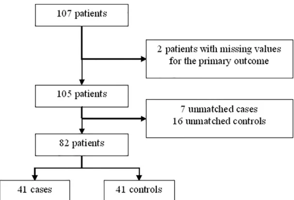
A total of 107 patients were selected and 82 were included in the analysis with data on primary
outcome (Fig 1). After matching on age, sex and BMI, 41 pairs were constituted by patients
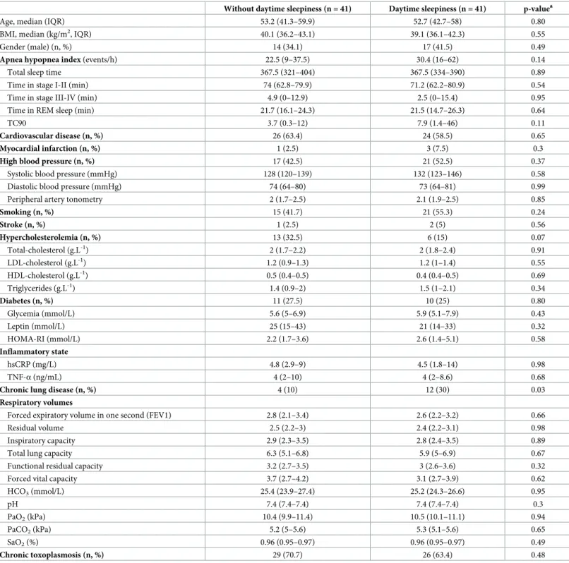
with sleepiness (cases) or not (controls). Patients had a median age of 53 years, (IQR: (41–60),
were mainly female (62%) and had a high median BMI (39; IQR: 35–44) (Table 1). The only
significant difference between patients with and without excessive daytime sleepiness was a higher proportion of patients with chronic respiratory disease in the group of patients with
excessive daytime sleepiness (p = 0.03) (Table 1). The prevalence of a chronic toxoplasmosis
was 70.7% and 63.4% in patients with excessive daytime sleepiness or without, respectively and
was not significantly different between both groups (p = 0.48) (Table 1andS1 Table). After
univariable analysis, the following variables were included in the multivariate analysis: hyper-cholesterolemia, other history of chronic diseases, history of respiratory diseases, history of cardiovascular diseases, pH (as binary variable), and AHI as adjustment factor to assess the association between chronic toxoplasmosis and excessive daytime sleepiness. Only other his-tory of chronic diseases was kept in the final multivariate model. Multivariable regression anal-ysis showed no significant effect of chronic toxoplasmosis on sleepiness: OR = 0.76, 95% confidence interval: [0.52; 2.33], p = 0.64).
Effect of chronic toxoplasmosis on OSA syndrome severity
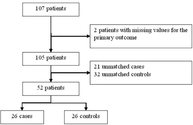
By considering patients with severe OSA syndrome, 26 cases (severe OSA, AHI>30) were
matched to 26 controls (no and mild OSA) (Fig 2,Table 2andS2 Table). By design there were
significant differences between groups in apnea hypopnea index (AHI), time below 90%
Fig 1. Flow chart of matching to study correlation between toxoplasmosis and effective daytime sleepiness (EDS). https://doi.org/10.1371/journal.pone.0235463.g001
Table 1. Descriptive data of patient’s criteria for matching (case-control study).
Without daytime sleepiness (n = 41) Daytime sleepiness (n = 41) p-valuea
Age, median (IQR) 53.2 (41.3–59.9) 52.7 (42.7–58) 0.80
BMI, median (kg/m2, IQR) 40.1 (36.2–43.1) 39.1 (36.1–42.3) 0.55
Gender (male) (n, %) 14 (34.1) 17 (41.5) 0.49
Apnea hypopnea index (events/h) 22.5 (9–37.5) 30.4 (16–62) 0.14
Total sleep time 367.5 (321–404) 367.5 (334–390) 0.89
Time in stage I-II (min) 74 (62.8–79.9) 71.2 (62.2–80.9) 0.54
Time in stage III-IV (min) 4.9 (0–12.9) 2.5 (0–15.4) 0.95
Time in REM sleep (min) 21.7 (16.1–24.3) 21.5 (14.7–26.3) 0.64
TC90 3.7 (0.3–12) 7.9 (1.4–46) 0.11
Cardiovascular disease (n, %) 26 (63.4) 24 (58.5) 0.65
Myocardial infarction (n, %) 1 (2.5) 3 (7.5) 0.3
High blood pressure (n, %) 17 (42.5) 21 (52.5) 0.37
Systolic blood pressure (mmHg) 128 (120–139) 132 (123–146) 0.58
Diastolic blood pressure (mmHg) 74 (64–80) 73 (64–81) 0.99
Peripheral artery tonometry 2 (1.7–2.5) 2.1 (1.9–2.5) 0.85
Smoking (n, %) 15 (41.7) 21 (55.3) 0.24 Stroke (n, %) 1 (2.5) 2 (5) 0.56 Hypercholesterolemia (n, %) 13 (32.5) 6 (15) 0.07 Total-cholesterol (g.L-1) 2 (1.7–2.2) 2 (1.8–2.4) 0.91 LDL-cholesterol (g.L-1) 1.2 (0.9–1.3) 1.2 (1–1.4) 0.55 HDL-cholesterol (g.L-1) 0.5 (0.4–0.5) 0.4 (0.4–0.5) 0.69 Triglycerides (g.L-1) 1.4 (0.9–2) 1.5 (1–2.1) 0.34 Diabetes (n, %) 11 (27.5) 10 (25) 0.80 Glycemia (mmol/L) 5.6 (5–6.9) 5.9 (5.1–7.9) 0.43 Leptin (mmol/L) 25 (15–43) 21 (14–33) 0.32 HOMA-RI (mmol/L) 2.2 (1.7–3.6) 2.6 (1.4–5.1) 0.58 Inflammatory state hsCRP (mg/L) 4.8 (2.9–9) 4.5 (1.8–14) 0.98 TNF-α (ng/mL) 4 (2–10) 4 (2–8.6) 0.68
Chronic lung disease (n, %) 4 (10) 12 (30) 0.03
Respiratory volumes
Forced expiratory volume in one second (FEV1) 2.8 (2.1–3.4) 2.6 (2.2–3.2) 0.66
Residual volume 2.5 (2.2–3) 2.4 (2.2–3.1) 0.98
Inspiratory capacity 2.9 (2.3–3.5) 2.8 (2.4–3.5) 0.89
Total lung capacity 6.3 (5.1–6.8) 5.9 (5–6.9) 0.67
Functional residual capacity 3.2 (2.7–3.5) 3 (2.6–3.6) 0.32
Forced vital capacity 3.7 (2.7–4.2) 3.1 (2.7–3.9) 0.62
HCO3(mmol/L) 25.4 (23.9–27.4) 25.2 (24.3–26.6) 0.95 pH 7.4 (7.4–7.4) 7.4 (7.4–7.4) 0.3 PaO2(kPa) 10.4 (9.9–11.4) 10.5 (10.1–11.1) 0.94 PaCO2(kPa) 5.2 (5–5.6) 5.3 (5.1–5.6) 0.65 SaO2(%) 0.96 (0.95–0.97) 0.96 (0.95–0.97) 0.49 Chronic toxoplasmosis (n, %) 29 (70.7) 26 (63.4) 0.48
Descriptive data related to sleep and ventilation during sleep, cardiovascular associated factors, inflammatory state, respiratory factors and repartition of patients with chronic toxoplasmosis among the two groups (excessive daytime sleepiness or not). Values are expressed in effective (frequency) for qualitative data and median (interquartile range) for quantitative data.
a
P-value: chi-square or exact Fisher test for qualitative value, Mann-Whitney test for quantitative value.
BMI, body mass index; IQR, interquartile range; REM, rapid eye movement; TC90, time during which arterial O2 saturation was less than 90%; hsCRP, high-sensitivity C-reactive protein, TNF-α, tumor necrosis factor-α, HCO3 serum bicarbonates, PaO2and PaCO2for arterial partial pressure in oxygen and carbon dioxide respectively
and SaO2 hemoglobin oxygen saturation.
(TC90) and mean SpO2 during sleep. As could be expected the OSA severity impact on sleep macrostructure with a decrease in slow wave sleep in more severe patients. Only PaO2 of
arte-rial blood gazes was different with a lower value in severe OSA (10.4 kPa [9.9; 11.0]vs 11.1 kPa
[10.2; 11.9], p = 0.03).
There was no significant difference for chronic toxoplasmosis between groups of OSA severity (univariable OR = 1.75, 95%CI: [0.51; 5.98], p = 0.37). No variable was significantly associated with OSAS severity to be introduced in a multivariable analysis.
Discussion
This study brings out new data in the fashionable thematic of “neurological diseases and chronic toxoplasmosis”. Herein, we provide an evaluation of the potential link between chronic toxoplasmosis and excessive daytime sleepiness in OSA patient as a primary outcome and between chronic toxoplasmosis and OSA syndrome severity in obese patients. In this case-control study of 107 patients, no significant link between chronic toxoplasmosis and sleepiness and OSA severity was demonstrated. Only the presence of chronic lung disease in medical history was associated with an increase in sleepiness. The most appropriate biological tools to answer the question “Is there a link between chronic toxoplasmosis and neurological disorders?” are serological tools within case-control studies. This study stands out for the qual-ity of the enrolment of patients and the qualqual-ity of serological analyses (two serological tech-niques and results interpreted by experts) [50]. Indeed, some of the studies showing a link between chronic toxoplasmosis and neurological troubles have low scientific evidence-based level and a real lack of high quality and these results must be confirmed by methodological rel-evant studies.
Fig 2. Flow chart of matching to study correlation between toxoplasmosis and OSA syndrome severity. https://doi.org/10.1371/journal.pone.0235463.g002
Table 2. Descriptive data of no severe versus severe OSA patient’s.
No severe OSA (n = 26) Severe OSA (n = 26) p-valuea
Age, median (IQR) 52.2 (42.7–58.4) 54.2 (41.3–59.8) 0.64
BMI, median (kg/m2, IQR) 39.2 (35.3–45.3) 40 (35.2–43.7) 0.96
Gender (male) (n, %) 7 (26.9) 7 (26.9) 1
Apnea hypopnea index (events/h) 14.3 (5.6–23.3) 46.9 (38–82.3) < .01
Total sleep time 367.5 (315–407) 363.5 (326.5–391) 0.41
Time in stage I-II (min) 70 (62.8–74.5) 76.8 (66.8–88.6) 0.03
Time in stage III-IV (min) 9.3 (4.7–16.9) 0 (0–10.9) 0.02
Time in REM sleep (min) 21.2 (16.7–24.9) 17.3 (10.2–23) 0.09
TC90 1 (0.2–4.6) 19.3 (1.7–60) 0.02
Cardiovascular disease (n, %) 17 (65.4) 18 (69.2) 0.77
Myocardial infarction (n, %) 1 (4) 1 (4) 1
High blood pressure (n, %) 12 (48) 15 (60) 0.39
Systolic blood pressure (mmHg) 124.3 (114–136) 130.5 (118–140) 0.28
Diastolic blood pressure (mmHg) 72.5 (64–81) 70 (60–76) 0.25
Peripheral artery tonometry 2.2 (1.8–2.5) 2.2 (1.9–2.4) 0.88
Smoking (n, %) 8 (34.8) 12 (52.2) 0.23 Stroke (n, %) 1 (4) 1 (4) 1 Hypercholesterolemia (n, %) 7 (28) 4 (16) 0.31 Total-cholesterol (g.L-1) 1.9 (1.7–2.2) 2 (1.7–2.4) 0.55 LDL-cholesterol (g.L-1) 1.2 (1–1.3) 1.2 (0.9–1.4) 0.85 HDL-cholesterol (g.L-1) 0.4 (0.4–0.5) 0.4 (0.3–0.5) 0.87 Triglycerides (g.L-1) 1.4 (0.8–2.1) 1.5 (1.1–2.1) 0.73 Diabetes (n, %) 8 (32) 8 (32) 1 Glycemia (mmol/L) 5.5 (5–6.4) 6.1 (5.1–8.7) 0.22 Leptin (mmol/L) 28 (14–50) 24 (14–34) 0.34 HOMA-RI (mmol/L) 1.9 (1.5–3.6) 2 (1.7–5.6) 0.42 Inflammatory state hsCRP (mg/L) 3.8 (2.9–9) 5.7 (3–14) 0.64 TNF-α (ng/mL) 4 (2–8.5) 3.5 (1.8–9) 0.43
Chronic lung disease (n, %) 7 (28) 7 (28) 1
Respiratory volumes
Forced expiratory volume in one second (FEV1) 2.5 (2.2–3.1) 2.7 (2.1–3.4) 0.56
Residual volume 2.5 (2.1–3.1) 2.3 (1.9–2.8) 0.28
Inspiratory capacity 2.8 (2.4–3.5) 2.8 (2.1–3.4) 0.74
Total lung capacity 5.5 (4.9–6.5) 6 (4.8–6.6) 0.74
Forced vital capacity 3 (2.7–3.8) 3.1 (2.7–4.1) 0.55
HCO3(mmol/L) 25.8 (24.2–26.9) 25.2 (24.2–26.6) 0.57 pH 7.4 (7.4–7.4) 7.4 (7.4–7.4) 0.15 PaO2(kPa) 11.1 (10.2–11.9) 10.4 (9.9–11) 0.03 PaCO2(kPa) 5.2 (5–5.5) 5.4 (5.2–5.6) 0.23 SaO2(%) 1 (1–1) 1 (1–1) 0.02 Chronic toxoplasmosis (n, %) 14 (53.8) 17 (65.4) 0.4
Descriptive data related to sleep and ventilation during sleep, cardiovascular associated factors, inflammatory state, respiratory factors, and repartition of patients with chronic toxoplasmosis among the two groups (severe OSA or not). Values are expressed in effective (frequency) for qualitative data and median (interquartile range) for quantitative data.
a
P-value: chi-square or exact Fisher test for qualitative value, Mann-Whitney test for quantitative value.
BMI, body mass index; IQR, interquartile range; REM, rapid eye movement; TC90, time during which arterial O2 saturation was less than 90%; hsCRP, high-sensitivity C-reactive protein; TNF-α, tumor necrosis factor-α HCO3 serum bicarbonates, PaO2 and PaCO2 for arterial partial pressure in oxygen and carbon dioxide respectively and SaO2 hemoglobin oxygen saturation.
A prolific literature has been published showing the relationship between toxoplasmosis
infection and change in rodent behavior making them more vulnerable to feline predators [16,
51,52]. The fact that the parasite can change the behavior of its host has led to hypothesize a potential link between chronic toxoplasmosis infection and some human neurologic diseases.
The most studied hypothesis is between schizophrenia and toxoplasmosis [53,54]. To this
extent there is component evidence that chronic inflammation due toT. gondii in patients
with genetic susceptibility may develop neurologic disorders and specifically schizophrenia [55].
Human is dead-end host forT. gondii but like all intermediary hosts infected by the parasite
have Toxoplasma cysts in brain. Interestingly there are some structural and functional changes
in the brain upon chronicToxoplasma infection. This may be a strong hypothesis that supports
the link betweenToxoplasma infection and neurologic symptoms [39]. We hypothesized that
the presence of Toxoplasma cysts in brainstem could be involved both in sleepiness and upper airway muscle control, chronic toxoplasmosis infection may modulate OSA symptoms or severity. But our population analysis demonstrates that there is no relationship between these
OSA features and chronicT. gondii infection.
Several limitations of this study need to be pointed out. Firstly, this study involves a cohort of obese patients, mainly female, and results cannot be largely extrapolated to all OSA patients, young OSA patients highly symptomatic. This preliminary survey could be enlarged to other categories of patients including non-obese patients, in which the mechanism of OSA in more linked to abnormalities of upper airways muscles control compared to obese patient. Secondly, the size of the cohort (n = 107) is relatively limited and the statistical power may be insufficient to unmask any effect of toxoplasmosis on the severity of OSA.
In conclusion, chronic toxoplasmosis in humans does not appear to be a risk factor of OSA severity and sleepiness-related OSA in obese patients. Despite a challenging hypothesis, we
demonstrate that there is no relationship between well-definedToxoplasma infection and
sleepiness and OSA severity, contrarily to what has been attempted to be demonstrated for many other behaviors or pathologies.
Supporting information
S1 Table. Individual data of the 82 patients used for matching (case-control study).
(XLSX)
S2 Table. Individual data of the 52 patients with no severe and severe OSA.
(XLSX)
Acknowledgments
The authors thank the volunteers for their participation in this study. Herve´ Pelloux is member of the European Study Group for Clinical Parasitology of the European Society for Clinical Microbiology and Infectious Diseases.
Author Contributions
Conceptualization: Ce´line Dard, Jean-Louis Pe´pin, Herve´ Pelloux, Renaud Tamisier. Data curation: Se´bastien Bailly.
Formal analysis: Ce´line Dard, Se´bastien Bailly, Marie Peeters.
Investigation: Ce´line Dard, Jean-Louis Pe´pin, Renaud Tamisier.
Methodology: Se´bastien Bailly, Jean-Louis Pe´pin, Herve´ Pelloux, Renaud Tamisier. Project administration: Jean-Louis Pe´pin, Renaud Tamisier.
Software: Se´bastien Bailly.
Supervision: Jean-Louis Pe´pin, Marie-Pierre Brenier-Pinchart, Herve´ Pelloux, Renaud
Tamisier.
Validation: Ce´line Dard, Se´bastien Bailly, Marie-Pierre Brenier-Pinchart, He´lène Fricker-Hidalgo, Herve´ Pelloux, Renaud Tamisier.
Writing – original draft: Ce´line Dard.
Writing – review & editing: Se´bastien Bailly, Jean-Louis Pe´pin, Marie-Pierre
Brenier-Pinch-art, He´lène Fricker-Hidalgo, Herve´ Pelloux, Renaud Tamisier.
References
1. Netchitaïlo M, Destors M, Bosc C, Pe´pin JL, Tamisier R. Obstructive sleep apnea syndrome. Diagnostic strategies in various clinical settings. Presse Med. 2017; 46(4):404–412.https://doi.org/10.1016/j.lpm. 2016.09.005PMID:28126505
2. Young T, Peppard PE, Gottlieb DJ. Epidemiology of obstructive sleep apnea: a population health per-spective. Am J Respir Crit Care Med. 2002; 165(9):1217–39.https://doi.org/10.1164/rccm.2109080
PMID:11991871
3. Gasa M, Tamisier R, Launois SH, Sapene M, Martin F, Stach B, et al. Residual sleepiness in sleep apnea patients treated by continuous positive airway pressure. J Sleep Res. 2013; 22(4):389–97.
https://doi.org/10.1111/jsr.12039PMID:23409736
4. Tregear S, Reston J, Schoelles K, Phillips B. Obstructive sleep apnea and risk of motor vehicle crash: systematic review and meta-analysis. J Clin Sleep Med. 2009; 5(6):573–81. PMID:20465027
5. Alchanatis M, Deligiorgis N, Zias N, Amfilochiou A, Gotsis E, Karakatsani A, et al. Frontal brain lobe impairment in obstructive sleep apnoea: a proton MR spectroscopy study. Eur Respir J. 2004; 24 (6):980–6.https://doi.org/10.1183/09031936.04.00127603PMID:15572542
6. Vernet C, Redolfi S, Attali V, Konofal E, Brion A, Frija-Orvoen E, et al. Residual sleepiness in obstructive sleep apnoea: phenotype and related symptoms. Eur Respir J. 2011; 38(1):98–105.https://doi.org/10. 1183/09031936.00040410PMID:21406511
7. Kingshott RN, Engleman HM, Deary IJ, Douglas NJ. Does arousal frequency predict daytime function? Eur Respir J. 1998; 12(6):1264–70.https://doi.org/10.1183/09031936.98.12061264PMID:9877475
8. Alvarado-Esquivel C, Rico-Almochantaf YDR, Herna´ndez-Tinoco J, Quiñones-Canales G, Sa´nchez-Anguiano LF, Torres-Gonza´ lez J, et al. Toxoplasma gondii exposure and neurological disorders: an age- and gender-matched case-control pilot study. Eur J Microbiol Immunol. 2017; 7(4):303–9.https:// doi.org/10.1556/1886.2017.00033
9. Montoya JG, Liesenfeld O. Toxoplasmosis. Lancet. 2004; 363(9425):1965–76.https://doi.org/10.1016/ S0140-6736(04)16412-XPMID:15194258
10. Kamerkar S, Davis PH. Toxoplasma on the brain: understanding host-pathogen interactions in chronic CNS infection. J Parasitol Res. 2012; 2012:1–10.https://doi.org/10.1155/2012/589295
11. Rougier S, Montoya JG, Peyron F. Lifelong persistence of Toxoplasma cysts: a questionable dogma? Trends Parasitol. 2017; 33(2):93–101.https://doi.org/10.1016/j.pt.2016.10.007PMID:27939103
12. Watts E, Zhao Y, Dhara A, Eller B, Patwardhan A, Sinai AP. Novel approaches reveal that Toxoplasma
gondii bradyzoites within tissue cysts are dynamic and replicating entities in vivo. mBio. 2015; 6(5):
e01155–15.https://doi.org/10.1128/mBio.01155-15PMID:26350965
13. Kim K. A bradyzoite is a bradyzoite is a bradyzoite? Trends Parasitol. 2015; 31(12):610–2.https://doi. org/10.1016/j.pt.2015.10.005PMID:26515047
14. Sinai AP, Watts EA, Dhara A, Murphy RD, Gentry MS, Patwardhan A. reexamining chronic Toxoplasma
gondii infection: surprising activity for a “dormant” parasite. Curr Clin Microbiol Rep. 2016; 3(4):175–85.
https://doi.org/10.1007/s40588-016-0045-3PMID:28191447
15. Berdoy M, Webster JP, Macdonald DW. Fatal attraction in rats infected with Toxoplasma gondii. Proc Biol Sci. 2000; 267(1452):1591–4.https://doi.org/10.1098/rspb.2000.1182PMID:11007336
16. Vyas A, Kim S-K, Giacomini N, Boothroyd JC, Sapolsky RM. Behavioral changes induced by
Toxo-plasma infection of rodents are highly specific to aversion of cat odors. Proc Natl Acad Sci U S A. 2007;
104(15):6442–7.https://doi.org/10.1073/pnas.0608310104PMID:17404235
17. Wohlfert EA, Blader IJ, Wilson EH. Brains and brawn: Toxoplasma infections of the central nervous sys-tem and skeletal muscle. Trends Parasitol. 2017; 33(7):519–531.https://doi.org/10.1016/j.pt.2017.04. 001PMID:28483381
18. Celik T, Kartalci S, Aytas O, Akarsu GA, Gozukara H, Unal S. Association between latent toxoplasmosis and clinical course of schizophrenia—continuous course of the disease is characteristic for Toxoplasma
gondii-infected patients. Folia Parasitol (Praha). 2015; 62: pii: 2015.015.https://doi.org/10.14411/fp. 2015.015
19. Dickerson F, Boronow J, Stallings C, Origoni A, Yolken R. Toxoplasma gondii in individuals with schizo-phrenia: association with clinical and demographic factors and with mortality. Schizophr Bull. 2007; 33 (3):737–40.https://doi.org/10.1093/schbul/sbm005PMID:17314085
20. Prandota J. Possible link between Toxoplasma gondii and the anosmia associated with neurodegenera-tive diseases. Am J Alzheimers Dis Other Demen. 2014; 29(3):205–14.https://doi.org/10.1177/ 1533317513517049PMID:24413543
21. Ngoungou EB, Bhalla D, Nzoghe A, Darde´ M-L, Preux P-M. Toxoplasmosis and epilepsy—systematic review and meta analysis. PLoS Negl Trop Dis. 2015; 9(2):e0003525.https://doi.org/10.1371/journal. pntd.0003525PMID:25695802
22. Lester D. Toxoplasma gondii and homicide. Psychol Rep. 2012; 111(1):196–7.https://doi.org/10.2466/ 12.15.16.PR0.111.4.196-197PMID:23045862
23. Duffy AR, Beckie TM, Brenner LA, Beckstead JW, Seyfang A, Postolache TT, et al. Relationship between Toxoplasma gondii and mood disturbance in women veterans. Mil Med. 2015; 180(6):621–5.
https://doi.org/10.7205/MILMED-D-14-00488PMID:26032378
24. Pearce BD, Kruszon-Moran D, Jones JL. The relationship between Toxoplasma gondii infection and mood disorders in the third national health and nutrition survey. Biol Psychiatry. 2012; 72(4):290–5.
https://doi.org/10.1016/j.biopsych.2012.01.003PMID:22325983
25. Flegr J, Havlı´cek J, Kodym P, Maly´ M, Smahel Z. Increased risk of traffic accidents in subjects with latent toxoplasmosis: a retrospective case-control study. BMC Infect Dis. 2002; 2:11.https://doi.org/10. 1186/1471-2334-2-11PMID:12095427
26. Flegr J, Klose J, Novotna´ M, Berenreitterova´ M, Havlı´cek J. Increased incidence of traffic accidents in
Toxoplasma-infected military drivers and protective effect RhD molecule revealed by a large-scale
pro-spective cohort study. BMC Infect Dis. 2009; 9:72.https://doi.org/10.1186/1471-2334-9-72PMID:
19470165
27. Flegr J, Dama M. Does the prevalence of latent toxoplasmosis and frequency of Rhesus-negative sub-jects correlate with the nationwide rate of traffic accidents? Folia Parasitol (Praha). 2014; 61(6):485–94.
https://doi.org/10.14411/fp.2014.061
28. Galva´n-Ramı´rez Mde L, Sa´nchez-Orozco LV, Rodrı´guez LR, Rodrı´guez S, Roig-Melo E, Troyo Sanro-ma´n R, et al. Seroepidemiology of Toxoplasma gondii infection in drivers involved in road traffic acci-dents in the metropolitan area of Guadalajara, Jalisco, Mexico. Parasit Vectors. 2013; 6(1):294.https:// doi.org/10.1186/1756-3305-6-294PMID:24499659
29. Kocazeybek B, Oner YA, Turksoy R, Babur C, Cakan H, Sahip N, et al. Higher prevalence of toxoplas-mosis in victims of traffic accidents suggest increased risk of traffic accident in Toxoplasma-infected inhabitants of Istanbul and its suburbs. Forensic Sci Int. 2009; 187(1–3):103–8.https://doi.org/10.1016/ j.forsciint.2009.03.007PMID:19356869
30. Yereli K, Balcioğlu IC, Ozbilgin A. Is Toxoplasma gondii a potential risk for traffic accidents in Turkey? Forensic Sci Int. 2006; 163(1–2):34–7.https://doi.org/10.1016/j.forsciint.2005.11.002PMID:16332418
31. Stibbs HH. Changes in brain concentrations of catecholamines and indoleamines in Toxoplasma gondii infected mice. Ann Trop Med Parasitol. 1985; 79(2):153–7.https://doi.org/10.1080/00034983.1985. 11811902PMID:2420295
32. Martin HL, Alsaady I, Howell G, Prandovszky E, Peers C, Robinson P, et al. Effect of parasitic infection on dopamine biosynthesis in dopaminergic cells. Neuroscience. 2015; 306:50–62.https://doi.org/10. 1016/j.neuroscience.2015.08.005PMID:26297895
33. Blanchard N, Dunay IR, Schlu¨ter D. Persistence of Toxoplasma gondii in the central nervous system: a fine-tuned balance between t ite, the brain he paras and the immune system. Parasite Immunol. 2015; 37(3):150–8.https://doi.org/10.1111/pim.12173PMID:25573476
34. Fabiani S, Pinto B, Bonuccelli U, Bruschi F. Neurobiological studies on the relationship between toxo-plasmosis and neuropsychiatric diseases. J Neurol Sci. 2015; 351(1–2):3–8.https://doi.org/10.1016/j. jns.2015.02.028PMID:25725931
35. Song ZY, Yi HL.Progress of dopamine in obstructive sleep apnea hypopnea syndrome. Zhonghua Er Bi Yan Hou Tou Jing Wai Ke Za Zhi. 2017; 52(7):549–551.https://doi.org/10.3760/cma.j.issn.1673-0860. 2017.07.018PMID:28728251
36. Dempsey JA, Veasey SC, Morgan BJ, O’Donnell CP. Pathophysiology of sleep apnea. Physiol Rev. 2010; 90(1): 47–112.https://doi.org/10.1152/physrev.00043.2008PMID:20086074
37. Berenreiterova´ M, Flegr J, Kuběna AA, Němec P.The distribution of Toxoplasma gondii cysts in the brain of a mouse with latent toxoplasmosis: implications for the behavioral manipulation hypothesis. PLoS One. 2011; 6(12): e28925.https://doi.org/10.1371/journal.pone.0028925PMID:22194951
38. Boillat M, Hammoudi PM, Dogga SK, Pagès S, Goubran M, Rodriguez I, et al. Neuroinflammation-asso-ciated aspecific manipulation of mouse predator fear by Toxoplasma gondii. Cell Rep. 2020; 30(2):320– 334.e6.https://doi.org/10.1016/j.celrep.2019.12.019
39. Tyebji S, Seizova S, Hannan AJ, Tonkin CJ. Toxoplasmosis: A pathway to neuropsychiatric disorders. Neurosci Biobehav Rev. 2019; 96:72–92.https://doi.org/10.1016/j.neubiorev.2018.11.012PMID:
30476506
40. Cannella D, Brenier-Pinchart MP, Braun L, van Rooyen JM, Bougdour A, Bastien O, et al. miR-146a and miR-155 delineate a MicroRNA fingerprint associated with Toxoplasma persistence in the host brain. Cell Rep. 2014; 6(5):928–37.https://doi.org/10.1016/j.celrep.2014.02.002PMID:24582962
41. Borel JC, Tamisier R, Gonzalez-Bermejo J, Baguet JP, Monneret D, Arnol N, et al. Noninvasive ventila-tion in mild obesity hypoventilaventila-tion syndrome: a randomized controlled trial. Chest. 2012; 141(3):692– 702.https://doi.org/10.1378/chest.10-2531PMID:21885724
42. Johns MW. Reliability and factor analysis of the Epworth sleepiness scale. Sleep. 1992; 15(4):376–81.
https://doi.org/10.1093/sleep/15.4.376PMID:1519015
43. Hori T, Sugita Y, Koga E, Shirakawa S, Inoue K, Uchida S, et al. Proposed supplements and amend-ments to “A manual of standardized terminology, techniques and scoring system for sleep stages of human subjects”, the Rechtschaffen & Kales (1968) standard. Psychiatry Clin Neurosci. 2001; 55 (3):305–10.https://doi.org/10.1046/j.1440-1819.2001.00810.xPMID:11422885
44. Sleep-related breathing disorders in adults: recommendations for syndrome definition and measure-ment techniques in clinical research. The report of an American Academy of Sleep Medicine Task Force. Sleep. 1999; 22(5):667–89. PMID:10450601
45. Berry RB, Budhiraja R, Gottlieb DJ, Gozal D, Iber C, Kapur VK, et al. Rules for scoring respiratory events in sleep: update of the 2007 AASM manual for the scoring of sleep and associated events. Delib-erations of the sleep apnea definitions task force of the American Academy of Sleep Medicine J Clin Sleep Med. 2012; 8(5):597–619.https://doi.org/10.5664/jcsm.2172PMID:23066376
46. Bonetti PO, Pumper GM, Higano ST, Holmes DR, Kuvin JT, Lerman A. Noninvasive identification of patients with early coronary atherosclerosis by assessment of digital reactive hyperemia. J Am Coll Car-diol. 2004; 44(11):2137–41.https://doi.org/10.1016/j.jacc.2004.08.062PMID:15582310
47. Nohria A, Gerhard-Herman M, Creager MA, Hurley S, Mitra D, Ganz P. Role of nitric oxide in the regula-tion of digital pulse volume amplitude in humans. J Appl Physiol(1985). 2006; 101(2):545–8.https://doi. org/10.1152/japplphysiol.01285.2005
48. Roux-Buisson N, Fricker-Hidalgo H, Foussadier A, Rolland D, Suchel-Jambon A-S, Brenier-Pinchart MP, et al. Comparative analysis of the VIDAS Toxo IgG IV assay in the detection of antibodies to
Toxo-plasma gondii. Diagn Microbiol Infect Dis. 2005; 53(1):79–81.https://doi.org/10.1016/j.diagmicrobio. 2005.04.005PMID:16054325
49. Dard C, Bailly S, Drouet T, Fricker-Hidalgo H, Brenier-Pinchart MP, Pelloux H. Long-term sera storage does not significantly modify the interpretation of toxoplasmosis serologies. J Microbiol Methods. 2017; 134:38–45.https://doi.org/10.1016/j.mimet.2017.01.003PMID:28093212
50. Dard C, Fricker-Hidalgo H, Brenier-Pinchart MP, Pelloux H. Relevance of and new developments in serology for toxoplasmosis. Trends Parasitol. 2016; 32(6):492–506.https://doi.org/10.1016/j.pt.2016. 04.001PMID:27167666
51. Ingram WM, Goodrich LM, Robey EA, Eisen MB. Mice infected with low-virulence strains of
Toxo-plasma gondii lose their innate aversion to cat urine, even after extensive parasite clearance. PloS One.
2013; 8(9):e75246.https://doi.org/10.1371/journal.pone.0075246PMID:24058668
52. Bezerra ECM, Dos Santos SV, Dos Santos TCC, de Andrade HF, Meireles LR. Behavioral evaluation of BALB/c (Mus musculus) mice infected with genetically distinct strains of Toxoplasma gondii. Microb Pathog. 2019; 126:279–86.https://doi.org/10.1016/j.micpath.2018.11.021PMID:30447421
53. Brown AS, Schaefer CA, Quesenberry CP, Liu L, Babulas VP, Susser ES. Maternal exposure to toxo-plasmosis and risk of schizophrenia in adult offspring. Am J Psychiatry. 2005; 162(4):767–73.https:// doi.org/10.1176/appi.ajp.162.4.767PMID:15800151
54. Blomstro¨m A, Karlsson H, Wicks S, Yang S, Yolken RH, Dalman C. Maternal antibodies to infectious agents and risk for non-affective psychoses in the offspring-a matched case-control study. Schizophr Res. 2012; 140(1–3):25–30.https://doi.org/10.1016/j.schres.2012.06.035PMID:22819777
55. Avramopoulos D, Pearce BD, McGrath J, Wolyniec P, Wang R, Eckart N, et al. Infection and inflamma-tion in schizophrenia and bipolar disorder: a genome wide study for interacinflamma-tions with genetic variainflamma-tion. PloS One. 2015; 10(3):e0116696.https://doi.org/10.1371/journal.pone.0116696PMID:25781172