Fungi, bacteria and soil pH: the oxalate–carbonate
pathway as a model for metabolic interaction
Gaëtan Martin,1,2†Matteo Guggiari,1,2†Daniel Bravo,1 Jakob Zopfi,2,3Guillaume Cailleau,2Michel Aragno,1 Daniel Job,1Eric Verrecchia2and Pilar Junier1* 1Laboratory of Microbiology, Institute of Biology, University of Neuchâtel, CH-2000, Neuchâtel, Switzerland.
2Biogeosciences Laboratory, Institute of Geology and Palaeontology, University of Lausanne, CH-1015, Lausanne, Switzerland.
3Laboratory of Aquatic Biogeochemistry, Institute of Environmental Sciences, University of Basel, CH-4056 Basel, Switzerland.
Summary
The oxalate–carbonate pathway involves the oxida-tion of calcium oxalate to low-magnesium calcite and represents a potential long-term terrestrial sink for atmospheric CO2. In this pathway, bacterial oxalate degradation is associated with a strong local alkalini-zation and subsequent carbonate precipitation. In order to test whether this process occurs in soil, the role of bacteria, fungi and calcium oxalate amend-ments was studied using microcosms. In a model system with sterile soil amended with laboratory cul-tures of oxalotrophic bacteria and fungi, the addition of calcium oxalate induced a distinct pH shift and led to the final precipitation of calcite. However, the simultaneous presence of bacteria and fungi was essential to drive this pH shift. Growth of both oxalo-trophic bacteria and fungi was confirmed by qPCR on the frc (oxalotrophic bacteria) and 16S rRNA genes, and the quantification of ergosterol (active fungal biomass) respectively. The experiment was replicated in microcosms with non-sterilized soil. In this case, the bacterial and fungal contribution to oxalate deg-radation was evaluated by treatments with specific biocides (cycloheximide and bronopol). Results showed that the autochthonous microflora oxidized calcium oxalate and induced a significant soil
alkali-nization. Moreover, data confirmed the results from the model soil showing that bacteria are essentially responsible for the pH shift, but require the presence of fungi for their oxalotrophic activity. The combined results highlight that the interaction between bacteria and fungi is essential to drive metabolic processes in complex environments such as soil.
Introduction
Oxalic acid (H2C2O4) and oxalate minerals are major sec-ondary products of plants, animals, fungi and bacteria present in soils (Tamer and Aragno, 1980). Calcium oxalate has been reported in more than 215 families of angiosperms and gymnosperms and occurs in the wood of more than 1000 genera of trees (Franceschi and Nakata, 2005), representing in some species more than 50% of their dry weight (Libert and Franceschi, 1987). Oxalate exudation in soils plays an important role by increasing the availability of phosphorous and micronutri-ents for plant uptake (Sahin, 2003).
Oxalotrophy, the metabolism of oxalate by bacteria, has been recognized as an important part of the biogeochemi-cal carbon cycle as it allows the precipitation of biogeochemi-calcium carbonate (CaCO3) in acidic tropical soils, which are, oth-erwise, free of primary carbonates. This process is central to the oxalate–carbonate pathway (Fig. 1), which couples the biogeochemical cycles of calcium and carbon, and is gaining increasing interest as a potential long-term sink for atmospheric CO2(Braissant et al., 2002; Garvie, 2003; Cailleau et al., 2004; 2011).
Three factors are required for an operating oxalate– carbonate pathway: Ca2+, oxalate and oxalate-degrading organisms. Oxalate degradation can be performed by a variety of plants and microorganisms (Dumas et al., 1995; Makela et al., 2002; Tuason and Arocena, 2009). However, in the case of its calcium salt (Ksp= 2.32 ¥ 10–9), spontaneous oxidation of oxalate is highly unlikely because of the high activation energy required. Therefore, any metal oxalate can be considered as a compound in a metastable equilibrium (Verrecchia et al., 2006). Only bacteria are so far undoubtedly known to be able to par-ticipate in the oxidation. Oxalate catabolism by bacteria is also associated with a strong pH increase (Jayasuriya, 1955; Braissant et al., 2004), due to the conversion of a *For correspondence. E-mail [email protected]; Tel.
(+41) 32 7182230; Fax (+41) 32 7182231.†These authors contributed equally to this study.
strong acid into a weaker one (Cromack et al., 1977). The influence of oxalotrophy on pH was first observed in faeces of plant litter detritivores (Van de Drift and Witkamp, 1960; McBrayer, 1973) and was deemed of interest for nutrient cycling, particularly of Ca and P (Cromack et al., 1977; Graustein et al., 1977).
Oxalotrophy is widespread and can be found in Gram-negative (Alpha-, Beta- and Gammaproteobacteria) and Gram-positive (Firmicutes and Actinobacteria) bacteria (Sahin, 2003). Thus, the study of the diversity and abun-dance of oxalotrophic bacteria cannot be based on a phylogenetic molecular marker such as the 16S rRNA gene. Instead, a gene directly involved in the metabolism of oxalate is a better candidate. At least two enzymes are involved in the catabolism of oxalate in aerobic and anaerobic bacteria. The first enzyme, the formyl coen-zyme A (CoA) transferase, is encoded by the frc gene and transfers a coenzyme A moiety to activate oxalic acid (Sidhu et al., 1997). The second enzyme is an oxalyl CoA decarboxylase, encoded by oxc, which decarboxylates the activated oxalate molecule (Lung et al., 1994). Recently, specific primers targeting frc have been designed and tested in a variety of oxalotrophic bacteria and environmental samples, and can be used for diversity or quantification studies (Khammar et al., 2009).
Bacteria alone are sufficient to shift the pH from acidic to alkaline in cultures with calcium oxalate as sole carbon source (Jayasuriya, 1955; Braissant et al., 2002; 2004). Under these experimental conditions, the pH shift allows the precipitation of calcium carbonate crystals (Braissant et al., 2002). To date, however, there is no direct evidence showing that bacteria can oxidize calcium oxalate under natural environmental conditions and induce the pH shift required for calcium carbonate precipitation. Furthermore, although fungi are recognized as major players in the oxalate cycle in soils (Dutton and Evans, 1996; Tuason and Arocena, 2009), their role in the oxalate–carbonate pathway, and more generally in the functioning of soils, needs to be clarified.
Consequently, two major questions were addressed in this study: (i) can oxalotrophic bacteria alone cause the shift in pH required for the precipitation of calcium car-bonate in soil? (ii) to what extent are fungi–bacteria interactions instrumental for the pH shift to occur? Micro-cosm experiments were conducted to fill the gap between experiments with pure cultures and field obser-vations in order to answer these two questions. In a first experiment, a sterile soil was inoculated with a mix of pure bacterial and fungal cultures. In a second one, fresh soil collected near an oxalogenic tree and contain-ing its own complex native microbial community was treated to selectively inhibit the activity of bacteria or/and fungi and to test their individual contribution to soil pH shift.
Fig. 1. Schematic representation of the oxalate–carbonate pathway
showing the main biological players and the chemical reactions involved (modified from Aragno and Verrecchia, 2012). The unknown contribution of fungi as calcium oxalate producers and the role of their interaction with bacteria are indicated by a question mark. CaOx= calcium oxalate. a = processes leading to the formation of CaOx in the plant and fungi, oxidation of CaOx by bacteria and arrows indicating the unknown contribution of fungi; b= effect of oxidation of CaOx on soil pH over time; c = final product of the oxalate–carbonate pathway. The reactions leading to the formation of calcium carbonate are indicated below.
Results
Shift in soil pH by an artificial microbial community A shift in soil pH towards alkaline conditions is a key element of the oxalate–carbonate pathway because it can trigger the precipitation of calcite in initially acidic soils (Braissant et al., 2004; Cailleau et al., 2004; 2005). We evaluated the influence of amendments with bacte-ria, fungi and oxalate on changes in soil pH in seven different microcosms (Fig. 2). For most of the systems assayed, pH values remained unchanged for more than 90 days. However, in the treatment amended with bac-teria, fungi and oxalate (FBox), an increase in soil pH was observed after 20 days of incubation. After 90 days, soil pH had reached a final value of 7.5, being 2.5 pH units higher than the initial value. This coincided simul-taneously with a decrease in the oxalate concentration from 34.2 ⫾ 24.5 mg g-1 to 7.9 ⫾ 5.2 mg g-1. This pH shift was sufficient to induce calcite precipitation, which was not observed in treatments where the pH remained constant. X-ray diffraction analysis revealed small yet characteristic peaks for calcite in FBox soil but not in SSox soil (Fig. S1).
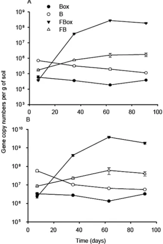
Development of inoculated bacteria and fungi in microcosms
Bacterial metabolism of oxalate has been shown to induce a pH increase in experiments on Petri dishes
(Jayasuriya, 1955; Braissant et al., 2004). Surprisingly, amendment of oxalotrophic bacteria was not sufficient to induce such a pH change in oxalate containing micro-cosms (Box). Addition of fungi alone (with or without oxalate) did not lead to a change in the soil pH either, raising the question as to whether the added microorgan-isms survived and developed in these microcosms. In order to test this, we followed copy numbers of frc and 16S rRNA genes of oxalotrophic and total bacteria, respectively, by qPCR (Fig. 3). As for soil pH, bacterial abundance increased only in the FBox treatment, where copy numbers of both marker genes increased by three to four orders of magnitude (frc: 3.7¥ 104to 2.8¥ 108; 16S rRNA gene: 2.3¥ 106to 3.8¥ 109). A statistical analysis shows a significant increase in frc copy number between 1 and 5, as well as 5 and 9 weeks (P-value= 0.0009 and 0.00006 respectively). In all other treatments containing bacteria (alone or in the presence of fungi), bacterial abundance remained close to the values recorded 7 days after inoculation (Fig. 3). In the bacteria-free microcosms,
0 20 40 60 80 100 pH 4.5 5.0 5.5 6.0 6.5 7.0 7.5 8.0 Box B FBox FB Fox F SSox Time (days)
Fig. 2. Evolution of pH in microcosms created with a sterile soil
and inoculated with allochthonous microbial community. The different treatments were: inoculation with bacteria and Ca-oxalate amendment (Box), inoculation with bacteria, without Ca-oxalate amendment (B), inoculation with bacteria and fungi, with Ca-oxalate amendment (FBox), inoculation with bacteria and fungi, without Ca-oxalate amendment (FB), inoculation with fungi and Ca-oxalate amendment (Fox), inoculation with fungi, without Ca-oxalate amendment (F), and finally sterile microcosms with Ca-oxalate amendment (SSox). Data points represent mean values of samples (⫾ standard deviations) from three independent microcosms.
Fig. 3. Quantification of frc gene (A) and 16S rRNA gene (B) copy
numbers by qPCR in microcosms with sterile soil and inoculated with allochthonous microbial community. Mean values and standard deviations of replicate quantification (n= 3) for three separate DNA extracts are given. For abbreviations see caption of Fig. 2.
frc gene concentration was below the detection limit (data not shown). Although the changes in the composition of the bacterial community were not the focus of this study, those could be indirectly observed based on the melting behaviour of frc PCR products (Fig. S2A). In absence of oxalate (B and FB), the melting temperature of the qPCR frc gene amplicons corresponded to those of the strains Pandoraea sp., Cupriavidus necator and Variovo-rax paradoxus (approximately 87°C). In contrast, in systems amended with oxalate (FBox and Box), melting values changed considerably over time, suggesting changes in the community composition. In the Box treat-ment, the melting temperature of the amplicons did not correspond to any specific taxon. In the presence of fungi (FBox), the melting temperature of the amplicons decreased over time and was close to those of Oxalici-bacterium flavum and Streptomyces violaceoruber (approximately 85°C). Changes in community composi-tion in FBox were confirmed by DGGE (Fig. S2B). Results suggest that among the non-oxalotrophic strains, only Escherichia coli remained detectable for the first 35 days of incubation. For the oxalotrophic bacteria, O. flavum was constantly present until 63 days of incubation, whereas S. violaceoruber, C. necator and Pandoraea sp. were detected only at two of the three time points. In the final point (91 days), a not easily identifiable band was observed.
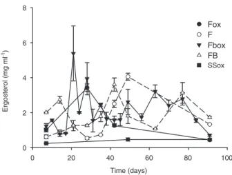
Active fungal biomass was assessed by the ergosterol content in soil. In contrast to bacteria, fungi developed in all microcosms independently of the presence of bacteria or oxalate (Fig. 4). However, in treatments containing fungi and bacteria (FBox and FB), ergosterol concentra-tions fluctuated over time compared with the treatments with fungi alone (Fox and F).
Shift in soil pH in microcosms with native microbial community
The results obtained for the microcosms with an artificially recreated microbial community prompted us to verify the findings in a less artificial system. Therefore, a second set of microcosms with tropical acidic soil containing a native microbial community was carried out. Previous tests, including cultivation of oxalotrophic bacteria and frc ampli-fication, suggested that this soil harboured an active guild of oxalotrophic bacteria (data not shown). In these micro-cosms, the various treatments of the previous experiment were mimicked by the addition of domain-specific bio-cides and changes in soil pH were again used as proxy for an operating oxalate–carbonate pathway (Fig. 5). In microcosms with the native microbial community, the pH rose by about one unit within 2 weeks after calcium oxalate addition but remained unchanged in the treat-ments with the biocide mix (SSox analogue; Fig. 5). Fur-thermore, maximum pH (pH 8) was reached in less than 10 days as compared with 8 weeks in the first experiment (Fig. 2). A shift in soil pH was observed under two experi-mental conditions: no biocide treatment (FBox analogue) and cycloheximide-treated soil (Box analogue).
Effect of biocides on bacteria and fungi
The results from the microcosms with native microbial community (FBox analogue, Fig. 5) confirmed those of the first microcosm series (Fig. 2). However, the pH shift in the Box analogue suggests that the bacterial activity alone acted as the driver of soil pH shift for a native microbial community. In order to confirm the effect of biocides, the abundance of bacteria and fungi was deter-mined at the end of the experiment. Although ergosterol was also measured, several additional peaks affected the interpretation of results and therefore fungal abundance
Time (days) 0 20 40 60 80 100 Ergosterol (mg ml -1) 0 2 4 6 8 Fox F Fbox FB SSox
Fig. 4. Quantification of ergosterol in microcosms with sterile soil
and inoculated with allochthonous microbial community. Mean values (⫾ standard deviations) of the quantification for three separate analyses. For abbreviations see caption of Fig. 2.
Time (days) 0 5 10 15 20 25 30 35 pH 4.5 5.0 5.5 6.0 6.5 7.0 7.5 8.0 8.5 Cycloheximide (Box) Water (FBox) Bronopol (Fox) Mix of biocides (SSox)
Fig. 5. Evolution of pH in soil microcosms with native microflora
and treated with specific biocides. Values are means (⫾ standard deviations) of measurements performed on three separate microcosms. For abbreviations in brackets see caption of Fig. 2.
was estimated by qPCR amplification of the 18S rRNA gene.
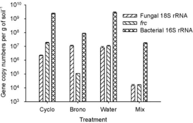
The results for the FBox analogue system confirmed the presence of bacteria, oxalotrophic bacteria and fungi in this soil (Fig. 6). The bacterial and fungal abundances in this treatment were at least three orders of magnitude higher than in the control (SSox analogue). In the micro-cosms treated with bronopol alone (Fox analogue), bac-terial abundance (total and oxalotrophic) dropped by one and a half orders of magnitude compared with the untreated soil (FBox). This system did not show a change in soil pH over time confirming the role of bacteria in the process. Finally, in the cycloheximide treated microcosm (Box analogue) fungal abundance decreased by less than an order of magnitude. A statistical analysis on the qPCR results shows that fungal abundance was significantly different between the water versus bronopol and cycloheximide treatments (P-value= 0.05), but not between water versus cycloheximide (P-value= 0.11), or bronopol (P-value= 0.54) alone. Therefore the cyclohex-imide treated microcosm (Box analogue) should be regarded as a less performing FBox analogue instead, still supporting the idea that bacteria and fungi are required simultaneously to cause the shift in soil pH observed in the oxalate–carbonate pathway.
Discussion
Experiments carried out with either an artificial or a native microbial community showed consistently that the simul-taneous presence of bacteria, fungi and oxalate is essen-tial to induce the alkalinization of the soil pH by up to 2.5 units and the precipitation of calcite, which are two keys effects observed for the oxalate–carbonate pathway in nature. These results confirm for the first time that the oxalate–carbonate pathway could be reproduced
artifi-cially in a microcosm, and that the parameters measured in the field (soil pH and presence of carbonates) effec-tively described an active pathway.
Although it is important to mention that the absolute frc copy numbers must be considered with caution due to methodological issues such as DNA extraction biases or the contribution of dead or lysed cells to the total DNA pool, the shift in soil pH was clearly associated with a significant increase of frc gene copy numbers of oxalo-trophic bacteria, which supports earlier studies proposing that the metabolism of oxalate by bacteria may hold the key to pH increase (Cromack et al., 1977; Braissant et al., 2002; Cailleau et al., 2004). In pure cultures, bacteria alone cause oxalate oxidation and the associated pH increase (Sahin, 2003; Braissant et al., 2004). However, the situation is less straightforward in complex environ-ments. In soils, bacteria needed the presence of fungi to thrive and oxidize calcium oxalate. In the case of the sterilized Ferralsol, it might be argued that the inoculated strains were not adapted to the acidic and nutrient poor conditions in the microcosm. However, in the presence of fungi (FBox), some of them were able to adapt and develop. In the Bolivian soil with its native bacterial com-munity, adaptation is not an issue, which explains the faster kinetics of the pH shift, even though the native microbial communities could have changed during trans-port and storage of soil prior to the experiment. A fungal-free microcosm (Box analogue) was intended by applying cycloheximide. As suggested previously, this fungicide had probably only a limited effect, (Griffin et al., 1978; Sugiura et al., 1999), which may explain the lack of a strong difference between the treatments with cyclohex-imide (Box analogue) and water (FBox analogue). Con-sidering that we cannot rule out some fungal activity in the microcosm treated with cycloheximide, the most con-servative interpretation of the results suggest that the simultaneous presence of bacteria and fungi was required in all the microcosms showing a soil pH shift. In the case of bronopol, it has been reported as a biocide with a broad spectrum of antibacterial activity (Shepherd et al., 1988). From the results obtained, it inhibited 97% of the bacterial community in the soil assayed, and it has as a synergistic effect in combination with cycloheximide (99.4% fungal and bacterial inhibition). The reasons for this synergistic effect are unknown. Although non-target effects of bro-nopol are known, it appears not to affect fungal growth and thus the results are surprising (Rousk et al., 2008).
Recent studies suggest that interactions between bac-teria and fungi contribute to the shaping of biological communities above and below Earth’s surface (Boer et al., 2005; Nazir et al., 2010). However, we still know little about the nature of this interaction and its effect on nutrient, niche or habitat exploitation. The mechanisms by which fungi interact with bacteria vary from one situation to another.
Fig. 6. Quantification of frc, 16S rRNA gene and 18S rRNA gene
copies at the end of the experiment performed in soils with native microflora and treated with biocides. Values are means (⫾ standard deviations) of quantifications performed with DNA from three replicate microcosms.
For example, fungal hyphae can act as transport vectors in unsaturated porous media such as soils. Bioremediation studies have shown that in a heterogeneous and complex environment, fungi act as highways for active bacterial dispersal towards pollutants (Kohlmeier et al., 2005; Wick et al., 2007; Furuno et al., 2010). Moreover, ecological studies have revealed specific interactions between fungal and bacterial species (Warmink et al., 2009; Warmink and van Elsas, 2009), as well as a ‘helper’ effect of some migratory bacteria on non-migratory bacteria (Warmink et al., 2011). We postulate that in our experiments fungi permitted a better and faster colonization of the micro-cosms by serving as a fungal highway and providing better access to the highly insoluble substrate Ca-oxalate. This is a key issue since the structure of soils used for the experi-ments were undoubtedly altered prior to inoculation or addition of calcium oxalate (e.g. drying, sieving and re-watering), making probably harder the dispersion of bacteria. We tested the bacterial and the fungal strains used in the amendment experiments for such a dispersion mechanism and found that Pandoraea sp., Ancylobacter polymorphus and C. necator were able to migrate from an unfavourable (straw) to a favourable substrate (nutrient agar) by crossing the liquid-air barrier with the aid of fungal hyphae (Fig. S3).
Facilitating access to nutrients, provision of growth factors and organic compounds (i.e. fungal exudates), as well as modification of the microenvironmental conditions, represent further beneficial effects of fungi on bacterial development. It has been suggested that soil fungi can affect the activity of fungi-associated bacteria by secretion of stimulatory or inhibitory compounds, or by changing soil structure (Johansson et al., 2004). Fungi have been shown to protect bacteria against acidic pH (Warmink and van Elsas, 2009), which could have been a critical factor in the amendment experiments with acidic Ferralsol, since soil bacteria need to be adapted to the natural pH of their habitat (Baath, 1996; Fernandez-Calvino and Baath, 2010). Consequently, we tested the effect of acidic pH on the growth of the chosen oxalotrophic strains. With the exception of Pandoraea sp., none of them grew on solid media at pH 5. At least, two additional species (A. poly-morphus and C. necator) could grow on the same medium when grown in co-culture with Pycnoporus cin-nabarinus, suggesting that this protective role of fungi may have played a role in the microcosms as well (Table S1).
In addition to the positive effects mentioned above, recent studies have suggested that fungi compete suc-cessfully with bacteria for the colonization of specific habi-tats. For example, white-rot fungi out-competed bacteria for the colonization of sterile beech wood blocks (Folman et al., 2008). In that study, a selective stimulation of spe-cific bacterial taxa, and particularly of potentially
oxalo-trophic bacteria, was observed on the fungal cords. This specific stimulation can be due to the fact that some white-rot fungi precipitate calcium oxalate crystals on the hyphal surface (Tuason and Arocena, 2009). Indeed, we have tested experimentally the production of calcium oxalate crystals by the three fungal species used in this study. All the species were able to produce calcium oxalate crystals, potentially providing an additional sub-strate for bacteria (Guggiari et al., 2011). However, the production of calcium oxalate of fungal origin during the experiments was not tested.
The oxalate–carbonate pathway is considered as a promising natural process for trapping atmospheric carbon. This study provides the first key towards opti-mizing the system in a natural environment. It confirms the role of bacteria as major calcium oxalate oxidizers. It also demonstrates that soil bacteria are able to adapt and to respond rapidly to a significant input of calcium oxalate despite the potential stress induced by it. Fur-thermore, fungi–bacteria interactions also appear to be a key factor for the development of frc-positive bacteria under stress conditions. The understanding of this inter-action, however, is still in its infancy and should be further explored in order to enhance the efficiency and the potential value of the oxalate–carbonate pathway worldwide. The observation of complex fungi–bacteria interactions in the oxalate–carbonate pathway may rep-resent general principles in microbial ecology and thus be of great interest for other fields of basic and applied research such as pest control, crop yield improvement by inoculation of plant growth promoting bacteria or bioremediation.
Experimental procedures
Bacterial strains and growth conditions
All bacterial strains are from the culture collection of the Laboratory of Microbiology at University of Neuchâtel. Nine bacterial species were selected. Six strains (Pandoraea sp.,
O. flavum T, A. polymorphus, S. violaceoruber, C. necator
and Alcaligenes paradoxus T) grew with oxalate as sole carbon source (Tamer and Aragno, 1980) and were positive for the amplification of the frc gene (Khammar et al., 2009). These species were considered to be oxalotrophic. Two species (Bacillus subtilis, Pseudomonas aeruginosa) were unable to oxidize oxalate and lacked the gene frc. Escherichia coli K12 was unable to oxidize oxalate (non-oxalotrophic), but was positive for the presence of the frc gene (Table 1). Bacteria
were cultivated in 1 l SchottTMbottles containing 400 ml of the
appropriate medium (Table 1) until late exponential phase. For the preparation of the inoculum, cells were harvested by centrifugation at 5000 g and 4°C for 15 min (O. flavum T,
Pandoraea sp. and S. violaceoruber were centrifuged for 1 h),
and then washed with sterile physiological salt solution (NaCl
0.9%). For each strain, cell suspensions with 8.3¥ 108
cells per ml were prepared separately, then mixed in equal
amounts, and finally added to the microcosms to a final
concentration of 107cells per species and per gram of soil.
Fungal strains and growth conditions
Three species of fungi, P. cinnabarinus, Trametes versicolor and Polyporus ciliatus were used as inoculum. They were maintained on malt agar plates. They were grown on
Mis-canthus sp. straw for 3 weeks at 25°C for inoculation of the
microcosms. Then, 2 cm3
of colonized straw were cut, placed on top of the soil and covered with sterile Miscanthus sp. straw for fungal nutrition. Sterile Miscanthus sp. straw was also included in microcosms without fungi. Each of the three strains was placed at an equal distance from each other.
Origin and characterization of soil samples
The first soil corresponded to an A0 horizon of a Ferralsol (Lismore, NSW, Australia), which was selected because of its acidic pH, absence of carbonates, and lack of an actively occurring oxalate–carbonate pathway. It was used as a model soil to conduct ‘additive’ experiments with an alloch-thonous microbial community. Before the experiment, the soil was sieved at 2 mm, autoclaved twice (25 min at 121°C), and gamma irradiated at 48.4–54.3 kGy (Studer AG Werk Hard, Däniken, Switzerland). Soil sterility was confirmed by inocu-lating a few soil aggregates in 20 ml of nutrient broth (Biolife, Milano, Italy). After sterilization, final soil pH was 5.2 and its
water holding capacity 0.45 ml g-1. According to its texture,
the soil from Australia was classified as a silty-clay soil, consisting of 45% clay and 51.6% silt. The mineralogical composition of the soil consisted of quartz, gibbsite, kaolinite and pumpellyite.
The second soil was a Cambisol collected under the canopy of the oxalogenic tree Ceiba speciosa (Malvaceae) in the region of Sapecho (Bolivia) at 10 cm in depth and 20 cm from the tree trunk. This soil corresponded to the B horizon of a Cambisol (Dystric). The soil was transported for 2 weeks before arrival to the laboratory, time during which it was stored
at room temperature. Upon arrival in the laboratory, the soil was sieved (2 mm mesh) and stored for four additional days at 4°C in order to preserve its natural microbial community. The relative humidity corresponded to 1.3% (98.7% dry material).
Soil pH at the beginning of the experiment was 5.5 (pHKCl4.4)
and the water holding capacity 0.29 ml g-1. The concentration
of oxalate initially was 0.40 mg kg-1. The soil texture consisted
of 64.5% silt, 21.8% sand and 13.8% clay. The mineralogical composition of the soil consisted of quartz, phyllosilicates, mica, feldspath, kaolinite, plagioclase, goethite, haematite and some traces of magnesium calcite.
Microscosm design
Microcosm experiments were conducted in autoclavable 375 ml plastic containers (Magenta V 8505; Sigma-Aldrich, Germany). The lids were perforated twice. A central hole of
0.5 cm in diameter allowed the insertion of a 0.2mm filter (X
50 Analypore PES 33 mm 0.2mm, Fischer Scientific,
Swit-zerland) for regular watering. A second hole with a diameter of 1.5 cm was covered with a cellulose stopper (16 mm top diameter, Semadeni, Switzerland) for aeration. Filters and stoppers were tightly sealed with silicone to prevent any contamination. All microcosms were filled with 50 g of soil.
Experimental design and sampling
Seven different combinations (in triplicates) were set up for the experiments. The Australian soil was inoculated with bac-terial and fungal cultures as follows: bacteria and oxalate (Box), fungi and oxalate (Fox), bacteria, fungi and oxalate (FBox), bacteria only (B), fungi only (F) and fungi and bacte-ria (FB), and finally, only with oxalate (i.e. organism-free,
SSox). Oxalate (4 mg g-1) was added as calcium oxalate
monohydrate (Acros Organics, Geel, Belgium) and mixed with the soil before addition of bacteria and fungi. This amount of oxalate used, which equal to the concentration used for the enrichment and isolation of oxalotrophic bacteria from soils (Tamer and Aragno, 1980), is higher than the
Table 1. Bacterial and fungal strains used for the inoculation of microcosms.
Ref. No. Strain Oxalotrophy frc gene Culture medium
Melting temperature of frc amplicon (°C)
23778 Bacillus subtilis - - NB NA
45* Pandoraea sp. + + NB 86.9
15506 Oxalicibacterium flavum T + + ST1+ 1% glycerol 85
1007* Escherichia coli - + NB 84.1 1023* Pseudomonas aeruginosa - - NB NA 18745 Ancylobacter polymorphus + + NB 85.9 40783 Streptomyces violaceoruber + + NB+ 0.1% SDS 85.1 428 Cupriavidus necator + + NB 86.9 30034 Variovorax paradoxus + + NB 87.1
NA Pycnoporus cinnabarinus NA NA Miscanthus straw NA
NA Trametes versicolor NA NA Miscanthus straw NA
NA Polyporus ciliatus NA NA Miscanthus straw NA
Ref. No. is for the reference numbers of the strain in the German Collection of Microorganisms and Cell Cultures (DSMZ), or in the in the culture collection at the University of Neuchâtel (*). NB, nutrient broth; ST1, standard medium 1 (Merck, Darmstadt, Germany); SDS, sodium dodecyl sulfate. Last column gives the value of the melting temperature of frc gene qPCR amplicons. NA, not applicable.
values found in nature that range from 0.015 to 0.175 mg g-1
in soil (Braissant et al., 2002; Cailleau et al., 2004) to 1.3 mg
g-1 in litter (G. Cailleau, unpublished). However, a higher
oxalate concentration was selected because it allowed repro-ducing and stimulating a phenomenon that in nature takes several tens of years. Incubation was carried out at 25°C in the dark. The water content was adjusted weekly to 30% of the soil’s holding capacity.
Three microcosms of each treatment were sampled in a destructive manner under sterile conditions at each sampling point. Soil was homogenized mechanically using a sterile weighing spoon. For DNA analysis, 4.5 g of soil were
sampled and kept at -80°C until DNA extraction. Three
samples of 15 ml were kept at -20°C for physicochemical
analyses. After 7 weeks of incubation, all remaining micro-cosms were opened for 10 min under sterile conditions to ensure gas equilibrium with the atmosphere and to avoid
accumulation of CO2.
The experiment with native microbial communities (Boliv-ian soil) was designed to be as similar as possible to the first one, yet using a subtractive approach instead of an additive one. The bacteria only, fungi only and sterile treatment were mimicked by using 1 mg of a specific biocide per gram of soil with cycloheximide (Box analogue; Sigma-Aldrich, St. Louis, MO, USA; Ingham and Coleman, 1984), bronopol (2-Bromo-2-nitro-1,3-propanediol, Fox analogue; Sigma-Aldrich; Bailey et al., 2003; Rousk et al., 2008), or a combi-nation of both (SSox analogue) respectively. Finally, a series of microcosms was prepared without biocides (FBox ana-logue). Biocides were dissolved in sterile deionized water and applied to the soil, which was incubated for 24 h at room temperature to allow biocides to act. After the incuba-tion, 2 g of calcium oxalate monohydrate were suspended in the watering solution before its addition to facilitate disper-sion. At this point, sterile water was added to reach 30% of the soil’s water holding capacity and the first sampling (0 days) was carried out. Incubation was carried out at 25°C in the dark. The water content was adjusted weekly to 30% of the soil’s holding capacity. Watering included the repeated addition of the appropriate biocide. For each treatment (trip-licates) sampling was carried out in a non-destructive way every week after watering. Approximately, 2 g of soil were
sampled for each microcosm and kept at -80°C. Soil pH
was monitored using a HELLIGE®Soil Reaction pH Tester
(Ben Meadows, USA) and the experiment was terminated once the soil pH stabilized.
Soil pH measurements
Soil was dried overnight at 105°C. A volume of 1.25 ml of
deionized water was added to 0.525 g (⫾ 0.025 g) of soil,
placed on a rotary shaker for 2 h and centrifuged at 16 000 g
for 1 min. Soil pHH2O was selected for the experiments
because it is supposedly of higher relevance for biological
systems (Gobat et al., 2004). However, pHKCl values were
also determined and corresponded to 0.2–0.3 pH units above
the pHH2Ovalues measured (data not shown). The pH was
measured in the supernatant with a pH microprobe (Biotrode, Metrohm, Zofingen, Switzerland). The significance of the dif-ference between values was assessed using bilateral Stu-dent’s t-test.
DNA extraction
DNA was extracted using the FastDNA® Spin Kit for Soil
(Qbiogene, Irvine, CA, USA) with a Fast-Prep™ bead-beating device (FP 120, Savant Instruments, HotBrook, NY, USA). Extractions were done according to the manufacturer’s
instructions. DNA extracts were quantified using a Nanodrop®
spectrophotometer (Thermo Fisher Scientific, Wilmington,
DE, USA) and conserved at -20°C. DNA concentration
ranged from 6 ngml-1to 165 ngml-1. Samples were diluted to
1/50 to limit the inhibitor effect of co-extracted soil humic acids. This 1/50 dilution factor was chosen experimentally after testing different dilutions (1/10 to 1/200) of soil DNA extract for the inhibition of qPCR on a constant quantity of a DNA solution of plasmid including a gene (ascV) absent in the Australian microcosms. The 1/50 dilution led to the highest Ct and an accurate quantification and therefore was chosen for all the soils. For the first set of microcosms, three DNA extractions were performed and analysed as individual repli-cates. For the second set, DNA was extracted from three individual microcosm replicates.
Quantification by qPCR
Quantification of the frc, 16S rRNA and 18S rRNA genes was
carried out using the QuantiTect SYBR® Green PCR Kit
(Qiagen®, Hilden, Germany). The experimental conditions
varied slightly for each gene target (Table 2). PCR was run on
a Rotor-GeneTM
6000 instrument. The fluorescence data were analysed with the Rotor-Gene 6 software (Corbett
Table 2. Primer sets and amplification conditions for the qPCR analysis of the frc, 16S rRNA and 18S rRNA genes.
Gene Primers Primer concentration (mM) DNA concentration (ngml-1) Program Reference frc F: 5′-CTSTAYTTCACSATGCTSAAC-3′ 1.25 1.6–4 40 cycles of 30 s–95°C; 60 s–56°C; 30 s–72°C Khammar et al. (2009) R: 5′-GDSAAGCCCATVCGRTC-3′
16S rRNA gene F: 5′-ACTCCTACGGGAGGCAGCAG-3′ 0.3 1.6–4 35 cycles of 10 s–95°C; 15 s–55°C; 20 s–72°C
Muyzer et al. (1993) R: 5′-ATTACCGCGGCTGCTGG-3′
18S rRNA gene F: 5′-GTA GGT GAA CCT GCR G-3′ 0.3 1.6–4 35 cycles of 10 s–95°C; 15 s–55°C; 20 s–72°C
Lopez-Garcia et al. (2001)a
R: 5′-CGC TGC GTT CTT CAT CG-3′ Fierer et al. (2005)
research, Sydney, Australia). Thresholds (Th), Ct values and derivatives of melting curves were determined using Rotor-Gene 6 software. For each gene, all extracts were analysed in a single qPCR run in order to minimize experimental error (Smith and Osborn, 2009). For quantification, three
inde-pendent standards series with 103 to 107 gene copiesml-1
were included. Standards were prepared with known amounts of plasmid DNA containing a fragment of the target gene. Obtained gene copy numbers in samples were cor-rected for differences in DNA extraction efficiencies by nor-malizing values to per gram of soil units. The significance of the difference between values was assessed as mentioned for the pH measurements.
Quantification of ergosterol
The method was adapted from Young (1995) and Larsen and colleagues (2004). Briefly, 4 ml of methanol and 1 ml of 2 N NaOH were added to 2 g of lyophilized soil and mixed by vortexing. The solution was placed in a water bath at 85°C for 30 min. After rapid cooling, 1 ml of 2 N HCl was added. The tubes were centrifuged at 400 r.p.m. for 2 min and the super-natant transferred to new tubes. Two millilitres of pentane was added for ergosterol extraction. Tubes were shaken manually and the liquid transferred to HPLC vials (Infochroma AG, Switzerland). The contents of the flasks were evaporated
under N2flow using a Techne concentrator Dri-Blocks dB 3D
(Techne, USA) at 37°C. The vials were preserved in the dark at -20°C. Before analysis, 100 ml of methanol was added.
Chro-matography (20ml injection volume) was performed at 35°C
on an EC 250/4 Nucleosil 100-5 C18 column (Macherey-Nagel, Oensingen, Switzerland) using a 95:5 v/v mixture of methanol and acetonitrile as mobile phase. Using a pump rate
of 1 ml min-1, ergosterol eluted after 8 min and was detected at
280 nm. Ergosterol (Sigma) stock solution and standards were prepared in methanol.
Acknowledgements
We thank N. Jeanneret, N. Linsig and N. Khammar from the University of Neuchâtel (CH), and Dr T. Adatte (University of Lausanne) for help and advice during the work. We also thank T. Adatte for X-ray diffraction analysis. Malcolm Clark, Southern Cross University, Australia, is acknowledged for providing Ferralsol soil samples. This research was mainly supported by the Swiss National Science Foundation through Grant No. K-23k1-118130/1 to E.V. and M.A. Additional funding came from the GeoNova project (Swiss Confedera-tion ‘Project for CooperaConfedera-tion and InnovaConfedera-tion’). Financial support for sampling Bolivian soil came from the EU-FP7 project CO2SolStock (Grant agreement No. 226306). We also thank three anonymous reviewers for the valuable com-ments on this manuscript.
References
Aragno, M., and Verrecchia, E. (2012) The oxalate–
carbonate pathway: a reliable sink for atmospheric CO2
through calcium carbonate biomineralization in ferralitic tropical soils. In Microorganisms in Environmental
Man-agement. Satyanarayana, T., Johri, B.N., and Prakash, A.
(eds). Dordrecht: Springer, pp. 191–200.
Baath, E. (1996) Adaptation of soil bacterial communities to prevailing pH in different soils. FEMS Microbiol Ecol 19: 227–237.
Bailey, V.L., Smith, J.L., and Bolton, H. (2003) Novel antibi-otics as inhibitors for the selective respiratory inhibition method of measuring fungal: bacterial ratios in soil. Biol
Fertil Soils 38: 154–160.
Boer, W., Folman, L.B., Summerbell, R.C., and Boddy, L. (2005) Living in a fungal world: impact of fungi on soil bacterial niche development. FEMS Microbiol Rev 29: 795–811.
Braissant, O., Verrecchia, E.P., and Aragno, M. (2002) Is the contribution of bacteria to terrestrial carbon budget greatly underestimated? Naturwissenschaften 89: 366–370. Braissant, O., Cailleau, G., Aragno, M., and Verrecchia, E.P.
(2004) Biologically induced mineralization in the tree Milicia
excelsa (Moraceae): its causes and consequences to the
environment. Geobiology 2: 59–66.
Cailleau, G., Braissant, O., and Verrecchia, E.P. (2004) Biom-ineralization in plants as a long-term carbon sink.
Natur-wissenschaften 91: 191–194.
Cailleau, G., Braissant, O., Dupraz, C., Aragno, M., and Ver-recchia, E.P. (2005) Biologically induced accumulations of
CaCO3in orthox soils of Biga, Ivory Coast. Catena 59: 1–17.
Cailleau, G., Braissant, O., and Verrecchia, E.P. (2011) Turning sunlight into stone: the oxalate–carbonate pathway in a tropical tree ecosystem. Biogeosciences 8: 1755– 1767.
Cromack, K., Jr, Sollins, P., Todd, R.L., Fogel, R., Todd, A.W., Fender, W.M., et al. (1977) The role of oxalic acid and bicarbonate in calcium cycling by fungi and bacteria: some possible implications for soil animals. Ecol Bull 25: 246– 252.
Dumas, B., Freyssinet, G., and Pallett, K.E. (1995) Tissue-specific expression of germin-like oxalate oxidase during development and fungal infection of barley seedlings. Plant
Physiol 107: 1091–1096.
Dutton, M.V., and Evans, C.S. (1996) Oxalate production by fungi: its role in pathogenicity and ecology in the soil envi-ronment. Can J Microbiol 42: 881–895.
Fernandez-Calvino, D., and Baath, E. (2010) Growth response of the bacterial community to pH in soils differing in pH. FEMS Microbiol Ecol 73: 149–156.
Fierer, N., Jackson, J.A., Vilgalys, R., and Jackson, R.B. (2005) Assessment of soil microbial community structure by use of taxon-specific quantitative PCR assays. Appl
Environ Microbiol 71: 4117–4120.
Folman, L.B., Gunnewiek, P.J.A.K., Boddy, L., and de Boer, W. (2008) Impact of white-rot fungi on numbers and com-munity composition of bacteria colonizing beech wood from forest soil. FEMS Microbiol Ecol 63: 181–191.
Franceschi, V.R., and Nakata, P.A. (2005) Calcium oxalate in plants: formation and function. Annu Rev Plant Biol 56: 41–71.
Furuno, S., Pazolt, K., Rabe, C., Neu, T.R., Harms, H., and Wick, L.Y. (2010) Fungal mycelia allow chemotactic disper-sal of polycyclic aromatic hydrocarbon-degrading bacteria in water-unsaturated systems. Environ Microbiol 12: 1391– 1398.
Garvie, L.A.J. (2003) Decay-induced biomineralization of the saguaro cactus (Carnegiea gigantea). Am Mineral 88: 1879–1888.
Gobat, J.M., Aragno, M., and Matthey, W. (2004) The Living
Soil. Fundamentals of Soil Science and Soil Biology.
Enfield, NH, USA: Science Publishers.
Graustein, W.C., Cromack, K., and Sollins, P. (1977) Calcium-oxalate – occurrence in soils and effect on nutrient and geochemical cycles. Science 198: 1252–1254. Griffin, D.H., Sullia, S.B., and Salkin, I.F. (1978) Resistance of
selected saprobic and zoopathogenic fungi to cyclohex-imide. J Gen Microbiol 105: 127–134.
Guggiari, M., Bloque, R., Aragno, M., Verrecchia, E., Job, D., and Junier, P. (2011) Experimental calcium oxalate crystal production and dissolution by selected wood rot fungi. Int
Biodeterior Biodegradation 65: 803–809.
Ingham, E.R., and Coleman, D.C. (1984) Effects of strepto-mycin, cycloheximide, fungizone, captan, carbofuran, cygon, and pcnb on soil-microorganisms. Microb Ecol 10: 345–358.
Jayasuriya, G.C.N. (1955) The isolation and characteristics of an oxalate-decomposing organism. J Gen Microbiol 12: 419–428.
Johansson, J.F., Paul, L.R., and Finlay, R.D. (2004) Microbial interactions in the mycorrhizosphere and their significance for sustainable agriculture. FEMS Microbiol Ecol 48: 1– 13.
Khammar, N., Martin, G., Ferro, K., Job, D., Aragno, M., and Verrecchia, E. (2009) Use of the frc gene as a molecular marker to characterize oxalate-oxidizing bacterial abun-dance and diversity structure in soil. J Microbiol Methods 76: 120–127.
Kohlmeier, S., Smits, T.H., Ford, R.M., Keel, C., Harms, H., and Wick, L.Y. (2005) Taking the fungal highway: mobiliza-tion of pollutant-degrading bacteria by fungi. Environ Sci
Technol 39: 4640–4646.
Larsen, T., Axelsen, J., and Weber Ravn, H. (2004) Simplified and rapid method for extraction of ergosterol from natural samples and detection with quantitative and semi-quantitative methods using thin-layer chromatography. J
Chromatogr A 1026: 301–304.
Libert, B., and Franceschi, V.R. (1987) Oxalate in crop plants.
J Agric Food Chem 35: 926–938.
Lopez-Garcia, P., Rodriguez-Valera, F., Pedros-Alio, C., and Moreira, D. (2001) Unexpected diversity of small eukaryo-tes in deep-sea Antarctic plankton. Nature 409: 603–607. Lung, H.Y., Baetz, A.L., and Peck, A.B. (1994) Molecular cloning, DNA sequence, and gene expression of the oxalyl-coenzyme A decarboxylase gene, oxc, from the bac-terium Oxalobacter formigenes. J Bacteriol 176: 2468– 2472.
McBrayer, J.F. (1973) Exploitation of deciduous leaf litter by
Apheloria montana (Diplopoda Eurydesmidae). Pedobio-logia 13: 90–98.
Makela, M., Galkin, S., Hatakka, A., and Lundell, T. (2002) Production of organic acids and oxalate decarboxylase in lignin-degrading white rot fungi. Enzyme Microb Technol 30: 542–549.
Muyzer, G., de Waal, E.C., and Uitterlinden, A.G. (1993) Profiling of complex microbial populations by denaturing gradient gel electrophoresis analysis of polymerase chain
reaction-amplified genes coding for 16S rRNA. Appl
Environ Microbiol 59: 695–700.
Nazir, R., Warmink, J.A., Boersma, H., and van Elsas, J.D. (2010) Mechanisms that promote bacterial fitness in fungal-affected soil microhabitats. FEMS Microbiol Ecol 71: 169–185.
Rousk, J., Demoling, L.A., Bahr, A., and Baath, E. (2008) Examining the fungal and bacterial niche overlap using selective inhibitors in soil. FEMS Microbiol Ecol 63: 350– 358.
Sahin, N. (2003) Oxalotrophic bacteria. Res Microbiol 154: 399–407.
Shepherd, J.A., Waigh, R.D., and Gilbert, P. (1988) Antibac-terial action of 2-bromo-2-nitropropane-1,3-diol (bronopol).
Antimicrob Agents Chemother 32: 1693–1698.
Sidhu, H., Allison, M., and Peck, A.B. (1997) Identification and classification of Oxalobacter formigenes strains by using oligonucleotide probes and primers. J Clin Microbiol 35: 350–353.
Smith, C.J., and Osborn, A.M. (2009) Advantages and limi-tations of quantitative PCR (Q-PCR)-based approaches in microbial ecology. FEMS Microbiol Ecol 67: 6–20. Sugiura, Y., Barr, J.R., Barr, D.B., Brock, J.W., Elie, C.M.,
Ueno, Y., et al. (1999) Physiological characteristics and mycotoxins of human clinical isolates of Fusarium species.
Mycol Res 103: 1462–1468.
Tamer, A.Ä., and Aragno, M. (1980) Isolement, caractérisa-tion et essai d’identifcacaractérisa-tion de bactéries capables d’utiliser l’oxalate comme seule source de carbone et d’énergie. Bull
Soc Neuchâtel Sci Nat 103: 91–104.
Tuason, M.M.S., and Arocena, J.M. (2009) Calcium oxalate biomineralization by Piloderma fallax in response to various levels of calcium and phosphorus. Appl Environ
Microbiol 75: 7079–7085.
Van de Drift, J., and Witkamp, M. (1960) The significance of the breakdown of oak litter by Enoicyla pusilla. Arch Neerl
Zool 13: 486–492.
Verrecchia, E.P., Braissant, O., and Cailleau, G. (2006) The oxalate–carbonate pathway in soil carbon storage: the role of fungi and oxalotrophic bacteria. In Fungi in
Biogeo-chemical Cycles. Gadd, G.M. (ed.). Cambridge, UK:
Cam-bridge University Press, pp. 289–310.
Warmink, J.A., and van Elsas, J.D. (2009) Migratory response of soil bacteria to Lyophyllum sp. strain karsten in soil microcosms. Appl Environ Microbiol 75: 2820– 2830.
Warmink, J.A., Nazir, R., and van Elsas, J.D. (2009) Univer-sal and species-specific bacterial ‘fungiphiles’ in the myco-spheres of different basidiomycetous fungi. Environ
Microbiol 11: 300–312.
Warmink, J.A., Nazir, R., Corten, B., and van Elsas, J.D. (2011) Hitchhikers on the fungal highway: the helper effect for bacterial migration via fungal hyphae. Soil Biol Biochem 43: 760–765.
Wick, L.Y., Remer, R., Wurz, B., Reichenbach, J., Braun, S., Schafer, F., et al. (2007) Effect of fungal hyphae on the access of bacteria to phenanthrene in soil. Environ Sci
Technol 41: 500–505.
Young, J.C. (1995) Microwave-assisted extraction of the fungal metabolite ergosterol and total fatty-acids. J Agric
Supporting information
Additional Supporting Information may be found in the online version of this article:
Fig. S1. XRD analysis showing the precipitation of calcite in the FBox treatment (down panel) compared with the sterile soil in presence of oxalate (SSox treatment; upper panel). Mineralogical composition determinations were performed using a Scintag diffractometer. X-ray diffractograms were analysed using Macdiff software V5.4.1. In the lower panel (FBox) only the signature for calcite is indicated. The other major components (quartz, gibbsite, kaolinite and pumpelly-ite) are also present but are not indicated.
Fig. S2. Changes in the composition of bacteria in the micro-cosms experiment with the Australian soil.
A. Melting curves of frc qPCR products obtained at different sampling time points. The melting analysis was carried out at the end of the frc qPCR by increasing the temperature from 72°C to 95°C.
B. DGGE analysis of the 16S rRNA gene for the FBox treat-ment at different time points. For DGGE the nearly complete 16S rRNA gene was amplified using the general bacterial primers GM3f and GM4r (Muyzer et al., 1995). The products were cleaned using a multiscreen plate (Millipore) and diluted 100 times to be used as template for a nested PCR amplifi-cation with the primers P3 (GC-clamped) and P2 (Muyzer
et al., 1993). A touchdown temperature programme was used
for nested PCR. A DCode System (Bio-Rad) was used for DGGE of the 16S rRNA gene PCR products. Separation was carried out in 7.5% polyacrylamide gels with a gradient of 35–65% of denaturants (100% denaturants contained 420 g
l-1 urea and 400 ml l-1 deionized formamide in 0.5¥ TAE)
during 5 h at 150 V and 60°C. Gels were stained with GelRed (BioTium). The ladder (L) consisted of 16S rRNA sequences from Pandoraea sp. NEU 45 (1), Oxalicibacterium flavum DSM 15507 (2), Ancylobacter polymorphus DSM 18745 (3),
Streptomyces violaceoruber DSM 40783 (4), Methylobacte-rium thiocyanatum NEU 1216 (5), Cupriavidus necator DSM
428 (6), and Escherichia coli NEU 1007 (7).
Fig. S3. Dispersion of the bacterial strains used for the micro-cosm experiment with Australian soil using the fungal highway.
A. Schematic representation of the experimental set-up. An inverted Petri dish containing nutrient agar (NA) as target medium and straw (on the cover) is used for the inoculation of bacteria alone or a combination of bacteria and fungi. To access the target medium bacteria must cross the air space between the inoculation and target medium. In the case of a fungal highway, hyphae provide a surface that can be used by bacteria to carry out the crossing.
B. Formation of fungal cords crossing the medium-air barrier between the straw and the target medium NA.
C. Growth tests carried out using bacteria alone (right panel) or in combination with the three fungal strains (left panel) listed in Table 1. 1. Bacillus subtilis; 2. Pandoraea sp.; 3.
Oxalicibacterium flavum; 4. Escherichia coli; 5. Pseudomonas aeruginosa; 6. Ancylobacter polymorphus; 7. Streptomyces violaceoruber; 8. Cupriavidus necator; 9. Alcaligenes paradoxus.
Table S1. Effect of pH on growth of the oxalotrophic bac-terial strains used in the microcosm experiments in both presence and absence of Pycnoporus cinnabarinus. The specific medium used previously was prepared with 1.3% agar and HCl was added to set the pH. Growth was tested simultaneously at pH 7 and 5. Strains unable to grow at pH 5 were then tested for their ability to overpass the pH stress if grown in the presence of P. cinnabarinus. For that purpose, the same media as described above were pre-pared. Prior to inoculation a circular cavity was prepared in the middle of each Petri dish, which was filled with sterile Miscanthus sp. straw. The fungus was cultured previously in
malt agar (12 g l-1, Biolife, Milan, Italy) and transferred to
the straw. Then, the bacterial strains were inoculated and incubated at 30°C in the dark. Growth was checked every 24 h for 20 days. For those strains that grew in the pres-ence of the fungus the number of days required for observ-ing colony formation is indicated in brackets. N.D., not determined.