HAL Id: tel-01468520
https://tel.archives-ouvertes.fr/tel-01468520
Submitted on 15 Feb 2017HAL is a multi-disciplinary open access
archive for the deposit and dissemination of sci-entific research documents, whether they are pub-lished or not. The documents may come from teaching and research institutions in France or abroad, or from public or private research centers.
L’archive ouverte pluridisciplinaire HAL, est destinée au dépôt et à la diffusion de documents scientifiques de niveau recherche, publiés ou non, émanant des établissements d’enseignement et de recherche français ou étrangers, des laboratoires publics ou privés.
Characterization of novel mitochondrial modulators for
the development of neuroprotective strategies
Imane Lejri
To cite this version:
Imane Lejri. Characterization of novel mitochondrial modulators for the development of neuroprotec-tive strategies. Neurons and Cognition [q-bio.NC]. Université de Strasbourg; Universität Basel, 2016. English. �NNT : 2016STRAJ019�. �tel-01468520�
UNIVERSITÉ DE STRASBOURG
ÉCOLE DOCTORALE des Sciences de la vie et de la santé (ED 414)
INSER UMR_S U1119 Biopathologies de la Myéline, Neuroprotection et
Stratégies Thérapeutiques
THÈSE
En cotutelle avec l’Université de Bâle, Suisse
présentée par :
Imane LEJRI
soutenue le : 27 Septembre 2016
pour obtenir le grade de :
Docteur de l’université de Strasbourg
Discipline/ Spécialité
: Neurosciences
Characterization of novel mitochondrial
modulators for the development of
neuroprotective strategies
THÈSE dirigée par :
M. MENSAH-NYAGAN Ayikoé, Guy Professeur, Université de Strasbourg, France
Mme. ECKERT Anne Professeur, Université de Bâle, Suisse
RAPPORTEURS :
M. KIRSCH Matthias Professeur, Albert-Ludwigs-Universität Freiburg, Allemagne
M. PAPASSOTIROPOULOS Andreas Professeur, Université de Bâle, Suisse
AUTRES MEMBRES DU JURY :
M. DUFOUR André Professeur, Université de Strasbourg, France
PREFACE
2
This present thesis was a part of an international research program, INTERREG IV Upper Rhine. This PhD work was performed within the framework of the Offensive Sciences on neuroprotection and neurogenesis led by the consortium Neuro-Rhine, which includes laboratories from three neighbouring countries (France, Switzerland and Germany) of the Upper Rhine Valley. This trinational research program was funded by the European Union (Fonds Européen de Développement Régional), Offensive Sciences, Région Alsace and INTERREG IV Rhin Supérieur.
The following dissertation was written by the author. The INTRODUCTION is based on the literature.
The RESULTS section of this dissertation consists of one revised manuscript, additional experiments, and two other manuscipts that will shortly be submitted for publication. Please refer to the author contributions section of the manuscripts, where the contribution of each co-author to this work is listed.
3
À mes chers parents, Latifa et Mohamed-Bachir LEJRI,
Aucun mot ne pourrait exprimer à leur juste valeur la gratitude et l'amour que je vous porte. Je mets entre vos mains, le fruit de longues années d'études. Votre soutien et votre encouragement m'ont toujours donné de la force pour persévérer dans la vie. Chaque ligne de cette thèse vous exprime la reconnaissance, le respect, et le merci d'être mes parents.
ACKNOWLEDGMENT
4
The present PhD has been performed between the INSERM unit U1119, Biopathologie de la Myéline, Neuroprotection et Stratégies Thérapeutiques, Fédération de Médecine Translationnelle de Strasbourg, Université de Strasbourg, under the supervision of Prof. Dr. Ayikoe Guy Mensah-Nyagan and the Neurobiology Laboratory for Brain Aging and Mental Health, Transfaculty Research Plateform, Molecular and Cognitive Neuroscience, University of Basel under the supervision of Prof. Dr. Anne Eckert.
I would like to express my sincere gratitude to my director Prof. Dr. Mensah-Nyagan for the continuous support during my PhD study and related research, for your patience, motivation, and immense knowledge. You gave me a great opportunity for performing this joint French-Swiss PhD, thank you a lot!
I would like to express my warmest thanks to Prof. Dr. Anne Eckert, who allowed me to work with her team for this joint PhD. Thank you for your advices, and I am very grateful for all the effort, support and help during my thesis. Your guidance helped me to move always forward even in less easy times. Thank you for teaching me all the methods and pushing me to give my best.
My deep appreciation and gratitude go to the members of my committee Prof. Dr. André Dufour, Dr. Matthias Kirsch, and Prof. Dr. Andreas Papassotiropoulos for taking the time to evaluate my dissertation.
I would like to thank all the members of the consortium for their insightful comments, scientific exchange, and encouragement during all the Neuro-Rhine’s meetings. A special thanks to Dr. Fréderic Bihel, Dr. Michel Miesch and Dr Philippe Geoffroy, our drug distributors. Thanks to Dr. Chantal Mathis and Aurélia Cès for all the behavioral studies and Prof. Hofmann and Dr. Matthias kirsch for all your scientific advices, discussions and support during the meetings.
I would like to thank the Basel team, which became a family to me; you all were there for me in my happy and sad moments. Dr. Amandine Grimm and Dr. Karen Schmitt, you are great
ACKNOWLEDGMENT
5
colleagues and nice friends. Thank you for all the help in everything – may it be scientific discussions or protocols. I will never forget our funny, crazy and amazing time together during the congress or just in our office! I would like to express my heartfelt sympathy to Ginette Baysang and Fides Meier. Thank you both for your help, advices, motivation and attention. Fides, you are my best Swiss-French translator! Ginette, I would like to thank you for all your technical support which made my life easier during the mice experiments.
Thank you to the rest of the great team, the master students (Melissa and Emanuela) and the “Zivi” guys (Stefano, Ricky, David, Fabio and Jonas), especially Robert, thank you for your help!
In addition, I would like to thank all the members of Prof. Mensah-Nyagan’s lab in Strasbourg for all the discussions during the meetings, advices and supports. Dr. Christian Klein, thank you for the brain sampling during the mice experiments.
Dr. Mona Karout and Stephanie Kraft, colleagues and friends, thank you for being there and for all your support and discussions (scientific or not) during my thesis work.
Je remercie tous mes amis pour leur soutien: Linda, Lamis, Souheila, Nora, Zohra, Adeline, Smane, Valérie, Roxana. Linda, la seule qui m’a suivi depuis la licence, merci pour ton écoute ton soutien et tout ton amour, c’est aussi ta thèse!
Le meilleur pour la fin, je remercie ma famille :
À mes frères pour leur indéfectible soutien et foi en moi, vous êtes mes exemples, c’est grâce à vous que je suis devenue la personne que je suis aujourd’hui,
À ma petite sœur, Sonya merci pour tous ce que tu as faits pour moi,
À mes belles sœurs Fatima et Wahiba, vous avez toujours été présente pour m’écouter, me rassurer comme des sœurs, merci,
À mes petits-neveux, Ella, Adam et Syrinne qui me redonnent chaque jour le sourire, merci mes petits monstres,
ACKNOWLEDGMENT
6
À mes parents, je n’ai pas trouvé de mots assez forts pour vous témoigner tout mon amour et ma reconnaissance pour tous ce que vous avez enduré et sacrifié pour nous élever de manière correcte et respectueuse, mes frères et moi. Vous êtes mon inspiration, ma motivation et ma force dans cette vie, du fond du cœur merci pour votre soutien inconditionnel, votre tendresse et votre patience.
Papa, c’est pour toi : « L’homme est un apprenti, la douleur est son maître, Et nul ne se
connaît tant qu’il n’a pas souffert »- Alfred de Musset.
Merci à tous du fond du cœur, Cette thèse est la nôtre.
TABLE OF CONTENT
7
TABLE OF ILLUSTRATIONS: INTRODUCTION ... 9
SUMMARY ... 10
I INTRODUCTION ... 15
A. Neurodegenerative diseases ... 16
B. Alzheimer’s disease ... 17
1. Clinical symptoms, risks and etiological factors ... 17
2. Neuropathological hallmarks of Alzheimer’s disease: ... 18
a) Neurofibrillary tangles (NFTs) ... 19
b) Amyloid beta plaques ... 20
i) The amyloid precursor protein... 20
ii) Enzymes involved in APP proteolysis ... 22
iii) Amyloid-β peptide ... 23
iv) Amyloid beta plaque formation and consequences ... 23
v) Toxicity of amyloid β oligomers ... 24
3. The amyloid cascade hypothesis... 25
4. Mitochondrial cascade hypothesis: attractive target for AD treatment strategy ... 26
C. Mitochondria ... 29
1. Structure of mitochondria ... 29
2. Mitochondria functions within the cells ... 30
a) Mitochondrial DNA ... 30
b) Cellular energy production ... 31
i) Cellular glycolysis ... 31
ii) Mitochondrial respiration ... 32
c) Mitochondria as sources and targets of reactive oxygen species ... 35
3. Age-associated changes of mitochondria: ... 37
4. Mitochondrial impairment in age-associated Alzheimer’s disease ... 39
D. Neurosteroids ... 42
1. Definition ... 42
2. Neurosteroids biosynthesized ... 43
3. Mechanism of action ... 46
4. Age-related changes in neurosteroid levels ... 48
5. Neurosteroid level disturbance in Alzheimer’s disease ... 48
E. Allopregnanolone ... 51
1. Allopregnanolone in psychiatric disorders... 51
2. Allopregnanolone in neuropathic pain ... 53
3. Neuroprotective effect of allopregnanolone in neurodegenerative disorders ... 53
4. Allopregnanolone, a therapeutic candidate for the treatment of Alzheimer’s disease pathology ... 54
F. Implication of TSPO ... 56
1. TSPO definition ... 56
2. Localization and structure of TSPO ... 56
TABLE OF CONTENT
8
4. TSPO ligands: ... 59
a) Endogenous TSPO ligands: ... 59
b) Synthetic TSPO ligands: ... 60
c) Therapeutic use of TSPO ligands ... 60
i) LTSPO in psychiatric disorders ... 60
ii) LTSPO in Alzheimer's disease ... 61
G. PhD project ... 63 1. Hypothesis ... 63 2. Objectives ... 65 3. Experimental models ... 65 a) In vitro model ... 65 i) ANS ... 66 ii) LTSPO ... 66 b) In vivo models ... 67 H. References ... 68 II RESULTS ... 83
A. Allopregnanolone and its analog BR 297 rescue neuronal cells from oxidative stress-induced death through bioenergetic improvement ... 84
B. Additional experiments ... 127
C. Neuroprotective effect of analogs of allopregnanolone on age-related mitochondrial dysfunctions ... 140
D. The future of TSPO ligands in Alzheimer’s disease ... 166
III DISCUSSION ... 191
1. Effect of allopregnanolone and its analog BR297 to rescue neuronal cells from oxidative stress-induced death through bioenergetic improvement ... 193
2. Effect of BR297 on mitochondrial function in Tg2576 ... 196
3. Effect of analogs of allopregnanolone on age-related mitochondrial deficits... 197
4. Effect of TSPO ligand on cellular energy in Alzheimer’s disease ... 200
IV CONCLUSION AND PERSPECTIVES ... 203
V DESCRIPTIF SYNTHÉTIQUE DES TRAVAUX DE LA THÈSE ... 216
VI ZUSAMMENFASSUNG DER DOKTORARBEIT ... 230
ABBREVIATIONS ... 245
TABLE OF ILLUSTRATIONS
9
Table of illustrations: Introduction
Figure Title Page
1 Amyloid plaques and neurofibrillary tangles in the cerebral cortex and
hippocampus of an Alzheimer’s disease patient 18
2
In AD, hyperphosphorylated tau may destabilize microtubule networks, impair axonal transport, and ultimately trigger neurofibrillary tangle
(NFT) formation and neuronal death
20
3 The amyloid precursor protein (APP) is a transmembrane protein
cleaved by secretase enzymes 21
4 Protein fibrilization: schematic model for amyloid β (Aβ) misfolding and
aggregation 24
5 APP cleavages are shown on the left side of the figure by α-, β- and γ
-secretase and the production of Aβ peptides 25
6 Conceptual representation of sequential events linking brain aging and
sporadic Alzheimer’s disease 28
7 Structure of a mitochondrion 30
8 A molecule of ATP 31
9 Bioenergetics of the electron transport chain and the Kerbs cycle 34 10 Production and disposal of mtROS 35
11 Signaling of mtROS 36
12 Model of mitochondrial dysfunction in aging 38 13 Positron emission tomography (PET) of a healthy subject (A), and of an
AD patient (B) 39
14 Protein oxidation of enzymes in energy metabolism 41
15
(A) Features of general structure of the neurosteroid. (B) Structure of pregnenolone, showing the
cycloperhydropentano-phenanthrene structure common to all steroids.
43 16 Biochemical pathways leading to neurosteroid biosynthesis 45 17 General structure of nuclear hormone receptors 46 18 Structure of GABAA (A) and NMDA (B) receptors 47
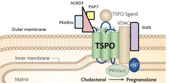
19 Biosynthetic pathway of allopregnanolone 52 20 Complex of proteins called transduceosome, necessary for the passage
of cholesterol in mitochondria 58
21
Ligands TSPO can stimulate the passage of cholesterol into the mitochondria and the synthesis of pregnenolone , the precursor of
neurosteroid biosynthesis
SUMMARY
10
SUMMARY
Mitochondria, known as the powerhouses of the cell, play a critical role in the neuronal survival and cell death, because they regulate both energy metabolism and apoptotic pathways. Mitochondria provide the majority of cellular energy in the form of adenosine triphosphate (ATP) and regulate the production of reactive oxygen species (ROS). Evidence shows that mitochondrial abnormalities are involved in the pathophysiology of Alzheimer's disease (AD). An increase in oxidative damage and decreased energy metabolism were observed at early stages of the disease before the generation of amyloid plaques and neurofibrillary tangles, representing the two main histopathological characteristics of AD. Therefore, current pharmacological concepts for the development of therapeutic strategies against AD have a particular interest in the track of the regulation of mitochondrial functions in neurons, including the control of ATP synthesis, mitochondrial respiration, and ROS production. The decreased neurosteroidogenesis and low brain concentrations of allopregnanolone have also been correlated with a cognitive decline seen in AD patients. These data have generated a strong interest in the development of therapeutic strategies based on the use of allopregnanolone. However, the pleiotropic nature of the effects of allopregnanolone does not facilitate the development of targeted therapy, particular focused on one specific indication, such as neuroprotection, while avoiding stimulation of cell proliferation that possibly could facilitate the emergence of glioma. Though, in cases of functional deficits related to a decline in neurogenesis, it might be interesting to produce chemically modified analogs that have a greater and more specific neurogenic action than that of allopregnanolone.
Regarding the data mentioned above, the main purpose of this thesis was to evaluate the ability of two families of new patented compounds that exert neuroprotective and / or neurogenic effects via the modulation of mitochondrial functions in nerve cells. These two families of compounds are the allopregnanolone analogs (ANS) and the ligands of the mitochondrial translocation of protein or translocator protein (LTSPO) which transfers cholesterol from the outer membrane to the inner mitochondrial membrane, where it is
SUMMARY
11
converted to pregnenolone by the cytochrome P450side-chain-cleavage, a major precursor in the biosynthesis of various neurosteroids. LTSPO are important modulators of neurosteroidogenesis.
To achieve our goal, the thesis was divided into two main parts:
1) The first part is divided into three subprojects (A), (B) and (C).Taking into account bibliographic data suggesting that allopregnanolone has a great neuroprotective potential, we decided to evaluate the ability of allopregnanolone and four of its analogs to improve mitochondrial functions in an in vitro cellular model of AD mimicking the accumulation of β -amyloid peptide (Aß) in human SH-SY5Y neuroblastoma, overexpressing the -amyloid precursor protein or APP (APP cells) (A) and in vivo in a transgenic mouse model (B) as well as in an mouse model of aging (C).
2) In the second part, we compared the effects of novel TSPO ligands to reference ligands with regard to improve the energy balance in APP and in control cells.
(A) Previous work from our team showed a decrease in ATP production in APP cells, which are more sensitive to oxidative stress than the control cells. In the first part of this thesis, the ability of allopregnanolone and its analogs (ANS) to modulate the bioenergetics in APP and control cells was investigated. Our results showed that at the concentration of 500 nM, allopregnanolone (AP) and its analog O-allyl-epiAP (BR297) stimulated the ATP production in APP and control cells. In contrast, the analogs O-allyl-AP (BR351), 12-oxo-epiAP (BR053) and 12-oxo-AP (BR338) did not alter the ATP synthesis. The powerful stimulatory effect on the ATP production exerted by BR297 has encouraged us to compare its capacity to protect against cell death, induced by oxidative stress generated by hydrogen peroxide (H2O2) with that of allopregnanolone. Both compounds, allopregnanolone and
BR297 were able to effectively protect APP cells against death induced by H2O2,while in
control cells only BR297 treatment was efficient. Our results also showed that BR297 and allopregnanolone, which significantly decrease the concentrations of cytosolic and mitochondrial ROS, strongly reduced the Aβ-related elevated anion superoxide levels in APP
SUMMARY
12
cells. Furthermore, our work shows that allopregnanolone and BR297 improved both oxygen consumption (OCR) and glycolysis (ECAR), increasing the bioenergetic activity in APP and control cells. In particular, our work showed that BR297 modulated the respiratory control ratio (RCR), an indicator of the respiratory capacity of mitochondria in both cell types, but allopregnanolone had no significant effect on APP cells.
Our results identify BR297 as an analog which is capable of inducing beneficial effects on mitochondrial bioenergetics without stimulating cell proliferation, as does allopregnanolone. These data give BR297 an interesting profile to develop neuroprotective strategies without the induction of proliferative effects.
(B) Notably, additional in vivo experiments confirmed the beneficial effects of BR297 in an AD transgenic mouse model (Tg2576) by ameliorating the cellular energy level as well as the mitochondrial complexes activities in the cortex and the hippocampus of Tg2576 up to the mitochondrial activity level of the wild-type (WT) group at a dose of 8 mg/kg of BR297. In line with our in vitro and in vivo findings, collaborators showed that BR297 had no effect on neurogenesis but that this analogue induced a positive trend to ameliorate the behavioral deficits in Tg2576 mice.
(C) Given that age is a risk factor for dementia, the protective effect of two analogs, O-allyl-epiallopregnanolone (BR297) and O-allyl-allopregnanolone (BR351), was investigated on age-related disturbances of energy maintenance in the brain. 7-month and 21-month old mice were subjected to a treatment with one of the analogs or the vehicle (control group). The treatment regimen involved three injections per week during four weeks at the doses of 2, 4, 8 mg/kg for BR297 and 1, 2, 4 mg/kg for BR351. Following the behavioural test of pattern separation, brains were collected for evaluation of the mitochondrial activity. Analysis of ATP levels as well as mitochondrial activity of complex I and complex IV were used to evaluate the effect on age-induced mitochondrial impairments in the cortex and hippocampus. Aged mice exhibited significant deficits in cellular ATP levels in the cortex and hippocampus and in mitochondrial ATP levels, especially in the hippocampus. Brains from
SUMMARY
13
21-month old mice only showed a trend to reduced complex activities. Notably, BR297 ameliorated cellular and mitochondrial ATP levels and complex I and IV activity in both brain regions. The beneficial effect of the BR351 treatment was more specific to the cortex by enhancing cellular and mitochondrial ATP levels. Concerning complex activities, BR351 increased only the activity of complex I but not that of complex IV. In total, BR297 exhibited a higher efficacy in the hippocampus with respect to the alleviation of age-related mitochondrial deficiency compared to BR351. These findings suggest that allopregnanolone analogs are promising compounds for the treatment of age-related neurodegenerative diseases.
The aim of the second part was to study the effects of new LTSPO analogs on mitochondrial functions. Few studies have suggested that LTSPO could exert neuroprotective action by stimulating the biosynthesis of neurosteroids as neuroprotective AP. However, most of the currently available LTSPO exhibit anxiolytic effects, low dissolution under physiological conditions and a lack of selectivity or specificity. Moreover, the involvement of LTSPO in neuroprotection processes, particularly their effects on mitochondrial bioenergetics in nerve cells, was not studied until now. Pre-tests by members of our group have shown that different new ligands induced the production of pregnenolone in glial cells. We therefore determined the effects of eleven new compounds and compared those to that of four LTSPO reference molecules, namely diazepam, XBD-173, SSR-180,575, and Ro5-4864 in APP cells. Out of eleven new TSPO ligands, two were very promising: 6a and 6b.The TSPO ligand 6b has a structure similar to 6a, but differs by a lateral chain dipropylamide instead of diethylamide. This structural difference gives the compound 6b a higher affinity for TSPO. Our results have demonstrated a non-toxic effect of the new analogs at 10 nM on cell survival. The higher affinity of 6b for TSPO is correlated to a greater increase in ATP levels than after a treatment with 6a in control and APP cells. In control cells, ATP levels were increased by both compounds but with a higher effect after 6b treatment. In APP cells, both compounds ameliorated the production of ATP to a similar extent. Generally, the new TSPO ligands 6a and 6b are more efficient in compensating the energy loss caused by an overexpression of Aβ peptide than the LTSPO described in the literature. More precisely, the lateral chain
SUMMARY
14
dipropylamide structure of the best ligand 6b allowed the highest amelioration of the energy production as well as an increase of affinity and selectivity for the TSPO receptor
Taking together, all the results showed that the imidazoquinazolinone structure seems to be importantl to alleviate the bioenergetic deficit observed in AD pathology.
In conclusion, this PhD work has identified new molecules that regulate mitochondrial functions and are promising for the development of optimized and better targeted neuroprotective strategies. The new analog of allopregnanolone, O-allyl-epiAP or BR297, which is able to exercise effective neuroprotective effects involving a strong upregulation of mitochondrial activity devoid of cell proliferation, seems to be a promising compound. Another new analog of allopregnanolone, BR351 or O-allyl-AP, has been identified to be also effective in neuroprotection, but its effect mainly results from neurogenic and / or proliferative action that does not seem to depend on a strong mobilization of mitochondrial functions. First
in vitro results already identified 6a and 6b as effective in ameliorating the cellular energy
homeostasis in APP cells. Ultimately, the results of this thesis will provide valuable preclinical data for the development of new therapeutic strategies against AD or neurodegenerative diseases.
15
INTRODUCTION
Neurodegenerative diseases
16
A.
Neurodegenerative diseases
Neurodegenerative diseases are amongst the major causes of disability, decreased quality of life and death worldwide [1]. They are defined as incurable, sporadic and hereditary conditions which are characterized by gradual nervous system dysfunction. Aging, a challenge to every living organism, is linked to accumulation of metabolism impairments and cell damage, causing disease development [2]. Neurodegenerative diseases often correlate with atrophy of the central or peripheral nervous system, where the nerve cells progressively degenerate or die. These diseases included disorders as Alzheimer’s disease (AD), Parkinson’s disease, Huntington’s disease, Motor Neurone disease, Prion diseases, Amyotrophic Lateral Sclerosis, Multiple Sclerosis and many other dementias. They all lead to progressive brain damage and neurodegeneration [3]. Parkinson’s disease and AD exhibit different clinical features but appear to be similar at the cellular level in the disease process. For example, Parkinson's disease depletes the basal ganglia of the brain of dopamine, which leads to bradykinesia, stiffness, rigidity, and tremors in the major muscles of the body [4]. In Alzheimer's disease, deposits of protein plaques and hyperphosphorylation of protein damage different parts of the brain and lead to progressive loss of memory.
Medication can only help to ameliorate the patients’ quality of life and reduce symptoms. For example, Levodopa can increase the brain’s dopamine levels to help alleviate symptoms of Parkinson’s disease with still many side effects and memantine or donepezil can sometimes slow the progression of dementia symptoms in some people with Alzheimer’s disease. The incidence of these age-related disorders is on rising and they are the attract of great deal of attention due to their irreversibility, accompanied with economic and social burden and the lack of effective treatment [2].
INTRODUCTION
Alzheimer’s Disease
17
B.
Alzheimer’s disease
1. Clinical symptoms, risks and etiological factors
The first form of dementia was described by the German neuropathologist and psychiatrist, Alois Alzheimer in 1906 and later became known as Alzheimer’s disease (AD) at the suggestion of Emil Kraepelin [5]. Alzheimer examined a woman from Frankfurt, Auguste Deter, who had shown a striking cluster of symptoms that included psychosocial incompetence, hallucinations, delusions, progressive cognitive deficit and focal symptoms. At necropsy, Alzheimer reported the histopathological finding of this disease: tangles of fibrils within the cytoplasm of neurons, widespread outside plaques pathology [6].
According to Alzheimer’s Association in the report 2015 Alzheimer’s Disease Facts and
Figures, AD is the most common cause of dementia. It account for 60 percent to 80 percent
of dementia cases and is the first leading cause of death. By 2050, there will be over 115 million people diagnosed with AD worldwide [7]. The triad of risk factors for AD are age, the apolipoprotein E ɛ4 (ApoE) and chromosomal sex [8]. The most important risk factor for Alzheimer’s disease is age above 65 years (National Institute of Aging 2015). People younger than 65 can also develop the disease but it is rare. The female prevalence of AD is correlated to a greater life span relative to men [9]. ApoE is a lipid binding protein that transports and coordinates the mobilization of cholesterol between and within the cells. ApoE is expressed mainly in the liver and brain [10]. Among AD patients, 60% carry at least one ɛ4 allele [11]. In AD, there is still a variability that is not explained by sex difference or ApoE genotype suggesting that the biologic system of aging is more likely the driving factor [8]. AD progresses generally slowly in three different stages: preclinical (early stage), mild cognitive impairment (middle-stage) and dementia (severe stage) [12]. In the preclinical stage there are no reliable and valid signs and symptoms for an early diagnosis. In the mild cognitive impairment, deficits in declarative memory are usually important and depressive symptoms are uncommon, but patients manage to live alone. In the severe stage, patients are completely dependent, develop neurological disturbances and need supervision, if
INTRODUCTION
Alzheimer’s Disease
18
perception and behaviour are affected [13]. In most people with Alzheimer’s disease which is caused in different ways, the experienced symptoms and the progress of the disease’s stages will be different.
2. Neuropathological hallmarks of Alzheimer’s disease:
By creating a continuum between the normal aging and AD, clinicopathological correlation studies generate hypotheses on the pathophysiology of AD that amyloid plaque development appears initially before the emergence of cognitive deficits, whereas neuronal loss, neurofibrillary tangles, and specifically synaptic loss appear at the same time as the evolution of cognitive decline. The neuropathological hallmarks of AD incorporate positive lesions like glial responses, neurofibrillary tangles, and amyloid plaques, and negative lesions such as synaptic and neuronal loss [14].
Neurofibrillary tangles formed by intraneuronal aggregation of hyperphosphorylated and misfolded tau protein, and amyloid plaques composed of the abnormal amyloid beta protein deposit as senile plaques, are considered to be the two major hallmarks of AD (Fig. 1) [15, 16].
Figure 1: Amyloid plaques and neurofibrillary tangles in the cerebral cortex and hippocampus of an Alzheimer’s disease patient: (A) The plaques of Alzheimer's disease are seen here with a bielchowski silver
stain. (B) Neurofibrillary tangles of Alzheimer’s disease appear as long pink filaments in the cytoplasm with a hematoxylin and eosin staining. Adapted from Neuro patho Flashcards by ProProfs and knowing neurones.
INTRODUCTION
Alzheimer’s Disease
19
a) Neurofibrillary tangles (NFTs)
Neurofibrillary tangles, described as intraneuronal filamentous inclusions within pyramidal neurons, occur in AD and neurodegenerative tauopathies [17]. Recently, the presence of twisted ribbon-like assemblies of tau fibrils was revealed thanks to modern molecular microscopy techniques. The major component of NFTs is the microtubule-associated protein tau, which is abnormally misfolded and hyperphosphorylated (Fig. 2) [18]. Normally, Tau is a microtubule-associated protein (MAPs) present in a soluble form in axons and promotes microtubule assembly and stability. Tau is implicated in the axonal transport of vesicles by binding and stabilizing the microtubules [14]. In pathological condition, tau is translocated in a cell soma and is hyperphosphorylated ~3-fold more than the protein tau in the normal brain [19]. This protein becomes insoluble and misfolded, self-aggregates into paired helical filaments (PHFs), destabilizes the microtubules and gives rise to neurofibrillary tangles (Fig. 2). Intermediate aggregations of insoluble protein tau are cytotoxic. This form induces axonal transport impairments and cognitive deficit [20]. The density of neurofibrillary tangles in the neocortex correlates with dementia, cognitive impairments [19]. Contrary to dementia with Parkinsonism where more than 30 mutations of tau have been detected, in AD tau mutations do not occur. The reductions in score of cognitive evaluation are correlated with the increase of total and phosphorylated tau in the cerebrospinal fluid [21, 22].
INTRODUCTION
Alzheimer’s Disease
20 Figure 2: In AD, hyperphosphorylated tau may destabilize microtubule networks, impair axonal transport, and ultimately trigger neurofibrillary tangle (NFT) formation and neuronal death. Adapted from [23]
b) Amyloid beta plaques
i) The amyloid precursor protein
APP is the most abundant transmembrane protein (106-130kDa) in the central nervous system (CNS) and it is also expressed in peripheral tissues such as blood cells and epithelium [24]. Through alternative splicing, APP presents isoforms ranging from 665 up to 770 amino acids, whereof APP695 is the most dominant form in the CNS. The physiological
role of APP is still unclear but implications in synaptic plasticity, cell adhesion, neurite outgrowth, and neuronal migration have been shown [25-28]. APP is metabolized by two distinct routes: (i) the non-amyloidogenic α-secretase pathway and, and (ii) the amyloidogenic β-secretase pathway (Fig. 3).
In the non-amyloidogenic pathway, APP is cleaved in the middle of the Aβ region by α -secretase releasing a soluble N-terminal APPsα ectodomain and a membrane bound carboxyl-terminal fragment (APP-CFT83). The C83 fragment is cleaved by γ-secretase
INTRODUCTION
Alzheimer’s Disease
21
releasing a C-terminal fragment C3 and an APP intracellular fragment called AICD [29]. The action of the APPSα has been considered beneficial by protecting neurons, exerting neurotrophic effects and promoting neurogenesis [30, 31]. The P3 fragment is not toxic but showed neuroprotective effects [32, 33].
In the amyloidogenic pathway, APP is cleaved by β-secretase, also named β-site APP cleaving enzyme 1 (BACE1), releasing a soluble N-terminal APPsβ fragment and a membrane bound carboxyl-terminal fragment (APP-CFT99), which is cleaved by γ-secretase, producing the full length β-amyloid peptides (Aβ) and an AICD fragment. AICD domain is shown to be involved in neuroprotection. Elevated levels of AICD are correlated with enhancement of memory and synaptic plasticity [34].
Figure 3: The amyloid precursor protein (APP) is a transmembrane protein cleaved by secretase enzymes. In the non-amyloidogenic pathway, APP is cleaved by α-secretase and then by the γ-secretase. In the amyloidogenic pathway, Aβ peptides are released after sequential cleavage of APP by β- and γ-secretases, and accumulate into oligomeric aggregates to form plaques. Adapted from [23, 35].
INTRODUCTION
Alzheimer’s Disease
22
ii) Enzymes involved in APP proteolysis
The production of Aβ peptide has been at the center of Alzheimer’s disease research by successive enzymatic cleavages of APP. The α-secretase activity is mediated by enzymes from the family of desintegrin and metalloproteinase domain proteins (ADAM), ADAM 9, 10, 17 and 19 to process four functions: proteolysis, cell adhesion, cell fusion and cell signalling [36]. The overexpression of ADAM 10 has been shown to reduce Aβ production and plaques deposition as well as decrease the cognitive impairments in a mouse model of AD [37, 38]. ADAM 17 has been shown to specifically cleave the precursor of tumor necrosis-α (TNF-α) [36]. APP is sequentially cleaved in the luminal and extracellular compartments by β-secretase and within the transmembrane domain by γ-secretase [39]. The BACE1 is a transmembrane aspartic protease and has been shown to be the major neuronal β-secretase [40]. The subcellular localization of BACE1 is within the endoplasmic reticulum and the trans-Golgi network [41]. The expression of BACE1 is increased in cellular stress situations such as energy deprivation but also during oxidative stress [42]. Recently, a study that supports the fact that Aβ causes microtubule disruption, showed that impaired axonal transport and the accumulation of BACE1 in peri-plaque dystrophies induces BACE1 cleavage of APP and exacerbation of amyloid pathology in AD [43]. γ-Secretase, a membrane-embedded protease, cleaves the transmembrane region of APP to generate Aβ [39]. γ-Secretase is a multi-subunit aspartyl protease complex. The γ-secretase activity interacts with four proteins: presenilin 1 or 2 (PSEN1 or 2) forming the catalytic subunit and having a protease activity, nicastrin (Nct), anterior pharynx-defective 1 (Aph-1) and presenilin enhancer 2 (Pen2) [39]. The three last partners are required for the assembly and the stabilization of the complex and for the presenilin endoproteolysis. PSEN1 is the dominant PSEN in the brain [38]. In AD, mutations in the PSEN gene have been shown to be a major risk factor [44]. In humans, at least six different possible γ-secretase complexes may have cell- or tissue specificity [45]. γ-Secretase complexes have been localized whithin the mitochondria by promoting apoptosis [46].
INTRODUCTION
Alzheimer’s Disease
23
iii) Amyloid-β peptide
Amyloid beta (Aβ) is a 40 or 42 amino acid peptide, a normal by product derived from sequential cleavage of amyloid precursor protein (APP) by β- and γ-secretase. Aβ is found in the cerebrospinal fluid (CSF) and secreted to the extracellular environment of the human brain. The physiological role of Aβ is related to modulate the synaptic activity, but it is still controversial. At picomolar range in normal physiological level, Aβ peptides positively regulate the synaptic function and learning [47]. Other suggested physiological roles of Aβ
include protection as antioxidant molecule against metal-induced oxidative damage, regulation of cholesterol transport, kinase activation, and transcriptional regulation of AD-associated genes [48-51]. Insufficient Aβ levels may induce a loss of normal function but excess Aβ could give rise to dysfunction [52]. The increase of Aβ peptide concentration induces pathological effects under pathological conditions such as stress, increased neuronal activity, or the presence of familial Alzheimer’s disease (FAD) mutations [47].
iv) Amyloid beta plaque formation and consequences
The protein aggregation (fibrillization) involves different conformations of the native protein, oligomers formation creating to protofibrils and finally mature fibrils (Fig. 4) [53]. While the aggregation features of Aβ42 and Aβ40 are equal, Aβ42 is more important than Aβ40 within the plaques. The additional amino acids confer an important rate of insolubility and fibrillization to Aβ42 [54]. A common conserved molecular architecture of Aβ (1-40) peptide fold is present for all the Aβ fibrils [55]. Diffuse plaques and dense-core plaques are the two types of amyloid plaques staining distinguishable by the β-sheet pleated conformation with specific dyes such as Thioflavin-S. The dense-core plaques are positive to Thioflavin-S staining and are associated with deleterious effects such as synaptic and neuronal loss [56].
INTRODUCTION
Alzheimer’s Disease
24 Figure 4: Protein fibrillization: schematic model for amyloid β (Aβ) misfolding and aggregation. Soluble
native protein is misfolded and associates in the form of oligomers and other intermediates that eventually give rise to fibrils. Adapted from [57]
v) Toxicity of amyloid β oligomers
Concerning oligomers and fibrils, a difference of toxicity is suggested by their distinct structure and morphology [58]. Aβ oligomers and its further conversion to fibrils induced toxic mechanisms formation such as disruption of membrane receptors, adsorption on membrane surface which alters the property of the membrane, formation of pore which causes the leakage of Ca
2+
, and the accumulation of intraneuronal Aβ (Fig. 5) [59]. In AD, accumulation
of Aβ forming intermediate soluble oligomers is synaptotoxic as well as cerebral angiopathy (primarly Aβ40) and insoluble β-sheet pleated amyloid fibrils [14]. After peptide solubilization, Aβ42 hexamers and dodecamers rapidly become the dominant oligomers, even at low (1
µM) concentrations. Aβ40 produces smaller oligomers [60]. The phosphorylation of Aβ
peptide by protein kinase A promotes the aggregation of this peptide. In AD pathogenesis, the stabilization of Aβ by phosphorylation might be a trigger for formation of toxic aggregates by increasing the concentration of Aβ in the brain [61].
INTRODUCTION
Alzheimer’s Disease
25 Figure 5: APP cleavages are shown on the left side of the figure by α-, β- and γ-secretase and the production of Aβ peptides. The following toxic mechanisms are illustrated: formation of Aβ oligomers and its further conversion to fibrils; disruption of membrane receptors; adsorption on membrane surface which alters the property of the membrane; formation of pore which causes the leakage of Ca
2+
; and the accumulation of intraneuronal Aβ [59].
3. The amyloid cascade hypothesis
The mutation in the gene coding for APP produces the Aβ peptide via sequential scission by the β-APP cleaving enzyme (BACE) and γ-secretase. In the amyloid cascade hypothesis, accumulation of Aβ peptide is considered as the primary influence driving AD pathogenesis. Glenner and Wong were the first to suggest this hypothesis after the isolation and identification of Aβ peptide in the brain of AD patients [15]. Several studies supported this hypothesis, and in 1992, Hardy and Higgins called it the amyloid cascade hypothesis [62]. The hypothesis combines genetic and histopathological information. This hypothesis was supported by the discovery that AD could also be induced by autosomal dominant mutations in three genes: presenilin (PSEN1), PSEN2, and APP, causing the early onset familial Alzheimer’s disease (FAD). The concept of soluble toxic oligomers has been proposed to explain the neurotoxicity of the amyloid-β peptide [63, 64]. The potential mechanism of
INTRODUCTION
Alzheimer’s Disease
26
toxicity called “aggregate stress” may lead to Aβ aggregation due to an imbalance between Aβ production and Aβ clearance, and having as consequences the formation of paired helical filaments (PHFs) of tau aggregates and finally neuronal loss, and dementia [65, 66]. This imbalance between Aβ production and Aβ clearance induces several pathogenic events including activation of microglia and astrocytes, impaired cell communication and synaptic function, oxidative injury and neuronal ionic homeostasis, altered kinase/ phosphatase activities leading to formation of neurofibrillary tangles and mitochondrial dysfunction [65, 67-70].
Over the years, this hypothesis has been modified as the correlation between dementia or other cognitive alterations and Aβ accumulation within the amyloid plaque in the brain is not linear, neither in mice nor in humans [71].
In contrast to the amyloid cascade hypothesis, other studies correlate NFTs pathology with cognitive deficit or dementia in AD patients [72, 73]. In another study also, the severity of the dementia was positively related to the NFTs in the neocortex and not to the degree of senile plaque deposition [74]. The tau hypothesis suggests that tau tangles pathology occurs prior to the Aβ plaque formation and is more correlated with the severity and progression of the disease [72].
4. Mitochondrial cascade hypothesis: attractive target for AD treatment strategy
Most AD researches were focused on the “amyloid-β cascade hypothesis” supposing that Aβ
plaques were the only cause of AD. However, this hypothesis was based on AD rare familial autosomal dominant cases, which explained only about 1% of the AD cases [75-77]. “Mitochondrial cascade hypothesis” was published by Prof Swerdlow and Prof Khan from the University of Virginia (USA) could explain the biggest part of common sporadic late-onset AD forms. This hypothesis present explanation for AD forms (sporadic, age-related) by considering more aspects of the disease [78, 79]. In the hypothesis, mitochondrial function is placed in the center of pathological events and correlated with the reactive oxygen species (ROS) coming principally from the electron transport chain. ROS are the cause of acquired
INTRODUCTION
Alzheimer’s Disease
27
age-related mitochondrial damage. The theory present that mutation in mtDNA accumulate all along a person’s life causing a slowly mitochondrial decline leading to a decrease in energy production and increase in oxidative stress. These considerations could be able to explain more AD related problems: damaged mitochondria may affect APP expression causing an accumulation of Aβ plaques. This causes an elevation of hyperphosphorylated tau protein and thus, together with Aβ plaques, disturbs mitochondria trafficking and function. The result is a negative vicious cycle, which implicates mtDNA mutation, oxidative stress, synaptic dysfunction and neuronal death by apoptosis [78, 80, 81]. Aging is marked by a gradual increase in brain oxidative stress and consequent damages (Fig. 6). The causes and/or consequences of mitochondrial deficit may be exacerbated by environmental and genetic factors and may lead to impaired mitochondrial bioenergetics and dynamics that, in turn, increase ROS production. This vicious cycle may be additionally fed by further vicious cycles, involving Aβ generation, microglial activation and tau hyperphosphorylation. The accumulation of damages initiates the collapse in cellular bioenergetics, cellular dysfunction, and ultimately, neuronal death and dementia by exceeding an individually different threshold (Fig. 6, dotted line) [82].
Based on these considerations, mitochondria represent an interesting target for AD treatment strategy and/or prevention.
INTRODUCTION
Alzheimer’s Disease
28 Figure 6: Conceptual representation of sequential events linking brain aging and sporadic Alzheimer’s disease. APP amyloid precursor protein; NFTs neurofibrillary tangles; ROS reactive oxygen species
INTRODUCTION
Mitochondria
29
C.
Mitochondria
Mitochondria are described as “the powerhouses of the cell”, producing a major part of the cellular energy via the adenosine triphosphate (ATP) generation through oxidative phosphorylation (OXPHOS) [83].
1. Structure of mitochondria
Mitochondria appear to originate 1.5 billion years ago of an endosymbiosis of an aerobic prokaryote into a eukaryotic cell which allowed essential aerobic life [84, 85]. The size, shape, and number of mitochondria in a cell vary from one tissue to another as well as to the physiological state of the cells. Mitochondria are ovoid bodies with a length of 1-2 µm (Fig. 7) [86]. The basic morphology of mitochondria is defined by the inner membrane, an inter-membrane space (IMS) and the outer inter-membrane [83]. The inner inter-membrane is divided into two domains: one juxtaposed closely to the outer membrane (“inner boundary membrane”), the second forms the cristae that are interpreted to form tubular and lamellar structures and to be connected to the inner boundary membrane by small tubular structures named crista
junctions [87]. Concerning the permeability of mitochondria, the outer mitochondrial
membrane (OMM) is easily permeable while the inner mitochondrial membrane (IMM) has a low permeability and contains cristae which have the role to increase the IMM surface. The big surface area of mitochondria induces their capacity to produce ATP. Mitochondria have a special feature which is the presence of their own mitochondrial DNA (mtDNA) composed of 37 genes, all of which are crucial for normal mitochondrial function. The matrix contains mtDNA and also a host of enzymes, as well as ribosomes for protein synthesis. These enzymes catalyze many critical steps of cellular respiration. In respiration, other proteins, including the enzyme that generates ATP, are embedded in the mitochondrial inner membrane. Folding of the cristae increases the surface area for hosting enzymes involved in cellular respiration.
INTRODUCTION
Mitochondria
30 Figure 7: Structure of a mitochondrion from [88]
2. Mitochondria functions within the cells
Mitochondria are special organelles present in every living cell. Their principal function is to supply the energy necessary to the cells to survive.
a) Mitochondrial DNA
In total, 37 genes compose the mitochondrial DNA and are essential for normal mitochondrial function. Thirteen of these genes have directive roles for the production of enzymes involved in oxidative phosphorylation. The OXPHOS process uses oxygen and simple sugars to create ATP, the cell's main energy source. The remaining genes provide the instructions to produce Transfer RNA (tRNA) and ribosomal RNA (rRNA). These RNA help piece together protein building blocks (amino acids) into functioning proteins [89]. One ATP molecule contains one adenosine, a ribose sugar and three phosphates, reach bonded by oxygen
INTRODUCTION
Mitochondria
31 (Fig. 8). This bond between the phosphates is highly energetic and release energy when it breaks [86].
Figure 8: A molecule of ATP (http://schoolworkhelper.net/wp-content/uploads/2010/07/atp-adp-structure.jpg)
ATP is essential for life and is generated in mitochondria via two pathways: glycolysis and oxidative phosphorylation.
b) Cellular energy production
i) Cellular glycolysis
Glycolysis, the first energy pathway, is a metabolic pathway that happens in the cytoplasm of cells that convert glucose molecules (coming from nutritional sources) in ten enzymatic steps into two pyruvates. In the process there is a release of free energy used to form the high-energy compounds nicotinamide adenine dinucleotide (NADH) and ATP. This high-energy is necessary for several cellular processes such as the synthesis of macromolecules (DNA, RNA, and proteins), their transport (endocytosis, exocytosis), cell signalling, and locomotion. Pyruvate molecules enter the mitochondria and are converted to an Acetyl coenzymeA. This molecule is transported to the mitochondrial matrix and enters in the citric acid cycle (Fig. 9) [86].
INTRODUCTION
Mitochondria
32 The Krebs cycle, also known as tricarboxylic acid cycle, is a cycle of enzymatic reactions,
whose net gain is three NADH and one flavin adenosine dinucleotide (FADH2), one
guanosine triphosphate (GTP). The newly produced molecules NADH and FADH2 will be
used in the oxidative phosphorylation (OXPHOS) pathway to generate ATP (Fig. 9) [86].
ii) Mitochondrial respiration
The second main pathway to meet the massive demand of ATP synthesis is called mitochondrial respiration or oxidative phosphorylation [85, 90]. The electrons carried by NADH and FADH2, coupled with protons pumped from the matrix to the intermembrane
space, are used to generate a chemical potential across the inner mitochondrial membrane (Fig. 9). During electron transfer, the gradient of protons generated is used to drive the synthesis of three additional ATP molecules for every electron that travels along the chain. The mitochondrial respiration takes place at the IMM mediated by a complex of proteins (encoded by mitochondrial or nuclear DNA) called electron transport chain (ETC). The ETC consists of five multi-subunit protein complexes connected by the mobile electron acceptor cytochrome c and electron donor ubiquinone. First, two electrons are donated by NADH to the respiratory complexes, complex I (CI) and NADH dehydrogenase. Next, they are passed to a lipid soluble redox carrier, coenzyme Q (Q). These transports are accompanied by the transfer of four protons from the matrix to the intermembrane space. Complex II (CII) and succinate dehydrogenase, also involved in the Krebs cycle, catalyse the reduction of FAD to FADH2. This reaction gives additional electrons into the quinine pool (Q). Compared to
complex I, complex II, the only complex that contains proteins exclusively encoded by nuclear DNA, does not pump protonfrom the matrix. Then the reduced coenzyme Q diffuses through the inner membrane and its electrons are transferred to the complex III (CIII or ubiquinol cytochrome c oxidoreductase). Coenzyme Q is oxidized by the enzyme and the liberated electrons are transferred to two molecules of cytochrome c, another soluble redox protein. Parallelly, the reaction is coupled by the translocation of protons toward the intermembrane space. Finally, cytochrome c removes electrons and transferred them to
INTRODUCTION
Mitochondria
33
molecular oxygen (O2), forming two molecules of water (H2O). Four protons are again
pumped from the matrix into the intermembrane space. ATP synthase, also called complex V (CV) is the last step of OXPHOS. Between IMS and the matrix, the proton gradient and the mitochondrial membrane potential generated by ETC ensure the rotation of the ATP synthase. This reaction creates ATP from adenosine diphosphate (ADP) and phosphate (Pi). The whole process allows the formation of 32 molecules of ATP.
In addition to their fundamental role in orchestrating energy production, mitochondria play also important roles including the amino acid metabolism, fatty acid and steroid metabolism, calcium homeostasis, and a particular function in ROS production.
INTRODUCTION
Mitochondria
34 Figure 9: Bioenergetics of the electron transport chain and the Kerbs cycle. Pyruvate is converted to
high-energy molecules such as NADH, GTP and FADH2 by TCA/Kerbs cycle enzymes through catalyzation. NADH
generated is shuttled to complex I and is converted to NAD+ driving oxidative phosphorylation. Transfer of electrons by the chain maintains the membrane potential via proton pumping into the IMS. ADP is phosphorylated to form ATP via complex V (ATP synthase). Adapted from [90]
INTRODUCTION
Mitochondria
35
c) Mitochondria as sources and targets of reactive oxygen species
Paradoxically, mitochondria are the major producers of ROS but at the same time the most susceptible targets of ROS toxicity (Fig. 10) [91]. Cell respiration requires oxygen and the majority of O2 is reduced into H2O by the ETC at the mitochondrial complex IV [92].
Nevertheless, the ETC lets escape a small amountof electron that react with oxygen producing superoxide radical (O2 .-). O2 .- can be easily transfomed into ROS, such as
hydroxyle radical (OH) and hydrogen peroxide (H2O2) through enzymatic reactions (Fig. 10)
[93, 94]. There is other sources of ROS, a small part derives from other enzyme systems [95].
Figure 10: Production and disposal of mtROS. Electrons (e-) donated from NADH and FADH2 pass through the
ETC and reduce O2 to form H2O at complex IV. MtROS are provided from the leakage of e-to form superoxide
(O2 .-) at complex I and complex III. O2 .- is produced within matrix at complex I, whereas at complex III O2 .- is
released towards the matrix and the intermembrane space. Next, O2
is dismutated to H2O2 by superoxide
dismutase 1 (SOD1) in the intermembrane space and by SOD2 in the matrix. H2O2 is fully reduced to water by
glutathione peroxidase (GPX). Both O2 .- and H2O2 are considered as mtROS. OM: outer membrane; IM: inner
membrane [96]
ROS can be produced in response to environmental stimuli such as growth factors, inflammatory cytokines, chemicals oxidants, chemotherapeutics, toxins hyperoxia, ionizing
INTRODUCTION
Mitochondria
36
radiation, and transition metals [97-100]. At low levels, mtROS are involved in the process of hypoxia adaptation by regulating the stability of hypoxia-inducible factor 1α (HIF-1α). Moderate levels of mtROS participate in regulation of the production of proinflammatory cytokines. High levels of mtROS are capable of inducing apoptosis and autophagy (Fig.11).
Figure 11: Signaling of mtROS [96]
Produced ROS could react with proteins, lipids, and nucleic acid causing oxidative damage to these macromolecules [101]. Mammalian cells have evolved a number of defense mechanisms to limit the cellular damage caused by ROS. In addition to the DNA lesions repairs (excision, double-strand break, and mismatch repairs), the damaging effects of ROS can be mostly neutralized via elevated antioxidant defense [102]. Indeed, to compensate or regulate the high ROS production, mitochondria possess essential endogenous antioxidants to face oxidative stress. The most important are superoxide dismutase, which can convert O2.-in O2, gluthation peroxidase, and catalase, which converts hydrogen peroxide into water
and oxygen [103].
Antioxidants should be in balance with the reactive species. Unbalance could induce harmful dysfunctions connected to oxidative stress, apoptosis, aging processes, mitochondrial abnormalities and Alzheimer’s disease can rise.
INTRODUCTION
Mitochondria
37
3. Age-associated changes of mitochondria:
The mitochondrial energy-transduction ability is crucial for the maintenance of neuronal activity. The hallmark of brain aging is the damage of energy metabolism and redox homeostasis, which is more important in early stages of neurodegenerative diseases. The communication between mitochondria and the rest of the cell is established by controlling cellular energy levels and the redox environment [104].
Aging is characterized by a progressive drop in physiological functions and an increase in mortality, often accompanied by pathological diseases [102]. A multitude of factors is involved in the complex process of aging. Mitochondrial dysfunction and oxidative stress are two important factors contributing to the aging process [102]. Neurodegeneration and aging present common impairments with declines in energy production in the brain and parallel changes in redox status with a pro-oxidant shift, partly due to the mitochondrial generation of O2·-and H2O2 (Fig. 12) [104]. In aging, the mitochondrial energy metabolism is tissue specific
and more prominent in tissues that contain mostly postmitotic cells, such as brain, heart, and skeletal muscle [104].
Mitochondria isolated from aged animals showed a partial loss of the energy transducing capacity. As has been described above mitochondrial OXPHOS is a process that includes the respiratory chain, and electron transfer through the complexes I, II, III and IV. The electron transport chain across the membrane, coupled to a proton gradient, drive the ATP synthesis through the complex V. In aged rat brains, electron transfer is decreased, which leads to a decreased mitochondrial inner membrane potential [105, 106]. Components of complex I and III are transported to generate O2.-. Elevated oxidant production is observed
since the activities of complex I, III, and IV decrease during aging: The rates of O2.-and H2O2
are higher with age [104]. Age induces a decrease of the F1-ATPase activity of complex V
due to the nitration of Tyr269 close to the Mg++ binding site of the F1β subunit [107]. State 3
(active respiration) and the related respiratory control ratio decreased in aging and the state 4 respiration increased, indicating lower energy-transducting efficiency [107, 108].
INTRODUCTION
Mitochondria
38 Figure 12: Model of mitochondrial dysfunction in aging. Toxic ROS generated during normal biological
activity impair cellular homeostatic pathways including the electron transfer chain (ETC) and mtDNA. Oxidative insults to mitochondria, in turn, impair energy transduction, biogenesis of metabolites, and regulation of redox biology with age, thereby contributing to a vicious cycle of accumulating mitochondrial damage that culminates in a mitochondrial functional crisis. This results in cell death and aging. Adapted from [109]
INTRODUCTION
Mitochondria
39
4. Mitochondrial impairment in age-associated Alzheimer’s disease
Mitochondria play an important role in the pathogenesis of neurodegenerative disease such as Alzheimer’s disease. Because of their high energy demand, neurons are especially vulnerable and sensitive to any abnormal mitochondrial activity.
The nervous system (NS) requires about 20 % of the body’s total basal oxygen consumption, showing the vital importance of mitochondria for this system. Mitochondrial dysfunction in the NS induces risk for neurones because of the energy deprivation. Analysis of brain positron emission tomography (PET) of an AD patient showed reduced glucose uptake and metabolism compared to a healthy brain (Fig. 13).
Figure 13: Positron emission tomography (PET) of a healthy subject (A), and of an AD patient (B). Red and
yellow colors show high level of glucose uptake of the brain. The AD-related decrease in energy metabolism is evident. Adapted from [110]
Second to oxidative stress, the impairment of brain resident energy metabolism is an essential event found to be altered already in early stages of AD. Decreased ATP production conducts to impairment of ATP-dependent processes, on which all cellular functions depend [111]. Tramutola and collaborators suggest that reduced glucose utilization, mitochondrial deficit, and decreased ATP production are early nitrosative stress (OS)-events (Fig.14). They contribute to the neurodegenerative process and its progression, culminating in AD pathology.
INTRODUCTION
Mitochondria
40
A growing body of evidence supports mitochondrial dysfunction as a prominent and early chronic oxidative stress-associated event that contributes to synaptic abnormalities and neuronal degeneration in AD [112, 113]. Grimm and collaborators highlighted the critical key role of mitochondria and the close inter-relationship of this organelle with the two main pathological features in the pathogenic process underlying AD: showing independent as well as synergistic effects of Aβ peptide and hyperphosphorylated tau on mitochondrial function by using a high-resolution respirometry system (Oxygraph-2k). The two hallmarks of AD, Aβ
and NFT in cells, have been shown to lead to mitochondrial dysfunction. The consequences of abnormal elevation of Aβ are diverse, including an increase in ROS production, alterations of OXPHOS, and interactions with mitochondrial dynamics and proteins leading to synaptic loss [81, 114]. Numerous studies have demonstrated mitochondrial deficit through the generation of ROS as an important factor involved in the pathogenesis of AD [115]. Aβ
peptide and the presence of trace metal ions have been identified as a potential source of OS [116, 117]. In AD, mitochondria are a target of Aβ toxicity, which may act directly or indirectly on several proteins, leading to mitochondrial deficit. The deregulation of the ETC leads to a decreased complex V activity and a lower ATP synthesis, in addition to an increased ROS generation [91].
INTRODUCTION
Mitochondria
41 Figure 14: Protein oxidation of enzymes in energy metabolism. Increased production of Aβ induces oxidative stress that causes the oxidation of glycolytic enzymes, highlighted in Red, and TCA enzymes, highlighted in Black. The oxidative modifications of these targeted enzymes culminate in reduced glucose metabolism and decreased synthesis of ATP in AD brain. From [111]
In mitochondria, NFT consequences are similar: mitochondrial transport is blocked, and a loss of energy and an increase in oxidative stress occurs. All these phenomena lead to neuronal death [118-120]. Concerning the neurites plaque, positive dense-cores plaques to thioflavin-S are correlated with impairments such as dystrophic neurites that contain abnormal mitochondria and dense bodies of probable mitochondrial and lysosomal origin [14, 121]. Deregulation of complex I is mainly tau dependent, while dysfunction of complex IV is Aβ dependent, at protein and activity level [91].
INTRODUCTION
Neurosteroids
42
D.
Neurosteroids
1. Definition
In the 80s, the term neurosteroid was defined as a sub-category of steroids which are synthesized de novo in the nervous system. Independent of peripheral steroidogenic glands, neurosteroids accumulate in the nervous system [122-124]. Baulieu and coworkers showed that the production of steroids in the brain [125]. They demonstrated that the concentration of diverse steroids such as pregnenolone, dehydroepiandrosterone (DHEA) and their sulfated derivatives is higher in the brain and peripheral nerves than in the plasma of rats and still high even after gonadectomy or adrenalectomy. Some studies showed that other steroids were synthesized within the brain. In neurons as well as glial cells enzymatic activities of proteins involved in steroidogenesis have been showed [124, 126-128]. To distinguish steroids synthesized within the nervous system from steroids derived from the periphery, such as gonads, adrenals, and placenta, they were named “neurosteroids”. Thus, the definition of neurosteroids considers them as endogenous steroidal molecules synthesized in glial cells or neurons of the CNS and PNS. Neurosteroids include steroids that still persist in substantial levels in the nervous system after removal of peripheral steroidogenic glands [124, 129]. This term refers to their site of production within the nervous system. Neurosteroids act in an autocrine or paracrine configuration and strongly influence system functions and reactions to injury or disease. Neurosteroids are composed of different groups of steroids. The first category is the non-exclusive neurosteroids that are steroidal hormones synthesized in glial cells, neurons or endocrine glands, such as pregnenolone (PREG), progesterone (PROG), or DHEA. The second is the semi-exculsive neurosteroids such as allopregnanolone that are mainly produced in the nervous system and in substantial amounts in the endocrine gland. The last is the exclusive neurosteroids that are steroids exclusively synthesized in nerve cells such as epiallopregnanolone [130, 131]. Neurosteroids derive from cholesterol that is translocated within mitochondria via the translocator protein (TSPO) and


![Figure 7: Structure of a mitochondrion from [88]](https://thumb-eu.123doks.com/thumbv2/123doknet/14439308.516627/31.892.227.663.161.574/figure-structure-mitochondrion.webp)