HAL Id: tel-02918150
https://tel.archives-ouvertes.fr/tel-02918150
Submitted on 20 Aug 2020
HAL is a multi-disciplinary open access
archive for the deposit and dissemination of sci-entific research documents, whether they are pub-lished or not. The documents may come from teaching and research institutions in France or abroad, or from public or private research centers.
L’archive ouverte pluridisciplinaire HAL, est destinée au dépôt et à la diffusion de documents scientifiques de niveau recherche, publiés ou non, émanant des établissements d’enseignement et de recherche français ou étrangers, des laboratoires publics ou privés.
of Trp37-G during the interaction of NCp7 protein of
HIV-1 with nucleic acids
Rajhans Sharma
To cite this version:
Rajhans Sharma. Characterization and targeting the dynamic recognition of Trp37-G during the interaction of NCp7 protein of HIV-1 with nucleic acids. Biophysics. Université de Strasbourg, 2018. English. �NNT : 2018STRAJ014�. �tel-02918150�
ÉCOLE DOCTORALE DES SCIENCES DE LA VIE ET DE LA SANTE
UMR CNRS 7021 Laboratoire de Bioimagerie et Pathologies
THÈSE
présentée par :
Rajhans SHARMA
soutenue le : 10th Avril 2018
pour obtenir le grade de :
Docteur de l’université de Strasbourg
Discipline/ Spécialité
: Sciences du vivant/Biophysique
Caractérisation et ciblage de la
reconnaissance dynamique de Trp37-G
lors de l’interaction de la protéine NCp7
de HIV-1 avec des acides nucléiques
THÈSE dirigée par :
M. MELY Yves Professeur, Université de Strasbourg
RAPPORTEURS :
Mme. MAHUTEAU-BETZER Florence Chargée de recherche, Institut Curie,Paris
Mme. GATTO Barbara Associate Professor, University of Padova
AUTRES MEMBRES DU JURY :
ÉCOLE DOCTORALE DES SCIENCES DE LA VIE ET DE LA SANTÉ
UMR CNRS 7021 Laboratory of Bioimaging and Pathologies
THESIS
presented by :
Rajhans SHARMA
Defense on : 10th April, 2018
For obtaining the degree of :
Doctorate of University of Strasbourg
Discipline/ Specialization
: Life sciences/Biophysics
Characterization and targeting the
dynamic recognition of Trp37-G during
the interaction of NCp7 protein of HIV-1
with nucleic acids
THESIS directed by :
Mr. MELY Yves Professor, University of Strasbourg
REPORTERS :
Mrs. MAHUTEAU-BETZER Florence Chargée de recherche, Institut Curie, Paris
Mrs. GATTO Barbara Associate Professor, University of Padova
OTHER MEMBERS OF JURY :
4
Acknowledgement ... 6
List of Abbreviations ... 8
1. Bibliographic Review ... 10
1.1. Human Immunodeficiency Virus Type 1 (HIV-1) ... 11
1.1.1. Overview ... 11
1.1.2. Origin Timeline ... 11
1.1.3. Classification and Transmission... 11
1.1.4. Structural Organization ... 12
1.1.5. Genomic Organization ... 13
1.1.6. Viral Proteins... 18
1.1.7. Replication Cycle of HIV-1 ... 23
1.1.8. Antiretroviral Therapy (ART) ... 28
1.2. Structure-Function Relationship of Nucleocapsid Protein ... 31
1.2.1. Introduction ... 31
1.2.2. Nucleic Acid Chaperoning of NC ... 32
1.2.3. Role of NC During HIV-1 Reverse Transcription ... 34
1.2.4. Role of NC Domain of Gag in Viral Assembly ... 38
1.3. Fluorescence and Nucleic Acids... 41
1.3.1. Introduction to Fluorescence ... 41
1.3.2. Introduction to Nucleic Acids ... 45
1.3.3. Fluorescence Nucleobase Analogues ... 52
1.3.4. Emerging Applications of Fluorescent Nucleobase Analogues ... 59
2. Research Objectives ... 62
3. Materials and Methods ... 66
3.1. Fluorescent Nucleobase Analogues ... 67
3.2. Peptides ... 68
3.3. Oligonucleotides ... 69
3.4. UV/Vis Absorption and Fluorescence Spectroscopy. ... 71
3.5. Time Correlated Single Photon Counting ... 74
3.6. Kinetic Measurements ... 76
5
4. Results and Discussions ... 80
4.1. CHAPTER 1: Photophysical Investigation of Fluorescent Nucleobase Analogues. 81 4.1.1. Publication 1: Tautomers Of A Fluorescent G Surrogate and Their Distinct Photophysics Provide Additional Information Channels. ... 81
4.1.2. Identifying the Fluorescence Lifetimes of thG Tautomers ... 84
4.1.3. Photostability of thG as substituted in (–)PBS ... 85
4.1.4. Publication 2: Conquering 2‑Aminopurine’s Deficiencies: Highly Emissive Isomorphic Guanosine Surrogate Faithfully Monitors Guanosine Conformation and Dynamics in DNA ... 90
4.2. CHAPTER 2: Fluorescent Nucleobase Analogues as a Tool for Characterizing Dynamics of Nucleocapsid Protein Promoted Annealing of (–)/(+)PBS. ... 92
4.2.1. Publication 3: Dynamic Interconversion of Nucleic Acid Structures Surveyed by Environment-Sensitive Fluorescent Nucleoside Analogues. ... 92
4.3. CHAPTER 3: Investigating the Role of Sugar Deoxyribose in Binding Polarity of HIV-1 Nucleocapsid Protein. ... 94
4.3.1. Introduction ... 94
4.3.2. Results and Discussions ... 101
4.3.3. Conclusions ... 128
4.4. CHAPTER 4: Screening the Inhibitors of Nucleocapsid Protein ... 131
4.4.1. Introduction ... 131
4.4.2. THINPAD ... 132
4.4.3. NCinh assay... 134
4.4.4. Site Specific Binding Assay ... 135
4.4.5. Conclusions ... 139 5. Conclusions ... 140 6. Perspectives... 146 7. Résume en Français ... 148 8. Appendix ... 156 9. References ... 168
6
I am immensely grateful to Prof. Yves Mély for giving me this opportunity to pursue the PhD research work under his supervision. Thank you for your guidance, discussions, sharing knowledge, pushing me to higher limits, improving my skills, encouragements and help me grow as a researcher. I admire your positivity, dynamism, sense of humor and winning attitude. Your ‘perfectionism’ has always inspired me to excel my inputs.
I am grateful to all the members of jury, Dr. Florence Mahuteau-Betzer, Dr. Barbara Gatto and Dr. Jerémié Leonard for accepting the proposal to evaluate this thesis work. I am grateful for your comments and suggestions.
I would like to thank our collaborators Prof. Yitzhak Tor, Dr. Mattia Mori and Dr. Roberto Importa for providing their scientific inputs. I would like to thank Prof. Alain Burger for careful reading of the thesis and providing important suggestions.
I am thankful to the permanent members of our lab for their suggestions during this work: Dr. Julien Godet for scientific discussions and designing the project; Dr. Pascal Didier for helping me solve complex mathematical calculations and playing football with us; Dr. Nicolas Humbert for teaching peptide purification (on papurus) and rewarding with choco pain; Dr. Ludovic Richert for giving me experimental guidance that made the long nights working with laser and
Brochon more bearable; Dr. Christian Boudier for his valuable insight on ITC and Stopped
flow results and additional thoughts on Game of thrones and Himalayas; Dr. Frédéric Przybilla for his suggestions and motivation, Dr. Real Elenore for teaching biological experiments; and Dr. Andrey Klymchenko and Dr. Youri Arntz for their excellent company and sharing never ending hilarious stories. Special thanks to the grand-father figure of our lab, Dr. Guy Duportail, who took care of the French administrative paper work and conundrums, and his huge support during the time when I underwent a surgery. I am thankful to Marlyse Wernert, Ingrid Barthel, Ludovic Four and Michel Morciano for their administrative and IT assistance.
I appreciate contribution from my PhD and Post-doc colleagues. I am hugely thankful to Dr. Marianna Sholokh, the first person who taught me experimental techniques, methods and critical analysis of results. I am thankful to the queen of Thinpad project and member of our ‘Thinpad patient team’, Dr. Lesia Kovalenko, for the advices, discussions and definitely the
cakes. I am also grateful to Dr. Kamal kant Sharma, Dr. Avisek Ghose and Dr. Krishna Gavvala
7
Jagganath, Caterina, Dima and others. As the leader of ‘‘4 o’clock tea time’’ group, I acknowledge contribution from its members: Sasha, for a wonderful friendship that certainly made life lot easier during these days and Taras for enriching our discussions with explicit contents. I would like to thank Nina for all the laughs, loads of cookies (free food) and particularly helping me in recover my health. Specially, I would like to thank Waseem and Tanveer for their true friendship. Back home in India, I am thankful to my friends Nachiketa, Himanshu and Aakriti for their kind support.
I will always be grateful to my parents. They have been a constant source of motivation and happiness in my life.
Thank you Rajhans Sharma.
8
A
AIDS: Acquired immunodeficiency syndrome
ALIX: ALG2-interacting protein X ART: Antiretroviral therapy
d2Ap7(–)/T12(+)PBS: duplex with 2Ap-T
match
C
CA or p24: Capsid protein CTD: C’-terminal domain
E
Env: Envelope proteins
ESCRT: Endosomal sorting complex required for transport
ESIPT: Excited state intramolecular proton transfer
λex: Excitation wavelengths
G
G: Guanine
gRNA: genomic RNA
H
HAART: Highly Active Anti-Retroviral Therapy
3HC: 3-hydroxychromones
3HCnt: 2-thienyl-3-hydroxychromone
I
IN: Integrase
ITC: Isothermal titration calorimetry IC: Internal conversion
ICS: Inter system crossing
IRES: Internal ribosome entry segment
K
Kd: Binding constant
M
MA or p17: Matrix protein MD: Molecular dynamics
MM-PBSA: Molecular Mechanics Poisson-Boltzmann Surface Area
N
NA: Nucleic acids
NC: Nucleocapsid protein nt: nucleotide
NC-ODN: NC-Oligonucleotide NTD: N’-terminal domain Nef: Negative regulatory factor
NRTIs/NtRTIs: Nucleoside/nucleotide reverse transcriptase inhibitors
NNRTIs: Non–nucleoside reverse transcriptase inhibitors
P
PBS: Primer binding site
(–)PBS: minus strand Primer binding site Δ(–)PBS: truncated (–)PBS without the overhangs
(–)/(+)PBS or (+)/(–)PBS: Duplex PBS PPT: Poly purine Tract
PPTC: Poly purine Tract central PR: Protease
PIC: pre-integration complex Phe or F: Phenylamine
Q
QY or Φ: Quantum yield
R
RT: Reverse transcriptase RRE: Rev response element
Rev: Regulator of virion Expression RTC: Reverse transcription complex
S
SU or gp120: Surface protein SL: Stem loop
SNP: Single nucleotide polymorphism
s: 2-amino-6-(2-thienyl)purine T
Trp or W: Tryptophan
TM or gp41: Transmembrane protein TAR: Transactivator region
TSG101: Tumor susceptibility gene 101
thG: thienoguanosine
dthG: deoxy-thG as substituted in DNA (or
mentioned with “underscore”DNA, like
thG7(–)PBS_DNA)
tRNA: Transfer RNA
U
UTR: Untranslated region
V
Vpr: Viral protein R
Vif: Viral infectivity factor Vpu: Viral protein U
VPS4: Vacuolar protein sorting 4
Z
10
11
1.1.
Human Immunodeficiency Virus Type 1 (HIV-1)
1.1.1. Overview
uman immunodeficiency virus (HIV) causes Acquired Immunodeficiency Syndrome (AIDS) that impairs the individual’s immune system during the lifetime. Since the beginning of the epidemic in early 1980’s, more than 70 million people have been infected, and a toll of 35 million people have died with HIV, that currently represents the fourth leading cause of death worldwide and has been projected to become the third leading cause by 2030 (UNAIDS and WHO). The rising epidemics led to a large influx of attention and funding among the scientific and medical community. For example, the US federal agency has spent an all-time maximum of 25 billion dollars only in 2015, 19 billion dollars in 2016 and an estimated 26 billion will be required in response to HIV in 2020.
1.1.2. Origin Timeline
The causative effect of HIV in humans was first detected in United States of America in early 1980’s. The patients diagnosed showed infections and rare malignancies. In 1983, Institut Pasteur, Paris became the first laboratory to isolate the lymphoadenopathy associated virus (LAV) (then termed name for HIV) from a culture derived from a lymph node biopsy sample of a patient with generalized lymphadenopathy (Barre-Sinoussi et al., 1983). Later in 2008, the Nobel Prize in Physiology and Medicine was awarded to Françoise Barré-Sinoussi and Luc Montagnier for this discovery. Their research led to the first diagnostic ELISA test for HIV infections that FDA allowed to sell commercially in 1985. Later, CD4 cell surface molecule was identified as the main receptors, and CXC- chemokine receptor 4 (CXCR4) and CC-chemokine receptor 5 (CCR5) as co-receptors for HIV. By the end of twentieth century, researchers gained insight into the life cycle of HIV, identifying targets of antiretroviral drugs. In 1989, AZT became the first commercially available drug that prevents HIV transfer from infected mother to child. Within next decade, combination antiretroviral therapy appeared and in 2009, doctors cured a HIV-infected patient from Berlin. However, the situation worsened with the discovery of the drug-resistant property of HIV virus that notably arises because of the error prone nature of its reverse transcription and the recombination occurring between the viruses.
1.1.3. Classification and Transmission
HIV is classified as a lentivirus that belongs to the family of the retroviruses. In general, retroviruses are spherical particles, enveloping a RNA genome, that infect the vertebrates. HIV
12
structure is similar to that of the Simian Immunodeficiency Virus (SIV), another lentivirus. Lentiviruses have the ability to infect dividing as well as non-dividing cells, ultimately causing neurological and immunological disorders.
Two types of HIV viruses, HIV-1 and HIV-2 were identified (Clavel et al., 1986; Guyader et al., 1987). They have similar gene organization, but their genome differs by about 55%. HIV-1 is more diverse, infectious and transmissible than HIV-2. They differ also geographically as HIV-1, which is widely spread, is mostly reported in North America, central Africa, Europe and Asia. HIV-2 is mainly localized in west Africa. HIV-1 and HIV-2 resemble to the strains of SIV in chimpanzees and sooty mangabeys, respectively. Hence, the virus might have cross transitioned during some point from primates to humans. HIV-2 infected areas are mostly being replaced by HIV-1 because the viral loads of HIV-2 is lesser than HIV-1, with a lower transmission rate and a nearly complete absence of mother to child transmission.
HIV-1 stains are further divided into four lineages, M (major), N (non M or non O), O (outliner) and P (new type) (Tebit and Arts, 2011). The group M is widely spread across the world and thus is largely studied. The other three groups represent less than 1 percent. Group M is further branched into nine subtypes (A-D, F-H, J, K) and more than 40 Circulating Recombinant Forms (CRFs) that were generated when multiple subtypes infected the same populations. HIV-2 exists in only eight distinct lineages, (A-H) in which only A and B have been identified in humans (Sharp and Hahn, 2011).
1.1.4. Structural Organization
HIV-1 forms a spherical, membrane enveloped virion, 100–150 nm in diameter, which contains two copies of single-stranded positive sense RNA genome (Ganser-Pornillos et al., 2012). The mature HIV-1 particle has several spikes on its lipid bilayer membrane that are composed of Envelope proteins (Env) (Checkley et al., 2011), which are trimers of Surface protein (SU, gp120) and Transmembrane protein (TM, gp41) . A canonically structured capsid membrane is formed of approximately 1500–2000 copies of Capsid protein (CA, p24), with an outer coating of 2000 copies of Matrix protein (MA, p17) separating it from the envelope membrane (Niedrig et al., 1994). The capsid membrane serves as a core that encloses two copies of unspliced viral RNA genome, coated with 2000 copies of Nucleocapsid protein (NCp7), 10–12 copies of tRNA3Lys (Isel et al., 2010), as well as with Protease (PR), Reverse
transcriptase (RT) and Integrase (IN). The immature virus is characterized by a thick layer of uncleaved Gag polyprotein that lies within the virion membrane. After proteolytic processing
13
of the Gag polyproteins, the specific structures of the mature viral particles are generated (Ganser-Pornillos et al., 2008).
Figure 1. Schematic representation of HIV-1 virion.
1.1.5. Genomic Organization
The viral genome of HIV-1 is single stranded positive sense RNA of 9,200 nucleotides long that exists in a dimer form within mature viral particles. The genomic RNA (gRNA) has 100–200 adenine residues at its 3’ end while at the 5’ end it has capping of trimethylguanosine (TMG) (Yedavalli and Jeang, 2010). The gRNA is poorly structured with several folding domains or motifs. These motifs form serval mismatches, bulges and hairpins. The gRNA has nine Open Reading Frames (ORFs) (Figure 2): gag, pol, env, tat, rev, vpr, vif, vpu and nef. The
gag, pol, env encodes for three polyproteins that are Gag, Pol and Env; tat and rev encode Tat
and Rev regulatory proteins; and vpr, vif, vpu, nef encode Vpr, Vif, Vpu and Nef accessory proteins, respectively. The proteolytic cleavage of Gag polyprotein further give rise to MA (Matrix protein), CA (Capsid protein), NC (Nucleocapsid protein), and p6; while Env proteins gives SU (Surface protein, gp120) and TM (Transmembrane protein, gp41), all these proteins are considered as structural components of HIV-1 particle. The three Pol proteins: PR (Protease), RT (Reverse Transcriptase), and IN (Integrase), provide essential enzymatic functions and encapsulated within the particle. HIV-1 encodes six additional proteins, often called accessory proteins, three of which (Vif, Vpr, and Nef) found in the viral particle. Two other accessory proteins, Tat and Rev, provide essential gene regulatory functions, and the last protein, Vpu, indirectly assists in assembly of the virion. The gRNA is divided into two regions, the coding region that codes for proteins and the non-coding region that plays crucial role in structural organization.
14
Figure 2. Genomic organization of HIV-1. The open reading frames are shown in shaded rectangles and the black lines corresponds to the connections between domain in polyprotein. Adapted from (Watts et al., 2009).
The Untranslated Region (UTR) of retrovirus gRNA is present on both 5’ and 3’ end. It comprises a number of folded sequences that play an important role in dimerization, genome packaging, and translation (Abbink et al., 2005a; Boeras et al., 2017; Wilkinson et al., 2008). The 5’UTR exists in two conformations that are in equilibrium: one that promotes the translation of Gag gene by Internal Ribosome Entry Segment (IRES) mechanism (Brasey et al., 2003); and second is dimmer promoting conformation (Abbink et al., 2005b; Keane and Summers, 2016).
15
Figure 3. Secondary structure of HIV‐1 5′‐UTR in monomer (a) and dimer‐promoting (b) conformations (Top section). The schematic diagram showing the formation of dimer conformer from monomer conformer (Below section). Adapted from (Keane et al., 2015).
The UTR is composed of:
A. The R region
The R region (repeat) is a 98 nucleotides region present at both ends of the HIV-1 gRNA. It is further sub-divided into two regions.
• Transactivator Region (TAR): This region consists of nucleotides from +1 to +59 in the HIV-1 UTR. Having a highly structured hairpin form, TAR is required for viral expression. The structural integrity of this region is critical for TAT activation (Heinicke et al., 2009; Wang et al., 1993). Moreover, TAR serves as a binding site for several cellular binding proteins, that regulate the basal expression of HIV-1 UTR (Henriet et al., 2005; Wang et al., 1993).
• poly(A): The poly(A) signal contains a polyadenylation signal, the cleavage site and part of the GU-rich downstream element (Edmonds, 2002; Klasens et al., 1999; Sükösd et al., 2015)
B. U5 Region
The U5 region has 83 nucleotides and represents the first part of the gRNA to be retro-transcribed. This region also contains the 18-nucleotide primer binding site (PBS) region which is present at the 3’ end of the U5 region. The role of PBS is crucial as it anneals to tRNALys3,
initiating reverse transcription (Sleiman et al., 2012).
C. ψ-site
The psi-site (ψ-site) is a 120 nt region, present between the PBS and the start codon. It has four stem loop motifs (SL1–SL4) that plays a crucial role in genome recognition and gRNA dimerization. Selective packaging of HIV-1 gRNA requires all four cis-elements (Lever et al., 1989; Lever, 2007; Pak et al., 2017).
• SL1: The structure of SL1 consists of an upper stem that has GC-rich loop and a lower stem having an intervening bulge which is highly conserved and required for packaging (Chung et al., 2010; Laughrea and Jetté, 1994) (Figure 4a). The eleven-nucleotide upper stem has a six-nucleotide palindromic sequence, usually GCGCGC (e.g., in the Lai
16
isolate) and GUGCAC (e.g., in the Mal isolate) that facilitates dimerization among two gRNAs (Lodmell et al., 2000). In addition, the portion having a GAAG bulge and the four nucleotide stem are mostly conserved among different HIV isolates (Mujeeb et al., 2007a). The function of SL1 is to ensure effective packaging by promoting dimer formation, through “kissing complex” interaction (Kieken et al., 2006), that facilitates the packaging of two copies of genome during virus assembly (Berkowitz et al., 1996; Lawrence et al., 2003; Lu et al., 2011) (Figure 4b,c). The importance of SL1 motif towards assuring effective packaging is assessed by introduction of mutations that resulted in a large decrease in packaging efficiency (Amarasinghe et al., 2000; McBride and Panganiban, 1997).
Figure 4. Stem loop structure of SL1(a). Proposed model for dimerization b) “pre-initiation model”, where the bases at the end of the stem mediates the primary interaction of DLS dimer formation and after the two molecules are sufficiently proximal, DIS interaction occurs. c) “Finalization model”, after interaction with DIS, two DLSs start to “melt” together by the viral nucleocapsid protein to form an extended duplex (Sakuragi et al., 2016)
• SL2: The SL2 region is a 19-nucleotide sequence that exists in the form of a hairpin (Amarasinghe et al., 2000). It has a tetraloop and a long stem with a single nucleotide bulge. SL2 loop contains the Splice Donor (SD) sequence that is required for the spliced mRNAs production. (Bazzi et al., 2012; Cruceanu et al., 2006b). SL2 plays an important role in genome packaging (McBride and Panganiban, 1997), by interacting with the NC domain of Gag (Amarasinghe et al., 2000; Belfetmi et al., 2016).
17
• SL3 is a 20-nucleotide hairpin having a tetraloop and a perfect double helix stem (Pappalardo et al., 1998). The loop of SL3 belongs to the A-family and is quite flexible. The structure of SL3 is highly conserved among the different strains of HIV-1 and its interaction with the NC domain of Gag during virus assembly is crucial for gRNA selection (Abd El-Wahab et al., 2014; Lever, 2007; Tanwar et al., 2017). Moreover, SL3 by itself is sufficient to drive the gRNA selection (Abd El-Wahab et al., 2014; Athavale et al., 2010; Belfetmi et al., 2016; Paoletti et al., 2002). Detailed discussion on SL3 is presented in section 4.3 of this thesis.
• SL4 or AUG: SL4 hairpin contains a stem with G-U pairs and a GAGA tetraloop (Kerwood et al., 2001). The SL4 tetraloop adopts a conformation similar to a classical GNRA form (Jucker et al., 1996) and the stem is quite unstable due to G-U pairs. The SL4 tetraloop participates in long range RNA-RNA interaction with the U5 element that connects polyA and PBS stem-loop (Abbink and Berkhout, 2003; Damgaard et al., 2004; Jucker et al., 1996; Lu et al., 2011; Watts et al., 2009).
D. U3 region
The U3 region at the 3’ end of gRNA is required for the regulation of transcription of the proviral DNA into viral RNA by the transcription machinery of the host cell (Calzado et al., 2004; Gaynor, 1992; Sgarbanti et al., 2002). The U3 region includes:
• The PPT (Poly Purine Tract) and PPTC (Poly Purine Tract Central): They are the purine rich domains of the gRNA that are resistant to RNase H activity. The PPT is present upstream of U3 sequence of 3’UTR of proviral genome, whereas the PPTC sequence is present in the ORF of the Pol gene (Charneau et al., 1992; Pollom et al., 2013; Rausch and Le Grice, 2004). Their resistance towards RNase H activity of reverse transcription is instrumental because these sequences serve as primers for the synthesis of the plus-strand DNA.
• Rev Response Element (RRE): it is a ~ 350 nucleotide-long sequence that contains several stem-loops and bulges (Watts et al., 2009). It binds with Rev protein to overcome the intron surveillance mechanism and facilitate the nucleo-cytoplasmic export of the mRNAs corresponding to the viral genome (Bartel et al., 1991; Rausch and le Grice, 2015; Sherpa et al., 2015).
18
1.1.6. Viral Proteins
The viral proteins are encoded from nine ORF that are in the coding sequence of gRNA.
Gag gene encodes for 55 kDa Gag polyprotein precursor (Pr55Gag) that upon cleavage
by protease (PR, p12) generates the MA, CA, NC and p6 protein.
Pol gene together with Gag gene code for a 160 kDa GagPol polyprotein precursor
(Pr160GagPol) that upon cleavage by PR results in all the three HIV-1 enzymes, namely PR (cleaved by auto-processing), RT and IN. The production of GagPol arises due to a -1 frame shift in the translation frame which occurs approximately 5% of the time. (Karn and Stoltzfus, 2012)
Env gene encodes for a 160 kDa Env polyprotein precursor (gp160) that upon cleavage
by a cellular furin-type protease in the Golgi, results in the synthesis of gp120 and gp41 glycoproteins.
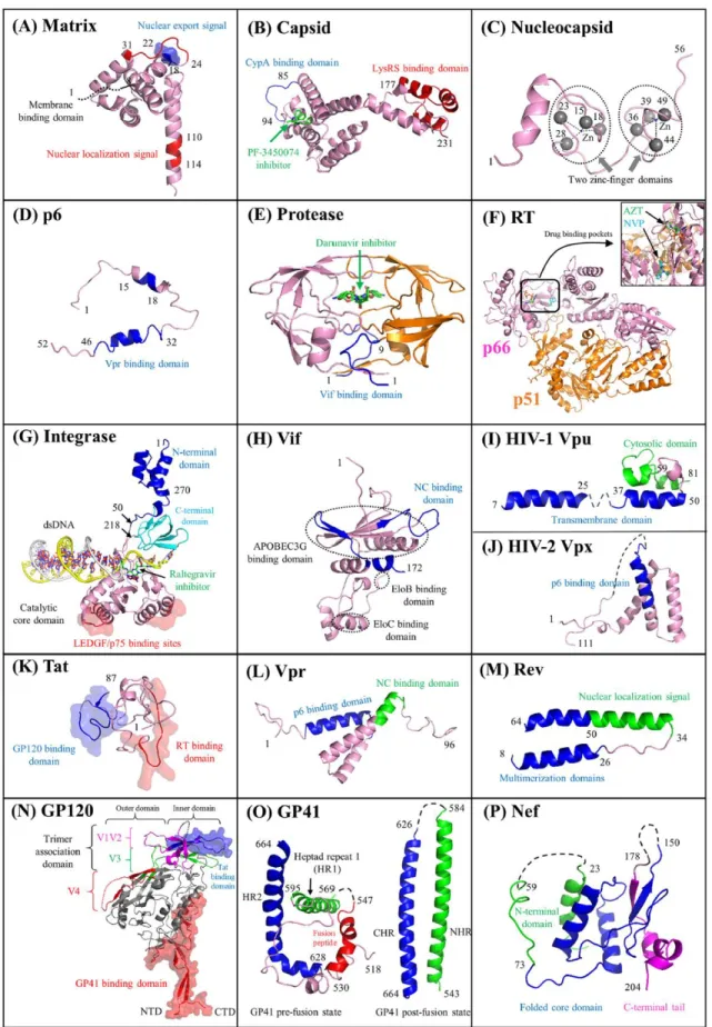
In addition, the HIV-1 genome also encodes for regulatory proteins (Tat, Rev) and accessory (Vpr, Vif, Vpu, Nef) proteins. Figure 5 represents the 3D structures of functional domain of all the HIV-1 proteins.
19
Figure 5. Functional domains of HIV proteins. Cartoon representations of HIV proteins (matrix, capsid, nucleocapsid, p6, protease, RT, integrase, Vif, Vpu, Vpx, Tat, Vpr, Rev, GP120, GP41, and Nef). In each panel, the protein domains involved with HIV
20
pairwise protein interactions are marked accordingly. Surface representations indicate protein interaction interfaces. The five small molecules shown in green correspond to protein inhibitors such as the capsid inhibitor PF-3450074 (503) (B), the protease inhibitor darunavir (E), the nucleoside analogue reverse transcriptase inhibitor zidovudine (AZT) (F), the nonnucleoside analogue reverse transcriptase inhibitor nevirapine (NVP) (F), and the integrase inhibitor raltegravir (G). Adapted from (Li and De Clercq, 2016)
Based on the structural and functional characteristics, the HIV-1 viral protein are classified in following sections:
A. Structural Proteins
• Matrix Protein (MA): The 17 kDa and 132 amino acid MA protein consists of 5 α-helices; two short α-helical stretches; and a three-stranded mixed β-sheet (Verli et al., 2007; Zeinolabediny et al., 2017). Its N-terminal myristate group and basic residues located within the first 50 amino acids are involved in membrane targeting residues (Alfadhli and Barklis, 2014; Tedbury and Freed, 2015). The MA proteins assemble into trimers that are required for viral assembly. Other than targeting the Gag and GagPol to the plasma membrane, MA also accommodates the cytoplasmic tail of the full length Env glycoprotein and incorporate it into the viral particles (Frankel and Young, 1998; Ganser-Pornillos et al., 2012)
• Capsid Protein (CA): It consists of two domains: N’-terminal domain NTD and C’-terminal domain CTD, that are linked together via a flexible linker. The 3D structure of NTD shows seven α-helices while the CTD has four α-helices (Gamble et al., 1997; Gitti et al., 1996; Pornillos et al., 2011). The CTD domain promote efficient viral assembly through CA dimerization and Gag oligomerization (Gamble et al., 1997), while the NTD domain participate in viral uncoating, through association with cyclophilin A (Luban, 2012). Moreover, CA protects the gRNA from cellular immune factors and ensures that RT is close enough to gRNA, in order to perform the reverse transcription (Campbell and Hope, 2015; Freed, 1998).
• Nucleocapsid Protein (NC): NC is a 7 kDa, 55 amino acid protein that has two highly conserved CCHC zinc finger motifs which are connected by a basic linker (RAPRKKG). The NC protein exerts many functions throughout the virus life cycle, mainly through its
21
nucleic acid chaperone activity (Darlix et al., 2007, 2014, 2011). The properties and functions of NC are discussed in detail in section 1.2.
• p6: It is a 6 kDa, 52 amino acid protein that has two helices connected by a flexible linker. One of the role of p6 is to bind and recruit Vpr (Votteler and Sundquist, 2013). Moreover the two late domains of p6 are responsible for recruitment of the cellular ESCRT (endosomal sorting complex required for transport) machinery that enables viral budding (Freed, 1998; Votteler and Sundquist, 2013).
B. Enzymatic Proteins
• Protease (PR): PR is a 6 kDa, 99 amino acid protein that exists in a homodimer form. The N- and C- terminal domains are arranged in a four-stranded antiparallel β-sheet, one loop and one helix (Navia et al., 1989). The catalysis site is highly conserved and comprises of amino acids Asp25-Thr26-Gly27 (Wlodawer et al., 1989). Protease is generated due to self-catalysis from Pr160Gag-pol and is responsible for the cleavage of Gag and GagPol polyprotein. This cleavage of Gag and GagPol by PR is necessary to form mature viral particle (Sundquist and Kräusslich, 2012).
• Reverse Transcriptase (RT): RT is a heterodimer of two subunits p66 and p51. The p66 subunit is a 66 kDa, 560 amino acid that has a catalytic function while the p51 domain is a 51 kDa, 440 amino acid domain that has structural function (Esnouf et al., 1995; Hsiou et al., 1996; Rodgers et al., 1995). The RNase H domain of RT cleaves the RNA portion of the RNA-DNA hybrids generated during reverse transcription (Das and Arnold, 2013; Hu and Hughes, 2012). RT also possess RNA-dependent and DNA-dependent polymerase activities and actively catalyze the reverse transcription process by synthesizing double strand DNA starting from a single stranded gRNA.
• Integrase (IN): It is a 32 kDa, 288 amino acid protein that has three domains: the N-terminal domain (NTD), the catalytic core domain and the C-N-terminal domain (CTD) (Cai et al., 1997; Dyda et al., 1994; Hare et al., 2010; Lodi et al., 1995). The role of IN is to insert the proviral DNA into the host cell DNA, by performing 3’ end processing activity (cleavage of two nucleotides from the two 3’ ends of the viral DNA) and strand transfer activity (Lesbats et al., 2016; Lusic and Siliciano, 2017).
22
C. Envelope Proteins
• Surface Glycoproteins (SU or gp120): It is a 515 amino acid highly glycosylated protein that is located at the surface of the viral particle. Structurally it has ten segments, five of them are conserved domains (C1–C5) and the rest are variable loops (V1–V5). It has three structural units, the inner unit that has two α-helices, five-stranded β-sandwichs and loops; the outer domain that has stacked barrel; and the bridging domain that has four-stranded β-sheets (Kwong et al., 1998; Pancera et al., 2010). The role of gp120 during the viral infection is to interact with the CD4 receptors of the target cell (Checkley et al., 2011; Haqqani and Tilton, 2013).
• Transmembrane Protein (TM or gp41): It is a 345-amino acid glycosylated protein that exists in a trimer form. It has a N-terminal ectodomain, a transmembrane domain, and a C-terminal cytoplasmic tail that orient themselves in antiparallel helices to form a trimer (Chan et al., 1997; Weissenhorn et al., 1997). The gp41 is involved in the fusion of virion membrane and plasma membrane during viral entry (Haqqani and Tilton, 2013).
D. Regulatory Proteins
• Trans activator of Transcription (Tat): It is a 9-11 kDa protein that exist either as an 86 amino acid or a 101 amino acid form. It has six regions: region I between 1–21 amino acid, region II between 22–37 which is Cys rich: region III between 38–48 amino acid which forms the core of the protein, region IV between 49–59 amino acids that is mostly occupied by basic amino acids, region V between 60–72 amino acids which is Gln rich region and region VI is between 73–101 amino acid which forms the C’-terminal of the Tat (Bayer et al., 1995; Peloponese Jr. et al., 2000). The main role of Tat is to promote efficient transcription of the viral genome (Johri et al., 2011).
• Regulator of Virion Expression (Rev): Rev is a 13 kDa, 116 amino acid protein with two domains, the N’-terminal domain having two α-helices and a loop, and the disordered C’-terminal domain. The NTD has nuclear localization signal, RNA binding domain and Rev multimerization domain. On the other hand the CTD domain has a Leu-rich motif needed for nuclear export (Daugherty et al., 2010; DiMattia et al., 2010). Rev facilitates the nuclear export of single-spliced and unspliced RNAs by binding to their RRE element (Pollard and Malim, 1998; Sherpa et al., 2015).
23
E. Accessory Proteins
• Viral Protein R (Vpr): It is a 14 kDa, 96 amino acid protein having three α-helices and flexible N’- and C’- terminal domains which tend to multimerize (Morellet et al., 2003). The N’- terminal domain of Vpr take part in G2 phase cell cycle arrest and apoptosis (Guenzel et al., 2014). While the C’-terminal domain functions in virion incorporation, nuclear localization and Vpr oligomerization (Freed, 1998; Ganser-Pornillos et al., 2012). • Viral Infectivity Factor (Vif): It is a 23 kDa, 96 amino acid protein that has three domains: a highly ordered N’-terminal domain, the central domain having Zn fingers motifs, and the disordered C’-terminal domain (Barraud et al., 2008). Vif play protective role against a cellular deaminase APOBEC3 (A3G and A3F). A3G halts viral progression as it mutates C to U residues during reverse transcription. Moreover, Vif also show nucleic acid chaperoning activity during virion assembly (Ennifar et al., 2007; J G Levin et al., 2010; Sleiman et al., 2014).
• Viral Protein U (Vpu): It is a 16 kDa, 81 amino acid protein that bind to internal membrane of the cell. It has two domains, N’-terminal domain which span through the membrane and C’-terminal domain which is present inside the cytoplasm (Zhang et al., 2015). The role of Vpu is to degrade CD4 receptors complexed with Env protein. Vpu also promote budding of the virus particle from the infected cell (Gonzalez, 2015).
• Negative Regulatory Factor (Nef): It is a 27–35 kDa, 206 amino acid protein with three domains: the N’-terminal domain, the core and the C’-terminal domain (Arold et al., 1997; Grzesiek et al., 1997). The role of Nef is to degrade the cell receptors, like CD4, CD28 and CxCR4 receptors. This in turn promote incorporation of Env protein into virion. It is also responsible for degradation of histocompatibility complex I and II on antigen presenting cells, and in recognition of infected cells by the immune system (Pereira and daSilva, 2016).
1.1.7. Replication Cycle of HIV-1
The life cycle of the HIV virus can be divided in two major phases, the early phase and the late phase.
1.1.7.1. Early Phase
Infection of the cell by HIV-1 particle start with protein-protein interaction on their surface (Figure 6). The CD4 receptors of the cell binds to HIV-1 gp120 surface glycoproteins. This interaction induce conformational change in CD4 receptors, which in turn promote the
24
interaction with the co-receptors i.e. chemokine receptors 5 (CCR5) chemokine receptor 4 (CXCR4) present on the surface of the target cell (Dean et al., 1996; Feng et al., 1999). Then, the conformational change in gp41 further promote its hydrophobic N’-terminal domain to cross into the plasma membrane and reach the cytoplasm of the cell target (Archin et al., 2014; Dr. Rainer Seitz, 2016). This step marks the completion of fusion between the cell membrane and viral envelope.
Figure 6. Early phase of the HIV-1 viral life cycle. Env, envelope glycoprotein; MA, matrix protein; CA, capsid protein; NC, nucleocapsid protein; RTC, reverse transcription complex; NPC nuclear pore complex; PIC, pre-integration complex. Adapted from (Campbell and Hope, 2015).
Fusion of the cell and virion membranes results in the translocation of the viral capsid
inside the cytoplasm. Reverse transcription begins in cytoplasm and convert single strand gRNA to double strand DNA. The steps of reverse transcription are discussed in section 1.2.3. During reverse transcription, the Reverse Transcription Complex RTC transform into pre-integration complex (PIC) for its migration to nucleus through nucleoporin embedded in the nuclear membrane. The disassembly of capsid as well as migration of RTC/PIC into nucleus are still debated (Campbell and Hope, 2015).
25
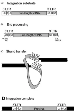
Figure 7. Integration of vDNA into the host chromosome is the final step of early infection. Adapted from (Thomas and Gorelick, 2008)
Integration of viral DNA into host genome performed by IN enzyme include two main
steps (Figure 7). First step occurs in the cytoplasm, where IN binds to two specifically recognized nucleotides at 3’ end of the UTR. Due to its endonuclease activity, these two nucleotides were removed creating a sticky end. This step is called as 3’ processing. The second step occurs in nucleus where IN catalyze the insertion of 3’ end processed viral DNA in to the host DNA (Campbell and Hope, 2015).
1.1.7.2. Late Phase
The late phase begins with transcription of viral DNA by the cellular transcription enzyme RNA polymerase II (Figure 8). Transcription starts from the R region at the 5’ end of UTR and Tat protein promotes the elongation. The transcription gives mRNAs with multiple splicing degrees that includes: unspliced, incompletely spliced and spliced mRNAs. Unspliced mRNA is used as gRNA (9 kb) which encodes both Gag and GagPol proteins. Incompletely spliced mRNAs (4 kb) encodes Env, Vpu, Vif, and Vpr proteins. Interaction between RRE and Rev protein facilitate transportation of unspliced and incompletely spliced mRNAs to the cytoplasm. The completely spliced mRNA has the simplest transfer mechanism as it is directly transferred to the cytoplasm (Karn and Stoltzfus, 2012; Pollard and Malim, 1998).
26
Figure 8. Late phase of the HIV-1 viral life cycle. Env, envelope glycoprotein; MA, matrix protein; CA, capsid protein; NC, nucleocapsid protein; ESCRT, endosomal sorting complex required for transport; ALIX, ALG2-interacting protein X; RER, rough endoplasmic reticulum. Adapted from (Freed, 2015).
Either free or membrane bound ribosomes execute the translation of mRNA in the cell. Translation of Env gene occur at the rough endoplasmic reticulum (Checkley et al., 2011). The unspliced mRNA encodes for Gag and GagPol polyproteins which further give rise to structural, functional and enzymatic proteins of the virus.
The assembly of viral particles starts with two important events that occurs in close time lapse of each other. First is the Gag oligomerization on gRNA and the second is the dimerization of the gRNA that is crucial for selection and packaging (Muriaux and Darlix, 2010; Paillart et al., 2004). Out of the pool of mRNAs in the cytoplasm, the Gag proteins select two unspliced gRNA. The dimerization of gRNA occurs through a series of correlated events happening at the 5’ UTR (D’Souza and Summers, 2005). Dimerization of two gRNA strands begins with NC-promoted annealing of SL1 sequences (Bernacchi et al., 2017; Grigorov et al., 2007; Lu et al., 2011). Due to this specific recognition between SL sequences in gRNA and NC as a domain of Gag, two competent gRNAs are selected for packaging. On the other hand,
27
Gag oligomers are responsible for trafficking the gRNA-Gag complex to plasma membrane (El Meshri et al., 2015). The number of Gag oligomers on gRNA increases as the Gag-gRNA complex moves closer to plasma membrane (Kutluay and Bieniasz, 2010; Mailler et al., 2016). At the plasma membrane, MA protein of the Gag domain interacts with negatively charged lipids, and notably with phosphatidylinositol-(4,5)-biphosphate (PIP2) phospholipids (Ono et al., 2004; Saad et al., 2006). This starts the formation of the immature virion (Briggs et al., 2009).
Budding of viral particle require p6 and NC domain of the Gag protein, which interact
with the cellular endosomal sorting complex required for transport (ESCRT) machinery. This recruitment process requires two-specific domains of p6, PTAP and YPXL domains, that binds to TSG101 (a tumor susceptibility gene 101) and ALIX (a ALG2-interacting protein X), respectively. The interaction of NC with Bro domain of ALIX facilitates the binding of Gag to ALIX. Later, ESCRT-I and ALIX interacts with ESCRT-III and AAA ATPase vacuolar protein sorting 4 (VPS4), respectively, which are required for membrane fission and virion release (Freed, 2015; Sundquist and Kräusslich, 2012).
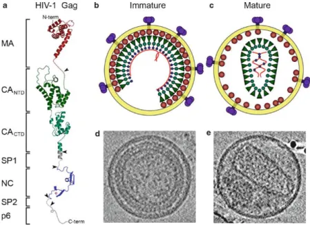
Figure 9 Organization of the immature and mature HIV-1 virions. Schematic structural model of full length HIV-1 Gag (a). Individual domains are in different colors and are labeled on the left. Schematic model and images of the immature virion (b, d) and mature virion (c, e). Adapted from (Ganser-Pornillos et al., 2008)
Maturation of the virion particle begins after it is released from the infected cell. An
28
being bound to the inner viral membrane and the C’-terminal of NC domain stretched towards the core of the virion. Transformation into the mature particle requires catalysis from the PR enzymes which are released from auto-processing of GagPol. PR cleaves the Gag and GagPol polyproteins to release MA, CA, NC, p6, PR, IN, and RT. As a result, the morphology inside the virion particle changes, giving rise to a conical shaped capsid (Freed, 2015; Ganser-Pornillos et al., 2012; Sundquist and Kräusslich, 2012).
1.1.8. Antiretroviral Therapy (ART)
The ever-increasing understanding on HIV-1 life-cycle is beneficial for antiviral drug development. Till date, FDA approved 28 drugs are in market that belongs to distinct classes. These drugs are classified based on their molecular mechanisms and resistance profiles: (1) nucleoside/nucleotide reverse transcriptase inhibitors (NRTIs/NtRTIs), (2) non–nucleoside reverse transcriptase inhibitors (NNRTIs), (3) integrase inhibitors, (4) protease inhibitors (PIs), (5) fusion inhibitors, and (6) coreceptor antagonists.
a) Nucleoside/Nucleotide Reverse Transcriptase Inhibitors (NRTIs/NtRTIs)
These were the first classes of drugs approved by FDA. Before enacting their antiviral effect, NRTIs require host cell entry and phosphorylation by cellular kinases (Mitsuya et al., 1985). NRTIs are 2’-deoxynucleosides analogues that lack 3’-hydroxyl at the sugar moiety. Reverse transcriptase is unable to differentiate between NRTIs and dNTPs, and thus they are taken up and incorporated in the nucleic acid chain. Due to lack of 3’-hydroxyl at the sugar moiety, it is unable to build the 3’-5’-phosphodiester bond that occurs between two natural dNTPs in the classical DNA synthesis. This results in termination of the growing viral DNA chain (Arts and Hazuda, 2012). Currently, there are eight FDA-approved NRTIs: abacavir (ABC, Ziagen), didanosine (ddI, Videx), emtricitabine (FTC, Emtriva), lamivudine (3TC, Epivir), stavudine (d4T, Zerit), zalcitabine (ddC, Hivid), zidovudine (AZT, Retrovir) and Tenofovir disoprovil fumarate (TDF, Viread). Two mechanisms mediate resistance to NRTIs: removal of NRTIs from the 3′-end of the nascent chain through an ATP-dependent pyrophosphorolysis and reversal of chain termination (Boyer et al., 2001).
b) Non–Nucleoside Reverse Transcriptase Inhibitors (NNRTIs)
NNRTIs are the backbone of ART because they show unique antiviral activity, high specificity and low toxicity that makes them a frequent choice for first-line therapy (Li et al., 2014). NNRTIs inhibit HIV-1 RT binding as it binds at the allosteric sites and changes the
29
spatial conformations for the substrate binding site. This eventually reduces the polymerase activity. The well-known structure of the hydrophobic pocket of RT allowed to tailor the design for substituting NNRTIs. The so-called NNRTI-binding pocket consists of hydrophobic residues (Y181, Y188, F227, W229, and Y232), and hydrophilic residues such as (K101, K103, S105, D192, and E224) of p66 and p51 subunit (Arts and Hazuda, 2012). Currently, there are five approved NNRTIs: etravirine, delavirdine, efavirenz, nevirapine and rilpivirine. NNRTI resistance generally results from the substitutions of L100, K101, K103, E138, V179, Y181, and Y188 amino acids in the NNRTI-binding pocket of RT (Rodgers et al., 1995).
c) Integrase Inhibitors
They are recently developed interfacial inhibitor drugs targeting intasome which consist of catalytic site for one IN polypeptide, the vDNA, and the two catalytic magnesium cations (Arts and Hazuda, 2012; Métifiot et al., 2013). Since 2007, FDA has approved three integrase inhibitors: Raltegravir, Elvitegravir, Dolutegravir. Resistance to Raltegravir is associated with different pathways or sets of mutations in the integrase at Y143, N155, or Q148 residues that reduce the susceptibility to Raltegravir (Arts and Hazuda, 2012; Fransen et al., 2009)
d) Protease Inhibitors
The role of HIV-1 protease is to cleave the Gag and GagPol polyproteins precursors during virion maturation. Thus, to prevent the transformation of immature virion into mature virion, the activity of protease is targeted by protease inhibitors. Ten protease inhibitors are currently approved: amprenavir (APV, Agenerase), atazanavir (ATZ, Reyataz), darunavir (TMC114, Prezista), fosamprenavir (Lexiva), indinavir (IDV, Crixivan), lopinavir (LPV), nelfinavir (NFV, Viracept), ritonavir (RTV, Norvir), saquinavir (SQV, Fortovase/Invirase), and tipranavir (TPV, Aptivus). More than 20 substitutions are associated with resistance of protease inhibitors (Arts and Hazuda, 2012; Rhee et al., 2010). Other pathways for resistance are based on mutations at the protease cleavage site in Gag and GagPol proteins. (Anderson et al., 2009; Fun et al., 2012)
e) Entry Inhibitors
Entry inhibitors target the interactions of gp120 with CD4 receptors and gp120 with coreceptors (CCR5 and CXCR4). Entry inhibitors are divided into two categories: Fusion inhibitors and small CCR5 antagonists. Fusion inhibitors: The two domains of gp41 interact each other to promote the fusion process. Thus, fusion inhibitors are designed, as synthetic
30
peptides that mimic the C’-helices of gp41 and disrupts the intermolecular interactions of the virus protein. Enfuvirtide, a 36 amino acid drug is the only FDA approved drug available as a fusion inhibitor. Resistance to fusion inhibitors appeared due to mutations in the amino-terminal heptad repeat region of gp41 (Rimsky et al., 1998). Small CCR5 antagonists are allosteric inhibitors that binds to the hydrophobic pockets within the transmembrane helices of CCR5 (Dhami et al., 2009). They stabilize the conformation of CCR5, preventing its recognition. Three antagonists (VCV, MVC, and Aplaviroc) have been shown to inhibit virus replication (Dorr et al., 2005). Binding of CCR5 coreceptors to gp41 can also be inhibited with natural chemokines that compete for binding. Natural ligands of CCR5, like MIP-1α, MIP-1β, RANTES (and its derivatives: Met-RANTES, AOP-RANTES, PSC-RANTES and 5P12-RANTES), have been tested for their antiviral activity.
f) Combinational Therapy
The ability of HIV-1 subtype to overcome drug resistance through mutation allowed clinicians to use instead of one drug, a combination of several drugs. These drugs may belong to same class or different classes of anti-retrovirals. A Highly Active Anti-Retroviral Therapy (HAART) is administered with combination of three drugs in which atleast two drugs should have two different targets. Although HAART is not a cure, but it has helped in prolongation of patient’s life expectancy.
31
1.2.
Structure-Function Relationship of Nucleocapsid
Protein
1.2.1. Introduction
Generated by proteolytic cleavage of the 55 kDa Gag precursor, there exists 1500–2000 copies of mature HIV-1 nucleocapsid protein NCp7 (NC) surrounding the viral genome. Multiple steps of proteolytic cleavage of the Gag, as shown in Figure 10, generate first a 71 amino acid intermediate (p9) containing NC (p7) and spacer peptide 2, and further a 55 amino acid mature form of NC (p7) (Thomas and Gorelick, 2008).
Figure 10 Proteolytic processing of HIV-1 Gag by PR (Thomas and Gorelick, 2008).
NC is a 55 amino acid, small (5–7 kD) retroviral protein consisting of a highly basic N-terminal region and two strictly conserved CX2CX4 HX4 C (CCHC) Zinc finger (ZF) motifs
connected by a basic linker (Berg, 1986; Mély et al., 1996; Rice et al., 1995; Summers et al., 1990) (Figure 11). The ZF motifs contain conservatively substituted hydrophobic and aromatic residues that forms a hydrophobic cleft on the surfaces of both the knuckles which plays crucial roles in binding to nucleic acids (NA) (specifically and non-specifically). Consequently, NC displays multifunctional roles during several steps of HIV-1 virus life cycle that are basically driven by its nucleic acids chaperoning properties (Darlix et al., 2011). It means that NC effectively remodels the NA so that the most thermodynamically stable conformations are gained. Such remodeling are achieved through certain properties of NC namely, (i) nucleic acid aggregation; (ii) duplex destabilization; (iii) rapid on-off kinetics. Nucleic acid aggregation is mainly driven by the basic residues while the duplex destabilization is largely administered through ZFs.
32
Figure 11 Amino acid sequence of HIV-1 NC(1–55). The zinc coordinating residues shown in red and the basic residues highlighted in blue. Adapted from (Levin et al., 2005).
1.2.2. Nucleic Acid Chaperoning of NC
NC has the ability to bind specifically and nonspecifically to nearly any NA sequence. However, the binding affinies varies with the nature, sequence and folding of the interacting sequences. Interestingly, through nonspecific low affinity interaction, NC is able to bind nearly any 5–8 nucleotide sequence due to its basic nature (Vuilleumier et al., 1999). Consequently, this binding mode is majorly defined in virus, where due to the small core, the concentration of NC and the relevant NAs are in millimolar range. Therefore, an extensive coating of 1500– 2000 copies of NC can be achieved ensuring its protection against cellular nucleases (Darlix et al., 2011).
33
Figure 12 Three-dimensional structure representations of free HIV-1 NC and bound to various RNA and DNA molecules. For the sake of clarity, only the major structural determinants of NC are shown here. The NC protein backbone is shown as a green tube and the two zinc atoms coordinated by the CCHC tetrad in each zinc finger are indicated as white spheres. Key hydrophobic interactions taking place between NC and the NA are highlighted here. In addition, the hydrophobic residues contacting the nucleic acid are illustrated as spheres. The F16 and W37 aromatic residues are in magenta and red, respectively, while other residues in ZF1 (V13, T24, A25) and ZF2 (Q45, M46) are in orange. The two nucleotides with the tightest hydrophobic interaction with the protein side chains are 2G residues in blue and yellow in (b) NC-SL3 RNA complex (De Guzman, 1998), T and G in (c) NC-(–)PBS_DNA complex (Sarah Bourbigot et al., 2008), and (d) NC-mini cTAR_DNA complex (Bazzi et al., 2011). Relative to free NC (a) (Morellet et al., 1998)), note that the hydrophobic interface is reorganized in the NC–NA complexes (b–d). In (b) the two guanines in blue and yellow are deeply inserted within the hydrophobic pocket formed by the zinc fingers. In (c) and (d), one guanine (G in yellow) is inserted in the ZF2 hydrophobic pocket while the thymine (T in blue) is on the exterior of the hydrophobic pocket. Also note that the T24, V13 andA25 residues are in close contact with the double stranded part of (–)PBS_DNA in (c), while there is no such contact in the NC-SL3 RNA complex in (b). Adopted from (Darlix et al., 2014)
NC exhibits also sequence specific binding to numerous defined single strand NA containing unpaired guanine residues (Levin et al., 2005). RNA apical loops with GNG sequences (De Guzman, 1998) and DNA apical loops with TG, GNG and TNG sequences (Avilov et al., 2012; Sarah Bourbigot et al., 2008; Vuilleumier et al., 1999) display high binding affinity for NC. The reason behind this strong binding was inferred from the resolved 3D structure of NC-NA complexes that demonstrated an active involvement of both the zinc fingers (Figure 12). The two ZFs are not functionally equivalent (Gorelick et al., 1993), ZF1 being more critical than ZF2 for the nucleic acid chaperone activity (Zargarian et al., 2014). But the specific binding of NC to these sequences are largely mediated through the hydrophobic platform formed at the top of two ZFs by the V13, F16, T24, A25, W37, Q45 and M46 residues. Importantly, all solved structures demonstrate that guanine binds deeply into the ZF1 and ZF2 hydrophobic pockets and stacks extensively with W37. While F16 in ZF1 stacks with guanines (in case where two guanines are available in NA sequences) or stacks partly with thymine or cytosine residues that remain outside of the hydrophobic pocket of ZF. Thus, such specific and
34
strong binding properties of NC result in recognition of target Stem loop (SL) ψ-encapsidation signal of genomic RNA among a large excess of cellular RNAs during virus assembly.
These interesting internal dynamics of NC protein that is essential for NA recognition through W37-G interaction has been studied in section 4.2 and 4.3 of results and discussion. Section 4.2 focuses on comprehending the kinetics of NC-promoted annealing of (–)PBS with its complementary (+)PBS during the reverse transcription step of HIV-1 life cycle. Whereas, the section 4.3 investigate the orientation of ZF1 and ZF2 along the NA chain in NC-RNA and NC-DNA complexes of SL3 and (–)PBS model.
1.2.3. Role of NC During HIV-1 Reverse Transcription
When a mature HIV-1 virion infects a susceptible target cell, interactions of the envelope glycoproteins with the coreceptors on the surface of the cell bring about a fusion of the two membranes of the host cell and the virion. This fusion introduces the contents of the virion into the cytoplasm of the cell, setting the stage for reverse transcription. Reverse transcription is a multistep process that leads to the synthesis of linear double stranded DNA copy from the single stranded viral RNA genome that is finally integrated to the host chromosomes (Herschhorn and Hizi, 2010). This process is an early phase event occurring in the cytoplasm of the cell and catalyzed by the virus encoded transcriptase (RT) enzyme. The sequential steps of reverse transcription are shown in Figure 13.
The multistep process begins with the annealing of a specific cellular tRNA to the primer binding site (PBS) and the role of NC at every step is discussed.
A. Initiation
Step 1: “tRNA primer placement” this step includes the hybridization of the 3’ end of 18 nt cellular tRNALys primer to the complementary primer binding site present at the 5’ end of the viral genome. This reaction is catalyzed by the NC domain in Gag (Cen et al., 1999; Feng et al., 1999) or NCp7 in its mature form (De Rocquigny et al., 1992; Saadatmand and Kleiman, 2012; Seif et al., 2015; Sleiman et al., 2012). The intrinsic dynamics of NC plays a dual role as the basic amino acids bring the complementary tRNA and PBS sequences together, whereas the two zinc fingers are responsible for destabilizing the organized viral RNA genome (Levin et al., 2005). There has been several studies suggesting the role of Gag and NC during the annealing of tRNA (Campbell and Rein, 1999; Cen et al., 1999). A two step annealing process was suggested in which the initial annealing is promoted by Gag while the final
35
primer/template remodeling is promoted by mature NC (Feng et al., 1999; Seif et al., 2015; Sleiman et al., 2013).
Step 2: RT catalyzes the synthesis of primer and extends it to 5’ end of the vRNA to form minus-strand strong-stop DNA (–)ssDNA. This (–)ssDNA contained the unique 5’ genomic sequence (U5) and R regions. The RNase activity of RT degrades the vRNA sequence that has already been reverse transcribed.
Figure 13 Schematic representation of reverse transcription (Sarah Bourbigot et al., 2008).
B. Minus strand transfer
Step 3: In order to extend this small (–)ssDNA in a full length (–)ssDNA copy of the viral RNA genome the “minus strand transfer” step is essential. This transfer is facilitated by the annealing of R region of short (–)ssDNA to the complementary R region at the 3’ end of the genome. Both TAR and polyA sequences are imperfect SLs having several conserved bulges, mismatches and internal double-stranded segments (Bazzi et al., 2011; Godet et al., 2006; Mougel et al., 2009). The role of NC is significant during annealing of cTAR TAR
36
sequences as it chaperones the NA and dramatically accelerates the annealing by 3,000-fold (Guo et al., 2000, 1998; Lapadat-Tapolsky et al., 1995; Vo et al., 2009, 2006; You and McHenry, 1994). The annealing of cTAR-TAR in the presence or absence of NC proceeds through a two-step reaction mechanism (via intermediate complex). In the absence of NC, the annealing intermediate of the kissing pathway is a loop–loop kissing complex involving six base-pairs (Kanevsky et al., 2011). Interestingly, the NC-assisted pathway can proceed through multiple pathways depending on the solution concentration and substrate used. At high concentrations of NC, the dominant zippering pathway involves annealing through the 3’ and 5’ ends of the stem-loop (Zeng et al., 2007). The reaction pathway is likely selected based on the available reactive intermediates which require least base pair melting before annealing (Kanevsky et al., 2005; Vo et al., 2009, 2006).
Step 4. In continuation of the minus strand transfer, elongation of (–)ssDNA and RNase degradation continues except for the PPT sequences that are RNase resistant.
Step 5. RT synthesizes the (+)ssDNA that is initiated from the PPT sequence. Thereafter, the RNase activity of RT degrades the tRNA and PPT primer regions from (– )ssDNA and (+)ssDNA, respectively.
C. Plus strand transfer
Step 6. Plus strand transfer is supported by annealing of the 18 nt PBS region of (– )DNA (or also called as (–)PBS) and the 3’ end of PBS region of (+)DNA.
Step 7, 8. Thereafter, RT resumes the formation and circularization of the DNA, resulting in linear double strand (ds) DNA strands. Annealing of short and stable (–)PBS and (+)PBS SLs is facilitated by NC. NMR resolved structure of Δ(–)PBS (truncated (–)PBS without the overhangs) and NC(12–55) proves crucial in comprehending the pivotal role of NC in successfully destabilizing and stretching the 5’-CTG-3’ NA in the loops thus making them available as an intermediate for annealing reaction (Sarah Bourbigot et al., 2008) (Figure 14). The 3D structure of NC-(–)PBS complex demonstrated the interaction of hydrophobic residues of ZF1, namely Val13, Phe16, Thr24, and Ala25, with the C5 and T6 bases, while the Trp37, Gln45, and Met46 residues of the second zinc finger interact with the G7 base. These interactions drive the T6 and G7 bases to the exterior of the loop, making them competent for annealing with (+)PBS loop.
37
Figure 14 NMR resolved structure of Δ(–)PBS (a), and complex with NC(12–55) (b).
Taking an insight into the annealing of (–)/(+)PBS reveals that the reaction proceeds through two different pathways (Figure 15). In the absence of NC, the nucleation occurs through the single strand overhangs of (+)PBS and spontaneously results in duplex formation. However, in the presence of NC the nucleation occurs via loop-loop interaction and the reaction kinetics is increased by 60-fold (Ramalanjaona et al., 2007). Moreover, NC mutants (SSHS) with unfolded ZFs were able to increase the annealing kinetics but were unable to modify the annealing pathway, clearly highlighting the need of the hydrophobic plateau at the top of ZF for specific loop-loop interaction.
38
1.2.4. Role of NC Domain of Gag in Viral Assembly
Assembly of HIV-1 particles requires selection of two copies of genomic RNA (gRNA) (Nikolaitchik et al., 2013) by the Gag polyprotein that contains all the signals and domains which orchestrate this process from the nucleation step to virus release by budding (Kutluay and Bieniasz, 2010; Muriaux and Darlix, 2010; Sundquist and Kräusslich, 2012). However, the mechanism implemented by Gag for selection of gRNA amidst large excess of cellular and spliced viral RNA is still unknown. Gag likely uses its NC domain to bind in nanomolar affinity to the psi specific region of gRNA (Cimarelli and Darlix, 2002; Lever, 2007; Muriaux and Darlix, 2010). The psi region corresponds to the SL1, SL2, SL3 and SL4 sequences present at the 5’ end of gRNA, and each SL sequence plays a crucial role in assembly. This nucleation event can certainly favor Gag-Gag interaction on dimeric RNA (Bernacchi et al., 2017; Cimarelli and Darlix, 2002). This is supported by the recent series of works presented by the Group of J.C. Paillart and R. Marquet in which the binding affinity of Gag to gRNA has been shown to be 10-fold stronger than to spliced RNA sequences (Abd El-Wahab et al., 2014; Houzet et al., 2007) (Figure 16). Due to the long-range interaction of NC and MA domains of Gag to the upstream and downstream of the psi region, SL1 is optimally exposed to Gag recognition. Moreover, SL1 was shown to be the only SL that binds with high affinity, that was surprising outcome since prior to this study SL3 was considered as the main packaging signal (Aldovini and Young, 1990; Lever, 2007).
Figure 16 Encapsidation signal (SL1-3) and Gag induced activity changes on them. Adapted (Abd El-Wahab et al., 2014)
1.2.4.1. NC Promoted RNA Dimerization
The Dimer initiation site in gRNA is a highly conserved and stable sequence of SL1. The process of dimer formation is characterized as a two step mechanism. The dimerization
39
begins via annealing of the auto complementary apical loop to form a metastable complex via loop-loop kissing mechanism. NC protein facilitates by refolding the NA in the stem thus resulting in its melting (Figure 17) (Muriaux et al., 1996, 1995; Shubsda et al., 2002; Yuan et al., 2003). By destabilizing the intramolecular base pairing, NC shifts the thermodynamics of the metastable kissing complex toward the stable dimer (Paillart et al., 2004; Rist and Marino, 2002; Weixlbaumer et al., 2004). Both RNA-RNA interaction and NC-promoted annealing of SL1 dimers are crucial steps required for dimerization, efficient packaging, viral maturation and, as a consequence, optimal viral infectivity (Mujeeb et al., 2007b).
Figure 17 NC mediated Dimerization. (Muriaux et al., 1996)
1.2.4.2. NC - SL3 Recognition
The 3D structure of NC bound to SL3 recognition element was solved by group of M.F. Summers in 1998 (Figure 12b) (De Guzman, 1998). Later, the group of P. Borer investigated the role of NC in specific recognition of SL3 motif of gRNA for encapsidation. The folding of SL3 GGAG tetraloop is quite flexible. As a result, the G10 and G12 residues are in unstacked orientations while the G9 and A11 residues are tightly constrained in the minor grove. In NC-SL3 structure, the N1H and C6O of unstacked 5’ end G10 and 3’ end G12 make hydrogen bonds with the N terminal W37 of ZF2 and the C-terminal F16 of ZF1, respectively. Meanwhile, A11 residue is in the exterior of the loop establishing hydrophobic contacts with the protein. Sequence specific binding of NC to SL3 tetra-loop was shown to lose its affinity by a factor 15–120 if A11 residue was replaced by any of the U, G or C residues (Paoletti et al., 2002). NC binds with SL3 in 1:1 binding stoichiometry. Their binding affinities were determined at several salt concentrations, but 100–200 mM of salt concentrations were preferred as the binding stoichiometry remained conserved (Athavale et al., 2010; Warui and Baranger, 2012). In recent work from the Group of J. Mak, first time a thermodynamic
40
investigation (binding affinity) of different domains of Gag protein, namely Gag, GagΔP6, GagΔCA, p15 and NCp7, were established with their interaction with SL3 and mutant sequences (Tanwar et al., 2017). They demonstrated specific interaction between Gag and Adenosine containing RNA motifs; and oligomerization of Gag; altogether they were found to be energetically favorable reactions that facilitate virion particle formation.
1.2.4.3. Gag-Gag oligomerization and trafficking to plasma membrane Gag-Gag oligomerization and its interaction with the plasma membrane initiates step towards formation of virus particle. Biophysical studies have reported that oligomerization of Gag is facilitated by its NC domain (El Meshri et al., 2015; Ott et al., 2009). The NC-RNA scaffold formation is likely a support for assembling of Gag molecules. Moreover, in recent work it has also been identified that the ribosomal protein RPL7 contribute in Gag oligomerization process by interaction with NC domain of Gag (Mekdad et al., 2016).
Binding of Gag to the plasma membrane is also thought to be driven by the interaction of NC with the lipids of plasma membrane (Kempf et al., 2015). Several works have highlighted the fact that by mutations in zinc finger of NC result in unsuccessful oligomerization and its trafficking to the plasma membrane (Cimarelli et al., 2000; Dawson and Yu, 1998; Robert J. Gorelick et al., 1999; Kafaie et al., 2008).
41
1.3.
Fluorescence and Nucleic Acids
1.3.1. Introduction to Fluorescence
Fluorescence is a form of luminescence with an emission of a photon from an excited state (S1) to the ground state (So). The process of fluorescence can be illustrated using the
Jablonski diagram (Figure 18).
Figure 18. Jablonski diagram.
An organic molecule is generally found in its lowest electronic singlet state or ground state (So). It stays in its ground state until it is excited with an energy higher than the energy
barrier between the ground and excited state. With an energy higher than the threshold energy, it absorbs the photons (absorption) to reach the excited state (Sn with n ≥ 1) within 10-15 s,
generating Franck Condon states. Subsequently, through vibrational relaxation the vibrationally excited molecule is quickly reduced (10-12–10-10 s) to the lowest vibrational state
through collision with the solvent molecules, generating a stokes shift. The molecules in the S1
state decay to the ground state through two processes, either fluorescence or phosphorescence. When the molecule from the S1 state decays to the S0 state, by emitting a photon (of lower
energy than absorbed), this radiative process (kr) is termed as fluorescence. Along with it, some part of energy is lost in form of heat that accounts for non-radiative processes. When the molecule is in S1 state, there is another probability that results in Intersystem crossing (kISC) of the molecule from S1 to T1 state, by changing the spin of an electron. Again, the molecule
undergoes vibrational relaxation to the lower vibrational state in T1 and thereafter, it reaches
the ground state by emitting a photon and this process is called as phosphorescence. However, phosphorescence does almost never happen at room temperature, due to the much higher