Actin network architecture and dynamics studied in vitro and in vivo
171
0
0
Texte intégral
(2) Sorbonne Université Ecole doctorale Complexité Du Vivant Laboratoire Physico Chimie Curie — Institut Curie. Actin network architecture and dynamics studied in vitro and in vivo Par Majdouline Abou-Ghali Thèse de doctorat de Biologie Dirigée par Julie Plastino Présentée et soutenue publiquement le 29 mars 2019. Devant un jury composé de : Rajaa. Boujemaa-Paterski. Maitre de conférence. Rapportrice. Olivia. Du Roure. Directrice de recherche. Examinatrice. Antoine. Jégou. Chargé de recherche. Examinateur. Christophe. Le Clainche. Directeur de recherche. Rapporteur. Julie. Plastino. Directrice de recherche. Directrice de thèse. François. Robin. Chargé de recherche. Examinateur.
(3) Acknowledgements. « Wear gratitude like a cloak and it will feed every corner of your life” Rumi. The acknowledgments are the last thing I write of this manuscript, yet the most difficult in my sense. Many people have accompanied me throughout this journey and showed their support at different level, for that I will eternally be grateful. The biggest share of thanks goes to Julie. Thank you believing in me; you literally taught me everything! Thank you for being honest, considerate, helpful whenever needed, and always present. Thank you for the long hours spent in your office or in a hallway discussing all kinds of topics from science to life issues, politics, travel, books, vampires, and latest songs. Thank you for being a real mentor who taught me how to be rigorous, attentive to details, and use all my senses when doing an experiment, for pushing me when needed, comforting and helping me come up with solutions when things were not working (aka: most of the time!), and teaching me when to stop trying. Most importantly, thank you for showing me that science is not only about experiments, it is as well about human interaction. A good PI is the one who listens, understands, and exchange as a human not only as a boss, you were the best PI in that sense. Thank you for treating me like an equal rather than an inferior, for giving me so many chances to travel with you and be part of some really cool experiences. Thank you for everything. For the Sykes/Plastino team: Cécile, Julie, Karine, Clement, Rodrigo, Antoine, Camille, Darine, Hanane, Patricia, Wylie, Valentina, Fabrice, I enjoyed every single moment we shared. You all taught me things beyond my expectations. It was truly enriching to interact with each one of you on a scientific and personal level. Cécile: Thank you for running (along with Julie) the team like you do, always making us feel part of a big family and always standing up for us. For giving me advice on how to overcome my fear of public speaking, for being an example on how to fight for what we want and never give up on what we believe in. For all the dinners/lunches you organized for us where we talked about everything but science. Karine: the best thing that happened to this team. Thank you for being a fun person with a big smile on your face and spreading positive energy. For always reminding me that things will be okay. Clément: Thank you for being the cool researcher that never failed to give advice on experiments along with music, for spreading your good vibes and love for broccoli. Many thanks go to my major support system on a daily basis: Antoine, Camille, Darine, and Hanane, you guys have been the best team mates anyone could ever ask for. Thank you for all the laughter we have shared and all the fun times we’ve had around a coffee doing crosswords, a meal, or in a park enjoying the sun. You are the most awesome “normal guys” on the planet. Darine: thank you for being you, my twin as I feel a lot of the times. The best thing that happened during my PhD was you joining the lab..
(4) To the CAMs: Antoine and Camille, you are more than amazing. I really don’t know how my life would have been without you! Thank you for helping me with the experiments when things were not working, for sharing your proteins and your advice, for making Pyrene and Bradford much more fun than what they really are, for teaming up to aliquot because it is never fun to aliquot alone! For being there every step of the way supporting me even through my writing phase, for sharing amazing music and singing out loud in the lab or in the office. For bringing joy to my life and making me want to come to the lab everyday even when it was difficult to find motivation. We have an indescribable bond that I never had with anyone, so thank you for being true friends and not only my lab mates. Special thanks to Camille, for all the late evening talks about literally everything! Thank you for being my sunshine. UMR168 has a unique atmosphere, my thanks go to everyone who made this place feel like home. Thank you Laura for being a friend I could count on who shared my route since the beginning of this journey, the tough times and the good times. For being such a good listener who had my back, was ready to punch someone for me, always found the right words and never failed to make me laugh. Fahima for being the soul of the lab who never failed to make me laugh and showed me real friendship. Thank you for always reminding me to stay real and humble, and of course for taking me out of my shell and making me interact with everyone in the lab. Thank you Joanna and Julien (along with Camille) for being the most adorable office mates who shared joy, food, and music and made us the coolest office in the building. Aude for always being the positive, heartwarming person who gives me faith in humanity. Michael for being the sweetest, most caring person I have ever met. Thanh for being kind, caring, funny, for making me discover so many restaurants and always amazing me with how much you can eat! Thank you Brigitte and Laurence for the administrative help and the big smile you always had. Thank you for my beloved market group: Remy, John, Alicia, Gert-jan, and occasional Efe. You made my saturday something to look forward to! Thank you for the lovely company between tomatoes and avocadoes, for the amazing brunches (mainly John’s), and for proving that scientists can be so much fun in a market or at a lake. Special thanks goes to Remy, thank you for turning into a best friend even though I was not looking for one! For always finding a positive side to any situation, for cooking for me while discussing existential issues or jut random places to visit on weekends. For my Palestinian friends, Maha, Layla, Maria, Haneen, Bisan, Majd S, Fawzya, Baraa, Ramz, Majd and Muna, thank you for being supportive all the way and always showing interest in what I do. For always being there and surrounding me even though we are miles apart. For my friends in Jordan, Ahmad R, Ahmad B, Moha, Rasha, and Yousef, for always taking the time even if it is a few hours to catch up and fill me with positive energy. Maha, my best friend, thank you for believing in me even when I did not believe in myself! For running with me around Ramallah to send my master application on time, for supporting me during my PhD as long as my life moments, the good and the bad ones. For keeping my feet on the ground and telling me to get real whenever I complain too much. Thank you for being my person..
(5) Last but not least, the biggest thank you for my family (mum, dad, Ismail): you are my rock. Along with my grandma and uncle, you all believed in me all along and pushed me towards my goals and dreams, and gave me all possible means to achieve them, thank you for always making me a priority. Thank you for absorbing all my stress, taking all my complains and sometimes breakdowns. You are a true blessing and I will never be thankful enough for having you. True unconditional love means helping the other spread their wings and fly even if it means flying away. Thank you for helping me spread my wings instead of cutting them, and lifting me higher and higher each time I fell..
(6) Table of Contents Preface ........................................................................................................................................5 Chapter 1: General Introduction to Cell Motility and the Actin Cytoskeleton...............................7 1.1 Cell shape changes and motility .........................................................................................7 1.2 Structures of cell motility ...................................................................................................7 1.2.1 Lamellipodia ................................................................................................................8 1.2.2 Filopodia .....................................................................................................................9 1.2.3 The cell cortex ...........................................................................................................10 1.2.4 Stress fibers and focal adhesions ...............................................................................10 1.3 Actin polymerization and dynamics .................................................................................11 1.3.1 Actin in general .........................................................................................................11 1.3.2 From monomers to filaments ....................................................................................12 1.3.3 Assembly dynamics ...................................................................................................13 1.4 Actin polymerization regulatory proteins .........................................................................16 1.4.1 G-actin binding proteins ............................................................................................17 1.4.2 F-actin regulating proteins.........................................................................................18 1.4.3 Cross-linkers of actin networks ..................................................................................18 1.4.4 Molecular motors ......................................................................................................19 1.4.5 Actin nucleating proteins ...........................................................................................19 1.4.6 Activators of actin polymerization .............................................................................21 1.4.7 Putting all the ingredients together ...........................................................................22 1.5 Biomimetic approaches to study actin dynamics and actin-based motility .......................24 1.5.1 Listeria monocytogenes motility ................................................................................25 1.5.2 Reconstitution of actin polymerization ......................................................................26 1.5.3 Symmetry breaking and movement generation .........................................................27 1.5.4 Diversity of biomimetic systems ................................................................................28 Chapter 2: Ena/VASP Proteins ...................................................................................................31 2.1 Ena/VASP proteins in general...........................................................................................31 2.3 Role of Ena/VASP proteins in cells and in vivo ..................................................................32 2.3.1 In lamellipodia and cell motility .................................................................................32 1.
(7) 2.3.2 In filopodia ................................................................................................................33 2.3.3 In cell-substrate adhesions and stress fibers ..............................................................33 2.3.4 In cancer ...................................................................................................................34 2.2 Ena/VASP domains and their functions ............................................................................34 2.2.1 EVH1 domain .............................................................................................................34 2.2.2 Proline rich domain ...................................................................................................35 2.2.3 EVH2 domain .............................................................................................................35 2.4 Modes of action of Ena/VASP and controversy ................................................................37 2.4.1 Nucleation activity .....................................................................................................37 2.4.2 Anti-capping activity ..................................................................................................37 2.4.3 Effect on barbed end elongation ...............................................................................40 2.4.4 Effect on Arp2/3 complex branching .........................................................................40 Chapter 3: Experimental Methods in vitro .................................................................................43 3.1. Actin network reconstitution on beads ..........................................................................43 3.1.1. DNA and proteins ....................................................................................................43 3.1.2. Bead preparation .....................................................................................................44 3.1.3. Actin polymerization on beads ................................................................................44 3.1.4 Two-color experiments ..............................................................................................44 3.1.5 Bead observation and data processing ......................................................................44 3.2 Actin polymerization assessment by pyrene assay ...........................................................45 3.3 Single filament assay by TIRF microscopy .........................................................................45 3.4 Photoswitchable Arp2/3 complex inhibitors ....................................................................46 Chapter 4: Ena/VASP Affects Polarized Actin Network Growth and Architecture ......................47 4.1 Introduction and open questions concerning the mode of action of Ena/VASP proteins ..47 4.2 Results .............................................................................................................................48 4.2.1 Mouse VASP restores polarized actin network growth in the absence of capping protein ...............................................................................................................................48 4.2.2 Mouse VASP is a barbed end elongation enhancement protein .................................50 4.2.3 Which VASP domains are necessary for restoring polarized growth in the absence of capping protein? ................................................................................................................51 4.2.4 Aggressive nucleation at the surface can compensate for the absence of capping protein ...............................................................................................................................53 2.
(8) 4.2.5 VASP can compensate for reduced Arp2/3 complex in the network polarity establishment. ...................................................................................................................55 4.2.6 Actin network density and Arp2/3 complex levels increase at the bead surface in the presence of VASP ...............................................................................................................56 4.3 Conclusion and perspectives ............................................................................................59 Chapter 5: Small Molecule Photoswitchable Inhibitors of the Arp2/3 Complex .........................61 5.1 Introduction to inhibition of the Arp2/3 complex.............................................................61 5.1.1 CK-666 .......................................................................................................................61 5.1.2 Photoswitchable Arp2/3 complex inhibitors based on CK-666 ...................................64 5.2. Results with photoswitchable Arp2/3 complex inhibitors ............................................65. 5.2.1 Effect of molecules on actin network polymerization in vitro ....................................65 5.2.2 Photoswitching LU06 .................................................................................................69 5.2.3 Attempts to improve solubility of LU06-type compounds ..........................................70 5.3 Conclusions and perspectives. .........................................................................................72 Chapter 6: Exploring Actin Architecture in vivo in Nematode Embryos ......................................73 6.1 Introduction .....................................................................................................................73 6.1.1 Goal of the study .......................................................................................................73 6.1.2 Asymmetric cell division ............................................................................................74 6.1.3 Symmetry breaking ...................................................................................................74 6.1.4 Polarity establishment ...............................................................................................76 6.1.5 Spindle positioning ....................................................................................................76 6.2 Preliminary results actin visualization ..............................................................................77 6.2.1 Actin labeling of live embryos ....................................................................................78 6.2.2 Phalloidin labeling of fixed samples ...........................................................................80 6.2.3 Conclusions actin visualization ..................................................................................83 6.3 First tests rheology of nematode embryos .......................................................................83 6.3.1 Optical trapping of endogenous granules ..................................................................83 6.3.2 Tests with bead injection ...........................................................................................85 6.3.3 Conclusions rheology.................................................................................................86 6.4 Overall conclusion and perspectives ................................................................................86 6.5 Procedures and solution recipes ......................................................................................87 3.
(9) 6.5.1 Worm manipulation and embryo isolation ................................................................87 6.5.2 Solutions ...................................................................................................................88 6.5.3 Polylysine slides.........................................................................................................88 General Conclusion ...................................................................................................................89 References ................................................................................................................................91 Annex 1: Review article Biochimica et Biophysica Acta 2015 ...................................................109 Annex 2: Research article Molecular Biology of the Cell 2015 ..................................................111 Annex 3: Research article Nature Physics accepted .................................................................113. 4.
(10) Preface Cell shape changes are crucial for different cell processes such as cell motility, cell division, wound healing and organ development, and are involved in pathologies like cancer. Cell shape is established by the cellular cytoskeleton. A key component of the cytoskeleton is actin, a biopolymer which interacts with many partners providing a high diversity of structures. Much effort has been made to understand actin cytoskeleton assembly and dynamics, however, the way it orchestrates some processes is still only partly understood. During my PhD I studied actin network architecture and dynamics both in vitro and in vivo. For the in vitro part, I used a reconstituted system of actin assembly to examine the role of the barbed end elongation enhancement protein, Ena/VASP. Specifically, I probed the interplay between Ena/VASP, the Arp2/3 complex (an actin polymerization nucleator) and capping protein in defining actin network polarity and growth. I also used this reconstituted system to test the effect of photoswitchable Arp2/3 complex inhibitors, in view to developing these reagents for general use in the actin field. These molecules, based on CK-666, can isomerize upon illumination with different wavelengths of light, giving active and inactive forms. Such drugs would give excellent spatial and temporal control over Arp2/3 complex activity in biological settings. For the in vivo part of my PhD studies, I investigated actin cytoskeleton architecture and rheological properties of the cytoplasm of the early embryo of evolutionarily distant nematode species. The goal of this project was to understand how all nematode embryos undergo a similar first asymmetric cell division, despite differences in cell shape changes and cytoplasmic characteristics. This thesis is organized into six chapters: the first two are introductory chapters, the third is a methods chapter to accompany results chapters four and five, and chapter six is another results chapter including methods pertaining to that chapter. A co-authored review article and two coauthored research articles are in the annexes. In the first chapter cell shape changes and more particularly cell motility is introduced, as well as the main actin structures used for motility. Actin, actin-binding proteins and their roles, polymerization activating proteins and the biomimetic approach are also described. The second chapter focuses on Ena/VASP protein, and reviews its role in different processes including cell motility, as well as what is known about its mode of action. The fourth, fifth and sixth chapters detail the results I obtained during my PhD. The fourth chapter shows the effect of Ena/VASP on actin network polarity establishment. The second chapter details the assessment of a series of putative photoswitchable Arp2/3 complex inhibitors and the validation of one of them. The sixth and last chapter is an exploratory chapter focused on actin network architecture in vivo in nematode embryos. The results of the first study will make up my first author publication which will be submitted soon. The other two studies are being continued by co-workers in the lab. 5.
(11) 6.
(12) Chapter 1: General Introduction to Cell Motility and the Actin Cytoskeleton 1.1 Cell shape changes and motility Cell shape changes are required for essential life processes, such as cell division, and cell motility during wound healing and morphogenesis. However, cell motility is also key for the development of certain pathologies, most notably cancer metastasis. How cells move is a complicated process, which can occur by different mechanisms, triggered by many different factors and involving the action of numerous proteins. One of the main motility modes involves the formation of a protrusion in the direction of movement with adhesions to the substrate and de-adhesion at the back of the cell, called mesenchymal cell motility (Figure 1.1). This process depends on the assembly of the biopolymer actin, and on the contractile activity of the molecular motor myosin. a). b). Figure 1.1 – Schematic representation of mesenchymal cell motility. a) In order to migrate, cells form protrusions in the direction of migration, and adhesions are formed to stabilize these protrusions. b) Adhesion disassembly and contraction at the rear of the cell lead to rear retraction. From (Ridley et al., 2003).. 1.2 Structures of cell motility Cells tightly control actin dynamics to produce structures that are unique both morphologically and functionally. Lamellipodia and filopodia are actin-dependent membrane protrusions at the front of the cell implicated in mesenchymal cell motility, while the cell cortex is the layer of actin interspersed with molecular motors, juxtaposed with the membrane at the back of the moving cell (Figure 1.2). Ventral stress fibers are contractile actin bundles that end in focal adhesions (Blanchoin et al., 2014).. 7.
(13) Figure 1.2 – Schematic representation of different actin architectures in a moving cell: the lamellipodium, filopodia, the cell cortex and stress fibers ending in focal adhesions (purple). The different structures are circled and zoomed in to see the details of the actin networks. From (Blanchoin et al., 2014).. 1.2.1 Lamellipodia The lamellipodium is a quasi-two-dimensional cellular protrusion with a thickness of about 200 nm and a depth of several microns, which forms at the front of a migrating cell, tens of microns wide (Small and Resch, 2005). The lamellipodium is considered as the major force driving mesenchymal cell migration in both 2D and 3D environments since it is what adheres to the substrate and pulls the cell forward (Caswell and Zech, 2018; Petrie et al., 2012). This protrusion is filled with a dense actin network mainly composed of branched filaments entangled with each other (Svitkina and Borisy, 1999) (Figure 1.3). For many years this structure was at the center of a debate as to whether it was really branched biochemically via a branching protein, the Arp2/3 complex, which will be described in the next section, or just appeared branched due to the crisscrossing of straight filaments (Urban et al., 2010). The debate was settled not long ago when a study reanalyzed electron microscopy data originally used to justify the crisscross/unbranched theory, and demonstrated the presence of branched actin filaments (Yang and Svitkina, 2011).. 8.
(14) a). c). d). b). Figure 1.3 – Electron microscopy images of the lamellipodium of a moving keratocyte. a) View of the actin network in the lamellipodia. b) The network is denser at the cell front (zoomed image shown in c) compared to the back (zoomed image shown in d). Scale bar is 1 m. From (Svitkina et al., 1997).. 1.2.2 Filopodia Filopodia are finger-like protrusions of bundled unbranched actin filaments at the front of the cell, several micrometers long and around 200 nm wide (Mogilner and Rubinstein, 2005) (Figure 1.4). Filopodia are widely considered as a sensor of the environment of the cell as they extend and retract with a speed of several m per minute (Mallavarapu and Mitchison, 1999). These structures form the first focal adhesions with the matrix and contacts between neighboring cells (Jacquemet et al., 2015).. 9.
(15) a). b). Figure 1.4 - Electron microscopy images of filopodia. a) A filopodium contains a tight bundle of actin filaments that separates at its root and becomes a part of the surrounding network. Filaments in the roots are long compared with the branching network of the adjacent lamellipodium (inset). b) Recently fused filopodium consists of two sub-bundles. Scale bar is 0.2 µm. From (Svitkina et al., 2003).. 1.2.3 The cell cortex The cell cortex is a layer of actin underneath the plasma membrane at the back of the cell that is highly contractile due to the presence of myosin motors. It is several hundred nanometers thick and is composed of a mixture of bundled and branched filaments, resulting in a mesh size in the range of 100 nm (Chugh and Paluch, 2018; Salbreux et al., 2012). 1.2.4 Stress fibers and focal adhesions In the migrating cell, there are three types of stress fibers: ventral stress fibers, transverse arcs, and dorsal stress fibers (Hotulainen and Lappalainen, 2006) (Figure 1.5). Ventral stress fibers are the most important for cell motility. They are made of bundled unbranched actin filaments, containing myosin motors along with various cross-linking proteins (Naumanen et al., 2008; Pellegrin and Mellor, 2007; Tojkander et al., 2011). Ventral stress fibers terminate in focal adhesions, sites of cell-substrate adhesion rich in actin and actin binding proteins (Ciobanasu et al., 2013). Ventral stress fibers play an important role in cellular 10.
(16) contractility and provide force for cell adhesion and migration (Ridley and Hall, 1992; Tojkander et al., 2012).. Figure 1.5 – Stress fibers in osteosarcoma cells. Actin is fluorescently labeled and ventral stress fibers are observed as bright bundles terminating in focal adhesions as visualized with vinculin staining. Bar, 10 μm. From (Hotulainen and Lappalainen, 2006).. 1.3 Actin polymerization and dynamics 1.3.1 Actin in general To grasp how cells produce the actin structures described in the previous sections, we need to understand basic actin dynamics. Actin is one of the most abundant proteins in the cell: it represents around 5% of the total protein in eukaryotic cells and it can attain 10% in specific types of cells like muscle and microvilli-containing cells (Lodish et al., 2000). In addition to giving cells their shape and driving movement, actin is essential for other processes like muscle contraction, cell division and gene transcription, which explains its abundance in many cell types. Actin is highly conserved through evolution (Gunning et al., 2015; Hanukoglu et al., 1983). From small unicellular eukaryotes, like yeast, that have only one gene encoding for actin to humans that have six genes encoding for several isoforms, few changes have occurred in the actin amino acid sequence (> 94% identity) (Vedula and Kashina, 2018). The six mammalian isoforms of actin are arranged into three families: α-actin (skeletal, smooth muscle and cardiac) is found in muscle cells, as is γ-smooth muscle actin, while β-actin and γ-actin are found in nonmuscle cells (Vedula and Kashina, 2018). Although very similar in amino acid sequence and overall 3D fold, actin isoforms play divergent roles in cells for reasons that are not entirely clear (Vedula and Kashina, 2018).. 11.
(17) 1.3.2 From monomers to filaments Monomeric actin (G-actin) Actin in its monomeric form is a globular protein of 42 kDa, roughly 5.5 nm in diameter (Kabsch et al., 1985). Composed of one polypeptide chain of 375 amino acids, actin is slightly acidic. Actin has two domains separated by a cleft, which binds two cofactors: a nucleotide, which can be either ATP or ADP, and a cation which can be either calcium (Ca +2) or magnesium (Mg+2) (Figure 1.6). Each of the two domains contain two other subdomains: subdomains I, II, III and IV. The accessible side of subdomains I and III are called the barbed end of the monomer, and the accessible side of subdomains II and IV are called the pointed end of the monomer (Figure 1.6). Monomers can assemble spontaneously to form filaments when placed in presence of nucleotide and a divalent cation at physiological salt concentrations. The dynamic of formation and the stability of filaments is highly dependent on the nucleotide state and the identity of the metal ion (Carlier, 1991). Pointed end (-)ADP. IV. II. Cation. III. Barbed end (+). I. Figure 1.6 – Representation of an ADP-bound actin monomer. Cation is in red and a molecule of ADP is in black. The monomer is composed of 2 lobes separated by a cavity that contains the ATP and the cation. Each lobe has two domains. Adapted from (Otterbein et al., 2001).. Filamentous actin (F-actin) F-actin is a linear chain of actin monomers arranged in a helix composed of two parallel protofilaments with a step size of 37 nm (Figure 1.7). Actin monomers assemble in a polar manner with barbed faces pointing in the same direction. As a result, the two ends of the filament expose different sides of the ultimate actin monomer, thus giving the filament a polarity with a pointed and a barbed monomer face exposed at each end. In fact, the nomenclature is inspired by the appearance of the filaments by transmission electron 12.
(18) microscope. When actin filaments are decorated with a fragment of the myosin II protein, myosin organizes into an arrowhead-type structure (Figure 1.7).. b). a). monomer. Figure 1.7 – a) Schematic representation of an actin double helix, adapted from (Alberts et al., 2002). b) Electron microscopy image of an actin filament decorated with myosin II heads. From (Pollard and Borisy, 2003).. 1.3.3 Assembly dynamics Actin polymerization has been reproduced in vitro using purified actin in order to study kinetics independently from other proteins. These studies revealed that the polymerization of actin takes place in three phases: the nucleation phase, the elongation phase, and the stationary phase (Figure 1.8).. Figure 1.8 - Actin polymerization over time. Polymerization is triggered by the addition of salt to the monomer solution. The formation of seeds or nuclei, composed of three actin monomers, is a kinetically slow step that can be avoided if the polymerization is started from a solution that already contains oligomers. Elongation from trimers to make filaments is rapid and occurs at both ends of the filament until the steady state is reached. From (Alberts et al., 2002).. 13.
(19) The nucleation phase is the stable association of three actin monomers to form an oligomer (also known as a nucleus or a seed), and this is the rate limiting step of the polymerization process. This oligomer serves as a nucleus to which monomers of actin will bind, rapidly elongating the filament. This phase can be bypassed by adding preformed actin oligomers in the solution. The elongation phase describes the addition of G-actin monomers to the oligomers, elongating the filament. Monomer association and dissociation rates at the two ends of the filament are not equal (see next subsection “Critical concentration and ATP hydrolysis”), and the elongation of the pointed and barbed ends are thus described by different equations where 𝑘𝑜𝑛,𝑏 and 𝑘𝑜𝑓𝑓,𝑏 pertain to monomer association and dissociation rate constants, respectively, at the barbed end, and 𝑘𝑜𝑛,𝑝 and 𝑘𝑜𝑓𝑓,𝑝 pertain to the same constants at the pointed end. C is the concentration of monomeric actin. 𝐺𝑟𝑜𝑤𝑡ℎ 𝑏𝑎𝑟𝑏𝑒𝑑 𝑒𝑛𝑑:. 𝑑𝑛𝑏 = 𝑘𝑜𝑛,𝑏 ∗ 𝐶 − 𝑘𝑜𝑓𝑓,𝑏 𝑑𝑡. 𝐺𝑟𝑜𝑤𝑡ℎ 𝑝𝑜𝑖𝑛𝑡𝑒𝑑 𝑒𝑛𝑑:. 𝑑𝑛𝑝 = 𝑘𝑜𝑛,𝑝 ∗ 𝐶 − 𝑘𝑜𝑓𝑓,𝑝 𝑑𝑡. These equations indicate that elongation of the barbed and pointed ends is the difference between assembly, which depends on the instantaneous actin monomer concentration, and disassembly, which is constant. At high actin monomer concentration at the beginning of the elongation phase, actin filaments elongate from both ends more rapidly than monomers dissociate. Over time the actin monomer pool becomes depleted, and at the “critical concentration” where Cc, b = koff, b / kon, b for the barbed end and Cc, p = koff, p/ kon,p for the pointed end, the ends stop elongating. In other words, the critical concentration is the monomer concentration at which association and dissociation are equal. The stationary phase. Once the critical concentration is reached, the concentration of Factin plateaus and the net growth of the filament is zero. In ADP actin this is a true equilibrium, where monomers continue to dissociate from both ends, transiently increasing the actin monomer concentration and allowing repolymerization. However, the presence of ATP monomers coupled with ATP hydrolysis in the filament produces a situation where the critical concentration of the barbed end is lower than that of the pointed end. In this case the barbed end will grow concomitant with shrinking of the pointed end, and filaments will turnover with no net change in the quantity of F-actin. This occurs with consumption of ATP, and is therefore a steady state not an equilibrium (Figure 1.8). This phenomenon, called “treadmilling”, was first illustrated by polymerization experiments with radioactively labelled G-actin (Wegner, 1976), and has been confirmed more recently by TIRF microscopy imaging of dynamic actin filaments (Fujiwara et al., 2002). 14.
(20) Critical concentration and ATP hydrolysis As mentioned, the presence of ATP in the polymerization assay changes the rate constants of monomer association and dissociation at both the barbed and pointed ends, and creates critical concentration differentials (Figure 1.9). Putting numbers to it, the critical concentration in ATP actin for the barbed end it is 0.12 µM, and for the pointed end 0.62 µM, whereas in ADP-actin, both ends have critical concentrations of 0.5 µM. The rate constants used to calculate these critical concentrations were originally measured by electron microscopy of elongating filaments, fixed at different time points (Pollard, 1986). In the past 20 years, new techniques to study unfixed filaments, such as TIRF microscopy, have confirmed these pioneering studies (Kuhn and Pollard, 2005). Overall the barbed end of the filament is more dynamic than the pointed end: the rate constants are higher for both polymerization and depolymerization (Figure 1.9). The nucleotide state of the monomer also alters association and dissociation constants. In particular ATP-actin dissociates from the barbed end of a filament slower than ADP-actin, but both of them dissociate slowly at the pointed end (Pollard, 1986). Although ADP-actin and even non-nucleotide bound actin can polymerize and assemble into filaments (De La Cruz et al., 2000), the polymerization rate of ATP-actin is much higher.. Figure 1.9 – Actin filament dynamics. Left: On rate constants (µM-1s-1), off rate constants (s-1) and critical concentrations (K, expressed in µM) in ATP and ADP actin at the barbed end (bottom) and pointed end (top). Middle and right: the time needed to hydrolyze ATP into ADP- Pi in an actin filament, and the time needed for phosphate to dissociate. From (Pollard and Borisy, 2003).. 15.
(21) Once in the filament, actin acts as an ATPase, hydrolyzing ATP to ADP and phosphate (Figure 1.9). This hydrolysis happens as filaments age, and triggers filament disassembly. ATP hydrolysis is a fast process, with a half-time of about 2 seconds (Blanchoin and Pollard, 2002), and irreversible (Carlier et al., 1988). ADP-Pi is a long-lasting intermediate as phosphate dissociation is slow (half-time of about 350 seconds) (Carlier and Pantaloni, 1986). If an actin monomer is followed over time, first it will incorporate into the barbed end of a filament in its ATP form, then ATP will be hydrolyzed to ADP-Pi, and after a while, phosphate will be released, allowing monomer to dissociate from the filament at the pointed end. From these in vitro studies of pure actin dynamics, if we assume that the cell is at steady state, motility is determined by the dissociation at the pointed end to replenish the actin monomer pool, so approximately 18 events per minute in ADP-actin. With each monomer addition contributing about 2.5 nm to filament length (5.5 diameter, but staggered because of the helix), cell speeds of 0.05 µm/min would be expected (Pollard and Borisy, 2003). This number is much slower than actual cells like keratocytes, which can move at 10 µm/min. On the other hand, actin monomer concentrations in some highly motile cells are on the order of hundreds of M (Pollard et al., 2000). Given barbed end elongation dynamics, one could expect, before steady state establishment, protrusion speeds of hundreds of µm/min. This is also never observed. The main explanation for such inconsistencies between in vitro estimations and in vivo observations is the activity of regulatory proteins.. 1.4 Actin polymerization regulatory proteins Actin regulatory proteins play key roles in the control of actin polymerization dynamics by directly binding either monomeric or filamentous actin and influencing the stability, nucleation, network formation and depolymerization of F-actin, or controlling monomer sequestering and delivery to barbed ends (Goley and Welch, 2006). There are hundreds of actin binding proteins that generate a vast panel of diverse structures that are essential for cellular functions (Figure 1.10). 16.
(22) Figure 1.10 – Overview of different actin binding protein families and their functions. Schematic illustrating monomer binding, filament severing, capping by capping protein, elongation, branching by Arp2/3 complex, cross-linking and bundling. From (Pollard, 2016).. 1.4.1 G-actin binding proteins In cells, the concentration of G-actin in the cytosol can be a thousand-fold more than the critical concentration of barbed ends in ATP-actin in vitro, depending on cell type (Pollard et al., 2000). This pool of actin is kept in monomeric form by monomer-binding proteins that are capable of binding globular actin and changing its interaction with filament ends and/or sequestering it and preventing it from polymerizing (Skruber et al., 2018). Profilin is a 14 kDa protein that binds monomeric actin, stimulating the exchange of ADP for ATP on actin monomers by increasing the rate of nucleotide dissociation by a 1000 fold (Goldschmidt-Clermont et al., 1991). Profilin also inhibits spontaneous nucleation in solution. In some cell types, it is sufficient to sequester all the free monomers in the cell (Kaiser et al., 1999). Profilin can also bind proline-rich domains of certain proteins that are partners of actin, like Ena/VASP proteins and formins (discussed later in the manuscript). Profilin complexes with actin by binding to the barbed face of the monomer leaving the side containing the nucleotide binding site free (Plastino and Blanchoin, 2018). Consequently, profilin favors polymerization at the barbed end of a filament and inhibits pointed end assembly. By the same mechanism profilin prevents formation of the actin trimer thus shutting down spontaneous nucleation of actin filaments. Thymosin-β4 is the most abundant actin sequestering protein in mammals. It is a 43 amino acid (~5 kDa) protein that forms a 1:1 complex with G-actin, and competes with profilin 17.
(23) for binding (Pollard et al., 2000). Thymosin-β4 inhibits actin nucleation, but unlike profilin, also inhibits polymerization and the exchange of nucleotide on monomers (Goldschmidt-Clermont et al., 1992; Xue et al., 2014) . Since its affinity for ATP-actin is 50 to 100 times higher than its affinity for ADP-actin (Carlier et al., 1993; Jean et al., 1994), nucleotide exchange presumably occurs before thymosin-β4 binds to recycling actin monomers (Plastino and Blanchoin, 2018). 1.4.2 F-actin regulating proteins Just as monomeric actin dynamics is tightly controlled by actin-binding proteins, so Factin-binding proteins modulate the dynamics and growth of filaments. One of the most important of these is capping protein, a heterodimer of structurally similar α- and β-subunits that bind with high affinity to the barbed end of actin filaments (Kd 0.1- 1 nM) and prevent their polymerization (Schafer et al., 1996). Capping is virtually irreversible with barbed ends remaining capped for a long time (half-time for dissociation is 30 minutes). Capping proteins thus limit the number of growing barbed ends and regulate filament length, generating short filaments more suitable for producing force to protrude the membrane during cell movement. (Iwasa and Mullins, 2007) (Kawska et al., 2012). ADF/cofilin or actin depolymerizing factor, is a 15 kDa protein that interacts with both actin monomers and filaments (Blanchoin and Pollard, 1998). Bound to actin monomers, ADF/cofilin inhibits nucleotide exchange (Nishida, 1985), but profilin can overcome this inhibition (Blanchoin and Pollard, 1998). ADF/cofilin binds the side of actin filaments, preferentially to ADP-actin rather than ATP-actin or ADP-Pi actin (Cao et al., 2006). ADF/cofilin changes the structure and the mechanical properties of filaments (Blanchoin and Pollard, 1999; McCullough et al., 2011; McGough et al., 1997). Due to the differential in mechanical properties, a filament partially decorated with ADF/cofilin is severed at the boundary between naked and decorated parts of the filament (Suarez et al., 2011). Recently, it was shown that Aip1, a small actin-interacting protein, cooperates with ADF/cofilin to induce severing of fully decorated actin filaments, the situation in vivo where ADF/cofilin concentrations can be high (Nadkarni and Brieher, 2014). Along with fragmenting the filament, binding of ADF/cofilin near barbed ends induces the dissociation of capping protein and blocks monomer addition while allowing dissociation, leading to filament depolymerization (Tanaka et al., 2018; Wioland et al., 2017). 1.4.3 Cross-linkers of actin networks Physical connections between actin filaments are induced by cross-linkers, resulting in a variety of different kinds of networks (Figure 1.11). To name the main cross-linkers, fascin, actinin and fimbrin are actin filament bundlers. Fascin tightly links parallel actin filaments to form polar bundles important for filopodia (Svitkina et al., 2003; Vignjevic et al., 2006), fimbrin is similar, but makes a looser bundle, while α-actinin can crosslink both parallel and anti-parallel filaments into loose bundles (Revenu et al., 2004; Stevenson et al., 2012). Filamin cross-links 18.
(24) disordered actin filaments into orthogonal arrays and spectrin binds several actin filaments at once, forming loose actin networks.. Figure 1.11 - Some actin cross-linking proteins and the network structures they form. Yellow represents actin binding domains, green the rest of the protein and actin filaments are shown in grey. Barbed and pointed ends are visible. Adapted from https://www.mechanobio.info/.. 1.4.4 Molecular motors The family of myosin motors contains about 25 different classes of proteins. Myosins use energy resulting from ATP hydrolysis to generate forces. Each attachment/hydrolysis cycle is coupled with a conformational change of the myosin heads that translates to the movement of the myosin motor along the filament when the catalytic cycle of the heads of the myosin dimer are coordinated. Myosins sense the polarity of actin filaments and move directionally. One member of this family is non-muscle myosin II present in the cortex of cells and essential for cell motility. Myosin II assembles into small, bipolar mini filaments of 10 to 30 myosins arranged in an anti-parallel manner. The heads at the mini filament extremities bind to anti-parallel oriented actin filaments and pull them together. Filaments slide past each other, giving a contraction of the network (Aguilar-Cuenca et al., 2014; Levayer and Lecuit, 2012; Vicente-Manzanares et al., 2009). 1.4.5 Actin nucleating proteins In vivo, the spontaneous formation of actin filaments from monomers is suppressed due to the activity of profilin and thymosin-4, as described in previous sections. Instead in cells filaments are nucleated by specific proteins leading to the formation of different kinds of actin networks. Formins are a homodimeric family of proteins that have a formin homology 2 (FH2) domain capable of interacting with barbed ends of actin filaments and an FH1 domain that 19.
(25) interacts with profilin and recruits profilin-bound actin monomers (Goode and Eck, 2007). Formins not only nucleate the formation of new filaments (Pruyne et al., 2002), but they subsequently track the barbed end of the polymerizing filament through their FH2 domain, while accelerating elongation by adding monomers to the barbed end using their FH1 domain in a processive fashion (Pring et al., 2003; Romero et al., 2004; Watanabe et al., 1997). Due to the interaction of its FH2 domain with barbed ends, formin protects them from capping protein, and is thus known as a leaky capper (Harris et al., 2004). Formins have been shown to be implicated in the formation and maintenance of filopodia (Schirenbeck et al., 2005) and also lamellipodia (Block et al., 2012). The Arp2/3 complex The Actin Related Protein complex, or Arp2/3 complex, is composed of 7 subunits of which the subunits Arp2 and Arp3 show 45% identity to actin (Machesky et al., 1994). To efficiently nucleate new filament formation, the Arp2/3 complex must be activated by the WASP/WAVE/Scar family of proteins (next section), and also requires the presence of a preexisting filament (Machesky et al., 1999). Activated Arp2/3 complex binds laterally to the side of a pre-existing actin filament and nucleates another filament to which it stays bound at its pointed end, creating a branch at 70o (Blanchoin et al., 2000; Machesky et al., 1999; Mullins et al., 1998; Rouiller et al., 2008) (Figure 1.12). The new barbed end grows until capped, generating force and movement such as for lamellipodial extension (Pollard and Borisy, 2003; Svitkina et al., 2003). In mammals, there exist Arp2/3 complexes with different properties, and this affects actin filament nucleation and dynamics (Abella et al., 2016). a)). b)b. ). c). Figure 1.12 - The Arp2/3 complex creates branches. a) Electron microscopy images of branches occurring at a 70o angle via the Arp2/3 complex. b) Electron microscopy image of the lamellipodium, gold beads coupled with antibodies decorate the Arp2/3 complex, scale bar 10 µm. c) Model of the structure of branches formed by the Arp2/3 complex. a) and b) adapted from (Mullins et al., 1998) and c) from (Rouiller et al., 2008).. 20.
(26) 1.4.6 Activators of actin polymerization Activators of the Arp2/3 complex are also called nucleation promoting factors (NPFs). NPFs are numerous and diverse, but they all have in common a highly conserved carboxyterminal domain, VCA (also called WA), which is the domain that binds and activates the Arp2/3 complex (Higgs and Pollard, 1999). The VCA domain consists of the V portion, which is a WH2 domain that binds monomeric actin, the C portion or cofilin homology sequence, and an acidic portion (A) that binds the Arp2/3 complex and promotes a conformational change to stimulate its nucleating activity (Espinoza-Sanchez et al., 2018; Higgs et al., 1999; Marchand et al., 2001; Symons et al., 1996). N-terminal to the VCA domain, NPFs contain a proline rich domain (PRD) that binds profilin-actin and delivers profilin-actin to adjacent growing barbed ends and/or to the WH2 domain (Bieling et al., 2018). The N-terminal part of NPFs have a role in the interaction with Rho family GTPases and lipids. The first NPF identified as an activator of Arp2/3 complex was the ActA protein, from Listeria (next section). ActA contains a VCA-like domain and a proline-rich domain, but these domains are organized differently: the VCA domain makes up the N-terminus of the protein (Skoble et al., 2000). The most important mammalian NPFs are WASP (Wiskott- Aldrich syndrome protein), and Scar (Suppressor of cyclic AMP repressor), similar to WAVE (WASPfamily verprolin-homologous protein), which will be described in the following (Machesky and Insall, 1998; Machesky et al., 1999). WASP and N-WASP family WASP is expressed in hematopoietic cells and contains several domains at its Nterminus that regulate its activity: a WASP homology domain (WH1), a basic region and a GTPase binding domain (GBD). WASP in its basal state is in a folded inactive conformation, autoinhibited by binding of the GBD to the VCA domain, preventing the interaction with and activation of Arp2/3 complex (Kim et al., 2000; Symons et al., 1996). This autoinhibition is released by the binding of Cdc42 to the GBD (Symons et al., 1996). The PRD of WASP has been shown to interact with Ena/VASP proteins (Chapter 2) (Castellano et al., 2001). N-WASP, or Neural WASP, is a ubiquitous protein present in a variety of cells (Miki et al., 1996). Highly similar to WASP, the particularity of N-WASP is its slightly modified VCA domain that contains two verprolin homology domains (VVCA) (Miki et al., 1996; Yamaguchi et al., 2000). The double V domain was originally thought to be at the origin of N-WASP’s increased Arp2/3 complex activating capacity as compared to WASP, but this was later shown to be incorrect, and to instead be due to the increased acidity of the A domain of N-WASP as compared to WASP (Zalevsky et al., 2001). Like WASP, N-WASP is autoinhibited via the interaction of the GBD and VCA domains. This inhibition is relieved differently that WASP: binding of either Cdc42 or phosphatidyl-inositol (4,5)- bisphosphate (PIP2) is sufficient to loosen 21.
(27) the inhibiting conformation and for the activation of the Arp2/3 complex (Prehoda et al., 2000; Rohatgi et al., 2000). WAVE/Scar family WAVE was discovered as a Dictyostelium discoideum homologue of WASP (Bear et al., 1998; Miki et al., 1998). In mammals there are three isoforms of WAVE: WAVE1 and WAVE3 are expressed mainly in the brain, while WAVE2 has ubiquitous expression. All three isoforms have a common structure, similar to WASPs: a basic domain, a proline rich region, and a VCA domain. Unlike WASPs, WAVEs have a basal actin nucleation activity (Machesky et al., 1999), and an Nterminal WAVE homology domain (WHD) instead of a GBD. WAVE is important in motility structures such as lamellipodia. It has been shown that it can interact, through its proline rich region, with partner proteins like Ena/VASP (Chapter 2) to enhance actin assembly and motility (Havrylenko et al., 2015). The basal activity of WAVE is regulated by a protein complex called the WAVE Regulatory Complex. This complex is composed of ABI1 (Abelson-interacting protein), NPA1 (Nck associated protein 1), SRA1 (specifically Rac associated 1), and HSPC300 (known as BRICK). In this complex, HSPC and ABI1 bind WAVE while NAP1 interacts with ABI1 and SRA1 (Gautreau et al., 2004). In vitro studies of the complex proved that it inhibits WAVE activity by masking its binding site to the Arp2/3 complex (Derivery et al., 2009; Ismail et al., 2009). Moreover, SRA1 sequesters the VCA domain of WAVE and prevents it from interacting with monomeric actin (Chen et al., 2010). Downstream of extracellular stimuli, active Rho GTPase Rac1 binds WAVE at the SRA1 subunit inducing a conformational change that liberates the VCA domain and enables it to interact with the Arp2/3 complex (Chen et al., 2010). Membrane localization of the WAVE Regulatory Complex is driven by its interaction with Rac1 (Chen et al., 2017). New studies revealed that the role of WAVE goes beyond its NPF function; it can also tether the branched actin network to the plasma membrane and accelerate filament elongation (Bieling et al., 2018). 1.4.7 Putting all the ingredients together The actin-binding proteins and their activities described in the preceding sections can be put together to describe a cell motility event like lamellipodial protrusion (Figure 1.13). External stimuli activate receptors on the cell membrane that signal to Rho family GTPases and PIP 2, which activate, in their turn, the WASP/WAVE/Scar proteins. Active NPFs bind the Arp2/3 complex at the membrane, activate its nucleation activity to create branches off the sides of mother filaments with a 70o angle. Due to local activation at the membrane of NPFs, nucleation of new filaments happens exclusively at the leading edge of the moving cell. Filaments grow in the system until capping proteins bind their barbed ends and terminate their polymerization. ADF/cofilin and profilin then work together, along with other proteins mentioned in text but 22.
(28) not pictured in Figure 1.12, to replenish the ATP-actin monomer pool for subsequent rounds of Arp2/3 complex-driven nucleation. In this scenario, all branches are depicted as pointing forward, with barbed ends oriented toward the cell membrane.. Figure 1.13 - Actin binding proteins at the front of a migrating lamellipodium. The schematic represents the dendritic model of actin polymerization. From (Pollard and Borisy, 2003).. In fact, branches are randomly oriented, and filaments therefore grow in all directions in the absence of capping protein (Achard et al., 2010). However, in the presence of capping protein, filaments are quickly capped, and the actin network away from the surface can therefore be described as a “dead zone” (Figure 1.14). New material is introduced into the network mostly by nucleation events at the surface, and this increase in material between the dead zone and the surface exerts forces on the plasma membrane thus pushing forward and generating motility.. 23.
(29) Figure 1.14 - Nucleation by primer model proposed by (Achard et al., 2010), giving random filament orientations leading to incorporation of new material in the form of branches beneath the membrane thus pushing it forward. From (Sykes and Plastino, 2010).. Several polymerization activating factors are involved in lamellipodia formation, but for a long time it was thought to be mainly activated by WAVE complex through Rac activation pathway (Chen et al., 2010). Relatively recently, WASP family proteins have been implicated; studies showed that Rac-independent N-WASP is the major polymerization regulator during cell motility in 3D (Petrie et al., 2012; Tang et al., 2013). This suggests that lamellipodia is mainly initiated by WAVE but that WASP can play that role as well. Other proteins are involved in regulating the dynamics of lamellipodia, like Ena/VASP (Krause and Gautreau, 2014).. 1.5 Biomimetic approaches to study actin dynamics and actin-based motility Living systems are diverse and structurally and biochemically complex as explained in the previous sections of this chapter. In cells, hundreds of protein-protein interactions are involved in cellular movement, rendering it difficult to perform controlled experiments and generate quantitative measurements. To tackle this complexity, both top-down and bottom-up approaches are used. The top-down approach starts with the cell and simplifies it by removing different proteins believed to be involved in the process of interest in order to better understand it. The bottom-up approach starts from purified components and proteins and recreates a target process. With this method, the minimal essential elements that are necessary and sufficient for a given function can be determined. This is also called a biomimetic approach. Biomimetic approaches have been extensively used to study how actin dynamics produces cell motility. 24.
(30) 1.5.1 Listeria monocytogenes motility The intracellular bacterial pathogen, Listeria, was the inspiration of the first biomimetic systems reconstituting actin-based motility. Listeria infects cells through an internalization process and then propels itself in the host cytosol by forming an actin structure known as a comet (Figure 1.15). This actin comet pushes the Listeria forward with enough force to deform the plasma membrane, and invade a neighboring cell (Figure 1.15). Listeria expresses several virulent factors on its surface. One of these agents is the ActA protein (Actin Assembly-inducing protein), distributed in a polarized manner and capable of activating the Arp2/3 complex (Boujemaa-Paterski et al., 2001a; Kocks et al., 1993; Welch et al., 1997). Once ActA activates the Arp2/3 complex, a network of actin forms at the surface of the pathogen making a comet. The structure of actin in the comet resembles the network that forms beneath the membrane in moving cells. In particular actin filaments have their barbed ends oriented in the direction of motion, towards the surface of the bacteria (Tilney et al., 1992) similar to that observed in the lamellipodium (Svitkina et al., 1997). ActA remained for a long time the only known activator of the Arp2/3 complex until WASP family proteins came into the picture (Welch et al., 1998). ActA is sufficient to produce movement in Listeria, it possesses all the essential elements to start the polymerization, and all other components for motility are hijacked from the host cytosol (Skoble et al., 2000). . a). b). Figure 1.15 - Listeria monocytogenes motility. a) Fluorescent microscopy image of Listeria propelled by an actin comet in an infected cell. Actin is labelled in red using phalloidin and the bacteria is in green. Scale bar 10 µm (Skoble et al., 2001). b) Schematic representation of the infection cycle of Listeria showing the role of ActA in initiatin actin polymerization around the Listeria, comet formation and the deformation of the plasma membrane to invade a neighboring cell. From (Tilney and Portnoy, 1989).. 25.
(31) 1.5.2 Reconstitution of actin polymerization From Listeria to beads Listeria can move using actin comets in the host cell cytoplasm, and this movement can also be reproduced in cellular extracts where membranes and organelles have been removed (Gouin et al., 1999; Kocks et al., 1995). From this observation emerged the idea of finding a minimal composition of proteins that could support Listeria movement. Mixing various purified actin-binding proteins in different amounts led to the definition of a minimal motility mix: actin, the Arp2/3 complex, ADF/cofilin, and capping protein (Loisel et al., 1999). Ena/VASP proteins and profilin were found to be not essential for motility, but increased the speed of Listeria movement. The system can be simplified further by replacing the bacteria with a polystyrene bead coated with the polymerization activator ActA (Figure 1.16). In this way the geometry and the coating of the surface can be controlled (Cameron et al., 1999; Carlier et al., 2003; Noireaux et al., 2000). Complete control of the motility mix protein composition, and the geometry and coating of the propelled object make the bead system a powerful system to understand cellular movement. Replacing ActA by mammalian Arp2/3 complex activators like WASP/WAVE/Scar makes the system more directly relevant for the moving cell, and shows that these activators are sufficient for the formation of branched Arp2/3 complex-dependent actin network that induces motility (Bernheim-Groswasser et al., 2002; Fradelizi et al., 2001). It has been shown in recent studies that these in vitro networks not only produce movement, but can sense force and adapt to it, similar to what has been observed in cells (Bieling et al., 2016; Mueller et al., 2017).. 26.
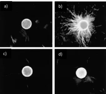
(32) a). b). c). d). Figure 1.16 - Actin comets on beads. a) Electron microscopy images of an actin comet assembled on a bead coated with ActA and placed in a Xenopus egg extract. b-d) Zoom on the boxed regions in a). Tarrows point the Y shaped junctions present in the comet. This dendritic network is similar to the one observed at the leading edge of migrating cells. Scale 1 µm. From (Cameron et al., 2001).. 1.5.3 Symmetry breaking and movement generation In order to produce actin comets from the surface of a bead that has been uniformly coated with activator, a symmetry breaking process has to occur. The steps are as follows: 1) Actin filaments polymerize uniformly from a bead surface coated with polymerization activator, forming an entangled network. Nucleation takes place only at the surface as the activator is present there, and barbed ends are rapidly capped, so growth of new actin is confined to the bead surface. As actin polymerizes, the old actin network is pushed away from the bead surface by the formation of the new network. 2) This stretches the old actin network and stress builds up (van der Gucht et al., 2005). This produces a break in the actin network, and it relaxes away from the site of rupture, giving rise to a comet (Figure 1.17) (van der Gucht et al., 2005). Many studies on actin based motility and actin network assembly have been conducted using the bead system. This system allows for the modulation of actin-binding proteins in the motility mix and on the bead surface, and for the quantification of the effect of these changes 27.
(33) on actin network growth, architecture and mechanics, and/or the effects on comet formation and bead speed.. Rupture of the gel. Figure 1.17 - Scheme illustrating steps of growth of the network on the surface of a bead leading to the symmetry breaking event and the formation of an actin comet that propels the bead forward. From (Plastino and Sykes, 2005).. 1.5.4 Diversity of biomimetic systems Beads have been useful, but during the past two decades, biomimetic systems to study actin polymerization have diversified in order to answer different questions (Figure 1.18). Glass rods coated with polymerization activators, and more recently, micro-patterning of activators on surfaces have been used to more closely mimic the quasi-2D lamellipodium (Achard et al., 2010; Boujemaa-Paterski et al., 2017; Carlier et al., 2003). Oil droplets coated with activators have been used to study mechanical deformation produced by the actin network (Boukellal et al., 2004; Trichet et al., 2007) . Liposomes coated with activators are also used to study mechanical parameters like forces exerted by an actin network on the plasma membrane and acto-myosin tension (Caorsi et al., 2016; Simon et al., 2018). Liposomes or water-in-oil emulsions encapsulating motility mixes and activators are also used to study actin network properties under geometrical and confinement conditions more like the cell (Abu Shah and Keren, 2014; Dürre et al., 2018; Pontani et al., 2009). As mentioned above, micro-patterning of activators has found its way into the field, and is being used to print actin polymerization in different motifs to see how geometry controls actin network organization, actin-binding protein activity and myosin motor function (Boujemaa-Paterski et al., 2017; Reymann et al., 2012; Reymann et al., 2010).. 28.
(34) a). c). b). Figure 1.18 - Diversity of biomimetic systems to study actin polymerization a) A glass rod (30 µm diameter) coated with an activator of actin polymerization. The flat, broad actin network mimics a lamellipodium. Phase contrast microscopy. Scale far 10 µm. From (Carlier et al., 2003). b) Oil droplet coated with a polymerization activator, placed in HeLa cell extract. The droplet deforms due to the stress generated by actin growth. Fluorescent microscopy; actin is fluorescently labelled. Scale bar 4 µm. From (Boukellal et al., 2004). c) Activator micropatterned in a ring shape in order to create bundles of actin. Fluorescent microscopy; actin is fluorescently labeled. Scale 10 µm. From (Reymann et al., 2010).. These different biomimetic systems are presented here only to provide a context for the bead system, and to show the activity in the field. In fact, for the experimental in vitro work that will be presented in Chapter 4 and 5 of this manuscript, the original bead system was used as it is still the simplest way to assess certain properties, including network polarity and actinbased motility.. 29.
(35) 30.
(36) Chapter 2: Ena/VASP Proteins 2.1 Ena/VASP proteins in general Ena/Vasodilator-stimulated phosphoprotein (Ena/VASP) proteins are actin-binding proteins that were one of the main subjects of study during my PhD, so I will describe them in detail in this chapter. Ena/VASP proteins have been variously attributed to have nucleation activity, the capacity to compete with capping protein for barbed ends (called anti-capping) and barbed end elongation enhancement activity (Trichet et al., 2008) (Krause and Gautreau, 2014). These activities will be explained more fully at the end of this chapter, along with a description of the controversies surrounding the mechanisms of Ena/VASP protein action. The first member of this family to be discovered was Drosophila Enabled (Ena), the gene for which was discovered as a dominant suppressor of lethal mutations in the tyrosine kinase gene abl, involved in axon guidance (Gertler et al., 1990). Based on this sequence, mammalian equivalents were identified, Mammalian Ena (Mena), Enabled/vasodilator-stimulated phosphoprotein-like protein (Evl) and Vasodilator-stimulated phosphoprotein (VASP), and shown to have roles in actin filament assembly (Gertler et al., 1996). VASP had been previously identified as a substrate of cyclic nucleotide-dependent kinase cAMP and cGMP in platelets (Haffner et al., 1995; Halbrügge and Walter, 1989). Ena/VASP proteins are highly conserved through evolution: in C. elegans the equivalent of Ena/VASP is UNC-34, and DdVASP in Dictyostelium (Han et al., 2002; Withee et al., 2004; Yu et al., 2002).. Figure 2.1 – Intracellular distribution of Ena/VASP proteins in a moving cell. VASP is mainly present in the front of the moving cell. VASP is fluorescently labelled in green, and actin in red. From (Bear and Gertler, 2009).. 31.
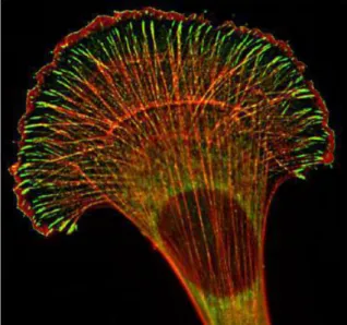
(37) 2.3 Role of Ena/VASP proteins in cells and in vivo 2.3.1 In lamellipodia and cell motility Ena/VASP proteins are found at the leading edge of lamellipodia, at the tips of filopodia, at cell-cell contacts, in cell-substrate adhesions, and in actin stress fibers (Gertler et al., 1996; Lanier et al., 1999; Reinhard et al., 1992; Rottner et al., 1999) (Figure 2.1). In lamellipodia-based cell motility, the local level of Ena/VASP recruitment at the membrane is proportional to transient protrusion rate of that portion of membrane (Rottner et al., 1999). When Ena/VASP proteins are artificially enriched at the front of a moving cell, a network of long unbranched actin filaments form under the membrane (Figure 2.2), and although these structures protrude rapidly, they are not persistent (Bear et al., 2002; Loureiro et al., 2002), and thus increased Ena/VASP sometimes has the effect of reducing overall cell motility (Bear et al., 2000). On the other hand, when Ena/VASP is reduced at the leading edge of the cell, lamellipodia protrude more slowly than wild type, and the actin network is composed of short highly-branched filaments (Bear et al., 2002; Loureiro et al., 2002). Similarly mislocalization of Ena/VASP protein in fish keratinocytes induces altered cell shape and less efficient migration (Lacayo et al., 2007). Ena/VASP reduced at the membrane a). Wild type cells b). Ena/VASP enriched at the membrane c). Figure 2.2 - Electron microscopy image of the actin network at the leading edge of migrating fibroblast cells. Accumulation of Ena/VASP at the leading edge using artificial targeting (c) results in an actin network with longer, less branched filaments than wild type (b). Reduction of the amount of Ena/VASP at the membrane produces a network of shorter, more highly branched filaments than wild type (a). Scale bar 100 nm. From (Bear et al., 2002).. In vivo in Drosophila oogenesis, border cells migrate to the posterior part of the egg chamber, and Ena mutation in these cells significantly reduces their migration speed (Gates et al., 2009). In the Drosophila embryo, Ena overexpression induces an increase in the rate of haemocyte migration, while Ena depletion decreases the rate of cell migration (Tucker et al., 2011). Deletion of C. elegans VASP, UNC-34, decreases the migration speed of leader cells during ventral enclosure, a WAVE-Arp2/3 complex dependent, lamellipodia-driven event (Havrylenko et al., 2014). T-cell movement through endothelial cell layers during extravasation in mice is also reduced by Ena/VASP protein deletion (Estin et al., 2017). 32.
(38) Listeria bacteria hijack Ena/VASP proteins of host cells to increase bacterial motility (Chakraborty et al., 1995; Geese et al., 2002; Skoble et al., 2001; Smith et al., 1996) . Likewise in the bead/comet system, when Ena/VASP proteins are recruited to the bead surface, they increase speed of movement (Castellano et al., 2001; Havrylenko et al., 2015; Plastino et al., 2004b; Samarin et al., 2003). Altogether, studies on cells in culture and in vivo suggest that Ena/VASP proteins promote cell migration, and this is confirmed in biomimetic systems. 2.3.2 In filopodia Ena/VASP proteins also play a role in the dynamics of filopodia. Ena/VASP deficient neurons have reduced filopodia formation in their growth cones (Bear et al., 2002; Lebrand et al., 2004), and Ena/VASP knockout completely suppresses filopodia formation in capping protein-deficient mouse melanoma cells and in Dictyostelium (Han et al., 2002; Mejillano et al., 2004; Schirenbeck et al., 2006) (Figure 2.3).. Ena/VASP knock-out a). Ena/VASP knock-out Capping protein knock-out b). Reintroduced GFP-Mena. VASP VASP knock-out GFP-VASP knock-out d). e). c). Figure 2.3- Role of Ena/VASP in filopodia dynamics. (a) Ena/VASP-deficient mouse melanoma cells, (b) the same cells lacking capping protein, and (c) with reintroduced GFP-Mena. Cells lacking Ena/VASP protein do not form filopodia in absence of capping proteins. Scale bar 10µm. From (Mejillano et al., 2004). (d) Dictyostelium knocked out VASP do not form filopodia. (e) Reintroducing GFP-VASP in this background induces filopodia formation. Scale bar 5µm. From (Schirenbeck et al., 2006).. 2.3.3 In cell-substrate adhesions and stress fibers Ena/VASP proteins play an important role in stress fibers and focal adhesions. Upon mechanical stress, VASP relocalizes from focal adhesions to stress fibers, and helps in their repair, thus restoring the structural integrity and the contractility of the stress fiber (Burridge and Guilluy, 2016; Smith et al., 2010; Yoshigi et al., 2005). VASP is also involved in remodeling stress fibers through cooperation with focal adhesion protein zyxin (Hoffman et al., 2006). Furthermore, Ena/VASP proteins are an integral component of focal adhesions (Kanchanawong et al., 2010). 33.
(39) 2.3.4 In cancer In the past two decades, several studies have emerged indicating a relation between Ena/VASP protein and cancer progression. Phosphorylation of Ena/VASP, which reduces its interaction with actin, inhibits the formation of invadopodia, essential structures for cancer cell invasion and metastasis, and thus reduces colon cancer cell circulation (Zuzga et al., 2012). Fibroblasts overexpressing Ena/VASP lose contact inhibition and are considered as potential tumorigenic cells (Liu et al., 1999), and Ena/VASP overexpression in lung adenocarcinoma cells is correlated with the progress of the tumor (Dertsiz et al., 2005). Mena is overexpressed in breast cancer cell lines, and in particular one splice form of Mena is associated with increased invasion and metastasis (Philippar et al., 2008; Roussos et al., 2010). In addition to its role in invasion, Ena/VASP plays a role in the vascularization of tumors: melanoma cancer cells transplanted into Ena/VASP deficient mice do not develop well, and tumors are smaller and significantly less vascularized (Kim et al., 2011). On a global scale, Ena/VASP proteins seem to be involved at multiple levels in the coordination of the development of metastasis.. 2.2 Ena/VASP domains and their functions All Ena/VASP family members share a conserved domain structure: an amino-terminal Ena/VASP homology 1 domain (EVH1), a central proline rich region, and a carboxy-terminal Ena/VASP homology 2 (EVH2) domain, encompassing G- and F-actin binding sites and a coiledcoil motif. Ena/VASP protein interacts with many partners and performs various functions via its different domains (Figure 2.4), as described in the following sections.. Regulatory Figure 2.4 – Schematic representation of Ena/VASP, showing its domains and their interacting partners. VASP binds to both monomeric and filamentous actin. The polyproline rich domain of VASP binds profilin.. 2.2.1 EVH1 domain The N-terminal EVH1 domain of Ena/VASP proteins is part of the pleckstrin homology (PH) domain superfamily, but unlike other members of this family, it does not bind phospholipid phosphatidyl inositol-(4,5)-bisphosphate (PIP2) (Prehoda et al., 1999; Volkman et al., 2002). The EVH1 domain binds to peptide ligands containing special poly-proline sequences with FPPPP-type sequences, such as those found in the Listeria ActA protein and in the proline34.
Figure
+7

Documents relatifs