RESEARCH OUTPUTS / RÉSULTATS DE RECHERCHE
Author(s) - Auteur(s) :
Publication date - Date de publication :
Permanent link - Permalien :
Rights / License - Licence de droit d’auteur :
Institutional Repository - Research Portal
Dépôt Institutionnel - Portail de la Recherche
researchportal.unamur.be
University of Namur
Assessment of the performances of AcuStar HIT and the combination with
heparin-induced multiple electrode aggregometry
Minet, V.; Bailly, N.; Douxfils, J.; Osselaer, J. C.; Laloy, J.; Chatelain, C.; Elalamy, I.;
Chatelain, B.; Dogné, J. M.; Mullier, F.
Published in:
Thrombosis Research
DOI:
10.1016/j.thromres.2013.06.004
Publication date:
2013
Document Version
Publisher's PDF, also known as Version of record
Link to publication
Citation for pulished version (HARVARD):
Minet, V, Bailly, N, Douxfils, J, Osselaer, JC, Laloy, J, Chatelain, C, Elalamy, I, Chatelain, B, Dogné, JM &
Mullier, F 2013, 'Assessment of the performances of AcuStar HIT and the combination with heparin-induced
multiple electrode aggregometry: A retrospective study', Thrombosis Research, vol. 132, no. 3, pp. 352-359.
https://doi.org/10.1016/j.thromres.2013.06.004
General rights
Copyright and moral rights for the publications made accessible in the public portal are retained by the authors and/or other copyright owners and it is a condition of accessing publications that users recognise and abide by the legal requirements associated with these rights. • Users may download and print one copy of any publication from the public portal for the purpose of private study or research. • You may not further distribute the material or use it for any profit-making activity or commercial gain
• You may freely distribute the URL identifying the publication in the public portal ?
Take down policy
If you believe that this document breaches copyright please contact us providing details, and we will remove access to the work immediately and investigate your claim.
Regular Article
Assessment of the performances of AcuStar HIT and the combination
with heparin-induced multiple electrode aggregometry:
A retrospective study
V. Minet
a,⁎
, N. Bailly
b, J. Doux
fils
a, J.C. Osselaer
c, J. Laloy
a, C. Chatelain
d, I. Elalamy
e, B. Chatelain
b,c,
J.M. Dogné
a, F. Mullier
a,ba
Department of Pharmacy, Namur Research Institute for LIfe Sciences (NARILIS), Namur Thrombosis and Hemostasis Center (NTHC), University of Namur, Namur, Belgium
b
Hematology Laboratory, Namur Research Institute for LIfe Sciences (NARILIS), CHU UCL Mont-Godinne, Dinant, Université Catholique de Louvain (UCL), Yvoir, Belgium
c
Blood Transfusion Center, CHU UCL Mont-Godinne, Dinant, Université Catholique de Louvain UCL, Yvoir, Belgium
dHematology Department, Namur Research Institute for LIfe Sciences (NARILIS), CHU UCL Mont-Godinne, Dinant, Université Catholique de Louvain (UCL), Yvoir, Belgium e
Hematology Department, Hôpital Tenon, Paris, France
a b s t r a c t
a r t i c l e i n f o
Article history:
Received 18 January 2013 Received in revised form 3 June 2013 Accepted 4 June 2013 Available online xxxx Keywords: Immunoassay Heparin-induced thrombocytopenia Platelets
Background: Early diagnosis of immune heparin-induced thrombocytopenia (HIT) is challenging. HemosIL® AcuStar HIT and heparin-induced multiple electrode aggregometry (HIMEA) were recently proposed as rapid diagnostic methods.
Objectives: We conducted a study to assess performances of AcuStar HIT-IgG (PF4-H) and AcuStar HIT-Ab (PF4-H). The secondary objective was to compare the performances of the combination of Acustar HIT and HIMEA with standardised clinical diagnosis.
Methods: Sera of 104 suspected HIT patients were retrospectively tested with AcuStar HIT. HIMEA was performed on available sera (n = 81). The clinical diagnosis was established by analysing in a standardized manner the patient’s medical records. These tests were also compared with PF4-Enhanced®, LTA, and SRA in subsets of patients. Thresholds were determined using ROC curve analysis with clinical outcome as reference.
Results: Using the recommended thresholds (1.00 AU), the negative predictive value (NPV) of HIT-IgG and HIT-Ab were 100.0% (95% CI: 95.9%-100.0% and 95.7%-100.0%). The positive predictive value (PPV) were 64.3% (95% CI: 35.1%-87.2.2%) and 45.0% (95% CI: 23.2%-68.6%), respectively. Using our thresholds (HIT-IgG: 2.89 AU, HIT-Ab: 9.41 AU), NPV of HIT-IgG and HIT-Ab were 100.0% (95% CI: 96.0%-100.0% and 96.1%-100.0%). PPV were 75.0% (95% CI: 42.7%-94.5%) and 81.8% (95% CI: 48.3%-97.7%), respectively. Of the 79 patients with a medium-high pretest probability score, 67 were negative using HIT-IgG (PF4-H) test at our thresholds. HIMEA was performed on HIT-IgG positive patients. Using this combination, only one patient on 79 was incorrectly diagnosed.
Conclusion: Acustar HIT showed good performances to exclude the diagnosis of HIT. Combination with HIMEA improves PPV.
© 2013 Elsevier Ltd. All rights reserved.
Introduction
Immune heparin-induced thrombocytopenia (HIT) is a severe immune-mediated adverse effect of heparin treatment [1,2]. Early diagnosis is essential and both missed diagnosis and erroneous diagnosis should be avoided[3]. A wrong diagnosis may lead to a discontinuation of the treatment with heparin and substitution by an anticoagulant which is actually not required. This substitution is expensive and could be associated with an increased risk of bleeding (10-20% of major haemorrhage) without effective antidote[3,4]. While
early diagnosis of HIT is essential to improve clinical management of patients, it remains challenging, especially in critically ill patients, in intensive care[5]. The current diagnostic relies on the use of a clinical scoring algorithm (“4T’s rule”) together with immunological and platelet activation assays [1,2,6]. This combination improves HIT diagnosis compared to its components considered independently[7]. However, the scoring system has to be used carefully and may require some exten-sive documentation of the patient medical history [8]. Non-specific enzyme-immunoassay (EIA) (IgG/A/M) and IgG-specific EIA can rule out the occurrence of HIT (their negative predictive value (NPV) is equal to 99%[2]). However, they are still lacking specificity (50-90%) and need a careful standardization of the optical density (OD) ranges[9]. HemosIL® AcuStar HIT and heparin-induced multiple electrode aggregometry (HIMEA) were recently proposed as new rapid methods for the diagnosis of HIT.
Thrombosis Research xxx (2013) xxx–xxx
⁎ Corresponding author at: University of Namur, Department of Pharmacy, Rue de Bruxelles, 61, B-5000 Namur, Belgium. Tel.: +32 81 724292; fax: +32 81 724299.
E-mail address:[email protected](V. Minet).
0049-3848/$– see front matter © 2013 Elsevier Ltd. All rights reserved.
http://dx.doi.org/10.1016/j.thromres.2013.06.004
Contents lists available atSciVerse ScienceDirect
Thrombosis Research
The HemosIL® AcuStar HIT-IgG (PF4-H) and AcuStar HIT-Ab (PF4-H) tests were developed by Legnani et al. as rapid, automated semi-quantitative immunoassays with very high sensitivity (100%) that can reliably be used to rule out the occurrence of a HIT, and a spec-ificity of 81-96%[10]. In addition, Elalamy et al. showed that HIMEA with the Multiplate® analyzer has a similar sensitivity for detection of the presence of HIT platelet activating antibodies as the conventional light transmission aggregometry (LTA) performed in citrated platelet-rich plasma (PRP)[11]. Consequently, HIMEA was proposed as a new diagnostic tool that simplifies the process of HIT diagnosis compared with LTA[11,12].
We assessed the performances of AcuStar HIT-IgG (PF4-H), AcuStar HIT-Ab (PF4-H), and HIMEA and compared them with the ones of PF4-Enhanced®, Light Transmission Aggregometry (LTA). These performances were assessed against the14C-Serotonin Release
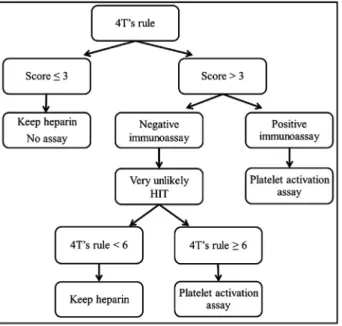
Assay (SRA), and a clinical diagnosis. Subsequently, we retrospectively assessed this combination in the HIT diagnosis strategy proposed by Greinacher et al. (Fig. 1)[2].
Materials and Methods Healthy subjects
The platelets were obtained from healthy volunteers O Rh +/-blood group donors who did not take any medicine which could potentially affect their platelet function for 10 days before the blood sampling. Platelet donors were selected on the basis of a good reactivity to HIT antibodies. No donor had to be excluded of the study because they all presented normal aggregation with the following reagents: adenosine diphosphate (ADP), collagen, arachidonic acid (AA) or thrombin receptor activating peptide (TRAP).
Patients
After approval by the local ethics committee, 106 patients with suspected diagnosis of HIT between October 2006 and July 2011 at CHU UCL Mont-Godinne-Dinant (Université Catholique de Louvain, Yvoir, Belgium) were included in this study (these include 104 inpa-tients, 2 outpainpa-tients, among them 94 patients underwent surgery). The primary diagnosis was based on a rapidly decreasing platelet count occur-ring in hospitalised patients and on the result of the“4T’s rule” (which
takes into consideration four criteria: a Thrombocytopenia, and its Timing, the occurrence of a Thrombosis and the exclusion of other causes of thrombocytopenia). Blood samples of these patients were sent to the He-matology Laboratory for laboratory assays. Samples for the study were collected independently of patient underlying disease. Samples were ei-ther tested as stored samples (samples before January 2008) or in real time (from January 2008). Routine laboratory diagnosis was based on PF4-Enhanced® (GTI Diagnostics, Waukesha, WI, USA) and LTA.
Criteria from the ACCP (American College of Chest Physicians) guidelines were used to make the clinical diagnosis of HIT: (1) Thrombocytopenia, defined as at least a 30% decline in the platelet count, with a platelet count increase after heparin cessation; (2) Timing of platelet count fall after the initiation of heparin occurring between 4 and 14 days, or occurring within 24 to 48 hours (in case of prior heparin exposure within 30 days); and (3) lack of other, predominant causes of thrombocytopenia[13,14]. Other causes of thrombocytopenia analysed in this study were: scalable neoplasia, lower extremity arterial disease, current pregnancy or postpartum, autoimmune disease, sepsis, disseminated intravascular coagulation, intra-aortic balloon pump counterpulsation, multitransfusion, multi-trauma, shock syndrome, in-flammatory syndrome and drug-induced thrombocytopenia (quinolone, β-lactam, vancomycin, teicoplanin, rifampicin, isoniazid, amphotericin, fluconazole, chemotherapy, anti-GPIIb IIIa; furosemide and proton pump inhibitor).
All those 3 clinical criteria have to be fulfilled for the confirmation of clinical HIT diagnosis. The medical history of each patient was extensively analysed. The following information was taken into consideration: pa-tient’s medical history, types (fractionated vs. unfractionated) and doses of heparin administered, thrombotic complications, alternative diagnoses, therapeutic attitude, clinical and platelet count evolution, co-suspected medications, and physician’s diagnoses[14]. These clinical criteria were independently assessed by two investigators (VM and FM), not aware of the results of the laboratory assays. Clinical diagnoses made by these 2 local investigators were 100% concordant.
Blood sampling and handling
Blood was collected with a 20 gauge needle via atraumatic antecubital venipuncture into polyethylene terephthalate tubes Venosafe® (Terumo Europe, Leuven, Belgium) containing buffered sodium citrate (109 mM, nine parts of blood to one part sodium citrate solution) or 25μg/mL recombinant hirudin (Verum Diagnostica GmbH, Munich, Germany, 1:10 v/v). The blood group was chosen as O. A discard tube was used to avoid thromboplastin contamination. Sera were used for all assays described in this study except for LTA.
When serum was used, we waited 30 min before performing cen-trifugation and 15 min after cencen-trifugation to reduce the interference of thrombin. We also controlled the absence of spontaneous aggrega-tion by mixing patients’ sera with whole blood from healthy volunteers without any agonist. For LTA, PRP was obtained from the supernatant fraction after centrifugation at 200 g during 10 min at room tempera-ture. The remaining blood was centrifuged at 2,200 g during 15 min at room temperature and the supernatant gave the platelet-poor plasma (PPP).
Laboratory testing Immunoassays
The presence of anti-PF4-heparin antibodies was investigated in 106 serum samples by using PF4-Enhanced® (GTI Diagnostics, Waukesha, WI, USA), AcuStar HIT-IgG (PF4-H) and AcuStar HIT-Ab (PF4-H) (Instrumentation Laboratory, Bedford, MA, USA)[10]according to the manufacturers’s recommendations. PF4-Enhanced® was performed in real time on fresh sera whereas AcuStar HIT-IgG (PF4-H) and AcuStar HIT-Ab (PF4-H) were performed afterwards on frozen sera (-80 °C). PF4-Enhanced® is an enzyme-linked immunosorbent assay (ELISA).
Fig. 1. Diagnostic algorithm for immune HIT.
The spectrophotometry threshold used in our study is the one recommended by the manufacturer (0.400 OD).
The HemosIL® AcuStar HIT-IgG (PF4-H) and HIT-Ab (PF4-H) are two chemiluminescent two-step immunoassays consisting of magnetic particles coated with PF4 complexed to polyvinyl sulfonate which bind the anti-PF4/H antibodies from the sample. This assay has been described by Legnani et al. [10]. The threshold used in our study (recommended by the manufacturer) is 1.00 AU.
Platelet reactivity and heparin-induced LTA
The platelet reactivity of the healthy donors was assessed by LTA. Aggregation tests were performed according to the Born's turbidi-metric method with a PACKS-4® aggregometer (Helena, Beaumont, Texas, USA)[15]. This assay has been described by Mullier et al.[16]. Platelet reactivity was performed by using three different agonists: ADP (final concentration: 5 μM), collagen (type 1, final concentration: from 40 and 190μg/mL) (Biodata Corporation, Horsham, PA, USA) and AA (final concentration: 0.5 mg/mL) (Biodata Corporation, Horsham, PA, USA). Heparin-induced LTA was performed with a range of un-fractionated heparin (UFH)final concentrations (0.5 IU/mL, 1 IU/mL, 10 IU/mL and 500 IU/mL) using unfractionated sodium heparin 5000 IU/mL (Heparin Leo®, Leo Pharma, Lier, Belgium) diluted with physiological saline (B Braun Medical, Diegem, Belgium).
The endpoint used for our study was the percentage of aggregation. The result was considered positive when platelet aggregation occurred in the presence of low UFH concentration (percentage of aggregation higher than 20% at 0.5 IU/mL or 1 IU/mL) but was partially inhibited (percentage of aggregation lower than 20%) by a high concentration of UFH (500 IU/mL).
Heparin-induced multiple electrode aggregometry (HIMEA)
HIMEA was performed retrospectively on all available frozen (-80 °C for maximum 18 months) sera (n = 81). Multiple electrode aggregometry (MEA) is a method for the assessment of platelet function in whole blood (Multiplate® analyzer; Dynabyte, Munich, Germany)
[17].
MEA is based on the principle of impedance aggregometry. Whole blood samples from three healthy volunteers O Rh+/- blood group were collected by venipuncture in hirudin tube.
The platelet reactivity of the healthy donors was assessed by Multiplate® using four different agonists: ADP (final concentration: 6.5μM), collagen (type 1, final concentration: 3.2 μg/mL) (Dynabyte, Munich, Germany), AA (final concentration: 0.5 mM) (Dynabyte, Munich, Germany) and TRAP-6 (final concentration: 32 μM) (Dynabyte, Munich, Germany). Our procedure is similar from the one described by Morel-Kopp et al.[12]but differs from volumes used. In our protocol, we mixed 300μL of saline with 150 μL of whole blood from healthy donors, 150μL of serum from HIT patients and 50 μL of UFH solution (Heparin Leo®). We used threefinal UFH concentrations of 1 IU/mL, 10 IU/mL and 385 IU/mL as well as with the addition of normal pooled plasma instead of serum of the patient. Heparin concentrations were chosen based on preliminary studies comparing 0.5 IU/mL and 1 IU/mL UFH (n = 32). We used 385 IU/mL UFH instead of 100 IU/mL UFH, often used in the literature, to avoid an additional dilution of the stock solution (5000 IU/mL). The HIMEA was expressed using the area under the aggregation curve (AUC). The result was considered to be positive when the platelet aggregation occurred in the presence of low heparin concentration (1 IU/mL UFH or 10 IU/mL UFH) but was partially inhibited (reduction > 80%) by a high concentration of heparin (385 IU/mL UFH). The negative control should beb100 AU.
14
C-serotonin release assay (SRA)
The SRA was performed retrospectively on frozen (-80 °C for maximum 18 months) sera.
The SRA was carried out according to previously published protocols in all available samples (n = 45)[18]. Two out of these 45 patients were excluded because of lack of clinical data.
Data analysis
Statistical analysis was performed using Medcalc software (version 10.4.8) (Gent, Belgium).
The AUC was computed with the corresponding 95% confidence intervals (95% CI). The sensitivity and specificity of HemosIL® AcuStar HIT-IgG (PF4-H) and HIT-Ab (PF4-H) were assessed using both SRA and clinical diagnosis as gold standards. We also assessed the performances of the methods using ROC curves.
Results
Of the 106 patients suspected of having developed HIT, 104 completed the clinical follow-up. Seventy three men and thirty one women aged from 26 to 101 years (median age 70 years), were included in our study. Nine patients (9/79 i.e. 11.4%) were considered to have developed a HIT based on our clinical diagnosis. Of these, 5 patients experienced a thrombosis (2 venous thrombosis, 2 arterial thrombosis, and 1 patient had both a venous and arterial thrombosis). One HIT patient died during his hospitalisation.
Determination of optimal thresholds from ROC curves (using clinical diagnosis as gold standard) and assessment of diagnostic performances
Compared to clinical diagnosis, we determined the optimal thresholds of AcuStar HIT-IgG (PF4-H), AcuStar HIT-Ab (PF4-H), PF4-Enhanced®, HIMEA at 1 IU/mL UFH and HIMEA at 10 IU/mL UFH using ROC curve analyses. These thresholds were 2.89 AU, 9.41 AU, 1.32 OD, 276 AU and 183 AU respectively. At the optimal cut-off provided by ROC Curves, we noted that the sensitivity of AcuStar HIT-IgG (PF4-H) and HIT-Ab (PF4-H) is respectively 100.0% (95% CI: 59.0%-100.0%) and 100.0% (95% CI: 66.4%-100.0%); a high sensitivity is a priority for screening assays. When clinical diagnosis was used as the reference, NPV and sensitivity were of 100.0% except for HIMEA at 10 IU/mL UFH for which the NPV and sensitivity were respectively 97.0% (95% CI: 91.4%-99.6%) and 75.0% (95% CI: 34.9%-96.8%). The specificity reached 96.8% (95% CI: 91.0%-99.3%), 97.9% (95% CI: 92.6%-99.7%), 96.8% (95% CI: 89.6%-98.8%), 97.3% (95% CI: 90.5%-99.7%), and 89.0% (95% CI: 79.5%-95.1%), respective-ly. The positive predictive values (PPV) were 75.0% (95% CI: 42.7%-94.5%), 81.8% (95% CI: 48.3%-97.7%), 75.0% (95% CI: 42.7%-94.5%), 80.0% (95% CI: 44.2%-96.3%) and 42.9% (95% CI: 17.1%-65.5%) (Table 1). Sensitivity, spec-ificity and PPV were lower with the HIMEA at 10 IU/mL UFH than with HIMEA at 1 IU/mL UFH, consequently we used the results of HIMEA at 1 IU/mL UFH. The recommended threshold for AcuStar HIT-IgG (PF4-H), AcuStar HIT-Ab (PF4-H) (i.e. 1.00 AU) and PF4-Enhanced® (i.e. 0.400 OD) yielded lower specificities of 94.7% (95% CI: 88.1%-98.3%), 88.4% (95% CI: 80.2%-94.1%), and 85.3% (95% CI: 76.5%-91.7%). The PPV were 64.3% (95% CI: 35.1%-87.2%), 45.0% (95% CI: 23.2%-68.6%) and 39.1% (95% CI: 19.8%-61.6%), respectively. The disagreements between the different tests are shown inTable 1.
Moreover, a preliminary study did not show any difference between 0.5 IU/mL and 1 IU/mL UFH (n = 32, p = 0.22).
Performances of the combination of AcuStar HIT-IgG (PF4-H) and HIMEA according the diagnostic algorithm proposed by Greinacher et al.[2]and using the optimised thresholds
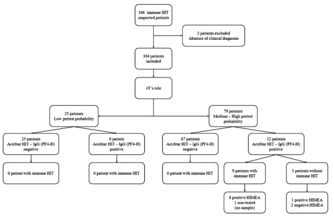
Twentyfive patients (25/104 i.e. 24%) included in the study were classified as low according to the 4T’s pre-test probability (PTP) rule, 76 as medium (73%), and 3 as high (3%).
Of the seventy nine patients with a medium-high pretest prob-ability score, AcuStar HIT-IgG (PF4-H) at our threshold excluded
the presence of HIT in 67 of these patients (Fig. 2). NPV of HIT-IgG (PF4-H) and HIT-Ab (PF4-H) assays for patients with a medium-high pretest probability, were 100.0% (95% CI: 93.2%-100.0%)
and 100.0% (95% CI: 92.7%-100%). The PPV of HIT-IgG (PF4-H) assay and HIT-Ab (PF4-H) were 75.0% (95% CI: 42.8%-93.3%) and 52.9 (95% CI: 28.5%-76.1%) (Table 1).
Table 1
Comparison of PF4-Enhanced®, HemosIL® AcuStar HIT-IgG (PF4-H) and HIT-Ab (PF4-H), LTA, HIMEA and SRA with the clinical diagnosis. Only patients with available clinical diagnosis were considered (n = 104).
Clinical diagnosis Sensitivity, % (95%CI) Specificity, % (95%CI) PPV, % (95%CI) NPV, % (95%CI) Negative Positive PF4-Enhanced (n = 104) Threshold : >0.40 OD Negative 81 0 100.0 (66.4-100.0) 85.3 (76.5-91.7) 39.1 (19.8-61.6) 100.0 (95.5-100.0) Positive 14 9 PF4-Enhanced (n = 104) Threshold : >1.32 OD Negative 92 0 100.0 (66.4-100.0) 96.8 (89.6-98.8) 75.0 (42.7-94.5) 100.0 (96.0-100.0) Positive 3 9 AcuStar IgG (n = 104) Threshold : >1.00 AU Negative 90 0 100.0 (66.4-100.0) 94.7 (88.1-98.3) 64.3 (35.1-87.2) 100.0 (95.9-100.0) Positive 5 9 AcuStar IgG (n = 104) Threshold : >2.89 AU Negative 92 0 100.0 (59.0-100.0) 96.8 (91.0-99.3) 75.0 (42.7-94.5) 100.0 (96.0-100.0) Positive 3 9 AcuStar IgG (n = 79) Threshold : >1.00 AU Negative 65 0 100 (62.9-100.0) 92.9 (83.4-97.3) 64.3 (35.6-86.0) 100.0 (93.0-100.0) Positive 5 9 AcuStar IgG (n = 79) Threshold : >2.89 AU Negative 67 0 100.0 (62.9-100.0) 95.7 (87.2-98.9) 75.0 (42.8-93.3) 100.0 (93.2-100.0) Positive 3 9 AcuStar Ab (n = 104) Threshold : >1.00 AU Negative 84 0 100.0 (66.4-100.0) 88.4 (80.2-94.1) 45.0 (23.2-68.6) 100.0 (95.7-100.0) Positive 11 9 AcuStar Ab (n = 104) Threshold : >9.41 AU Negative 93 0 100.0 (66.4-100.0) 97.9 (92.6-99.7) 81.8 (48.3-97.7) 100.0 (96.1-100.0) Positive 2 9 AcuStar Ab (n = 79) Threshold : >1.00 AU Negative 60 0 100 (62.9-100.0) 85.7 (74.8-92.6) 47.4 (25.2-70.5) 100.0 (92.5-100.0) Positive 10 9 AcuStar Ab (n = 79) Threshold : >9.41 AU Negative 62 0 100.0 (62.9-100.0) 88.6 (78.2-94.6) 52.9 (28.5-76.1) 100.0 (92.7-100.0) Positive 8 9 HIMEA (n = 81) Threshold : >276 AU (1 IU/mL UFH) Negative 71 0 100.0 (63.1-100.0) 97.3 (90.5-99.7) 80.0 (44.2-96.3) 100.0 (96.1-100.0) Positive 2 8 HIMEA (n = 81) Threshold : >183 AU (10 IU/mL UFH) Negative 65 2 75.0 (34.9-96.8) 89.0 (79.5-95.1) 42.9 (17.1-65.5) 97.0 (91.4-99.6) Positive 8 6 LTA (n = 46) Threshold: >20% (0.5 and 1 IU/mL UFH)
Negative 38 0 100.0 (39.8-100.0) 90.5 (77.9-97.4) 50.0 (26.1-74.0) 100.0 (95.8-100.0) Positive 4 4 SRA (n = 43) Threshold: >20% (0.1 and 0.3 IU/mL UFH)
Negative 36 1 80.0 (28.4-99.5) 94.7 (82.3-99.4) 66.7 (28.2-85.3) 97.3 (92.6-99.8) Positive 2 4
AcuStar IgG + HIMEA (n = 78) Threshold : >2.89 AU + > 276 AU (1 IU/mL UFH) Negative 69 0 100.0 (59.7-100.0) 98.6 (91.2-99.9) 88.9 (50.7-99.4) 100 (93.4-100.0) Positive 1 8
Fig. 2. Flow chart with results according to the HIT diagnosis strategy proposed by Greinacher et al.[2]. 4 V. Minet et al. / Thrombosis Research xxx (2013) xxx–xxx
Among the 12 positive AcuStar HIT-IgG (PF4-H), 9 patients were clinically confirmed HIT. 11 positive AcuStar HIT-IgG (PF4-H) were tested on HIMEA, 1 patient could not be tested (no sample available). HIMEA at 1 IU/mL UFH diagnosed correctly 10 patients of the 11 posi-tive AcuStar HIT-IgG (PF4-H). HIMEA at 1 IU/mL UFH confirmed the absence of HIT in 2 out of 3 patients not presenting a HIT (Table 1). The third non-HIT patient showed a platelet aggregation at 1 IU/mL UFH slightly above the threshold of 276 AU (306 AU). The sensitivity, specificity, PPV and NPV of this combination using our thresholds, for the 78 patients with a medium-high pretest probability and sample avail-able, were 100.0% (95% CI: 59.7%-100.0%), 98.6% (95% CI: 91.2%-99.9%), 88.9% (95% CI: 50.7%-99.4%) and 100.0% (95% CI: 93.4%-100.0%), respec-tively. Over the 104 patients, one patient was incorrectly diagnosed using our approach.
The individual cases considered to be HIT as well as SRA positive cases considered not to be HIT, are described in details inTable 3. Other analyses
Comparison of AcuStar HIT-IgG (PF4-H), AcuStar HIT-Ab (PF4-H) and PF4-Enhanced®
We have performed a comparison of the ROC curves for AcuStar HIT-IgG (PF4-H), AcuStar HIT-Ab (PF4-H) and PF4-Enhanced® before and after the optimization. Before optimization, the AUC was signi fi-cantly higher for AcuStar IgG (PF4-H) compared to AcuStar HIT-Ab (PF4-H) and PF4-Enhanced® (p = 0.0118 and 0.0017, respectively). The optimization of our thresholds improved significantly the AUC of AcuStar HIT-Ab (PF4-H) and PF4-Enhanced® (p = 0.0017 and 0.0009, respectively) but not the one of AcuStar HIT-IgG (PF4-H) (p = 0.1551). After the optimization, the differences were not statisti-cally significant between the 3 tests (p > 0.05).
Comparison of HIMEA with LTA
HIMEA and LTA were performed on the same samples of 45 patients with a suspected HIT. Compared to the clinical diagnosis, HIMEA at 1 IU/mL UFH, HIMEA at 10 IU/mL UFH and LTA showed, respectively, a sensitivity of 100.0% (95% CI: 63.1%-100.0%), 75.0% (95% CI: 34.9%-96.8%) and 100.0% (95% CI: 39.8%-100.0%) and their specificity were 97.3% (95% CI: 90.5%-99.7%), 89.0% (95% CI: 79.5%-95.1%) and 90.5% (95% CI: 77.9%-97.4%). The comparison of ROC curves did not show any statistically significant difference (p = 0.67).
Comparison with SRA
The SRA is one of the two gold standards (the second is the heparin induced platelet activation (HIPA) for the diagnosis of HIT)[19]. The SRA and the clinical diagnosis provided discordant results in 3 patients (out of 43 patients with completed clinical follow-up) (Table 2). Two of these 3 patients were positive with SRA and but considered not to have developed HIT according to our clinical diagnosis.
Discussion
In this study, we compared the performances of AcuStar HIT-IgG (PF4-H), AcuStar HIT-Ab (PF4-H), new automated semi-quantitative immunoassays, with the ones of PF4-Enhanced® as screening tests for immune heparin-induced thrombocytopenia (HIT). We also devel-oped HIMEA and compared its performances to LTA, as confirmation tests for the diagnosis of HIT. The reference used to confirm diagnosis of HIT was the clinical diagnosis as SRA does not entirely solve the dif fi-cult challenge of appropriate HIT diagnosis[18]. In addition, SRA does not entirely solve the issue of HIT misdiagnosis[14,20]. Our SRA proto-col was different from the one performed by Gruel et al. (different tem-perature and incubation time, different steps of wash and incubation time)[21]. The threshold used by Warkentin et al. (≥50%) differs from the threshold considered in our study (>20%)[22]. Consequently data of SRA in our study cannot be compared to other laboratories, as
previously mentioned by Tardy et al.[20]. Inconsistencies can also be explained by the absence of systematic use of at least two suitable donors in order to control sufficient reactivity of platelets, the absence of standardized quality control and the exclusion of intermediate or non-specific results in studies[20]. The performances of the combina-tion of AcuStar HIT-IgG and HIMEA for rapid diagnosis of HIT were also studied.
We showed that before redetermination of the thresholds, AUC of the ROC curve was significantly higher for AcuStar HIT-IgG (PF4-H) in comparison with AcuStar HIT-Ab (PF4-H) and PF4-Enhanced®. The new thresholds obtained by comparing the performances of these methods to the clinical diagnosis were higher than those recommended by the manufacturer. The thresholds observed are assay-dependent but most important is the choice of the gold standard used to determine these thresholds. The manufacturers provided thresholds following a study on healthy subjects and not on patients suspected of having developed HIT. The optimization of thresholds improves significantly the AUC of AcuStar HIT-Ab (PF4-H) and PF4-Enhanced®. After the optimization, sensitivity and NPV reached 100.0% for all three tests. The specificity reached 96.8% (95% CI: 91.0%-99.3%), 97.9% (95% CI: 92.6%-99.7%), and 96.8% (95% CI: 89.6%-98.8%), respectively. The PPV were 75.0%, 81.8% and 75.0%, respectively (Table 1). No statistically sig-nificant difference was observed between these 3 tests. However, from a practical perspective AcuStar HIT-IgG (PF4-H) and AcuStar HIT-Ab (PF4-H) have a running-time of about 30 min compared to 2-3 hours of ELISA assays (such as PF4-Enhanced®). In addition, this fully auto-mated machine can be run for a single sample testing. Consequently, considering these beneficial aspects, we suggest that this new immuno-logical assay could be widely used to screen patients with a suspected HIT.
In our study, AcuStar HIT-IgG (PF4-H) and AcuStar HIT-Ab (PF4-H) showed the same sensitivity as reported by Legnani et al.[10]. The use of local thresholds markedly improves the specificity of AcuStar HIT-Ab (PF4-H) from 81.2% (95% CI: 71.3%-88.8%) in Legnani study to 97.9% (95% CI: 92.6%-99.7%). Our results confirm that the IgG-specific EIA is more specific than the polyspecific assays[23–25]
in the absence of optimization.
The magnitude of a positive result (in optical density (OD) units) and the inhibition of reactivity at high heparin concentration are the two features of EIA reactivity that predict for presence of HIT antibodies. The probability of anti-PF4/heparin antibodies of being responsible for the low platelet count or new thrombosis increases with the magnitude of the EIA OD result[8,9,22,24,26]. However, the interpretation of ELISA test results according to afixed OD threshold is associated to the prob-lem that OD units are non-standardized units. They might differ consid-erably across photometers according to (i) the wavelength-dependent coefficient of extinction of the materials and fluids, (ii) the concentra-tion of the molecules in thefluid and (iii) the thickness of the assessed fluid layer[9]. Consequently, there is no clear threshold to distinguish between clinically relevant and non-relevant antibodies. For example, the literature reports contradictory results on the association between anti-PF4/heparin antibodies with a 0.40b OD b 1.00 in the PF4/heparin EIA with a positive SRA[22,27,28]. As long as there is no standard for anti-PF4/heparin antibodies, the OD of the ELISA could be used as an estimate for the likelihood of a clinically relevant antibody only.
Finally, the threshold may depend on the population characteris-tics[28]. For example, the 97.5% percentile OD value of sera from patients before cardiac surgery was described as 0.74[28]. This is likely explained by recent exposure to heparin, e.g. during coronary angiogra-phy[9]. In this population, increasing the OD threshold did not improve the usefulness of HIT EIA’s screening[28]. In the present study, we have also developed HIMEA, a technique recently proposed by 2 differ-ent teams for the rapid confirmation of the diagnosis of HIT[11,12]. HIMEA assay is based on the presence of a platelet aggregation at 1 IU/mL UFH or 10 IU/mL UFH with a significant (>80%) reduction at 385 IU/mL UFH. This high concentration leads to a dissociation of the
complex Ig-PF4-heparin and is therefore used to enhance the specificity of all platelet activation tests[2]. HIMEA at 1 IU/mL UFH offers better sensitivity and specificity than HIMEA at 10 IU/mL UFH. We have also compared the performances of HIMEA at 1 IU/mL UFH with the stan-dard LTA, as confirmation tests for the diagnosis of HIT on 45 suspected HIT patients. Both showed sensitivity of 100.0%. LTA performed in this study showed higher sensitivity in comparison with previous reports. However, these results should be confirmed in a larger series of patients. The specificity of HIMEA at 1 IU/mL UFH (i.e. 97.3%) is higher than LTA at 0.5 and 1 IU/mL UFH (i.e. 90.5%) although not significant. Galea et al. have recently shown that HIMEA at 1 IU/mL UFH is more sensitive (81% vs. 76%) and more specific (99% vs. 96%) than LTA, in comparison to SRA[18]. In addition, LTA requires both experience, as well as a specific expertise and is time-consuming (turn-around-time of about 90 min), which limits its routine use[12]. On the contrary, the use of HIMEA which requires whole blood provides results within 45 min and is an accessible assay to a large number of laboratories[11,12]. A limitation for performing of HIMEA is the immediate availability of a healthy compatible blood group donor. Results were also compared with SRA assay[29], on a limited series of patients. SRA also suf-fers from a series of well-known limitations: it is time-consuming,
technically demanding, uses radioactive material, and is not easily available in routine clinical laboratories. Some concerns about inter-laboratory variability and lack of standardization with SRA have also been described [30,31]. In addition, previous studies have already shown undetected cases by SRA[29,30,32]due to the fact that HIT anti-bodies to PF4 are heterogeneous regarding their affinity and specificity for target antigens[33,34]. Consequently, validation and standardiza-tion of SRA is still required before considering it as the gold standard for the diagnosis HIT[20]. This gold standard assay is still required to avoid missed diagnosis[30]and overdiagnosis of HIT [3]. Finally, we tested the algorithm recently proposed by Greinacher et al.[2]. As shown inFig. 1, this algorithm includes a PTP, a highly sensitive immunological assay and a specific confirmatory assay to improve the diagnosis of HIT.
In the present study, no patient with a low PTP (n = 25) presented a HIT confirming the effectiveness of the 4T’s rule at reducing the num-ber of laboratory tests (hence the costs). These patients should remain on heparin and no subsequent laboratory assay should be performed
[2]. In addition, all these patients tested negative with AcuStar HIT-IgG (PF4-H) and AcuStar HIT-Ab (PF4-H) demonstrating the high specificity of these tests. Among the 79 patients with a medium-high PTP, AcuStar
Table 2
Comparison of PF4-Enhanced®, HemosIL® AcuStar HIT-IgG (PF4-H) and HIT-Ab (PF4-H), HIMEA and LTA with SRA. SRA Sensitivity, % (95%CI) Specificity, % (95%CI) PPV, % (95%CI) NPV, % (95%CI) Threshold : >20% (0.1 and 0.3 IU) Negative Positive PF4-Enhanced (n = 45) Threshold : >0.400 OD Negative 32 2 66.7 (22.3-95.7) 82.1 (66.5-92.5) 36.4 (10.1-70.8) 94.1 (80.3-99.3) Positive 7 4 PF4-Enhanced (n = 45) Threshold : >1.32 OD Negative 36 2 66.7 (22.3-95.7) 92.3 (79.1-98.4) 57.1 (18.4-90.1) 94.7 (82.0-99.4) Positive 3 4 AcuStar IgG (n = 45) Threshold : >1.00 AU Negative 36 2 66.7 (22.3-95.7) 92.3 (79.1-98.4) 57.1 (18.4-90.1) 94.7 (82.0-99.4) Positive 3 4 AcuStar IgG (n = 45) Threshold : >2.89 AU Negative 37 2 66.7 (22.3-95.7) 94.9 (82.7-99.4) 66.7 (18.8-96.9) 94.9 (82.7-99.4) Positive 2 4 AcuStar Ab (n = 45) Threshold : >1.00 AU Negative 34 2 66.7 (22.3-95.7) 87.2 (72.6-95.7) 44.4 (12.3-80.6) 94.4 (81.3-99.3) Positive 5 4 AcuStar Ab (n = 45) Threshold : >9.41 AU Negative 37 2 66.7 (22.3-95.7) 94.9 (82.7-99.4) 66.7 (18.8-96.9) 94.9 (82.7-99.4) Positive 2 4 HIMEA (n = 34) Threshold : >276 AU (1 IU/mL UFH) Negative 26 2 60.0 (14.7-94.7) 89.7 (72.6-97.8) 50.0 (9.48-90.6) 92.9 (76.5-99.1) Positive 3 3 HIMEA (n = 34) Threshold : >183 AU (10 IU/mL UFH) Negative 26 2 60.0 (14.7-94.7) 89.7 (72.6-97.8) 50.0 (9.48-90.6) 92.9 (76.5-99.1) Positive 3 3 LTA (n = 32) Threshold : >20% (0.5 and 1 IU/mL UFH)
Negative 24 4 75.0 (19.4-99.4) 85.7 (67.3-96.0) 42.9 (9.89-81.6) 96.0 (79.1-99.9) Positive 1 3 Table 3
Clinical and laboratory data for the 9 patients with a positive diagnosis of HIT and the 2 SRA positive cases considered not to be HIT. (ND = Not determined). Patient Laboratory assays
Immunological assays Platelet functional assays Clinical data
PF4-Enhanced® (AU)
AcuStar HIMEA LTA SRA 4T's Clinical evolution from the HIT suspicion to the discharge from hospital
Clinical diagnosis IgG (AU) Ab (AU) 1 IU/mL (AU) 10 IU/mL (AU) 385 IU/mL (AU) Stop heparin Platelet count increase Thrombotic complications 1 >3 57.73 >128 495 612 48 / / 5 + + - HIT 2 2.798 40.94 68.29 714 197 20 / / 5 + + - HIT 3 >3 74.39 >128 335 84 0 / / 5 + + - HIT 4 2,061 7.11 20.95 660 62 0 positive / 8 + + - HIT 5 >3 >128 >128 946 586 97 / positive 5 + ND (death) +
(venous and arterial) HIT 6 >3 37.17 35.1 339 202 34 / positive 7 + + + (venous) HIT 7 1.5 4.45 11.86 - - - positive positive 5 + + - HIT 8 >3 61.15 108.91 338 216 95 positive positive 7 + + + (venous) HIT 9 >3 >128.00 >128.00 303 184 0 positive negative 6 + + + (arterial) HIT 10 0.353 0.03 0.79 151 174 27 positive positive 7 + - - No HIT 11 0.117 0.03 0.56 66 101 0 negative positive 3 - - - No HIT 6 V. Minet et al. / Thrombosis Research xxx (2013) xxx–xxx
HIT-IgG (PF4-H) excluded the diagnosis of HIT in 67 of them. Among the remaining 12 patients with a positive AcuStar HIT-IgG (PF4-H), 9 of them had a clinically confirmed HIT. HIMEA allowed the exclusion of a diagnosis of HIT in 2 out of 3 patients who did not actually develop HIT.
We have enrolled patients with suspected HIT regardless of their underlying disease or the indication for the treatment with heparin. The performances of our methods may also depend on the patient’s underlying condition e.g. pathologies requiring major surgery or hospitalisation in intensive care units. This could limit the generalization of our results and the use of our thresholds. Indeed, the threshold of AcuStar depends both on the instrumentation used to perform the test and on the population characteristics.
One of the strengths of our study relies on the comparison of the performances of the assays against a standardized clinical diagnosis. Most of the studies published so far simply assessed the correlation of the results obtained with PF4/heparin EIA ODs and washed platelet activation assays.
Our study suffers from several limitations. Firstly, our study was conducted retrospectively, design which can be associated with some (mostly historical) biases (e.g. lack of documentation of patient medical history, recollection biases…). Secondly, our study was conducted in one laboratory which does not allow for inter-laboratory comparisons. Thirdly, the size of our cohort is limited (it is in the same range or even larger than many prior studies conducted in the field). For that reason, we could not really study the extent and impact of misclassification associated with the use of a higher threshold than the ones recommended by the manufacturer. A selection bias may have affected our study considering that not all patients with a positive HIT EIA had confirmatory SRA. Samples were either tested as stored samples or in real time, which may affect the results as the influence of storage has not yet been studied. HIMEA and SRA were performed using different platelet donors at different times. Finally, we used a different HIMEA protocol than the one used in recently published proto-cols[11,12]. The threshold depends on variables like: volume of whole blood of the healthy subject, volume of sample of patients, volume and concentration of heparin, duration of the measurement and the platelet reactivity of the healthy donors. For example, we used a higher dilution of patient sample than described in the literature[11,12]. This could affect the sensitivity of the tests. However, in the present series, HIMEA correctly detected all (but one, due to an insufficient amount of sample) patients who developed a HIT.
Ideally, a multicentre study with a larger sample size using a standardized HIMEA procedure should be conducted. It should also be noted that HIMEA currently suffers from the absence of a positive control. Multiple positive controls have been described in the literature (Polyclonal antibodies to PF4 (Hyphen), positive plas-ma from a known confirmed HIT patient[11], confirmed anti-PF4-H platelet activating antibodies[12], antibodies cross-reacting with the major antigen in HIT[35], and murine monoclonal antibody that mimics heparin-induced thrombocytopenia antibodies[36]). However, there is no current consensus on the use of these positive controls.
Conclusion
In our retrospective study, we demonstrated that the combination of AcuStar HIT-IgG and HIMEA with optimized thresholds proves to be useful for the rapid and accurate diagnosis of immune HIT. However, the demonstration of this effectiveness should be confirmed in large multicentre prospective study.
Conflict of interest statement
The authors declare no competingfinancial interests.
Acknowledgments
The authors wish to thank Marie-Claire Vandendaele, Marie-Line Coulon and Maité Guldenpfennig for performing ELISA, Justine Baudar for LTA measurements, and François Maignen for helpful writing assistance and statistical advices.
Authorship Contributions
Contribution: F.M., C.C, J-M.D and B.C conceived the idea and designed the study protocol; V.M, F.M, N.B, J-C.O, I.E. and B.C collected, assembled data and interpreted the data; V.M, F.M performed statistical analysis; V.M, F.M, C.C., J-M.D and B.C wrote the manuscript; and all authors reviewed and approved the manuscript.
References
[1]Chong BH, Isaacs A. Heparin-induced thrombocytopenia: what clinicians need to know. J Thromb Haemost 2009;101:279–83.
[2]Greinacher A. Heparin-induced thrombocytopenia. J Thromb Haemost 2009;7(Suppl. 1):9–12.
[3]Lo GK, Sigouin CS, Warkentin TE. What is the potential for overdiagnosis of heparin-induced thrombocytopenia? Am J Hematol 2007;82:1037–43.
[4]Berry C, Tcherniantchouk O, Ley EJ, Salim A, Mirocha J, Martin-Stone S, et al. Overdiagnosis of heparin-induced thrombocytopenia in surgical ICU patients. J Am Coll Surg 2011;213:10–7.
[5]Crowther MA, Cook DJ, Albert M, Williamson D, Meade M, Granton J, et al. The 4Ts scoring system for heparin-induced thrombocytopenia in medical-surgical intensive care unit patients. J Crit Care 2010;25:287–93.
[6]Chong BH. Heparin-Induced Thrombocytopenia. In: Michelson AD, editor. Platelets. Elsevier Science; 2007. p. 861–86.
[7]Elalamy I, Lecrubier C, Horellou MH, Conard J, Samama MM. Heparin-induced throm-bocytopenia: laboratory diagnosis and management. Ann Med 2000;32(Suppl. 1): 60–7.
[8]Greinacher A, Althaus K, Krauel K, Selleng S. Heparin-induced thrombo-cytopenia. Hamostaseologie 2010;30:17–28.
[9]Greinacher A, Ittermann T, Bagemuhl J, Althaus K, Furll B, Selleng S, et al. Heparin-induced thrombocytopenia: towards standardization of platelet factor 4/heparin antigen tests. J Thromb Haemost 2010;8:2025–31.
[10]Legnani C, Cini M, Pili C, Boggian O, Frascaro M, Palareti G. Evaluation of a new automated panel of assays for the detection of anti-PF4/heparin antibodies in patients suspected of having heparin-induced thrombocytopenia. Thromb Haemost 2010;104:402–9.
[11]Elalamy I, Galea V, Hatmi M, Gerotziafas GT. Heparin-induced multiple electrode aggregometry: a potential tool for improvement of heparin-induced thrombo-cytopenia diagnosis. J Thromb Haemost 2009;7:1932–4.
[12]Morel-Kopp MC, Aboud M, Tan CW, Kulathilake C, Ward C. Whole blood impedance aggregometry detects heparin-induced thrombocytopenia antibodies. Thromb Res 2010;125:e234–9.
[13]Warkentin TE, Greinacher A. Heparin-induced thrombocytopenia: recognition, treatment, and prevention: the Seventh ACCP Conference on Antithrombotic and Thrombolytic Therapy. Chest 2004;126:311S–37S.
[14]Tardy B, Presles E, Akrour M, de Maistre E, Lecompte T, Tardy-Poncet B. Experts’ opinion or serotonin release assay as gold standard for the diagnosis of heparin induced thrombocytopenia (HIT)? Abstracts of the XXIII Congress of the International Society on Thrombosis and Haemostasis with the 57th Annual SSC (Scientific and Standardization Committee) Meeting July 23–28 2011, ICC Kyoto, Kyoto, Japan J Thromb Haemost 2011;9(Suppl. 2):543–4.
[15]Born GV, Cross MJ. The aggregation of blood platelets. J Physiol 1963;168:178–95.
[16]Mullier F, Bailly N, Cornet Y, Dubuc E, Robert S, Osselaer JC, et al. Contribution of platelet microparticles generation assay to the diagnosis of type II heparin-induced thrombocytopenia. Thromb Haemost 2010;103(6):1277–81.
[17]Solomon C, Traintinger S, Ziegler B, Hanke A, Rahe-Meyer N, Voelckel W, et al. Platelet function following trauma. A multiple electrode aggregometry study. J Thromb Haemost 2011;106:322–30.
[18] Galea V, Khaterchi A, Robert F, Gerotziafas G, Hatmi M, Elalamy I. Heparin-induced multiple electrode aggregometry is a promising and useful functional tool for heparin-induced thrombocytopenia diagnosis: Confirmation in a prospective study.Platelets Sep 2012. http://dx.doi.org/10.3109/09537104.2012.724736[Epub ahead of print].
[19]Sachs UJ, von Hesberg J, Santoso S, Bein G, Bakchoul T. Evaluation of a new nanoparticle-based lateral-flow immunoassay for the exclusion of heparin-induced thrombocytopenia (HIT). Thromb Haemost 2011;106:1197–202.
[20]Tardy B, Presles E, Akrour M, de Maistre E, Lecompte T, Tardy-Poncet B. Ex-perts' opinion or the serotonin release assay as a gold standard for the diagno-sis of heparin-induced thrombocytopenia (HIT)? J Thromb Haemost 2011;9: 1667–9.
[21]Pouplard C, Amiral J, Borg JY, Vissac AM, Delahousse B, Gruel Y. Differences in specificity of heparin-dependent antibodies developed in heparin-induced thrombocy-topenia and consequences on cross-reactivity with danaparoid sodium. Br J Haematol 1997;99:273–80.
[22]Warkentin TE, Sheppard JI, Moore JC, Sigouin CS, Kelton JG. Quantitative interpreta-tion of optical density measurements using PF4-dependent enzyme-immunoassays. J Thromb Haemost 2008;6:1304–12.
[23]Cuker A, Ortel TL. ASH evidence-based guidelines: is the IgG-specific anti-PF4/heparin ELISA superior to the polyspecific ELISA in the laboratory diagnosis of HIT? Hematology Am Soc Hematol Educ Program 2009:250–2.
[24]Morel-Kopp MC, Aboud M, Tan CW, Kulathilake C, Ward C. Heparin-induced thrombocytopenia: evaluation of IgG and IgGAM ELISA assays. Int J Lab Hematol 2011;33:245–50.
[25]Van Hoecke F, Devreese K. Evaluation of two new automated chemiluminescent assays (HemosIL(R) AcuStar HIT-IgG and HemosIL(R) AcuStar HIT-Ab) for the detection of heparin-induced antibodies in the diagnosis of heparin-induced thrombocytopenia. Int J Lab Hematol 2012;34:410–6.
[26]Whitlatch NL, Perry SL, Ortel TL. Anti-heparin/platelet factor 4 antibody optical density values and the confirmatory procedure in the diagnosis of heparin-induced thrombocytopenia. Thromb Haemost 2008;100:678–84.
[27]Juhl D, Eichler P, Lubenow N, Strobel U, Wessel A, Greinacher A. Incidence and clinical significance of anti-PF4/heparin antibodies of the IgG, IgM, and IgA class in 755 consecutive patient samples referred for diagnostic testing for heparin-induced thrombocytopenia. Eur J Haematol 2006;76:420–6.
[28]Chan CM, Corso PJ, Sun X, Hill PC, Shorr AF. Evaluating the role for the optical density in the diagnosis of heparin-induced thrombocytopenia following cardiac surgery. Thromb Haemost 2011;106:934–8.
[29]Pouplard C, Amiral J, Borg JY, Laporte-Simitsidis S, Delahousse B, Gruel Y. Decision analysis for use of platelet aggregation test, carbon 14-serotonin release assay, and heparin-platelet factor 4 enzyme-linked immunosorbent assay for diagnosis of heparin-induced thrombocytopenia. Am J Clin Pathol 1999;111:700–6.
[30]Cuker A, Arepally G, Crowther MA, Rice L, Datko F, Hook K, et al. The HIT Expert Probability (HEP) Score: a novel pre-test probability model for heparin-induced thrombocytopenia based on broad expert opinion. J Thromb Haemost 2011;8: 2642–50.
[31]Price EA, Hayward CP, Moffat KA, Moore JC, Warkentin TE, Zehnder JL. Laboratory testing for heparin-induced thrombocytopenia is inconsistent in North America: a survey of North American specialized coagulation laboratories. Thromb Haemost 2007;98:1357–61.
[32]Walenga JM, Jeske WP, Wood JJ, Ahmad S, Lewis BE, Bakhos M. Laboratory tests for heparin-induced thrombocytopenia: a multicenter study. Semin Hematol 1999;36:22–8.
[33]Amiral J, Pouplard C, Vissac AM, Walenga JM, Jeske W, Gruel Y. Affinity purification of heparin-dependent antibodies to platelet factor 4 developed in heparin-induced thrombocytopenia: biological characteristics and effects on platelet activation. Br J Haematol 2000;109:336–41.
[34]Prechel MM, McDonald MK, Jeske WP, Messmore HL, Walenga JM. Activation of platelets by heparin-induced thrombocytopenia antibodies in the serotonin release assay is not dependent on the presence of heparin. J Thromb Haemost 2005;3:2168–75.
[35]Krauel K, Potschke C, Weber C, Kessler W, Furll B, Ittermann T, et al. Platelet factor 4 binds to bacteria inducing antibodies cross-reacting with the major antigen in heparin-induced thrombocytopenia. Blood 2011;117:1370–8.
[36]Arepally GM, Kamei S, Park KS, Kamei K, Li ZQ, Liu W, et al. Characterization of a murine monoclonal antibody that mimics heparin-induced thrombocytopenia antibodies. Blood 2000;95:1533–40.