RESEARCH OUTPUTS / RÉSULTATS DE RECHERCHE
Author(s) - Auteur(s) :
Publication date - Date de publication :
Permanent link - Permalien :
Rights / License - Licence de droit d’auteur :
Bibliothèque Universitaire Moretus Plantin
Institutional Repository - Research Portal
Dépôt Institutionnel - Portail de la Recherche
researchportal.unamur.be
University of Namur
New phenomenon in the channels of mesoporous silicate CMI-1: quantum size effect
and two-photon absorption of ZnO nanoparticles
Bouvy, Claire; Chelnokov, Evgeny; Marine, Wladimir; Sporken, Robert; Su, Bao-Lian
Published in:Applied Physics A
Publication date: 2007
Document Version
Early version, also known as pre-print
Link to publication
Citation for pulished version (HARVARD):
Bouvy, C, Chelnokov, E, Marine, W, Sporken, R & Su, B-L 2007, 'New phenomenon in the channels of mesoporous silicate CMI-1: quantum size effect and two-photon absorption of ZnO nanoparticles', Applied Physics A, vol. 88, no. 1, pp. 105-109.
General rights
Copyright and moral rights for the publications made accessible in the public portal are retained by the authors and/or other copyright owners and it is a condition of accessing publications that users recognise and abide by the legal requirements associated with these rights. • Users may download and print one copy of any publication from the public portal for the purpose of private study or research. • You may not further distribute the material or use it for any profit-making activity or commercial gain
• You may freely distribute the URL identifying the publication in the public portal ? Take down policy
If you believe that this document breaches copyright please contact us providing details, and we will remove access to the work immediately and investigate your claim.
Laboratoire de Physique des Mat´eriaux Electroniques, The University of Namur (FUNDP), 61, Rue de Bruxelles, 5000 Namur, Belgium
Received: 17 October 2005/Accepted: 13 January 2007 Published online: 27 March 2007 • © Springer-Verlag 2007 ABSTRACT Quantum size effect (QSE) and very efficient laser action were observed in ZnO-mesoporous CMI-1 silica nanocomposites prepared through the incorporation of ZnO nanoparticles inside the channels of a silica mesoporous mate-rial CMI-1. The incorporation was made by direct impregnation of the mesoporous material in a zinc nitrate aqueous solution followed by calcination. The PL spectrum of the nanocom-posites presents a significant blue-shift corresponding to the enhancement of the semiconductor band gap. Being excited by incident photons with energy of 1.82 eV, the ZnO nanoparticles exhibit spontaneous emission due to the excitonic recombina-tion. In our samples, spontaneous emission turns to stimulated emission when the pumping intensity reaches a threshold value. This effect implies a two-photon excitation which was never ob-served with ZnO nanoparticles. This paper deals with results about the characteristics of the matrix and the nanocomposites and the lasing effect. A two-photon excitation phenomenon has been observed.
PACS81.05.Zx; 78.45.+h; 78.55.-m; 78.30.Fs; 72.80.Tm; 61.43.Gt; 61.46.+w; 61.66.Fn
1 Introduction
Among all the direct wide band-gap semiconduc-tors studied up to now, zinc oxide ZnO is obviously the most interesting binary semiconductor (3.37 eV) [1] with very im-portant optical properties which can be used in the fields such as short wavelength lasers, blue light emitting diodes, UV detectors, gas sensors, etc. It is believed that ZnO with a large excitation binding energy (60 meV) and bio-safe and biocompatibility could be the next most important nanoma-terials after carbon nanotubes. A recent and growing inter-est has been devoted to assemblies made of semiconductors incorporated in mesoporous materials such systems are of-ten called nanocomposites. Different preparation techniques u Fax: +32-81-725414, E-mail: bao-lian.su@fundp.ac.be
∗FRIA fellow
such as sol–gel [2, 3], impregnation [4, 5] and molecular cap-ping [6, 7] have been used for the dispersion of ZnO particles in inorganic [2–5] or polymeric [6, 7] matrices. However, the aggregation of ZnO particles is unavoidable. The discovery of highly ordered mesoporous materials [8] makes the con-trol of nanoparticle size possible since the nanocrystallites are well confined into the pore system whose size is tunable by different methods. This should lead to potential applications in optoelectronics since size-dependent optical properties are expected as a result of quantum size effects.
2 Experimental
2.1 Synthesis
The mesoporous matrix CMI-1 was prepared as de-scribed elsewhere [9]. In order to introduce ZnO nanoparticles inside the channels of the mesoporous material CMI-1, the in-cipient wetness method [5] was employed. 0.5 g of the sample CMI-1 was wetted in 20.0 ml of a 2.5 M zinc nitrate aqueous solution during 2 h under stirring (300 rpm). After drying, the powder was calcined at 550◦Cin a flow of nitrogen for 12 h and then in a flow of oxygen during 6 h. The sample obtained from this procedure is labeled as ZnO/mesoporous CMI-1. 2.2 Characterization
Nitrogen adsorption–desorption isotherms were carried out at −196◦Cover a wide relative pressure range from 0.01 to 0.995 with a volumetric adsorption analyzer TRISTAR 3000 manufactured by Micromeritics. The sample was degassed under vacuum for several hours at 320◦Cbefore adsorption measurements. The pore diameter and the pore size distribution were determined by the BJH (Barret, Joyner, Halenda) method [10]. The morphology of the mesoporous materials was studied using a Philips XL-20 scanning electron microscope (SEM). A thin layer of gold (Au) was deposited to avoid charging and increase the secondary electron yield. The TEM micrographs were taken using a 100 kV Philips Tecnaï microscope. TEM specimens were prepared by embedding the nanocomposite powder in an epoxy resin which is sec-tioned with an ultramicrotome. The thin films were supported
106 Applied Physics A – Materials Science & Processing
FIGURE 1 Nitrogen adsorption isotherm (a) and pore size distribution (b) of samples CMI-1 (solid curve) and ZnO-CMI-1 (dotted curve)
on copper grids coated with carbon to improve stability and reduce electrostatic charging. The photoluminescence of the sample was excited by an Hg lamp with the wavelength of 254 nmand detected by a cooled photomultiplier linked to a grating monochromator. Lasing effect was produced in our sample with a femtosecond laser (pulse duration 100 fs, rep-etition rate 1 kHz) focused onto the sample with a spot of ca. 3× 10⁻⁴cm2.
3 Results and discussion 3.1 Information about texture
Results about textural properties of both the start-ing CMI-1 silica and the obtained ZnO/mesoporous CMI-1 nanocomposite are given in Fig. 1a. The isotherm curves are type IV, characteristic of mesoporous materials according to the BDDT [11] classification. The mesoporosity of the ma-terial is thus maintained during the incorporation of ZnO. At relative pressure between 0.3 and 0.4 the adsorption increases due to capillary condensation. From the slope near 0.3–0.4, we can conclude that the pore size is rather homogenous as confirmed by the pore size distribution determined by the BJH [1] method and shown in Fig. 1b. The volume adsorbed and the specific area of sample ZnO-CMI-1 is of course less than those of the CMI-1 matrix because the channels are oc-cupied by ZnO nanoparticles.
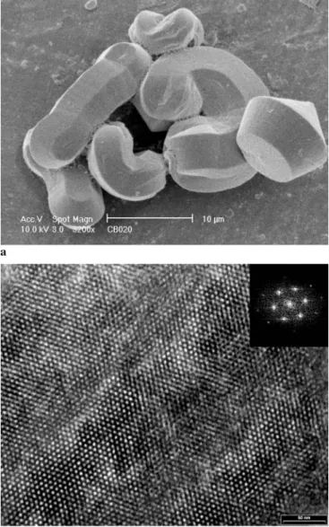
The typical morphology of mesoporous CMI-1 materials, such as gyroids, toroids and ropes is observed and remains unchanged during the incorporation of ZnO (Fig. 2a). The TEM image depicted in Fig. 2b demonstrates that our CMI-1 matrix contains a honeycomb-like pore arrays. The Fourier transform of TEM picture exhibits 6-fold symmetry and the measured angles between two bright spots are very close to 60◦, confirming the hexagonal symmetry in the stacking of the channels of our CMI-1 material. No changes in pore ar-rays after incorporation of ZnO nanoparticles were revealed by TEM technique.
3.2 Information about optical properties
In order to highlight quantum size effect in ZnO nanocrystals, we have first studied the photoluminescence (PL) of the mesoporous matrix with an excitation source of
FIGURE 2 SEM (a) and TEM (b) images of the starting material and ZnO/mesoporous CMI-1 nanocomposite. The inset of (b) is the Fourier transform picture of TEM image
254 nm(photon energy of 4.88 eV). The corresponding spec-trum is shown in Fig. 3 and is composed of two wide PL bands centred at 2.94 and 3.89 eV. The decomposition of the PL spectrum into three Gaussian functions allows us to give
FIGURE 3 Photoluminescence spectrum (λ = 254 nm) of the mesoporous material CMI-1 with its Gaussian decomposition
Maximum (eV) Full width half Intensity maximum (eV) (arb. units)
1.97 0.18 210
2.94 0.62 1840
3.89 0.56 1050
TABLE 1 Results of the Gaussian decomposition of the experimental spectrum of the mesoporous material CMI-1
accurate values for the three observed bands (Table 1). The PL band centred at 1.97 eV comes from the second order of diffraction of the monochromator and is a replica of the 3.89 eV band. The origin of the two other bands is the ex-istence of structural defects [12, 13] inside the silica frame-work of the mesoporous material CMI-1. According to Saku-rai [14], the 2.94 eV band is caused by silanol groups present inside the channels of the material. PL studies have been per-formed on CMI-1 mesoporous silicas in order to determine the origin of these two PL bands [15]. The observations have led to the conclusion that both PL bands, at 2.94 and 3.89 eV, are generated by the same defects associated with silanol groups. The photoluminescence band of ZnO particles is expected to be located at 3.28 eV [16] and comes from the excitonic recombination causing spontaneous emission. In our case, the PL spectroscopy gives a very wide PL band in the range 3.2–3.4 eV (Fig. 4).
In this wide band, we have to distinguish the contribu-tion of the ZnO nanoparticles and of the mesoporous matrix. Table 2 shows the result of a decomposition of the spectrum into three Gaussian peaks.
The position and the FWHM of the matrix PL bands remain unchanged. The 3.41 eV PL band corresponds, of course, to spontaneous emission from ZnO and is shifted to higher energies compared with the photoluminescence of bulk ZnO (3.28 eV). This shift is related to the quantum size ef-fect and is due to the small size of the ZnO nanoparticles. This observation confirms the presence of ZnO inside the channels of the mesoporous material and indicates that the particle size is small enough to produce the quantum size ef-fect. Concerning the interaction between the surface of the nanoparticles and the silica matrix, we believe that, as the
par-FIGURE 4 Photoluminescence spectrum (λ = 254 nm) of sample ZnO-CMI-1 with its Gaussian decomposition
Contribution Maximum (eV) Full-width half Intensity maximum (eV) (arb. units)
Matrix CMI-1 2.96 0.62 1480
ZnO 3.41 0.58 2885
Matrix CMI-1 3.86 0.55 830
TABLE 2 Results of the Gaussian decomposition of the experimental PL spectrum of the sample ZnO-CMI-1
ticles are well adjusted on the channels and although the SiO2 matrix is electrically neutral, the weak acidic silanol groups at the internal surface of the mesochannels can interact with the ZnO nanoparticles and can further stabilize the particle size of ZnO. However, it is very difficult to observe experimen-tally the existence of this interaction which could be a very interesting subject of investigation.
3.3 Lasing effect
By submitting our samples to a 1.82 eV femtosec-ond laser excitation (which correspfemtosec-onds to a wavelength of 680 nm, approximatively half of the value of the band gap of
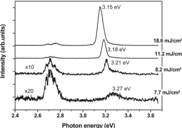
FIGURE 5 Emission spectra (λ = 680 nm, E = 1.82 eV) of sample ZnO-CMI-1 for various pumping intensities
108 Applied Physics A – Materials Science & Processing
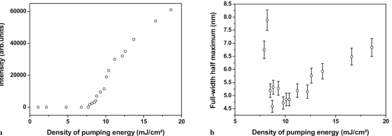
FIGURE 6 Variation of the intensity (a) and of the FWHM (b) of the emission band of ZnO with the density of the pumping energy of the femtosecond laser (λ = 680 nm)
ZnO bulk), we manage to observe at low exciting intensities (7.7 mJ/cm2) a broad emission band centred at 3.27 eV which is attributed to the spontaneous emission of the ZnO nanopar-ticles (Fig. 5).
When the pumping intensity reaches a threshold value (ca. 8.2 mJ/cm2), the broad emission turns to a much narrower peak corresponding to stimulated emission. The threshold for the transition from spontaneous to stimulated emission can be accurately determined by studying the intensity vari-ation and the full-width at half maximum (FWHM) of the band in function of the energy delivered by the laser. Accord-ing to both diagrams, the threshold value equals 8.2 mJ/cm2 (Fig. 6a and b) and represents the energy of the transition from spontaneous to stimulated emission from the nanoparticles of ZnO incorporated in the channels of the mesoporous mate-rial CMI-1. At low pumping intensities, the emission intensity is proportional to the square of the pumping fluence. Above 12 mJ/cm2, the absorption process changes. When increasing the fluence, a progressive spectral shift of the stimulated emis-sion (of about 100 meV) and a broadening of the spectra (of about 2 nm) were observed (Fig. 6b). These effects indicate a very strong influence of the band gap renormalisation [17] on the emission properties of the ZnO nanoparticles.
It is worth indicating that the photon energy used is not sufficiently high (1.82 eV) to excite one electron from the va-lence band to the conduction band of ZnO (gap= 3.37 eV). However, this phenomenon occurs indeed. This can only be produced by an excitation process implying two photons. The first photon sends one electron on a virtual level located in the band gap (lifetime of around 10−16s) and then the second photon excites the electron to the conduction band. Once the electron is excited, spontaneous or stimulated emission can take place. The two-photon absorption occurs only if the time delay between the two-photons is shorter than the lifetime of the electron on the virtual level. The two-photon absorption process is well-known [18], but was never reported in ZnO in mesoporous materials. It is for the first time observed that the stimulated emission is produced after a two-photon absorp-tion with ZnO (two-photon lasing effect). However, no cavity modes which could confirm lasing effect were observed in our experiments.
Thus it is very possible that the mechanism of lasing is based on light scattering within the mesoporous matrix where
active ZnO nanoparticles were introduced. The spontaneously emitted photons scatter from one particle to another and fi-nally form a loop. The photons can be trapped and amplified, resulting in stimulated emission. This mechanism has been clearly described in literature [19–22].
4 Conclusion
In summary, incorporation of ZnO nanoparticles inside the channels of a mesoporous material CMI-1 has been achieved via the incipient wetness method. N2 adsorption– desorption, TEM and SEM have demonstrated a decrease of the specific area of the matrix, indicating the real incorpora-tion of ZnO nanoparticles in the channels of CMI-1 and the retention of the mesoporosity after the incorporation process. Moreover, photoluminescence spectroscopy provides a spec-trum with one wide PL band corresponding to the contribution of both silica matrix and ZnO particles. It is observed a shift for the ZnO PL band compared to the PL of ZnO bulk which can be attributed to the quantum size effect observed when the size of the particles is small enough. On another hand, stim-ulated emission in a disordered medium like nanoparticles of ZnO introduced inside the channels of a mesoporous mate-rial CMI-1 is demonstrated. Transition between spontaneous and stimulated emission is characterized by a threshold which represents the minimum energy necessary to observe lasing effect. Moreover, this lasing effect with ZnO nanoparticles has been observed after an absorption process implying two pho-tons: two-photon lasing effect. It is interesting in the future to study the lifetime of the stimulated emission and its behaviour at low cryogenic temperature.
ACKNOWLEDGEMENTS C. Bouvy thanks the FRIA (Fonds National de la Recherche Scientifique) for a doctoral fellowship. This work was realized in the frame of the Interuniversity Attraction Poles Program – Belgian State – Belgian Science Policy. Financial supports from the Uni-versity of Namur (FUNDP) and from the European SOXESS network were gratefully acknowledged.
REFERENCES
1 C. Klingshirn, Phys. Stat. Solidi B 71, 547 (1975)
2 C. Cannas, C. Casu, A. Musinu, A. Lai, G. Piccaluga, J. Mater. Chem. 9, 1765 (1999)