HAL Id: tel-02296215
https://tel.archives-ouvertes.fr/tel-02296215
Submitted on 25 Sep 2019
HAL is a multi-disciplinary open access archive for the deposit and dissemination of sci-entific research documents, whether they are pub-lished or not. The documents may come from teaching and research institutions in France or abroad, or from public or private research centers.
L’archive ouverte pluridisciplinaire HAL, est destinée au dépôt et à la diffusion de documents scientifiques de niveau recherche, publiés ou non, émanant des établissements d’enseignement et de recherche français ou étrangers, des laboratoires publics ou privés.
Morphogenesis and Growth
Valeria Davì
To cite this version:
Valeria Davì. Cell Wall Dynamics in the Regulation of Cell Morphogenesis and Growth. Subcellular Processes [q-bio.SC]. Université Paris-Saclay, 2018. English. �NNT : 2018SACLS295�. �tel-02296215�
Dynamique de la paroi cellulaire
dans la régulation de la
morphogenèse et de la croissance
cellulaire
Thèse de doctorat de l'Université Paris-SaclayPréparée à l’Université Paris-Sud École doctorale n°577 : Structure et Dynamique des Systèmes Vivants (SDSV) Spécialité : Science de la Vie et de la Santé Thèse présentée et soutenue à Paris, le 24 Septembre 2018, par
Valeria Davì
Composition du Jury :
M. Olivier Hamant, DR Président et
ENS de Lyon Rapporteur
(Laboratoire Reproduction et Développement des Plantes)
M. Mohan Balasubramanian, Professeur Rapporteur
Université de Warwick
(Département de Sciences Biomédicaes)
M. Giuseppe Baldacci, Professeur Examinateur
Université Paris-Diderot, CNRS (Institut Jacques Monod)
M.me Florence Chapeland-Leclerc, MC Examinateur
Université Paris Decartes
(Laboratoire Interdisciplinaire des Energies de Demain)
M. Alexis Peaucelle, CR Examinateur
INRA, AgroParitech
(Institut Jean-Pierre Bourgin)
M. Nicolas Minc, DR Directeur de thèse
CNRS
(Institut Jacques Monod)
Prof. Arezki Boudaoud, Professeur Invité
Université de Lyon, ENS de Lyon
(Reproduction et Développement des Plantes)
NNT : 20 18 SAC LS2 95
Université Paris-Saclay Espace Technologique / Immeuble Discovery
1
Abstract
Cells in nature develop in a wide range of forms, following diverse growth patterns. Despite the importance of these fundamental processes, how cells regulate their growth and morphogenesis is still poorly understood. In this thesis, I explored these processes, focusing my investigations on tip growing walled cells and in particular, by exploiting the fission yeast Schizosaccharomyces pombe, adopting mainly a biomechanical approach. To this aim, I first developed novel methods to measure key cell wall mechanical parameters in vivo at a large population scale, which allowed the very first observations of cell wall dynamics. This revealed that the cell wall is softer and highly variable at growing poles, and almost stable and stiffer at non-growing sites. During elongation, there is an interplay between wall mechanics and cell growth, whose active control allows cell expansion while preserving cell integrity. In addition, I observed that there is a strong correlation between cell wall mechanics and cell morphology, and ectopic perturbations of wall properties directly affect shape establishment and maintenance. Together my results show that the regulation of wall mechanics is fundamental in the determination of cell dynamics in tip growing walled cells. Moreover, this suggests that dynamic observation of cell surface mechanics is crucial for a complete understanding of multifactorial and complex processes, such as growth and morphogenesis.
Resumé
Les cellules dans la nature se développent dans un large éventail de formes, suivant divers modèles de croissance. Malgré l'importance de ces processus fondamentaux, la façon dont les cellules régulent leur croissance et leur morphogenèse est encore mal comprise. Dans cette thèse, j'ai exploré ces aspects, avec une approche principalement biomécanique, en concentrant mes investigations sur des cellules à paroi et en exploitant en particulier la levure fissipare
Schizosaccharomyces pombe. J'ai d'abord développé de nouvelles méthodes pour mesurer les
paramètres mécaniques clés de la paroi cellulaire in vivo, ce qui a permis les premières observations de la dynamique des parois cellulaires. Ceci a révélé que la paroi cellulaire est plus souple et très
2
variable au niveau des pôles de croissance, et presque stable et plus rigide dans les sites sans croissance. Au cours de l'allongement, il existe une interaction entre la mécanique des parois et la croissance cellulaire, dont le contrôle actif permet l'expansion cellulaire tout en préservant l'intégrité des cellules. De plus, j'ai observé qu'il existe une forte corrélation entre la mécanique des parois cellulaires et la morphologie cellulaire, et que des perturbations des propriétés de la paroi affecte directement l'établissement et la maintenance de la forme. Ensemble, mes résultats montrent que la régulation de la paroi est fondamentale dans la détermination de la dynamique cellulaire dans les cellules à parois. Globalement, cela suggère que l'observation dynamique de la mécanique de surface cellulaire est essentielle pour une compréhension complète des processus multifactoriels et complexes comme la croissance et la morphogenèse.
3
Table of Contents
Aims of the study ... 5
INTRODUCTION ... 7
Chapter 1 – General principles of cell morphogenesis ... 9
1.1 General physical principles regulating living (and non-living) matter ...12
1.2 Cell polarity and cell shape ...13
1.2.1 Molecular components in cell polarity ...14
1.2.2 Cell polarity establishment and maintenance ...18
1.3 Cell surface mechanics ...20
1.3.1 Surface mechanics of non-walled cells ...21
1.3.2 Surface mechanics of walled cells...23
1.4 Mechanosensing mechanisms for shape regulation ...25
Chapter 2 – Elongation and morphogenesis in tip-growing walled cells ...29
2.1 Mechanics of walled cells ...35
2.2 Turgor pressure regulation ...41
2.1 Cell wall properties ...44
2.1.1 Cell wall composition ...44
2.1.2 Cell wall synthesis ...48
2.1.3 Cell Wall Integrity pathway ...52
2.2 Influence of cell wall mechanics on morphogenesis and growth ...54
Chapter 3 – Schizosaccharomyces pombe, morphogenesis and growth ...61
3.1 Different stages of fission yeast life cycle ...64
3.2 Polarity machinery during tip elongation ...68
3.3 Turgor regulation...73
3.4 Cell wall properties ...74
3.4.1 Cell wall composition ...74
3.4.2 Cell wall synthesis ...75
3.4.3 Wall integrity pathway ...77
3.5 Cell mechanics ...79
RESULTS...81
1- Mechanosensation Dynamically Coordinates Polar Growth and Cell Wall Assembly to Promote Cell Survival ...83
4
2- Mechanical Control of Cell Shape and Size in Fission Yeast ... 85
DISCUSSION ... 87
METHODS ... 97
WALLTRACKER ... 99
WALLTRACKER 2.0 ... 109
ANNEX 1 ... 127
Mechanics and morphogenesis of fission yeast cells ... 127
ANNEX 2 ... 129
Procede de mesure en temps reel de l’epaisseur de la paroi et ses applications ... 129
RÉSUMÉ ... 131
5
Aims of the study
Cells in nature comes with very different forms and grow with profoundly diverse patterns. These processes are the output of complex genetic and biochemical regulations, which tune local delivery of proteins, synthesis of new material, deformation and remodeling, to ultimately modify the mechanics of the cell surface, in order to acquire a certain shape or expand in a specific manner. However, the complex interplay between all these layers of regulation is still largely unclear. In most living organisms, from plants to bacteria and fungi, the cell surface is composed of a stiff structure covering the cell boundaries, named cell wall. Those cells are also characterized by a very high internal turgor pressure, which is the driver of growth. Despite the rigidity of their surfaces, walled cells can undergo very dynamic processes of fast-polarized growth, reorientation towards/against stimuli, and acquire profoundly different shapes, both as single cells or in tissues. This suggests that cell walls must continuously modify their structures, to allow for these dynamic processes. Therefore:
what is the dynamical behavior of cell walls?
To answer this question, in my work, I focused on a subset of walled cells, that grow isolated as single cells (e. i. not part of a tissue) by tip extension generating tubular shapes, and in particular, on the rod shaped fission yeast Schizosaccharomyces pombe, which among them is one of the best described. So far, mainly because of technological limitations, there was very little access to cell wall dynamics. My first achievement was the setup of novel methods suitable to follow cell wall mechanical parameters in vivo. This allowed, the very first observations of cell wall dynamics, showing that the cell wall is stable at non-growing regions, but highly variable at growing ones. Those results led to the following questions:
How is cell wall dynamics regulated to ensure growth while preserving cell integrity?
In fact, tip growing cells are in a constant precarious condition, because during growth, the deposition of new wall material must constantly balance the internal turgor pressure that without a sufficient support may cause cell lysis. To investigate this aspect, in the first part of my results, I characterized wall dynamics and wall synthesis regulators in wild type cells, as well as in mutants of highly conserved pathways involved in cell integrity maintenance, uncovering an interplay between cell elongation and wall thickness and one mechanism for its regulation.
6
How do wall mechanical properties influence cell shape?
By exploiting the stereotypical rod shape of fission yeast, in the second part of my results, I investigated the correlation between wall mechanical properties and cell diameter, in different wild type conditions and in tens of mutants defective in diameter or wall synthesis. In addition, I observed diameter changes after perturbation of wall properties, uncovering a putative mechanism for diameter regulation.
The overall aim of this work is to lay the foundation for the understanding of how cell walls are dynamically regulated, and how this can influence fundamental processes, such as growth and shape establishment in walled cells.
7
9
Chapter 1 – General principles of cell morphogenesis
In this chapter, I describe the basic principles of cell shape determination. I start by introducing the physiological importance of cell morphogenesis in full organisms and single cells. I then focus on the physical principles that are involved in assembly of both living and non-living matter. Then, I introduce the important principle of cell polarity, the ability of a cell to define local differences in shape, structure or function, based on the local organization of molecular machineries. Thereafter, I focus on the mechanical properties of cell surface, the structure that ultimately confers a defined form. Finally, I describe mechanosensing pathways, involved in the regulation and homeostasis of cell shape, differentiation and response to external perturbations.
Table of contents
Chapter 1 – General principles of cell morphogenesis ... 9
1.1 General physic principles regulating living (and non-living) matter ...12
1.2 Cell polarity and cell shape ...13
1.2.1 Molecular components in cell polarity ...14
Cytoskeleton ...14
Membrane trafficking ...15
RhoGTPases ...16
1.2.2 Cell polarity establishment and maintenance ...18
1.3 Cell surface mechanics ...20
1.3.1 Surface mechanics of non-walled cells ...21
1.3.2 Surface mechanics of walled cells ...23
1.4 Mechanosensing mechanisms for shape regulation ...25
11
M
orphogenesis, from the Greek terms “morphê”, shape, and “genesis”, creation, literallymeans the generation of shape. In biology, this term concerns the fundamental question of how biological form and structure are generated, from the rather simple cell level, to the more complex tissues, or the higher order assembly of organs and full organisms.
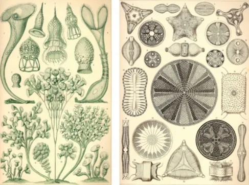
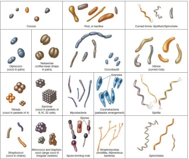
Already at the cell level we can appreciate that shapes can vary from simple spheres to rods, disks, cubes, branched trees, up to the very complex shapes of protists or diatoms (Figure 1.1). Cell morphology is fundamental, not only for single cell functionality, but also for the correct assembly of tissues and folding of organs, and therefore for the viability of organisms.
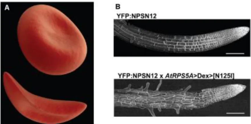
Defects in cell shape are associated with loss of function or disease. For instance, the concave shape of erythrocytes confers high plasticity and their ability to flow through very narrow vessels (Figure 1.2A above). Defects in hemoglobin generate the sickle like shape, typical of drepanocytosis (sickle anemia): cells become more rigid, easily undergo hemolysis and can occlude vessels, not being able to deform in small capillaries (Figure 1.2A below) (Pauling et al., 1949). Another example is represented by the very organized cubic cells that, growing in an oriented direction, define a tubular
Arabidopsis thaliana root, necessary for anchoring and nutrition of a plant (Figure 1.2B above).
Inhibition of polarized trafficking in these roots lead to protrusions of several extra fronts of growth and the root will end up being very small and branched, and consequently lowering its overall
Figure 1.1. Drawing of single cell and colonies of protozoa (left) and diatoms (right) from Ernst Haeckel’s Art Forms
12
Figure 1.2. Examples of cell shape degenerations. (A) An erythrocyte in normal shape (top) and in sickle anemia
(bottom). (B) A. thaliana root in a control (top) or after inhibition of polarized exocytosis by RAB delocalized activation (bottom – AtRPS5A dexamethasone inducible promoter, induce RAB activation at root sides). YFP fluorescence in lateral roots expressing the PM marker YFP:NPSN12. Scale bar 100 m. Adapted from (Kirchhelle et al., 2016).
fitness of the plant. (Figure 1.2B below) (Kirchhelle et al., 2016).
1.1 General physical principles regulating living (and non-living) matter
The variety of shapes in nature is evident and under the eyes of everyone, but the understanding of how these shapes are generated is still fragmentary.
Biological structures are characterized by several layers of organization: at nanometrical scales, macromolecules, such as structural proteins, or lipids, together with enzymatic activity arrange in order to define micrometric structures such as complex lipid bilayers, organelles and cells. Cells interact and organize, forming millimetric tissues, which folds into organs that will ultimately define full organisms. Therefore, the generation of an organism, with precise shape and form, relies primarily on the nanometric interplay of biochemical molecules, enzymatic activity and signaling. Many principles regulating forms of inorganic material are also common in living matter. Two proposed mechanisms that allow for the formation of biological structures are self-assembly and self-organization (Dumont and Prakash, 2014).
Self-assembly is the effect of passive interactions of subunits, that assemble because their association is energetically favorable; there is no consumption of energy and the system can reach an equilibrium. Typical structures generated by this mechanism are nuocleoli, P-bodies or amyloids (Brangwynne, 2011).
Self-organization occurs when subunits interact by dissipating energy; in a living system this energy comes from the metabolism, moving the system out of equilibrium, giving rise to
13 continuous structural transformations. Examples of self-organized structures are acto-myosin and microtubules networks, that form by dissipating ATP or GTP both during filament growth and movement of motors (Nedelec et al., 1997).
However, these two principles alone cannot explain the very organized and reproducible structures of living organisms. For example, actin can self-organize in filaments, but how can these filaments be localized in the right cell location to promote cytokinesis? To understand how a complex shape is defined in biology we must understand all the layers of regulation that are involved in these phenomena.
1.2 Cell polarity and cell shape
As briefly mentioned before, the ability of a cell to define a specific shape relies, in the first place, on the correct distribution of its components at a biochemical level; this process is named “cell polarity”. In other words, cell polarity is the capacity to deliver the correct molecules at the correct place, promoting the definition of a geometric axis, and defining specialized domains of action that can be dedicated to specific tasks.
Cell polarity is prominent in most cell types, from prokaryotes to plants, fungi and mammals. It is essential for a countless number of functions, from growth, to asymmetric cell division, cell migration, extending to embryogenesis, tissue and organ development.
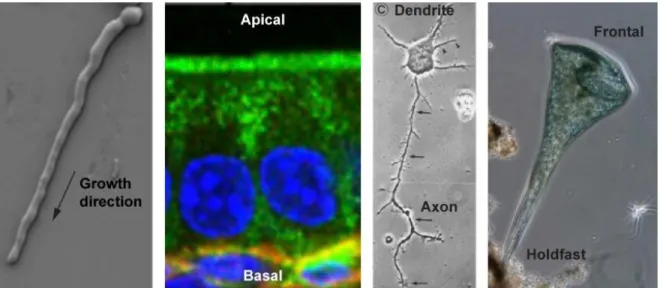
Clear examples are found in tip-growing cells, such as fission yeast, filamentous fungi or pollen tubes of plants: all localize their growth machinery at one or more protrusions, restricting growth to discrete regions of the cell, resulting in rod/branched shaped structures necessary to explore the substrate. Another more complex example of cell polarity is the gigantic cell Stentor coeruleus, a trumpet like shaped protist that exhibits a highly polarized and specialized structure, with the larger part covered in cilia, and the smaller developed in a holdfast. In animals, hippocampal neurons elongate from the soma short dendritic protrusions, specialized for synaptic signal reception, and long axon that can releases synaptic vesicles. Cell polarity is also essential for organism development, being involved in embryogenesis and differentiation, as well as in the correct arrangement and functionality of tissue and organs. For example, cells in epithelial tissues possess a distinct polarized internal organization, which defines the assembly of membrane domains, named apical and basolateral: these domains differentiate the functions of the two sides of the tissue and are essential for its correct folding (Figure 1.3).
14
Figure 1.3. Examples of cell polarity. From the left germtubes growth of Neurospora crassa (Lichius et al., 2014);
immunostaining of small intestinal epithelial cells in WT mice. In green Ezrin, in blue Nuclei, in red laminin (Toya et al., 2016). Cultured hippocampal rat neuron (Dotti et al., 1988). Stentor coeruleus cell (From Bill Porter youtube channel). Every image have been adapted from the reference.
1.2.1 Molecular components in cell polarity
At the molecular level, cell polarity is normally defined by two fundamental properties in eukaryotes: the orientation of the cytoskeleton along the polarity axis and the asymmetric accumulation of proteins in discrete regions of the cell (Li and Gundersen, 2008).
Cytoskeleton
Microtubules and actin filaments are the major components of the cytoskeleton. Both polymers are polarized, due to the asymmetric properties of their subunits.
Microtubules are composed of hetero dimers of - and -tubulin, which bind head to tail through GTP hydrolysis. Initial nucleation of microtubules occurs at centrosomes or other microtubule organizing centers (MTOCs) that are normally close to the cell center. The so-called minus end of the filament is anchored to those structures, and therefore the polarized direction of microtubules is determined by the regulation of the dynamic polymerization at the plus end. Actin filaments are composed of monomers of globular actin that bind hydrolyzing ATP (Figure 1.4).
Actin nucleation is promoted by actin nucleator factors, such as formins, which nucleate and elongate linear filaments from their barbed ends, or the Arp2/3 complex, which produces branched filaments. Actin nucleation factors can be distributed at the cell surface, therefore their localization is the main mechanism regulating acting orientation for cell polarization (Pollard, 2007).
15
Figure 1.4. Schematic structure of actin filament (a) and microtubule (b), including the dynamic of free polymerization
and the direction of molecular motors. In (b) above GTP-dimer stabilize the microtubule structure. Loss of the GTP cap induces de-polymerization (below) (Li and Gundersen, 2008)
Both actin and microtubules are extremely dynamic, with differing polymerization and de-polymerization rates on the two ends of the filaments, both depending on various regulators of elongation that contribute to their dynamism. The ability to reorganize in a fast manner is very important to respond rapidly and locally to arising polarity signals (Li and Gundersen, 2008). Motor proteins are a class of cytoskeleton-associated proteins highly linked to cell polarity. These proteins can move along the polymer tracks by hydrolyzing ATP. During their “walk”, they can carry cargoes, such as molecules, vesicles or organelles within the cell. Microtubule motors include dynein and kinesins. While dynein moves towards the minus end, most kinesins move to the plus end. Actin-associated motors are myosins, which normally moves towards the barbed end (with some exceptions). One example is MyoV that due to its high processivity (ability to be associated to the filament for several steps), can transport cargoes on long distances (Figure 1.4) (Kincaid and King, 2006). Hence, once the cytoskeleton is polarized, it can direct trafficking to specific cellular locations in the cell, promoting or reinforcing polarity.
Membrane trafficking
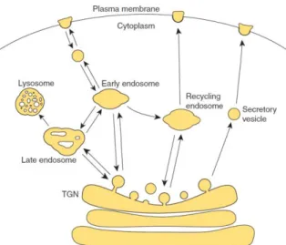
Polarized accumulation of proteins at specific regions of the plasma membrane can be highly influenced by delivery and fusion of vesicles containing proteins (exocytosis) and internalization
16
Figure 1.5. Membrane trafficking to and from the plasma membrane. TGN is the trans Golgi network (Orlando and
Guo, 2009).
and membrane recycling (endocytosis). For exocytosis, proteins containing vesicles that reach the plasma membrane originate from the trans-Golgi network, or through recycling of previously internalized membranes. They are tethered to specific locations at the plasma membrane by a specific set of proteins, such as the exocyst, an evolutionary conserved octameric protein complex (consisting of Sec3, Sec5, Sec6, Sec8, Sec10, Sec15, Exo70, and Exo84). Tethered vesicles can fuse to the membrane, in a process mediated by SNARE protein complexes.
Proteins at the plasma membrane can be recycled through endocytosis, in a process that creates clathrin-coated vesicles departing from the membrane, which can then be delivered to the endosome either for recycling or for degradation (Figure 1.5) (Orlando and Guo, 2009). Membrane trafficking and cytoskeleton are linked, for example transport from the trans-Golgi network to the plasma membrane can be facilitated by motor-dependent delivery along cytoskeletal tracks (Porat-Shliom et al., 2013); in addition, endocytosis is actin dependent in some organisms, such as budding and fission yeasts (Aghamohammadzadeh and Ayscough, 2009; Basu et al., 2014; Mooren et al., 2012).
Although vesicle traffic is important to generate local domains of polarity regulators, it can be directly modulated by some of those regulators as well; some examples will be given in the next paragraph.
RhoGTPases
In eukaryotes, RhoGTPases, small G proteins part of the Rho family, have emerged as central regulators of cell polarity signaling. Within this family there are Cdc42, Rac, Rho and Rop.
17
Figure 1.6. Schematic of Rho-GTPase cycle (Etienne-Manneville, 2002)
They are membrane-associated proteins, prenylated at their C-terminus domain. Like all GTPases they can function as molecular switches, being active in their GTP bound state and inactive after GTP hydrolysis. GTPase-activating proteins (GAPs) can promote GTP hydrolysis with a consequent inhibitory effect. GTP exchange-factors (GEFs) on the contrary, can promote GDP-GTP exchange, leading to activation. Moreover, guanine nucleotide dissociation inhibitors (GDIs) can bind to their GDP form, and sequestrate them in the cytosol, by masking their membrane anchoring binding site (Figure 1.6) (Etienne-Manneville, 2002).
RhoGTPases act in very diverse stages of cell shape determinations, interacting directly or indirectly with other molecular players of cell polarity. In both mammals and plants, RhoGTPases can interact with actin structures: Ras can interact with Arp2/3 via the WAVE complex, promoting actin elongation and branching (Basu et al., 2008; Wittmann and Waterman-Storer, 2001; Yanagisawa et al., 2013). During fibroblasts migration, it has been shown that Cdc42 and Rac promote membrane protrusions through actin polymerization, while Rho promotes membrane retraction through actin depolymerization (Etienne-Manneville, 2002). Moreover, Cdc42 can promote directional migration in macrophages (Allen et al., 1998). Furthermore, RhoGTPases can also interfere with microtubules: during cell migration, Rac can indirectly promote microtubule stabilization, while Cdc42 can orient the Microtubule Organizing Center (Wittmann and Waterman-Storer, 2001). There is also proof of direct interactions between RhoGTPases and the secretory pathway: Cdc42 in yeast interacts with Sec3, while in mammals Ral, a small G protein of the Rac family, can interact with Sec5, therefore with the exocyst (Moskalenko et al., 2002; Sugihara et al., 2002). In addition, in mammal zygotes, Cdc42 can interact with Par6, promoting the correct orientation of vegetative and animal poles (Lin et al., 2000). Finally, in fungi
18
RhoGTPases control polarized growth, but also the local assembly of cell wall, by the direct regulation of cell wall synthases (Drgonova et al., 1996; Perez and Rincón, 2010).
In conclusion, cell polarization is regulated by a complex communication between different classes of proteins and cellular processes, including cytoskeletal components, membrane
recycling and small GTPases, which are mutually regulated and differentially distributed within a cell.
1.2.2 Cell polarity establishment and maintenance
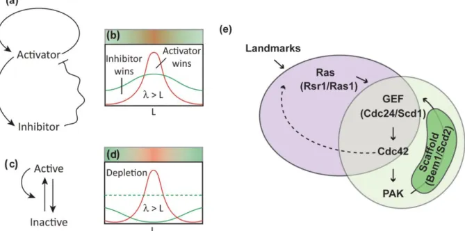
Very often in cells, symmetry breaking, the initiation of cell polarity, occurs in specific areas of the cell surface due to the presence of polarity cues provided by molecules that act as landmarks, by directing RhoGTPases recruitment. However, in several organisms, the depletion of polarity cues may impair the correct orientation of the polarity domain, but does not prevent the formation of polarity domains at random positions. For instance, in Saccharomyces cerevisiae the budding pattern is highly conserved: in haploid cells, each new bud will form next to a previous site of budding, a bud scar. This is regulated by the landmark Rsr1 that recruits downstream polarity machinery. However, when Rsr1 is bypassed either by deletion, or by the constitutive activation of
Figure 1.7. Self-assembly. A-D. Pattern formation in chemical systems, with activator/inhibitor (a-b), or with long range
inhibition (c-d) Adapted from (Goehring and Grill, 2013). E. Self-amplification of Cdc42 in budding and fission yeasts. Cdc42-GTP can bind a PAK protein, that bind a scaffold protein. The scaffold is also binding a GEF of Cdc42, promoting its further activation. If this happen in the presence of a landmark, can occur at specific location of the cell, through activation of the GEF by the landmark itself. Dotted line is a putative, but yet not described, positive feedback. Adapted from (Martin, 2015).
19 Cdc42 with a Cdc42Q61L allele, cells lose the correct budding pattern, but are still able to bud at random positions (Irazoqui et al., 2003; Wedlich-Soldner et al., 2003). This demonstrates the ability of the system to self-assemble.
The first significant study on pattern self-assembly, is the reaction diffusion model from Alan Turing (Turing, 1953). He proposed that in a uniform starting condition some chemical species, “morphogens”, can interact, leading to the formation of new species that have a different rate of diffusion, creating instability. The system will find its equilibrium in very specific and organized patterns.
This theory was later refined by Gierer and Meinhardt (Gierer and Meinhardt, 1972), and included the existence of short-range positive feedbacks, together with long-range negative feedbacks. The positive feedback can be due to activators acting at a short range, while the negative feedbacks caused by inhibitors acting at a longer range. Differences in diffusion between activators and inhibitors, with inhibitors diffusing faster, can lead to patterns formations, such as polarity domains (Figure 1.7 A-B). Moreover the inhibitor can be dispensable, if the molecules diffusion rate varies, moving slow when active and fast when inactive creating an oscillatory dynamic (Figure 1.7 C-D) (Goehring and Grill, 2013; Mogilner et al., 2012).
The best examples of these patterns are found in budding and fission yeasts, during budding in the first one and polarized growth in the second. Cdc42, with or without spatial cue, can form a polarity domain, through the formation of a complex, stabilized by a scaffold protein that can bind both a PAK protein and the GEF of Cdc42. The PAK protein binds Cdc42-GTP, while the GEF activates more Cdc42, providing a positive feedback loop. In Schizosaccharomyces pombe, it has been shown that this feedback can be further increased by a low diffusion rate of Cdc42-GTP compared to its inactive form. Moreover, the scaffold diffuses slower when bound to the membrane, than in its cytosolic form, recapitulating the second model represented in Figure 1.7 C-D. (Figure 1.7 E) (Bendezú et al., 2015; Martin, 2015)
To conclude, polarity is established in a self-regulated manner, either at random positions or in specific areas of the cell, when landmarks or external cues are present. An intricate interplay between the cytoskeleton, vesicles trafficking, RhoGTPases regulation and all their possible effectors can lead to the establishment of specialized polarity domains that will orchestrate the building and remodeling of cell structures.
20
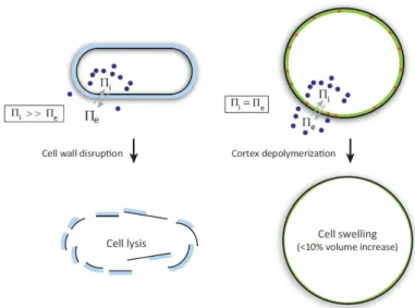
Figure 1.8. The actomyosin cortex versus the cell wall. In walled cells the internal turgor pressure is very high. The
cell wall act as a mechanical barrier: its disruption lead to immediate cell lysis. In non walled cells difference in internal and external osmotic pressure is small. Actin cortex still balance the osmotic pressure, but its depolymerization cause only a moderate swelling (From (Salbreux et al., 2012)).
1.3 Cell surface mechanics
Once all the biochemical machinery has been delivered in the right location of a cell, it must polymerize/remodel/rearrange the surface, in order to obtain the correct mechanical properties, to achieve a specific shape.
In fact, as proposed by the pioneering work “On Growth and Forms” in 1917 by D’Arcy W. Thompson, any geometrical structure, even if part of a living system, will follow the mathematical and physical rules of any matter: “cell and tissue, shell and bone, leaf and flower, are so many portion of matter, and it is in obedience to the laws of physics that their particles have been moved, molded and conformed” (Thompson, 1942). He suggested that problems of form are in the first instance mathematical problems, problems of growth are physical problems, and therefore the morphologist must be a student of physical science. One of the main intuition that he had was that, despite the large variety of cell shapes existing in nature, they all follow similar physical principles. He dedicated one entire chapter on the forms of cells, considering them as simple entities comparable to soap bubbles. Like in soap bubbles, surface tension is the leading rule that brings about the minimization of surface area through taking on spherical shapes. By integrating Plateau’s work on surface tension, he demonstrated that this rule is not just applicable to soap bubbles but also to many other more complex cell shapes can fall in this definition, under certain physical constrictions. Thompson’s work, despite its 101 years and the obvious lack of informations on the
21 molecular players involved, remains an extremely important milestone in morphology studies, introducing the importance of mechanics, for the complete understanding of morphogenesis. Thus, to unveil the mechanisms of shape control we need to understand how the cell regulates the mechanical properties and the forces generated at its surface.
In this context, we can divide cells in two families with different boundary composition: walled and non-walled cells.
1.3.1 Surface mechanics of non-walled cells
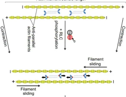
In most of the non-walled cells, from animal cells to amoeba, the most important regulator of surface mechanics is the actin cortex (Paluch and Heisenberg, 2009). This is a complex thin network made of proteins, located below the plasma membrane. It is mainly composed of actin filaments, parallel to the cell surface, with a mesh size ranging from 20 to 250 nm. Many actin-binding proteins bind to the cortex, among the most important are proteins involved in actin contractility, such as myosin or tropomyosin (Salbreux et al., 2012). The main myosin involved in actin cortex contractility is the non-muscle Myo II, which filaments can associate with each other in an anti-parallel fashion. This allows them to crosslink and slide actin filaments during ATP binding and hydrolysis, directly influencing network contraction (Figure 1.9) (Newell-Litwa et al., 2015).
Figure 1.9. Non-muscle myosin II promote sliding of actin antiparallel filaments. Myo II filaments (blue) associate
with each other in an anti-parallel fashion, allowing them to crosslink and slide actin filaments past each other. RLC (regulatory light chains) Ser19 phosphorylation increases Myo II ATPase activity, leading to contraction of actin filament bundles, and phosphorylation of both Ser19 and Thr18 increases Myo II ATPase activity, driving the
22
association of multiple actin filaments into actomyosin filament bundles, often referred to as stress fibers. Adapted from (Newell-Litwa et al., 2015).
The elasticity of the cortex has been estimated to be of the order of 103 Pa in cultured fibroblasts (Tinevez et al., 2009) and may balance cell osmotic pressure, in fact, in mitotic HeLa cells cortex depolymerization leads to small increase in cell volume (Stewart et al., 2011) (Figure 1.8 right). One of the key characteristics of the actin cortex, that underlies the biggest difference with walled cells, is its capacity for rapid turnover of its molecules, which allows for rapid remodeling, with a consequent high plasticity of the mesh. Local changes in cortex mechanical properties, particularly in cortical tension, drive cellular deformations, permitting very fast cell shape changes, necessary in these cells for both amoeboid (Charras et al., 2008) or lamellipodial migration (Vicente-Manzanares et al., 2009), rounding for cell division (Stewart et al., 2011), and also for shape modification at the tissue level, such as epithelial contraction (Levayer and Lecuit, 2012; Salbreux et al., 2012). A recent work showed that actin cortex thickness influences surface mechanics, with putative consequences on cell deformation. The authors showed that, in the transition between interphase and mitosis, during cell rounding, actin cortex thickness decreases together with an increase in surface tension. By affecting filament length regulators, actin cortex thickness can increase or decrease, but in both cases tension in mitosis decreases. By coupling this observation with a theoretical model, the authors suggested that only an actin cortex with the right thickness possesses filaments of the optimal length to maximize myosin binding and contractility. This suggests, first, that the length of actin filaments directly influences cortex tension, but also that surface thickness can be an important parameter to study the dynamic of morphogenetic changes (Figure 1.10) (Chugh et al., 2017).
Figure 1.10. Actin filament length influences actin cortex tension. Only intermediate filament lengths, networks are
sufficiently connected my Myo II motors for tension generation, and sufficiently compliant to promote tension increase (center). Adapted from (Chugh et al., 2017).
23
1.3.2 Surface mechanics of walled cells
Cells of plants, bacteria and fungi, have their cells surrounded by a very different structure, the cell wall. The cell wall is composed of different kind of polymers according to cell type, but in general, the main structural components are glucans chains and/or proteins. The first role of the cell wall is to act as a mechanical barrier, balancing the large internal turgor pressure typical of walled cells. Removal of the cell wall lead to immediate cell lysis (Davì and Minc, 2015; Flor-Parra et al., 2013) (Figure 1.8 left). For this reason, cell walls are much stiffer than the actin cortex. Wall elasticity in plants and fungi has been estimated to be on the order of 104/105 Pa (Minc et al., 2009a; Steudle and Zimmermann, 1974; Triboulot et al., 1995). Despite the typical rigidity of their surface, walled cells are highly dynamic and can grow in the most variable forms and sizes. Large morphological variations are also observed in bacteria, and their shape has often been used for classification.
24
Spherical/ball shaped bacteria are called cocci, but cocci can also be oval or pointed. Bacilli are rod shaped and variable in length/radius ratio. Plump and short rods are called coccobacilli. Bent rods are normally named vibrios. These simple shaped cells can often grow into multicellular structure, such as chains or squares. Both spirillia and spirochete are characterized by a curved spiral shape, but while the former are rigid, the latter are more flexible. Streptomycetes can grow micelia composed of hyphae, long tubular cells, and develop small rounded spores. The size of bacteria varies from the almost one micron spirillia and spirochete of some Ricketsias, to 40-50 m in length for spirillia and spirochete (Figure 1.11) (Park Talaro and Talaro, 2001).
Plant cells, in tissues, can also acquire very diverse forms. One example, previously mentioned, is the very organized array of cubic and rectangular shape, typical of roots (Figure 1.2B). These cells can be longer than 100 m in their long axis. Another beautiful example are leaf epidermal pavement cells, where multipolar growth patterns emerge to generate complex irregular cell shapes, that in some species create indentations in their anticlinal cell wall which has been likened to a puzzle piece. The longest axis of these cells can be longer than 50 m. In the same tissue, much smaller guardian cells surround stomata pores, and are kidney shaped (Figure 1.12) (Ivakov and Persson, 2013).
After synthesis, the wall is deformed by turgor pressure and some enzymatic activity, leading to a very stable structure that is often referred as a shell.This rigid structure literally shapes the cell underneath, as clearly shown by the enzymatic removal of the cell wall in osmo-balanced conditions: the resulting protoplast (cell without wall) totally lose cell morphology becoming round, while often an intact “ghost” shape is left behind, that is nothing but the remnant of the wall (Davì and Minc, 2015; Flor-Parra et al., 2014).
Figure 1.12. Leaf epidermal pavement cells on the abaxial surface (underside) of an Arabidopsis cotyledon. Stomata
are pores that are surrounded by a pair of kidney-shaped guard cell. Pores appear as black voids in this image. The epidermal surface between stomata is occupied by pavement cells with undulating edges. Scale bar: 50 λm. (Dolan and Langdale, 2004)
25 Therefore, in a walled cell the correct polarized distribution of wall deposition, the precise control of material properties and the rate of remodeling are key for the definition of a precise cell shape, that will have little margin of change after synthesis, compared to non-walled cells (Cosgrove, 2016; Geitmann and Ortega, 2009). I will discuss with greater completeness, composition, assembly and mechanics of cell walls in the next chapter.
1.4 Mechanosensing mechanisms for shape regulation
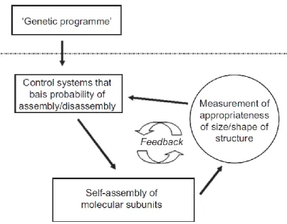
In order to generate the correct subcellular structures, a cell must sense and directly control its own shape, not only for the already complex initial shape establishments, but also to control transient or permanent shape changes, to correct for eventual mistakes and to respond to eventual signals from their environment. A generalized view of a morphogenetic program can be oversimplified in the schematic proposed by J. Davies (Figure 1.13), which suggests that the presence of negative feedback is a key principle in morphogenesis (Davies, 2013).
Many mechanosensing feedback regulating morphogenesis and differentiation of specialized structures, have been described in a wide range of organisms, from amoeba, to animal, plants and fungi.
In Dictyostelium, during mitotic cell shape change, an active recruitment of myosin II has been suggested to counteract cell shape deformation, to correct for cell asymmetries. This recruitment
26
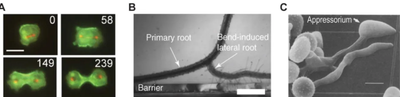
Figure 1.14. Mechanosensing mechanisms regulating morphogenesis and differentiation. A. Redistribution of
GFP-MyoII during mitosis progression in Dyctostelium. In red RFP-Tub1.Time is in second, scale bar 10 m (Effler et al., 2006). B. Lateral root formation in A. thaliana primary root, induced experimentally by placing a coverslip perpendicular to the axis of growth. Scale bar 500 m (Monshausen and Gilroy, 2009). C. Scanning electron microcgraph of two germlings of U. appendiculatus grown on polystyrene substrate presenting ridges. The appressorium differentiate at site of contact with the ridge. Scale bar 10 m (Zhou et al., 1991). Images adapted from indicated references.
is also activated after ectopic deformation of the cell surface, suggesting that a mechanosensing mechanism may perceive strain or cell shape, recruiting MyoII to correct for deformation. However, putative mechanosensors involved in this mechanism have not been uncovered yet (Figure 1.14A) (Effler et al., 2006).
In some instances, the signal can come from external cues, as in plants, where lateral root formation can be triggered by mechanical bending of a primary root. After this mechanical stimulus, auxin can redistribute and lead to the emergence of a front of growth. This mechanism can be crucial during growth through the soil: if a root encounters a barrier, it adopts an avoidance response to circumnavigate the obstacle and increase root spreading (Figure 1.14B). Despite this mechanism being known from the early 1900, the mechanosensing mechanism involved in this event remains to be established (Monshausen and Gilroy, 2009).
In the pathogenic fungus Uromyces appendiculatus, germ tubes form appressoria, differentiated structures involved in host invasion. Appressoria form only on specific topological areas of leaves, in particular when the germ tube encounters ridges of the height of stomatal guard cells. It has been proposed that, when germ tube/ridge contact occurs, this may activate mechanosensitive ion channels that trigger differentiation (Figure 1.14C) (Zhou et al., 1991).
Although several pathways involved in response to mechanical stimuli have been described or suggested, it is still not clear how these signals are perceived. Some classes of proteins or protein complexes have been identified as mechanosensors, such as ion channels, G proteins coupled receptors (GPRC), complexes linking the extracellular matrix to the cytoskeleton, or trans-wall proteins in fungi. However, the precise mechanical change sensed is not always clear (tension,
27 stress, strain), as well as the link between sensing and response pathway. Moreover, mechanosensing is often studied under mechanical stress, but cells must sense their boundaries and shape even during physiological conditions, to develop correctly and maintain homeostasis.
29
Chapter 2 – Elongation and morphogenesis in tip-growing walled cells
In this chapter, I introduce the main focus of my work, which is the morphogenesis of tip growing walled cells. As described below, in these cells the process of growth and shape establishment are tightly linked. I mainly focus on plant pollen tubes and root hairs, on fungal hyphae and tip growing yeasts. I start with a general introduction on tip growth. Then I describe tip growing walled cells from a purely mechanical point of view, introducing the main parameters that influence cell mechanics. Thereafter, I focus on how these cells regulate these mechanical parameters. For instance, I describe how cells regulate their turgor pressure and the properties of their walls. Finally, I summarize several models, both theoretical and experimental, proposed to link wall mechanical properties, growth and morphogenesis. I highlight differences and similarities between these models, their strengths and weaknesses, trying to define a general scenario on which we can base further studies, to better understand the process of tip elongation.
Table of contents
Chapter 2 – Elongation and morphogenesis in tip-growing walled cells ...29 2.1 Mechanics of walled cells ...35 2.2 Turgor pressure regulation ...41 2.3 Cell wall properties ... Errore. Il segnalibro non è definito. 2.3.1 Cell wall composition ... Errore. Il segnalibro non è definito. 2.3.2 Cell wall synthesis ...48 2.3.3 Cell Wall Integrity pathway ...51 2.4 Influence of cell wall mechanics on morphogenesis and growth ...54
31
O
rganisms ranging from plants to bacteria and fungi have their cells encased in a stiff structure, the cell wall. This wall, mainly composed of glucans and/or proteins, constitutes the full surface of the cell. One essential role of the cell wall is to prevent lysis, as walled cells are normally under very high internal osmotic pressure that is also the driving force for growth.As mentioned in the previous chapter (1.3.1), in walled cells the structure of the wall is shaping the cell underneath. This is evident by the enzymatic removal of the cell wall in osmotically supported conditions, whereby the generated protoplast loses its shape, becoming round, and sometimes leaves behind the remnants of the cell wall in an intact and defined shape (Davì and Minc, 2015; Flor-Parra et al., 2014).
Similarly, the cell cannot grow without assembly of new wall material, and this assembly may directly influence the direction and velocity of growth. Moreover, during growth, the cell will acquire a new shape that is nothing but the shape of the newly assembled cell wall.
Therefore, in these cells, the process of growth and morphogenesis are tightly linked, and one cannot fully understand how growth and morphogenesis work, without understanding the properties of the cell wall, its composition, mechanics and dynamic regulation.
Tip growing walled cells are interesting models to directly study the links between wall properties, elongation and shape determination. In these cells, the extremely polarized growth typically generates tubular shapes (Figure 2.1).
Figure 2.1. Examples of tip growing walled cells. Microbacterial filament from A. Kitasatospora setae and B.
Streptomyces coelicolor A3(2). Bar is 1 m. C. Allomyces hyphal growth. D. Lily root hair. Adapted from (Goriely, 2017)
32
Figure 2.2. Apical and lateral branching in Neurospora crassa hyphae. On the right, the central part of the colony
presents hyphal fusions (stars). Scale bars 20 m (Pandey et al., 2004).
Tip elongation is a mechanism that has evolved several times in diverse taxonomic kingdoms (Figure 2.1). It has important physiological functions, to give some examples: in mycelial bacteria as well as in filamentous fungi, tip growth is necessary for the correct spreading of the colony (Flardh, 2003; Sanati Nezhad and Geitmann, 2013). In some fungal pathogens, tip elongation of the hyphae is necessary for host infection (Brand, 2012). Tip elongation is at the base of tropism, the ability of the cell to grow towards (or away from) a specific stimulus (chemicals, light, pheromones). Moreover, tip growth is necessary for root and root hair development, therefore for plant anchorage, and also for fertilization in seed plants, during which pollen can grow a germ tube that explores relatively long distances towards the ovules (Rounds and Bezanilla, 2013).
Despite the structural rigidity of the surface in walled cells, tip growth and morphogenesis are highly variable and dynamic processes. In fact, these are between the fastest growing cells in nature, reaching growth rates up to several tens of microns per minute (Lew, 2011). Growth rate can vary depending on several factors such as nutrients or temperature (Hedhly et al., 2005; Sevilla and Odds, 1986). Even within the same cell, growth rate variation can occur over time, as in pollen tubes, which alternate between phases of stable and oscillatory growth (Pierson et al., 1996; Rojas et al., 2011).
Shapes of growing tubes are highly variable. Diameters vary between species ranging from 10-15
m of pollen tubes, to less than 1 m in tip growing bacteria (Figure 2.1). Many filamentous fungi create apical or lateral branches, fundamental for the development of mycelia that often show high
33
Figure 2.3. Appressoria development. A Cryo-SEM of conidia (C) from Blumeria graminis f.sp. hordei growing on
barley leaf, developing a primary germ tube (PGT), the secondary appressoria germ tube (AGT) and the appressoria (Ap) (Prats et al., 2006). B Electron micrographs of Magnaporthe grisea peg formation from an appressorium, invading cellophane membrane (Deising et al., 2000).
variability in diameter, even within the same colony (Figure 2.2) (Harris, 2008). A single cell can change the direction of growth, hence shape, in response to external cues, such as the presence of chemo-attractant (e.g. pheromone). A beautiful example of how these cells can develop different shapes over growth and differentiation is the generation of appressoria in pathogenic fungi, which are specialized structures involved in host invasion first described by Frank in 1883. They originally develop from conidia, non-sexual spores of fungi, which initially germinate creating a primary thin germ tube, probably used for nutrient uptake. A second germ tube is then elongated, which can swell at the apex, creating a much larger structure that Frank calls an “organ”, divided from the germ tube by a septum. This structure tightly adheres to the host surface, develops a penetration peg, perpendicular to the host surface and initiates invasion (Figure 2.3) (Bastmeyer et al., 2002; Deising et al., 2000; Prats et al., 2006).
Another process requiring the dynamical behavior of tip growth is cell fusion, an event that entails a large amount of wall digestion and remodeling to allow for plasma membrane fusion. This happens during mating in yeasts, where cells of compatible mating types can fuse to undergo sexual differentiation (Merlini et al., 2013). In some filamentous fungi, like Neurospora crassa, both germinating conidia and hyphae can grow towards each other and fuse, a process that has been associated with increased fitness of the mycelial colony (Figure 2.4) (Herzog et al., 2015).
34
Figure 2.4. Conidia germination and fusion in Neurospora crassa. Time is in minutes, scale bar 5m (Herzog et al., 2015).
Therefore, the cell wall must be continuously remodeled and reshaped to permit all these dynamic changes. Tip growth comes with a very polarized synthesis of wall material that is then remodeled by turgor pressure and in some cases by enzymatic activity. This newly deformed wall is then shifted to the side of the tip, while further new wall is synthesized (Figure 2.5).
Without regulation, tip growth can be dangerous for the cell: errors in synthesis rate or in wall composition can cause for example too soft walls that may be pierced by pressure, and consequently cell lysis and death. Similar effects can be caused by defects in wall extension and turgor pressure. On the contrary, walls that are too hard or when pressure is too low, may also cause growth arrest.
Moreover, polarized synthesis and deformation of wall establishes per se the typical tubular shape of tip growing cells (Figure 2.1), suggesting that defects in synthesis and remodeling can cause aberrations in cell morphogenesis.
For these reasons, the process of tip growth is thought to be highly regulated, to allow for the correct balance between turgor pressure, synthesis and remodeling, able to preserve cell integrity and to establish a correct morphology.
In this chapter, I describe growth and morphogenesis of tip growing cells, focusing on yeast, filamentous fungi, pollen tubes and root hair. I first introduce walled cells from a purely
mechanical point of view, presenting some of the experimental measurements of their mechanical properties.
35
Figure 2.5. During tip growth new wall material (light blue) is deposited at the tip of the cell and then remodeled by
internal turgor pressure (P), to be irreversibly incorporated in the structure. Adapted from (Davì and Minc, 2015). I then focus on how these cells can control their mechanical parameters, for instance by regulating wall properties and turgor pressure. Finally, I outline the different proposed theories to explain how cell wall mechanics can influence growth and shape generation.
2.1 Mechanics of walled cells
The main features that define mechanically a walled cell are the properties of its surface (the wall), and the internal osmotic pressure: these can be seen as the simplest set of components defining cell shape and growth patterns (Figure 2.6).
Walled cells are often compared to balloons. In fact, they are composed of a relatively thin external layer, under tension from high pressure. Turgor pressure is recognized as the driver of growth in walled cells (Rojas and Huang, 2018). This is a hydrostatic pressure, arising from the ability of these cells to increase (or decrease) their intracellular concentration of osmotically active molecules, such as ions, sugars or amino acids. Osmotic adjustment leads to influx (or efflux) of water from the milieu, increasing (or decreasing) turgidity (more insights on turgor pressure regulation in 2.2). Direct measurement of turgor can be performed by a pressure probe, an oil filled microcapillary directly inserted in the vacuole of studied cells (Green, 1968). Due to the size of this capillary this direct measurements are restricted to large cells, such as giant algae or plant cells, where different studies have reported values between 0.1 and 1 MPa (Steudle and Zimmermann, 1974; Triboulot et al., 1995). In smaller cells, such as yeast and filamentous fungi, turgor pressure can be estimated through indirect approaches, such as Atomic Force Microscopy (AFM) (Deng et al., 2011), osmotic treatments coupled with laser ablation (Atilgan et al., 2015), or elastic deformation of microchambers (Minc et al., 2009a). These different methods gave similar
36
Figure 2.6. Mechanics of tip growing cell. The wall is represented in green. P is the isotropic turgor pressure; Y is the
bulk elastic modulus of the wall, while h is thickness. represent wall deformation over growth (strain) estimated values of turgor, in the order of 1-2 MPa for yeasts and filamentous fungi.
Although turgor is the effective driving force for growth and wall deformation, its sole contribution cannot explain the polarized shape of tip growing cells, since its force is equal in all directions (Figure 2.6). Moreover, in lily pollen tubes, oscillations in growth rate can occur without variations in turgor (Benkert and Bentrup, 1997; Zerzour et al., 2009). Therefore, to understand the polarized shape and the mechanism of growth of these cells, the mechanics of their cell wall must be investigated.
In a simplified view, the mechanics of the cell wall can be separated in two independent components, thickness and bulk elastic modulus.
Cell wall thickness does not depend on wall composition and is the most relevant component of wall geometry. Wall thickness is highly variable amongst different organisms. It ranges from few nanometers in gram negative, to tens on nanometers in gram positive bacteria (Vollmer et al., 2008), reaching 100-300 nm in fungi (Ghamrawi et al., 2015; Osumi, 2012), up to 500 nm in tip growing cells of plants (Lancelle and Hepler, 1992). Significant variations in thickness have been reported within species, the extreme of which can be found in lily pollen tube which is in a range of 500 nm (Lancelle and Hepler, 1992; McKenna et al., 2009), while in tobacco the wall measures around 250 nm (Lancelle and Hepler, 1988). Wall thickness also varies in different structures of the same organism, a simple example being the difference between hyphal and conidial wall in fungi, such as in Scedosporium boydii, where they present thickness of about 80 nm and more than 200 nm respectively (Figure 2.7) (Ghamrawi et al., 2015). Also growth and metabolic condition can influence wall thickness, as has been observed in pollen grains of pea, in which a thickness increase of two fold is reached between 25°C and 36°C, together with a decrease in germination efficiency
37
Figure 2.7. Transmission electron micrographs showing thickness and structural difference of the cell wall between
hypha (A) and conidia (B) in Scedosporium boydii (Ghamrawi et al., 2015).
(Jiang et al., 2015). Similarly, differences in carbon source may affect thickness, for instance in
Candida albicans, yeasts growing on glucose have a 100 nm thick structural layer of the wall, that
halves if cells grow on lactose (Ene et al., 2015). However, so far, observation of the dynamic behavior of cell wall thickness have been highly hindered as thickness is typically below the resolution of light microscopy. The classical method to measure cell wall thickness is Transmission Electron Microscopy (TEM) that is a powerful tool to study the ultrastructure of cells. However, TEM has several limitations for the study of cell wall: first, TEM can be performed only on fixed samples that are afterwards embedded in resins; in these processes, cell structures often shrink and deform, leading to potential aberration in measurements. Second, for observation, samples are sliced by ultra-tomography: a cut out of plane or slightly oblique can lead to mistakes in thickness spatial distributions. Third, and most important, fixation precludes any information on cell wall dynamics. Despite the aforementioned observation of thickness variation, this technological limit is probably at the base of the idea that wall thickness is quasi-constant over time, and this assumption has been used in many models of tip growth. However, there are few attempts to study wall thickness in live. One method is based on the expression of His tagged membrane sensors, engineered to have variable measured length, used as molecular rulers, by coupling single molecule Atomic Force Microscopy (AFM). Only long enough sensors can be detected by the AFM tip.
38
Through this method the authors could detect thickness variation after enzymatic digestion of the wall, in
Figure 2.8. Different approaches for live cell wall thickness measurements. A. Schematic of the approach used by
Dupres et al.: a transmembrane sensor with different length (grey+yellow) is His tagged (green). The AFM tip (yellow), functionalized for His interaction can recognize only sensors with length higher than wall thickness (Dupres et al., 2010). B. DIC images of lily pollen tubes. The sharp gradient in at the front of the tip is used as a direct measure of local wall thickness (McKenna et al., 2009).
budding yeast. However, this approach is particularly complicated, the expected thickness must be known to engineer the right ruler size, and only one molecular ruler can be expressed in one strain, with the consequence of hiding local variation below the size of the molecule. Moreover, modified expression of sensors may have an impact on wall integrity and alter its properties (Figure 2.8 left) (Dupres et al., 2010). The second study exploited the thick cell wall of lily pollen tubes (500 nm): thickness variations can be detected by difference in DIC images. This analysis showed that during oscillating growth of pollen tubes, apex thickness also oscillates in a range between 400 and 800 nm, with phases of thickening preceding fast growth (Figure 2.8 right) (McKenna et al., 2009). Despite the limitation of this method being only applicable to very thick walls, it is clearly evident that thickness is a highly dynamic parameter of wall mechanics that should be considered as such in the studies of dynamic behavior of walled cells. More technological advancements are needed to measure this parameter in a live experimental setting.
The bulk elastic modulus, or Young modulus, is an intrinsic property of the material, directly related to its composition and independent of geometry, and has units of pressure. For the cell wall, this modulus can directly depend on glucan properties, polymers length or cross-links. An evident proof of cell wall elasticity has been observed in S. pombe cells, where cells forced in bent shape
39 in microchambers can come back to their normal rod shape within seconds, if they pop out of the well (Minc et al., 2009a; Minc et al., 2009b).
A classical method to measure wall elastic modulus is AFM. This has been applied on isolated cell walls, or on intact cell surfaces, to study cell wall ultrastructure and measure its elastic modulus. However, only very accurate technical approaches can allow for the correct estimation of cell wall elastic modulus in live cells, successfully separating the influence from wall geometry and turgor (Milani et al., 2013). Other indirect approaches have used deformation of elastomeric microchambers (Minc et al., 2009a), or have coupled modeling with cell deformation under osmotic treatment (Abenza et al., 2015; Atilgan et al., 2015), or with estimation of the other mechanical parameters (Goriely and Tabor, 2006). These estimations revealed values of elastic modulus on the order of 10-100 MPa in tip growing cells (Goriely and Tabor, 2006; Minc et al., 2009a; Vogler et al., 2013). Indentation analysis have shown a local softening of the wall at cell tip, in pollen tubes (Geitmann and Parre, 2004; Zerzour et al., 2009), supported also by different composition of the apical area (discussed below Errore. Il segnalibro non è definito.). Similar heterogeneities have been reported also for hyphal elongation (Ma et al., 2005) and for the extreme case of polar growth in mating protrusion of budding yeast (Goldenbogen et al., 2016).
Other important mechanical parameters are derived from this initial set, plus cell geometry, for each cell type.
The surface modulus of the cell wall is the product of bulk elastic modulus and thickness. This parameter best represents the “stiffness” of the wall, containing together the information on mechanical composition and geometry.
Surface tension is the tendency of a fluid to acquire the minimal surface area possible. Defined as the force exerted per unit area by the surrounding molecules, in a cell it does not depend on surface properties. Instead, it depends on cell geometry, and in a cylindrical cell it is defined as:
𝜇 = ∆𝑃𝑅
where P is turgor pressure and R is cell radius. As discussed in 1.3, non-walled cells modify their surface tension, through actin cortex rearrangement, to permit morphological changes (Salbreux et al., 2012). Moreover, in Bacillus subtilis, a gram positive bacteria, it has been recently proposed that membrane surface tension regulates the rate of cell wall synthesis, modulating responses to hypo- or hyper-osmotic stress (Rojas and Huang, 2018). This suggests that some cells can regulate
40
their homeostasis by controlling this parameter, therefore its estimation may help to understand these strategies.
Surface stress is defined as the ratio of the force acting on a cross-section of the material scaled by the area of the material resisting the force, and in walled cells can be written as:
𝜎 =∆𝑃𝑅
ℎ
where h is the thickness of the wall, while the elastic strain of the wall corresponds to its deformation, and can be defined as:
𝛾 =∆𝑃𝑅
𝑌ℎ where Y is the bulk modulus of the wall (Figure 2.6).
These last two parameters are interesting to understand how cells can balance turgor pressure, by modulating shape and wall mechanical parameters, to maintain homeostasis and integrity. For instance, a recent work proposed that the highly lobed puzzle shape of leaf epidermal pavement cells is the consequence of isotropic growth, and is obtained in order to minimize mechanical surface stress. In support of this model, the authors show that, in early meristems, cells unable to undergo geometrical changes, because of microtubules de-polymerization, increase in size until they burst (Sapala et al., 2018). In another recent work, that will be better discussed in the last paragraph of this chapter, the authors showed that, during polarized growth of mating projections in budding yeasts, cells must sense their strain, in order to maintain cell integrity. In fact, mutants putatively impaired in this sensing can lyse (Banavar et al., 2018).
Therefore, monitoring more complex parameters of surface mechanics can be a good approach to understand how cells maintain integrity, while ensuring growth and expansion.
To summarize, tip-growing cells have very peculiar mechanical properties, with turgor pressure up to 1-2MPa, and elastic moduli similar to that of rubber. We can compare their mechanical properties to that of a bike tire. In these cells, growth and wall deformation are driven by turgor, while shape is ultimately determined by the mechanics of the cell wall. Cells adopt different strategies, by modulating surface mechanics, for the maintenance of their homeostasis.
Despite current knowledge being sparse and incomplete, estimations of cell mechanical parameters start to give an idea of wall asymmetries that may regulate tip growth expansion, and they have been used in several theoretical and experimental models that attempt to explain tip growth and shape determination. I focus on these works in the last paragraph of this chapter. For a better
41 understanding of mechanical parameter regulation, in the next paragraph, I describe how cells establish and modulate turgor and wall structure.
2.2 Turgor pressure regulation
Turgor pressure is the mechanical driver of cell growth, therefore, it is not surprising that cells carefully regulate it. Furthermore, many walled cells are sessile, and they can be subjected to massive and uncontrolled change in environmental osmotic conditions. For example, floods can cause hypo-osmotic stress, with consequent increase of water intake and risk of cells bursting. On the other hand, a microorganism that is growing on a fruit can be subjected to hyper-osmotic shock, as soon as the fruit starts to dry and its concentration in sugars increase to saturation levels. This causes turgor reduction, water efflux and cell shrinkage, all conditions deleterious for growth. Similarly, drought or pathogen infections can cause water exit and turgor loss. Moreover, modulation of turgor pressure is fundamental for several physiological functions, for example, to breach the hosts interior, in appressoria, pressure can increase up to 8MPa before penetration (Howard et al., 1991). Dynamic changes in turgor are also associated to organ movements in plants, as well as circadian movements of leaves (Beauzamy et al., 2014; Forterre, 2013).
To contrast osmotic stress and/or to modulate pressure under physiological conditions, walled cells have developed diverse signaling cascades to tune the concentration of intracellular osmolytes, in order to control the water content. So far, the best described system is the HOG MAP kinase cascade in S. cerevisiae (Figure 2.9). This pathway, is not essential for growth in standard