HAL Id: hal-02797029
https://hal.archives-ouvertes.fr/hal-02797029
Submitted on 5 Jun 2020HAL is a multi-disciplinary open access archive for the deposit and dissemination of sci-entific research documents, whether they are pub-lished or not. The documents may come from teaching and research institutions in France or abroad, or from public or private research centers.
L’archive ouverte pluridisciplinaire HAL, est destinée au dépôt et à la diffusion de documents scientifiques de niveau recherche, publiés ou non, émanant des établissements d’enseignement et de recherche français ou étrangers, des laboratoires publics ou privés.
Anisotropy links cell shapes to tissue flow during
convergent extension
Xun Wang, Matthias Merkel, Leo Sutter, Gonca Erdemci-Tandogan, M. Lisa
Manning, Karen Kasza
To cite this version:
Xun Wang, Matthias Merkel, Leo Sutter, Gonca Erdemci-Tandogan, M. Lisa Manning, et al.. Anisotropy links cell shapes to tissue flow during convergent extension. Proceedings of the National Academy of Sciences of the United States of America , National Academy of Sciences, 2020, 117 (24), pp.13541-13551. �10.1073/pnas.1916418117�. �hal-02797029�
1
Classification: Biological Sciences, Developmental Biology; Physical Sciences, Biophysics and Computational Biology
Anisotropy links cell shapes to tissue flow during convergent extension
Xun Wang1,4, Matthias Merkel2,3,4, Leo B. Sutter 2,4, Gonca Erdemci-Tandogan2, M. Lisa
Manning2, Karen E. Kasza1,*
1 Department of Mechanical Engineering, Columbia University, New York, New York 10027,
USA
2 Department of Physics and BioInspired Institute, Syracuse University, Syracuse, New York,
13244, USA
3 Aix Marseille Univ, Université de Toulon, CNRS, CPT, Turing Center for Living Systems,
Marseille, France
4 These authors contributed equally: Xun Wang, Matthias Merkel, Leo B. Sutter
* To whom correspondence should be addressed. Email: [email protected]
2
Abstract
Within developing embryos, tissues flow and reorganize dramatically on timescales as short as minutes. This includes epithelial tissues, which often narrow and elongate in convergent extension movements due to anisotropies in external forces or in internal cell-generated forces. However, the mechanisms that allow or prevent tissue reorganization, especially in the presence of strongly anisotropic forces, remain unclear. We study this question in the converging and extending
Drosophila germband epithelium, which displays planar polarized myosin II and experiences
anisotropic forces from neighboring tissues, and we show that in contrast to isotropic tissues, cell shape alone is not sufficient to predict the onset of rapid cell rearrangement. From theoretical considerations and vertex model simulations, we predict that in anisotropic tissues two experimentally accessible metrics of cell patterns—the cell shape index and a cell alignment index—are required to determine whether an anisotropic tissue is in a solid-like or fluid-like state. We show that changes in cell shape and alignment over time in the Drosophila germband predict the onset of rapid cell rearrangement in both wild-type and snail twist mutant embryos, where our theoretical prediction is further improved when we also account for cell packing disorder. These findings suggest that convergent extension is associated with a transition to more fluid-like tissue behavior, which may help accommodate tissue shape changes during rapid developmental events.
Significance
Cells and tissues dramatically change shape to form functional tissues and organs during embryonic development. It is not well understood how mechanical and biological factors influence whether a developing tissue flows like a fluid or instead resists shape changes like a solid. Combining experimental studies in the fruit fly embryo with modeling approaches, we show that the shapes and alignment of cells within tissues can help to elucidate and predict how tissues change shape during development and how defects in these processes can result in abnormalities in embryo shape. Because many genes and cell behaviors are shared between fruit flies and humans, these results may reveal fundamental mechanisms underlying human development.
3
Introduction
The ability of tissues to physically change shape and move is essential to fundamental morphogenetic processes that produce the diverse shapes and structures of tissues in multicellular organisms during development (1, 2). Developing tissues are composed of cells that can dynamically change their behavior and actively generate forces to influence tissue reorganization and movement (3–8). Remarkably, tissues dramatically deform and flow on timescales as short as minutes or as long as days (6). Recent studies highlight that tissue movements within developing embryos can be linked with the tissue fluidity (8–11), and computational models assuming predominantly fluid-like tissue behavior predict aspects of tissue movements (12, 13). Fluid-like tissues accommodate tissue flow and remodeling, while solid-like tissues resist flow. Yet, the mechanisms underlying the mechanical behavior of developing tissues remain poorly understood, in part due to the challenges of sophisticated mechanical measurements inside embryos and the lack of unifying theoretical frameworks for the mechanics of multicellular tissues (6, 7, 14). Epithelial tissue sheets play pivotal roles in physically shaping the embryos of many organisms (2), often through convergent extension movements that narrow and elongate tissues. Convergent-extension is highly conserved and used in elongating tissues, tubular organs, and overall body shapes (15). Convergent extension movements require anisotropies in either external forces that deform the tissue or asymmetries in cell behaviors that internally drive tissue shape change. Indeed, an essential feature of many epithelia in vivo is anisotropy in the plane of the tissue sheet, a property known as planar polarity, which is associated with the asymmetric localization of key molecules inside cells (16–19). For example, during Drosophila body axis elongation, the force-generating motor protein myosin II is specifically enriched at cell edges in the epithelial germband tissue that are oriented perpendicular to the head-to-tail body axis (20, 21) (Fig. 1A). Planar polarized myosin is required for cell rearrangements that converge and extend the tissue to rapidly elongate the body and is thought to produce anisotropic tensions in the tissue (12, 13, 21–25). In addition, the Drosophila germband experiences external forces from neighboring tissues, including the mesoderm and endoderm, which have been linked to cell shape changes in the germband during convergent extension (26–29) (Fig. 1A). Despite being fundamental to epithelial tissue behavior in vivo, it is unclear how such anisotropies arising from internal myosin planar polarity and external forces influence epithelial tissue mechanical behavior, particularly whether the tissue behaves more like a fluid or a solid.
Vertex models have proven a useful framework for theoretically studying the mechanical behavior of confluent epithelial tissues (30, 31), including the packings of cells in tissues (32–34) and the dynamics of remodeling tissues (23, 32, 35–37). Recent studies of the energy barriers to cell rearrangement in isotropic vertex models, which assume no anisotropy in either internal tensions at cell-cell contacts or in external forces, have revealed a transition from solid to fluid behavior, which depends on whether large or small contacts are favored between neighboring cells. The transition is indicated by a single parameter describing cell shape, 𝑝𝑝̅, which is the average value in the tissue of cell perimeter divided by the square root of cell area (38–40). When cells prefer smaller contacts with neighbors, 𝑝𝑝̅ is small and the tissue is solid-like. Above a critical value of 𝑝𝑝̅, 𝑝𝑝𝑜𝑜∗, the tissue becomes fluid-like. The isotropic vertex model successfully predicts that cell shapes identify the transition from fluid-like to solid-like behavior in cultured primary bronchial epithelial tissues; initial modeling work suggested the critical cell shape 𝑝𝑝𝑜𝑜∗ is close to 3.81 (39), in good agreement with the experiments (41). Such a simple way to infer tissue behavior from static images
4
is appealing, particularly for tissues that are inaccessible to mechanical measurements or live imaging.
However, subsequent work has shown that the precise value of 𝑝𝑝𝑜𝑜∗ depends on specific features of the cell packing, such as the number of manyfold coordinated vertices (42) or the distribution of neighbor numbers in the packing (43–45), though the latter feature has never been studied systematically. In addition, these previous vertex model studies did not account for effects of anisotropy, potentially limiting their use in the study of converging and extending tissues.
Here, we combine confocal imaging and quantitative image analysis with a vertex model of anisotropic tissues to study epithelial convergent extension during Drosophila body axis elongation. We show that cell shape alone is not sufficient to predict the onset of rapid cell rearrangement during convergent extension in the Drosophila germband, which exhibits anisotropies arising from internal forces from planar polarized myosin and external forces from neighboring tissue movements. Instead, we show that for anisotropic tissues, such as the
Drosophila germband, anisotropy shifts the predicted transition between solid-like and fluid-like
behavior and so must be taken into account, which can be achieved by considering both cell shape and cell alignment in the tissue. We find that the onset of cell rearrangement and tissue flow during convergent extension in wild-type and mutant Drosophila embryos is more accurately described by a combination of cell shape and alignment than by cell shape alone. Moreover, we use experimentally accessible features of cell neighbor relationships to quantify cell packing disorder and pinpoint 𝑝𝑝𝑜𝑜∗ , which further improves our predictions. These findings suggest that convergent extension is associated with a transition from solid-like to more fluid-like tissue behavior, which may help to accommodate dramatic epithelial tissue shape changes during rapid axis elongation.
Results
Cell shape alone is not sufficient to predict the onset of rapid cell rearrangement in the
Drosophila germband epithelium. To explore the mechanical behavior of a converging and
extending epithelial tissue in vivo, we investigated the Drosophila germband, a well-studied tissue that has internal anisotropies arising from planar polarized myosin (20–25, 46) and also experiences external forces from neighboring developmental processes that stretch the tissue (26, 27). The germband rapidly extends along the anterior-posterior (AP) axis while narrowing along the dorsal-ventral (DV) axis (Fig. 1A), roughly doubling the length of the head-to-tail body axis in just 30 min (47) (Fig. 1C). Convergent extension in the Drosophila germband is driven by a combination of cell rearrangements and cell shape changes (Fig. 1B,C). The dominant contribution is from cell rearrangement (21, 22, 28, 47), which requires a planar polarized pattern of myosin localization across the tissue (20, 21) that is thought to be the driving force for rearrangement (21, 23, 24, 46). Cell stretching along the AP axis also contributes to tissue elongation and coincides with movements of neighboring tissues (26–28, 48, 49), indicating that external forces play an important role in tissue behavior. Despite significant study of this tissue, a comprehensive framework for understanding its mechanical behavior is lacking, in part because direct mechanical measurements inside the Drosophila embryo, and more generally for epithelial tissues in vivo, continue to be a challenge (50–52).
To gain insight into the origins of mechanical behavior in the Drosophila germband epithelium, we first tested the theoretical prediction of the vertex model that cell shapes can be linked to tissue
5
mechanics. In the isotropic vertex model, tissue mechanical behavior is reflected in a single parameter, the average cell shape index 𝑝𝑝̅ (38–41). To quantify cell shapes in the Drosophila germband, we used confocal time-lapse imaging of embryos with fluorescently-tagged cell membranes (53) and segmented the resulting time-lapse movies (28) (Fig. 2A, SI Appendix Fig. S1). Prior to the onset of tissue elongation, individual cells take on roughly isotropic shapes and become more elongated over time (Fig. 2A,B), consistent with previous observations (26–28, 54). Ten minutes prior to tissue elongation, the cell shape index 𝑝𝑝̅ averaged over 8 wild-type embryos is just above 3.81. Eight minutes before the onset of tissue elongation, 𝑝𝑝̅ starts to increase before reaching a steady value of 3.98 about 20 min after the onset of tissue elongation (Fig. 2B). The average cell shape index prior to tissue elongation, 𝑝𝑝̅ = 3.81 (dashed line, Fig. 2B), is close to the value associated with isotropic solid-like tissues in previous work (38–40), suggesting that the tissue may be solid-like prior to elongation.
We next asked how these cell shapes vary among the individual embryos and correlate with tissue mechanical behavior. As an experimentally accessible read-out of tissue fluidity, we used the instantaneous rate of cell rearrangements occurring within the germband tissue (Fig. 1C), where higher rearrangement rates are associated with more fluid-like behavior and/or larger driving forces. Plotting instantaneous cell rearrangement rate versus 𝑝𝑝̅ at each time point from movies of individual wild-type Drosophila embryos, we find that the onset of rapid cell rearrangement occurs at different values of 𝑝𝑝̅ for each embryo, ranging from 3.83 to 3.90 for a cutoff rearrangement rate per cell of 0.02 min-1 (Fig. 2C). We verified that we observe a similar variation in the values of 𝑝𝑝̅ for different cutoff values (SI Appendix Fig. S2). This suggests that in the germband epithelium, comparing the cell shape index 𝑝𝑝̅ to a fixed critical value (e.g. 3.81) is not sufficient to predict tissue behavior.
Cellular packing disorder is not sufficient to predict the onset of rapid cell rearrangement in the germband. Recent vertex model simulations suggest that 𝑝𝑝̅ = 3.81 is often insufficient to
separate solid from fluid tissue behavior, as the precise location of the solid-fluid transition depends on how exactly cells are packed in the tissue (42–45). A hexagonal packing has no packing disorder, while each cell with neighbor number different from six increases the packing disorder in the tissue. In the modeling literature, this disorder is typically generated either by allowing manyfold coordinated vertices (i.e. vertices at which more than three cells meet) or using simulation preparation protocols that create cell neighbor numbers other than six. Including manyfold vertices in simulations is natural, as they are observed in the germband epithelium (54), and are often formed during cell rearrangements involving four or more cells (21, 22). Moreover, recent theoretical work has predicted how the presence of manyfold vertices increases the critical shape index (42).
We wondered whether cell packing disorder quantified by the vertex coordination number z could explain the observed embryo-to-embryo variability in 𝑝𝑝̅ at the transition point in wild-type embryos (Fig. 2C, SI Appendix Fig. S2). To test this idea, we plotted 𝑝𝑝̅ versus z at each time point and color-coded the data based on the instantaneous cell rearrangement rate, pooling the data from all wild-type embryos (Fig. 2F). To isolate the changes in mechanical behavior of the germband during convergent extension from later developmental events, we focus on times t ≤ 20 min after the onset of tissue elongation, well before cell divisions begin in the germband. If vertex coordination were sufficient to explain the germband behavior, then the theoretically determined
6
line (dashed line) should separate regions with low cell rearrangement rate (blue symbols) from regions with high cell rearrangement rate (red, orange, and yellow symbols) (Fig. 2F). However, this is not the case, indicating that the prediction from Ref. (42) alone is not sufficient to account for the germband behavior during this stage.
Next, we asked if other aspects of packing disorder could affect tissue fluidity. Even without manyfold vertices, it is possible to generate packings in silico with differences in packing disorder just by altering the preparation protocol. Since this has not been systematically studied, we performed a large number of vertex model simulations where we varied the packing disorder (SI
Appendix Fig. S3A,B). In our simulations, the transition point is well predicted by the fraction of
pentagonal cells, i.e. cells that have exactly five neighbors, with a linear dependence (Fig. 2D, SI
Appendix Fig. 3C,D). In particular, without any pentagonal cells, we recover the previously
predicted transition point of ≈3.72 for tissues consisting only of hexagonal cells (33, 38). In comparison, the previously reported value of 3.81 corresponds to a fraction of ≈15% pentagonal cells (Fig. 2D). While additional aspects of cell packing likely affect the transition, these results suggest that the fraction of pentagonal cells may also be a good predictor for the transition point in isotropic tissues.
To test whether this second measure of packing disorder could explain the variability in 𝑝𝑝̅ at the transition in wild-type embryos, we plotted 𝑝𝑝̅ versus the fraction of pentagonal cells at each time point, again color-coding the data based on the instantaneous cell rearrangement rate and pooling data from all wild-type embryos (Fig. 2E, SI Appendix Fig. S2). We find that the packing disorder quantified by the fraction of pentagonal cells is also insufficient to explain the onset of cell rearrangements.
Our results suggest that two measures of packing disorder, the vertex coordination number and fraction of pentagonal cells, have at least partially independent effects on the isotropic vertex model transition point. However, neither of them is sufficient to understand the transition to high cell rearrangement rates in the Drosophila germband.
Theoretical considerations and vertex model simulations predict a shift of the solid-fluid transition in anisotropic tissues. To study whether anisotropies in the germband could affect the
relation between the cell shape index and cell rearrangement rate, we used vertex model simulations to test how tissue anisotropy, introduced into the model in different ways, affects tissue fluidity.
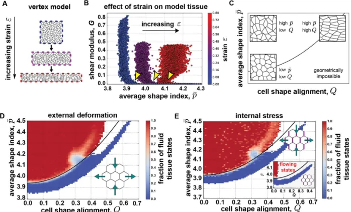
First, we introduced anisotropy by applying an external deformation, mimicking the effects of forces exerted by neighboring morphogenetic processes, and then studied force-balanced states of the model tissue (Fig. 3A). As a metric for tissue stiffness, we measured the shear modulus of the model tissue, which describes with how much force a tissue resists changes in shape. A vanishing shear modulus corresponds to fluid behavior, where the tissue flows and cells rearrange in response to any driving force, whereas a positive shear modulus indicates solid behavior, where the tissue does not flow so long as the driving force is not too large. We then analyzed how the shear modulus correlates with 𝑝𝑝̅ for different amounts of global tissue deformation, quantified by the strain 𝜀𝜀 (Fig. 3B). For small strain, we recover the behavior of the isotropic vertex model. The shear modulus is finite when 𝑝𝑝̅ is small and vanishes above a critical cell shape index, which is 𝑝𝑝𝑜𝑜∗ = 3.94 for our simulations (Fig. 3B, blue symbols). For larger strains, we find that the critical value of the shape
7
index at the transition between solid-like and fluid-like behavior generally increases with the amount of strain (Fig. 3B). Indeed, 𝑝𝑝̅ for cells in a deformed, solid tissue can be higher than for cells in an undeformed, fluid tissue. This suggests that anisotropy affects the critical shape index at which the tissue transitions between solid and fluid behavior.
Some of us recently developed a theoretical understanding for a shift in the critical shape index when deforming a vertex model tissue (45). In the limit of small deformations by some strain ε and without cell rearrangements, the critical value of 𝑝𝑝̅ increases from 𝑝𝑝𝑜𝑜∗ to 𝑝𝑝𝑜𝑜∗ + 𝑏𝑏ε2, where b is a constant prefactor. To compare this formula to the vertex model simulations (Fig. 3A,B), we need to take into account that cell rearrangements occur in our simulations. Removing their contribution from the overall tissue strain ε leaves us with a parameter Q (Fig. 3C) (SI Appendix, SI Materials and Methods), which can be quantified using a triangulation of the tissue created from the positions of cell centers (SI Appendix, SI Materials and Methods) (55, 56). We term Q a “shape alignment index”, as Q is non-zero only when the long axes of cells are aligned. We emphasize that, unlike the nematic order parameter for liquid crystals, the cell alignment parameter Q is additionally modulated by the degree of cell shape anisotropy; tissues with the same degree of cell alignment but more elongated cells have a higher Q (Fig. 3C). In other words, Q can be regarded as a measure for tissue anisotropy. After accounting for cell rearrangements, we expect the transition point in anisotropic tissues to shift from the isotropic value 𝑝𝑝𝑜𝑜∗ to (SI Appendix, SI Materials and Methods): 𝑝𝑝̅𝑐𝑐𝑐𝑐𝑐𝑐𝑐𝑐 = 𝑝𝑝𝑜𝑜∗ + 4bQ2 . (1)
Indeed, comparing this equation to vertex model simulations yields a good fit with the simulation results (Fig. 3D, solid line), with fit parameters 𝑝𝑝𝑜𝑜∗ = 3.94 and b = 0.43 (SI Appendix Fig. S4). We confirmed that cell area variation did not significantly affect these findings (SI Appendix Fig. S5). In principle, we expect both the transition point 𝑝𝑝𝑜𝑜∗ and the precise value of b to depend on the packing disorder, but our best-fit value for b is consistent with previously published results (45). Therefore, we used b = 0.43 for the remainder of this study. Hence, for external deformation, the solid-fluid transition point in the vertex model increases quadratically with tissue anisotropy Q. We also tested how the model predictions change when we introduce anisotropy generated by internal forces into the vertex model. We modeled myosin planar polarity as increased tensions on “vertical” cell-cell contacts (Fig. 3E, SI Appendix Fig. S6) (23), and focus again on stationary, force-balanced states. We investigated simulations of model tissues with internal forces, both with (Fig. 3E) and without (Fig. 3E, inset) externally applied deformation. We find that in both cases solid states exist for larger cell shape indices than the isotropic 𝑝𝑝𝑜𝑜∗ = 3.94, and our results are again consistent with the fit from Fig. 3D (solid lines in Fig. 3E and inset). With finite anisotropic internal tensions only, we obtain states in the fluid regime that do not reach a force-balanced state (detailed discussion in SI Appendix), and this explains the white region devoid of stable states in the upper middle region of Fig. 3E, inset. Taken together, these findings demonstrate that a combination of cell shape 𝑝𝑝̅ and cell shape alignment Q in the vertex model indicates whether an anisotropic tissue is in a solid-like or fluid-like state, regardless of the underlying origin of anisotropy.
Cell shape and cell shape alignment together indicate the onset of cell rearrangement during
Drosophila axis elongation. We returned to our experiments to test whether a combination of 𝑝𝑝̅
and Q would be a better predictor for the behavior of the Drosophila germband during convergent extension. We quantified alignment Q using the triangle method (Fig. 4A) and found that prior to
8
the onset of tissue elongation, which begins at t = 0 min, alignment is not very high (Fig. 4B). Q begins to increase just prior to elongation, peaking at t = 1 min (Fig. 4B, SI Appendix Fig. S7), which is consistent with observations using other cell pattern metrics (23, 26, 28, 29). This peak in Q corresponds to stretching of cells along the dorsal-ventral axis, perpendicular to the axis of germband extension, and coincides with the time period during which the presumptive mesoderm is invaginating (29, 53). Q relaxes back to low levels during axis elongation (Fig. 4B). Plotting 𝑝𝑝̅ vs Q at each time point from movies of individual wild-type embryos reveals common features, despite embryo-to-embryo variability (Fig. 4C, inset). Initially, we see a concomitant increase of 𝑝𝑝̅ and Q prior to the onset of convergent extension. Above 𝑝𝑝̅ = 3.87, Q decreases drastically as 𝑝𝑝̅ continues to increase, indicating that further increases in 𝑝𝑝̅ are associated with randomly oriented cell shapes (cf. Fig. 3C). Thus, cell shapes in the germband are transiently aligned around the onset of convergent extension.
We next asked whether this temporary increase in alignment could help resolve the seeming contradiction between the measured cell shapes and cell rearrangement rates. To this end, we investigated how 𝑝𝑝̅ and Q correlate with the instantaneous rate of cell rearrangements occurring within the germband, with higher rearrangement rates associated with more fluid-like behavior and/or larger active driving forces (Fig. 4C). The anisotropic vertex model predicts that the solid or fluid behavior of the tissue should depend on both 𝑝𝑝̅ and Q according to Eq. (1), with only two adjustable parameters, 𝑝𝑝𝑜𝑜∗ and b. We fit Eq. (1) to our experimental data by minimizing a quality of fit measure defined as the number of experimental data points on the wrong side of the theoretical transition line, and for simplicity vary only 𝑝𝑝𝑜𝑜∗ while keeping the theoretically determined value for b. Varying the value b leads at most to a slight improvement of our fit (SI
Appendix Fig. S8). To differentiate between solid-like and fluid-like tissue behavior in the
experimental data, we need to choose a cutoff value for the cell rearrangement rate. Choosing a cutoff of 0.02 min-1 per cell yields a best fit with 𝑝𝑝𝑜𝑜∗ = 3.83 (solid line, Fig. 4C). To confirm that our prediction of a quadratic dependence on Q is supported by the data, we also identify the best fit to a null hypothesis of a Q-independent transition point (horizontal dashed line, Fig. 4C). Using our quality of fit measure, we find that the Q-dependent fit is always better, independent of the chosen cell rearrangement rate cutoff (SI Appendix Fig. S8).
Comparing the trajectories of individual embryos (Fig. 4C, inset) to the predicted transition in the anisotropic vertex model (Fig. 4C), we see that during early times, when 𝑝𝑝̅ and Q are both increasing, the tissue stays within the predicted solid-like regime. The subsequent rapid decrease in Q brings embryos closer to the transition line. As 𝑝𝑝̅ further increases, individual embryos cross this transition line, which coincides with increased rates of cell rearrangement, at different points (𝑄𝑄, 𝑝𝑝̅). Thus, compared to the isotropic model, the anisotropic vertex model better describes the onset of rapid cell rearrangement and tissue flow during convergent extension with two metrics of cell patterns, 𝑝𝑝̅ and Q, that are both easy to access experimentally.
Accounting for cell shape alignment and cell packing disorder allows for a parameter-free prediction of tissue behavior. While the above results confirm that tissue anisotropy must be
taken into account to predict the onset of rapid cell rearrangement, the theoretical prediction in Fig. 4C still required a fit parameter 𝑝𝑝𝑜𝑜∗. Theoretical results suggest that this fit parameter, which is the isotropic transition point in the absence of anisotropic forces, should depend systematically on cell packing disorder quantified by vertex coordination (42) and fraction of pentagonal cells (Fig. 2D).
9
Therefore, we analyzed the 𝑝𝑝̅ and Q data for each embryo individually, by fitting them to Eq. (1) with b=0.43 where we again use 𝑝𝑝𝑜𝑜∗ as the only fit parameter (Fig. 4D, inset). We compared the 𝑝𝑝𝑜𝑜∗ obtained for each embryo (purple point, Fig. 4D inset) to the average vertex coordination number in the tissue at the time of the transition (green point, Fig. 4D inset) and found a clear correlation (dashed line, Fig 4F), which fits well with the previous theoretical prediction (42), with no fit parameters.
Combining this previous theoretical prediction of the effects of vertex coordination on the solid-fluid transition in isotropic tissues with our prediction for how cell shape alignment shifts this transition in anisotropic tissues in Eq. (1) generates the following parameter-free prediction of the critical shape index for tissue fluidity:
𝑝𝑝̅𝑐𝑐𝑐𝑐𝑐𝑐𝑐𝑐 = 3.818 + (𝑧𝑧 − 3)/𝐵𝐵 + 4𝑏𝑏𝑄𝑄2, (2)
where 𝑧𝑧 is the measured average vertex coordination number, and the other parameters are universally determined a priori from vertex model simulations: 𝐵𝐵 = 3.85 (42), and 𝑏𝑏 = 0.43. To test this prediction, we plot the cell shape index corrected by the vertex coordination number, 𝑝𝑝̅𝑐𝑐𝑜𝑜𝑐𝑐𝑐𝑐 = 𝑝𝑝̅ − (𝑧𝑧 − 3)/𝐵𝐵, versus cell shape alignment Q in the germband of wild-type embryos, and compare it to the theoretical curve given by 𝑝𝑝̅𝑐𝑐𝑜𝑜𝑐𝑐𝑐𝑐 = 3.818 + 4𝑏𝑏𝑄𝑄2 (solid line, Fig. 4D). Remarkably, this parameter-free prediction describes our experimental data well. We compared the quality of fit to alternative parameter-free predictions and found that Eq. (2) consistently provides the best prediction for a wide range of cell rearrangement rate cutoffs (SI Appendix Fig. S8).
Some embryos deviate from the theoretical prediction from Ref. (42) (Fig. 4F), suggesting that perhaps alternate features of packing disorder may play an important role in those embryos. Thus we also compared 𝑝𝑝𝑜𝑜∗ obtained from the individual-embryo fits to the respective fraction of pentagons at the time of the transition, and found a strikingly clear correlation well described by a linear relation (Fig 4E, dashed line is a linear fit). This relationship quantitatively differs from what we extracted from our vertex model simulations (Fig. 2D), indicating again that other aspects of packing disorder may also play a role. Nevertheless, using this linear fit to correct the shape index for each data point by the fraction of pentagonal cells, we obtain an improved prediction of our data (compare Fig. 4D to SI Appendix Fig. S9) at the expense of requiring two fit parameters. Taken together, these results show that we can quantitatively predict the behavior of the germband tissue in wild-type embryos, with no fit parameters using Eq. (2), from an image of cell patterns in the tissue. To do so, we needed to quantify three observables: cell shapes, cell alignment, and cell packing disorder. We found that vertex coordination and the fraction of pentagonal cells are both good proxies for packing disorder, in vertex model simulations and the germband.
Cell shape, alignment, and tissue behavior in snail twist and bnt mutant embryos. Since the Drosophila germband experiences both internal forces due to myosin planar polarity and external
forces from neighboring tissues, we wondered whether our theoretical predictions hold when altering the nature of the forces in the germband. To dissect the effects of internal and external sources of tissue anisotropy, we studied cell patterns in snail twist mutant embryos, which lack genes required for invagination of the presumptive mesoderm (57), and in bcd nos tsl (bnt) mutant
10
embryos, which lack patterning genes required for planar polarized patterns of myosin localization and axis elongation (22, 47).
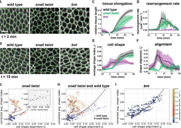
First, we analyzed cell shapes and cell shape alignment in the germband of snail twist mutant embryos in which the presumptive mesoderm does not invaginate. In snail twist embryos, we observe that the germband tissue elongates (Fig. 5C) and cell rearrangements occur (Fig. 5D), similar to prior studies (28) although at somewhat reduced rates compared to in wild-type embryos. However, in contrast to wild-type embryos, we find that the cell shape alignment Q is significantly reduced between t = -5 min and t = +8 min (Fig. 5A,F), similar to previous reports of other metrics for cell stretching (28). The cell shape index 𝑝𝑝̅ is also reduced during this period (Fig. 5E). These observations are consistent with the idea that external forces from mesoderm invagination produce the transient cell shape elongation and alignment observed in wild-type embryos.
Next, we tested whether our theoretical predictions would describe tissue behavior in snail twist embryos, even with their significantly reduced cell alignment. We found that the onset of rapid cell rearrangement in snail twist embryos is also well predicted by Eq. (2) (Fig. 5G). This is corroborated by comparing the parameters 𝑝𝑝𝑜𝑜∗ of the individual snail twist embryo fits to the vertex coordination number at the transition (Fig. 5G, inset), which is close to the previous theoretical prediction (dashed line) (42). Hence, our prediction also holds in embryos with reduced cell shape alignment Q, where the transition to rapid cell rearrangement occurs at a lower cell shape index 𝑝𝑝̅ compared to in wild-type embryos (Fig. 5H).
To investigate how disrupting other forces in the germband affects tissue behavior, we studied cell patterns in bnt mutant embryos, which lack anterior-posterior patterning genes required for axis elongation. These mutant embryos do not display myosin planar polarity, although there is significant myosin present at the apical cortex of cells (SI Appendix Fig. S10). The bnt embryos have severe defects in tissue elongation (Fig. 5C), cell rearrangement (Fig. 5D), and endoderm invagination, but still undergo mesoderm invagination (13, 20, 22, 26, 28, 47). 𝑝𝑝̅ displays an initial increase (Fig. 5E), concomitant with an increase in Q (Fig. 5F), similar to in wild-type embryos. After t = 1 min, 𝑝𝑝̅ does not increase further and takes on a steady value of 3.87 (Fig. 5E). This supports the idea that the further increase in 𝑝𝑝̅ in wild-type embryos is due to internal anisotropies associated with myosin planar polarity or external forces associated with endoderm invagination. Interestingly, Q returns more slowly to low levels in bnt compared to wild-type embryos (Fig. 5F), suggesting a potential role for myosin planar polarity, cell rearrangements oriented along the AP axis, or endoderm invagination in relaxing cell shape alignment along the DV axis. The bnt tissues do not transition to a state of rapid cell rearrangement. This is not consistent with the predictions of Eq. (2) (Fig. 5I), which predicts some fluid-like tissue states in the germband of bnt embryos, suggesting that either the driving forces are too small or that there are additional barriers that prevent rapid cell rearrangement in these embryos.
Taken together, these findings demonstrate that external forces associated with mesoderm invagination contribute to tissue anisotropy in the germband and that the onset of rapid cell rearrangement can be predicted from cell shape and alignment, even in the absence of forces associated with mesoderm invagination.
11
In this work, we show that cell shape, cell alignment, and packing disorder can be used to understand and predict whether an anisotropic tissue flows and remodels like a fluid or instead maintains its shape like a solid. Importantly, in contrast to isotropic tissues, the mechanical behavior of the converging and extending Drosophila germband cannot be predicted by cell shape and packing disorder alone. Instead, we show via theoretical analysis and simulation that in anisotropic tissues three experimentally accessible metrics—the cell shape index 𝑝𝑝̅, the cell alignment index Q, and packing disorder quantified by either vertex coordination or fraction of pentagonal cells—are required to determine whether an anisotropic tissue flows and remodels or not. We demonstrate that the onset of rapid cell rearrangement in wild-type Drosophila embryos is indeed more accurately described by a combination of these three cell pattern metrics, using an equation with no fit parameters, than by cell shape or packing disorder alone. We further tested this prediction in snail twist mutant embryos in which the presumptive mesoderm does not invaginate and found that our parameter-free prediction successfully predicts the onset of rapid cell rearrangement and tissue flow in this case as well. These findings suggest that convergent extension of the Drosophila germband might be viewed as a transition to more fluid-like behavior to help accommodate dramatic tissue flows. This raises the possibility that the properties of developing tissues might be tuned to become more fluid-like during rapid morphogenetic events. A fluid-to-solid jamming transition has recently been reported in mesodermal tissues during zebrafish body axis elongation (8). In contrast to the zebrafish mesoderm in which the transition to more solid-like behavior is associated with an increase in cellular volume fraction (proportion of the tissue occupied by cells), the Drosophila germband epithelium comprises tightly packed cells and its mechanical behavior changes in the absence of any change in cell volume fraction. Future studies will be needed to explore how the properties of epithelial cells might be regulated during development to tune the mechanical behaviors of the tissues in which they reside.
The vertex model predictions of tissue behavior are independent of the underlying origin of anisotropy, and therefore can be used to predict mechanical behavior of tissues from cell shape patterns, even when external and internal stresses cannot be directly measured. Although our current simulations were not able to access some of the tissue states driven by internal stresses, we found that the cases that were accessible were fully consistent with our simulation results without internal stresses. Importantly, the average cell shape index 𝑝𝑝̅, cell shape alignment index Q, and metrics for packing disorder are easy to access experimentally from snapshots of cell packings in tissues, even in systems where time-lapse live imaging of cell rearrangement and tissue flow is not possible. Thus, this approach may prove useful for studying complex tissue behaviors in a broad range of morphogenetic processes occurring in developing embryos in vivo or organoid systems
in vitro.
In our analysis, we characterized the mechanical state of the germband epithelial tissue using the rate of cell rearrangement as the observable. We made this choice because direct measurements of the mechanical properties of the germband remain a significant experimental challenge (6, 7, 14). Generally, higher rates of cell rearrangement could be due to more fluid tissue properties or a stronger driving force, which is the sum of externally applied forces and internally generated mechanical stresses. Based on our Eq. (2) result, the cell shape index and alignment predict the onset of rapid cell rearrangement in the germband. While this would be consistent with the tissue becoming more fluid, it is also possible that the observed increase in cell rearrangement rate is at least in part due to an increase in the driving force while the tissue remains solid.
12
To parse this possibility further, it is useful to consider a solid tissue, where the tissue will flow only if it is pulled with a force above some threshold called the yield stress. If the tissue is deeply in the solid state, far from the solid-fluid transition, and the applied force is far above the yield stress, one would expect cells to acquire elongated shapes and transiently form manyfold vertices during cell rearrangements in response to the applied force. The rearrangement rate would correlate with the cell shape index, after accounting for packing disorder and alignment, which is similar to what we predict with our fluid-solid model. However, based on our vertex model simulations we would not expect to see tissue states with high shape index 𝑝𝑝̅ and low alignment Q associated with high rearrangement rates for solid tissues. Since we do observe such tissue behavior during germband extension, this suggests that the germband is more fluid-like during these periods with high cell rearrangement rates.
Of course, it could be that the tissue is a very weak yield stress solid, so that it becomes fluid-like under very small applied forces. This is consistent with the observations that the large majority of rearrangements are oriented along the head-to-tail body axis (21, 22, 46, 47, 58) and the time period of rapid cell rearrangement (Fig. 1C) coincides with the period of planar polarized myosin (13, 25, 46). Direct mechanical measurements of the germband have not been conducted during axis elongation, but ferrofluid droplet and magnetic bead microrheology measurements have probed the mechanical behavior of the epithelium prior to germband extension in the cellularizing embryo. These studies report that tissue behavior is predominantly elastic (solid-like) over timescales less than several minutes and suggest fluid-like behavior on the longer ~30 min timescales relevant for germband extension (51, 52). These measurements might also be consistent with a weak yield-stress solid, an interpretation that would be supported by the near absence of cell rearrangements prior to germband extension. Taken together, these observations suggest that over the time period that we describe the germband as “fluid-like”, it could actually be a very weak yield-stress solid.
Though there is often little functional difference between a fluid and weak yield stress solid, the difference may be relevant for mutant bnt embryos, whose behavior is not well-captured by our theoretical predictions. In particular, we observe bnt tissues with 𝑝𝑝̅, Q, and cell packing disorder that would be predicted to display fluid-like behavior but do not undergo rapid cell rearrangement. This suggests that in these embryos the driving forces are not sufficient to overcome the yield stress. One obvious explanation for this is that the germband in bnt embryos experiences altered forces associated with disrupted myosin planar polarity (22) and defects in endoderm invagination, which would contribute to a reduced driving force. Alternatively, additional barriers to cell rearrangement in bnt mutants, of the sort described in Ref. (59), could also explain this behavior. Similarly, our vertex model does not predict the observed decrease in cell rearrangement rates after 20 minutes of axis elongation (Fig. 1C). Given the observed high values of 𝑝𝑝̅ and low values of Q, our model would still predict fluid-like behavior. Just as in the bnt mutants, this discrepancy could be explained by a decreased driving force or additional barriers to cell rearrangement. The former explanation is supported by the observation that myosin planar polarity reaches a maximum 5-10 min after the onset of axis elongation and then decreases during the rest of the process (25, 28, 46), while the latter could potentially be explained by maturation of cell junctions or changes to adhesive interactions over the course of embryonic development (60, 61).
13
Consistent with the notion of additional barriers to cell rearrangement, recent work suggests that local remodeling of active junctional tension at cell-cell contacts only occurs above a critical strain threshold in cultured epithelial cells (59, 62). This is consistent with a growing body of work that points toward important roles for membrane trafficking and E-cadherin turnover in junctional remodeling during Drosophila epithelial morphogenesis (11, 63–65). Indeed, such a mechanism of mechanosensitive barriers to junctional remodeling and cell rearrangement can be added to standard vertex models to explain such weak yield-stress behavior (59).
Moving forward, it will be interesting to explore experimentally how the nature of internal and external forces contributes to tissue mechanics, cell rearrangement, and tissue flows in the germband and other developing epithelial tissues. Incorporating these features into more sophisticated vertex models will contribute to understanding the diverse behaviors of living tissues, and the approaches we develop here will be useful for interrogating these questions.
Methods
Embryos were generated at 23℃ and analyzed at room temperature. Cell outlines were visualized with gap43:mCherry (53), Spider:GFP, or Resille:GFP cell membrane markers. Embryos were imaged on a Zeiss LSM880 laser scanning confocal microscope.Time-lapse movies were analyzed with SEGGA software in MATLAB (28) for quantifying cell shapes and cell rearrangement rates, PIVlab version 1.41 in MATLAB (66) for quantifying tissue elongation, and custom code for quantifying cell alignment using the triangle method (55, 56, 67). The vertex model describes an epithelial tissue as a planar tiling of N cellular polygons, where the degrees of freedom are the vertex positions (33). Forces in the model are defined such that cell perimeters and areas act as effective springs with a preferred perimeter p0 and a preferred area of one, which is implemented via an effective energy functional (45). Unless otherwise noted, error bars are the standard deviation. The data that support the findings of this study are included in the paper and SI
Appendix. The custom code used in this study to extract the average triangle-based Q tensor from images segmented using SEGGA (28) is available at https://github.com/mmerkel/triangles-segga.
Details can be found in the SI Appendix, SI Materials and Methods.
Acknowledgements
The authors thank Erik Boyle for assistance with data processing; Dene Farrell and Jennifer Zallen for the use of SEGGA, a segmentation and quantitative image analysis toolset; Adam Martin for the sqh-gap43:mCherry fly stock; and the Bloomington Drosophila Stock Center (BDSC) for fly stocks. We would like to thank an anonymous reviewer of our manuscript for suggesting that we develop a more quantitative analysis of packing disorder for our data, ultimately resulting in a significant improvement in our ability to predict tissue flow. This work was supported by the National Science Foundation CMMI 1751841 to K.E.K., DMR-1352184 and POLS-1607416 to M.L.M, and DMR-1460784 (REU) to L.B.S. M.L.M., M.M., and G.E.T. acknowledge support from Simons Grant No. 446222 and 454947, and NIH R01GM117598. K.E.K. holds a BWF Career Award at the Scientific Interface, Clare Boothe Luce Professorship, and Packard Fellowship.
14
References
1. Keller R (2012) Physical biology returns to morphogenesis. Science 338(6104):201–203. 2. Gilmour D, Rembold M, Leptin M (2017) From morphogen to morphogenesis and back.
Nature 541(7637):311–320.
3. Zhou J, Kim HY, Davidson LA (2009) Actomyosin stiffens the vertebrate embryo during crucial stages of elongation and neural tube closure. Development 136(4):677–688. 4. Davidson LA (2011) Embryo mechanics: balancing force production with elastic
resistance during morphogenesis. Curr Top Dev Biol 95:215–241.
5. Campàs O, et al. (2013) Quantifying cell-generated mechanical forces within living embryonic tissues. Nat Methods 11(2):183–189.
6. Herrera-Perez RM, Kasza KE (2018) Biophysical control of the cell rearrangements and cell shape changes that build epithelial tissues. Curr Opin Genet Dev 51:88–95.
7. Stooke-Vaughan GA, Campàs O (2018) Physical control of tissue morphogenesis across scales. Curr Opin Genet Dev 51:111–119.
8. Mongera A, et al. (2018) A fluid-to-solid jamming transition underlies vertebrate body axis elongation. Nature 561(7723):401–405.
9. Lawton AK, et al. (2013) Regulated tissue fluidity steers zebrafish body elongation. Dev 140(3):573–582.
10. Tetley RJ, et al. (2019) Tissue fluidity promotes epithelial wound healing. Nat Phys:1–9. 11. Iyer KV, Piscitello-Gómez R, Paijmans J, Jülicher F, Eaton S (2019) Epithelial
viscoelasticity is regulated by mechanosensitive E-cadherin turnover. Curr Biol 29(4):578-591.e5.
12. Dicko M, et al. (2017) Geometry can provide long-range mechanical guidance for embryogenesis. PLoS Comput Biol 13(3):e1005443.
13. Streichan SJ, Lefebvre MF, Noll N, Wieschaus EF, Shraiman BI (2018) Global
morphogenetic flow is accurately predicted by the spatial distribution of myosin motors.
Elife 7:e27454.
14. Campàs O (2016) A toolbox to explore the mechanics of living embryonic tissues. Semin
Cell Dev Biol 55:119–130.
15. Walck-Shannon E, Hardin J (2014) Cell intercalation from top to bottom. Nat Rev Mol
Cell Biol 15(1):34–48.
16. Vichas A, Zallen JA (2011) Translating cell polarity into tissue elongation. Semin Cell
Dev Biol 22(8):858–864.
17. Butler MT, Wallingford JB (2017) Planar cell polarity in development and disease. Nat
Rev Mol Cell Biol 18(6):375–388.
18. Simons M, Mlodzik M (2008) Planar cell polarity signaling: from fly development to human disease. Annu Rev Genet 42(1):517–540.
19. Hale R, Strutt D (2015) Conservation of planar polarity pathway function across the animal kingdom. Annu Rev Genet 49(1):529–551.
20. Zallen JA, Wieschaus E (2004) Patterned gene expression directs bipolar planar polarity in Drosophila. Dev Cell 6(3):343–355.
21. Bertet C, Sulak L, Lecuit T (2004) Myosin-dependent junction remodelling controls planar cell intercalation and axis elongation. Nature 429(6992):667–671.
22. Blankenship JT, Backovic ST, Sanny JS, Weitz O, Zallen JA (2006) Multicellular rosette formation links planar cell polarity to tissue morphogenesis. Dev Cell 11(4):459–470.
15
23. Rauzi M, Verant P, Lecuit T, Lenne P-F (2008) Nature and anisotropy of cortical forces orienting Drosophila tissue morphogenesis. Nat Cell Biol 10(12):1401–1410.
24. Fernandez-Gonzalez R, Simões S, Roper JC, Eaton S, Zallen JA (2009) Myosin II dynamics are regulated by tension in intercalating cells. Dev Cell 17(5):736–743. 25. Tetley RJ, Blanchard GB, Fletcher AG, Adams RJ, Sanson B (2016) Unipolar
distributions of junctional myosin II identify cell stripe boundaries that drive cell intercalation throughout Drosophila axis extension. Elife 5:e12094.
26. Butler LC, et al. (2009) Cell shape changes indicate a role for extrinsic tensile forces in
Drosophila germ-band extension. Nat Cell Biol 11(7):859–864.
27. Lye CM, et al. (2015) Mechanical coupling between endoderm invagination and axis extension in Drosophila. PLoS Biol 13(11):e1002292.
28. Farrell DL, Weitz O, Magnasco MO, Zallen JA (2017) SEGGA: a toolset for rapid automated analysis of epithelial cell polarity and dynamics. Development 144(9):1725– 1734.
29. Rauzi M, et al. (2015) Embryo-scale tissue mechanics during Drosophila gastrulation movements. Nat Commun. doi:10.1038/ncomms9677.
30. Fletcher AG, Osterfield M, Baker RE, Shvartsman SY (2014) Vertex models of epithelial morphogenesis. Biophys J 106(11):2291–2304.
31. Alt S, Ganguly P, Salbreux G (2017) Vertex models: from cell mechanics to tissue morphogenesis. Philos Trans R Soc B Biol Sci 372(1720):20150520.
32. Nagai T, Honda H (2001) A dynamic cell model for the formation of epithelial tissues.
Philos Mag Part B 81(7):699–719.
33. Farhadifar R, et al. (2007) The influence of cell mechanics, cell-cell interactions, and proliferation on epithelial packing. Curr Biol 17(24):2095–2104.
34. Spencer MA, Lopez-Gay J, Nunley H, Bellaïche Y, Lubensky DK (2018) Multicellular actomyosin cables in epithelia under external anisotropic stress. Available at:
http://arxiv.org/abs/1809.04569.
35. Staple DB, et al. (2010) Mechanics and remodelling of cell packings in epithelia. Eur
Phys J E Soft Matter 33(2):117–127.
36. Aigouy B, et al. (2010) Cell flow reorients the axis of planar polarity in the wing epithelium of Drosophila. Cell 142(5):773–786.
37. Krajnc M, Dasgupta S, Ziherl P, Prost J (2018) Fluidization of epithelial sheets by active cell rearrangements. Phys Rev E. doi:10.1103/PhysRevE.98.022409.
38. Bi D, Lopez JH, Schwarz JM, Manning ML (2014) Energy barriers and cell migration in densely packed tissues. Soft Matter 10(12):1885–90.
39. Bi D, Lopez JH, Schwarz JM, Manning ML (2015) A density-independent rigidity transition in biological tissues. Nat Phys 11(12):1074–1079.
40. Bi D, Yang X, Marchetti MC, Manning ML (2016) Motility-driven glass and jamming transitions in biological tissues. Phys Rev X 6(2):021011.
41. Park J-A, et al. (2015) Unjamming and cell shape in the asthmatic airway epithelium. Nat
Mater 14(10):1040–1048.
42. Yan L, Bi D (2019) Multicellular rosettes drive fluid-solid transition in epithelial tissues.
Phys Rev X 9(1):011029.
43. Sussman DM, Merkel M (2018) No unjamming transition in a Voronoi model of biological tissue. Soft Matter 14(17):3397–3403.
16
for confluent 3D tissues. New J Phys 20:022002.
45. Merkel M, Baumgarten K, Tighe BP, Manning ML (2019) A minimal-length approach unifies rigidity in underconstrained materials. Proc Natl Acad Sci U S A 116(14):6560– 6568.
46. Kasza KE, Farrell DL, Zallen JA (2014) Spatiotemporal control of epithelial remodeling by regulated myosin phosphorylation. Proc Natl Acad Sci 111(32):11732–11737.
47. Irvine KD, Wieschaus E (1994) Cell intercalation during Drosophila germband extension and its regulation by pair-rule segmentation genes. Development 120(4):827–841.
48. Collinet C, Rauzi M, Lenne P-F, Lecuit T (2015) Local and tissue-scale forces drive oriented junction growth during tissue extension. Nat Cell Biol 17(10):1247–1258. 49. Yu JC, Fernandez-Gonzalez R (2016) Local mechanical forces promote polarized
junctional assembly and axis elongation in Drosophila. Elife 5:10757.
50. Serwane F, et al. (2016) In vivo quantification of spatially varying mechanical properties in developing tissues. Nat Methods 14(2):181–186.
51. Doubrovinski K, Swan M, Polyakov O, Wieschaus EF (2017) Measurement of cortical elasticity in Drosophila melanogaster embryos using ferrofluids. Proc Natl Acad Sci 114(5):1051–1056.
52. D’Angelo A, Dierkes K, Carolis C, Salbreux G, Solon J (2019) In vivo force application reveals a fast tissue softening and external friction increase during early embryogenesis.
Curr Biol 29(9):1564–1571.
53. Martin AC, Gelbart M, Fernandez-Gonzalez R, Kaschube M, Wieschaus EF (2010) Integration of contractile forces during tissue invagination. J Cell Biol 188(5):735–749. 54. Zallen JA, Zallen R (2004) Cell-pattern disordering during convergent extension in
Drosophila. J Phys Condens Matter 16(44):S5073–S5080.
55. Etournay R, et al. (2015) Interplay of cell dynamics and epithelial tension during morphogenesis of the Drosophila pupal wing. Elife 4:e07090.
56. Merkel M, et al. (2017) Triangles bridge the scales: Quantifying cellular contributions to tissue deformation. Phys Rev E 95(3):032401.
57. Leptin M, Grunewald B (1990) Cell shape changes during gastrulation in Drosophila.
Development 110(1):73–84.
58. Paré AC, et al. (2014) A positional Toll receptor code directs convergent extension in
Drosophila. Nature 515(7528):523–527.
59. Staddon MF, Cavanaugh KE, Munro EM, Gardel ML, Banerjee S (2019)
Mechanosensitive Junction Remodeling Promotes Robust Epithelial Morphogenesis.
Biophys J. doi:10.1016/j.bpj.2019.09.027.
60. Harris TJC (2012) Adherens junction assembly and function in the Drosophila embryo.
International Review of Cell and Molecular Biology, pp 45–83.
61. Sun Z, Toyama Y (2018) Three-dimensional forces beyond actomyosin contraction: lessons from fly epithelial deformation. Curr Opin Genet Dev 51:96–102.
62. Cavanaugh KE, Staddon MF, Munro E, Banerjee S, Gardel ML (2020) RhoA Mediates Epithelial Cell Shape Changes via Mechanosensitive Endocytosis. Dev Cell.
doi:10.1016/j.devcel.2019.12.002.
63. Jewett CE, et al. (2017) Planar polarized Rab35 functions as an oscillatory ratchet during cell intercalation in the Drosophila epithelium. Nat Commun 8(1):476.
64. Sumi A, et al. (2018) Adherens Junction Length during Tissue Contraction Is Controlled by the Mechanosensitive Activity of Actomyosin and Junctional Recycling. Dev Cell.
17
doi:10.1016/j.devcel.2018.10.025.
65. Kale GR, et al. (2018) Distinct contributions of tensile and shear stress on E-cadherin levels during morphogenesis. Nat Commun. doi:10.1038/s41467-018-07448-8.
66. Thielicke W, Stamhuis EJ (2014) PIVlab – Towards user-friendly, affordable and accurate digital particle image velocimetry in MATLAB. J Open Res Softw 2(1):e30.
67. Etournay R, et al. (2016) TissueMiner: A multiscale analysis toolkit to quantify how cellular processes create tissue dynamics. Elife 5:e14334.
18
Figures and captions
Figure 1. Cell shapes and cell rearrangements in the converging and extending Drosophila germband epithelium during axis elongation. (A) Schematic of Drosophila body axis
elongation. The germband epithelium (dark gray) narrows and elongates along the head-to-tail body axis in a convergent extension movement. The tissue is anisotropic, experiencing internal stresses from planar polarized patterns of myosin II (red) within the tissue as well as external stresses (orange) due to the movements of neighboring tissue. (B) Schematic of oriented cell rearrangement and cell shape change. (C) The germband epithelium doubles in length along the head-to-tail AP axis in 30 min (black). Cell rearrangements are thought to drive tissue elongation (magenta), and cell shape changes also contribute (green). Tissue elongation begins at t = 0. The cell rearrangement rate includes cell neighbor changes through T1 processes and higher order rosette rearrangements. Relative cell length along the AP axis is normalized by the value at t = -10 min. Mean and standard deviation between embryos is plotted (N=8 embryos with an average of 306 cells analyzed per embryo per time point).
19
Figure 2. Cell shape and packing disorder alone are not sufficient to predict the onset of cell rearrangements in the Drosophila germband. (A) Confocal images from time lapse movies of
epithelial cell patterns in the ventrolateral region of the germband tissue during Drosophila axis elongation. Cell outlines visualized using the fluorescently-tagged cell membrane marker, gap43:mCherry (53). Anterior left, ventral down. Images with overlaid polygon representations used to quantify cell shapes (green). Scale bar, 10 µm. See SI Appendix Fig. S1. (B) The average cell shape index 𝑝𝑝̅ in the germband before and during convergent extension. The cell shape index,
p, is calculated for each cell from the ratio of cell perimeter to square root of cell area, and the
average value for cells in the tissue, 𝑝𝑝̅, is calculated at each time point. The mean and standard deviation between embryos is plotted. Dashed line denotes the previously reported value for the solid-fluid transition in the isotropic vertex model, 𝑝𝑝̅= 3.81. See also SI Appendix Fig. S2. (C) The instantaneous rate of cell rearrangements per cell versus the average cell shape index 𝑝𝑝̅ from movies of individual embryos at time points before and during convergent extension in 8 wild-type embryos (different symbols correspond to different embryos). Small green arrows indicate the values of 𝑝𝑝̅ at the onset of rapid cell rearrangement (>0.02 per cell per min, dashed line) in different embryos. Shaded region denotes values of 𝑝𝑝̅ for which different embryos display distinct behaviors, either showing rapid cell rearrangement or not. Thus, a fixed value of 𝑝𝑝̅ is not sufficient to determine the onset of rearrangement. (D) In vertex model simulations, the solid-fluid transition depends on exactly how cells are packed in the tissue (SI Appendix, Materials and Methods and Fig. S3). In model tissues, we find a linear dependence of the critical cell shape index on the fraction of pentagonal cells f5, which is a metric for packing disorder. The dashed line represents a linear fit to this transition: 𝑝𝑝𝑜𝑜∗ = 3.725 + 0.59𝑓𝑓5 . (E) The relationship between 𝑝𝑝̅ and f5 for 8 wild-type embryos, with each point representing a time point in a single embryo. The dashed line is the prediction from vertex model results (same as in panel D). (F) The relationship between 𝑝𝑝̅ and vertex coordination number for 8 wild-type embryos, with each point representing a time point in a single embryo. The dashed line is the prediction from Ref. (42). (E-F) Instantaneous cell rearrangement rate per cell in the tissue is represented by the color of each point, with blue indicating low rearrangement rates and red to yellow indicating high rearrangement rates.
20
Figure 3. The solid-to-fluid transition in a vertex model of anisotropic tissues. (A) We study
the effect of anisotropies on the solid-fluid transition in the vertex model by externally applying an anisotropic strain ε. An initially quadratic periodic box with dimensions L0 ✕ L0 is deformed into a box with dimensions eεL
0 ✕ e-εL0. (B) Vertex model tissue rigidity as a function of the average cell shape index with different levels of externally applied strain ε (values for ε, increasing from blue to red: 0, 0.4, 0.8). For comparison, the strain in the wild-type germband between the times 𝑡𝑡 = 0 min and 𝑡𝑡 = 20 min is ε ≈ 0.6. For every force-balanced configuration, the shear modulus was analytically computed as described in the SI Appendix, SI Materials and Methods. For zero strain, we find a transition at an average cell shape index of 𝑝𝑝̅ = 3.94 from solid behavior to fluid behavior. For increasing strain, the transition from solid to fluid behavior (i.e. the shear modulus becomes zero for a given strain) occurs at higher 𝑝𝑝̅ (approximate positions marked by yellow arrows). Thus, a single critical cell shape index is not sufficient to determine the solid-fluid transition in an anisotropic tissue. (C) Cell shape and cell shape alignment can be used to characterize cell patterns in anisotropic tissues. Cell shape alignment Q characterizes both cell
shape anisotropy and cell shape alignment across the tissue. While a high cell shape index 𝑝𝑝̅ correlates with anisotropic cell shapes, the cell shape alignment Q is only high if these cells are also aligned. Conversely, low 𝑝𝑝̅ implies low cell shape anisotropy and thus low Q. (D-E) Vertex model simulations for the case of an anisotropic tissue arising (D) due to externally induced deformation (cf. panels A and B), (E) due to internal active stresses generated by an anisotropic cell-cell interfacial tension combined with externally applied deformation, and (E, inset) due to internal active stresses without any externally applied force (SI Appendix). The fraction of tissue configurations that are fluid is plotted as a function of 𝑝𝑝̅ and Q. For both internal and external sources of anisotropy, the critical shape index 𝑝𝑝̅ marking the transition between solid states (blue) and fluid states (red) is predicted to depend quadratically on Q. White regions denote combinations
21
of 𝑝𝑝̅ and Q for which we did not find force-balanced states. In particular, in the case of finite tension anisotropy, we did not find any stable force-balanced fluid states, and the red fluid states in panel E all correspond to the limiting value of zero tension anisotropy. In SI Appendix, Materials and Methods we explain how the lack of fluid states for finite tension anisotropy can be explained analytically. Our findings quite generally suggest that stationary states of fluid tissues with an anisotropic cell-cell interfacial tension are difficult to stabilize even when preventing overall oriented tissue flow via the boundaries. In panel D, the solid line shows a fit of the transition to Eq. (1) with 𝑝𝑝𝑜𝑜∗ = 3.94 and b = 0.43; panel E and inset show this same line. In panel D, a deviation from Eq. (1) is only seen around 𝑝𝑝̅ ≈ 4.15 and Q ≈ 0.3, where we observe an abundance of solid states, which is likely due to the occurrence of manyfold vertices in this regime (SI Appendix Fig. S4), which are known to rigidify vertex model tissue (42).
22
Figure 4. Cell shape and cell shape alignment together predict the onset of cell rearrangements during Drosophila convergent extension. (A) Confocal images from time lapse
movies of epithelial cell patterns in the ventrolateral region of the germband during Drosophila axis elongation. Cell outlines were visualized with gap43:mCherry (53). Anterior left, ventral down. Scale bar, 10 µm. Images of cells with overlaid triangles that were used to quantify cell shape anisotropy. Cell centers (green dots) are connected with each other by a triangular network (red bonds). Cell shape stretches are represented by triangle stretches (blue bars), and the average cell elongation, Q, is measured (56). (B) The cell shape alignment index Q (red) and average cell shape index 𝑝𝑝̅ (black, same as Fig. 2B) for the germband tissue before and during axis elongation.
Q was calculated for each time point, and the mean and standard deviation between embryos is
plotted (N=8 embryos with an average of 306 cells analyzed per embryo per time point). The onset of tissue elongation occurs at t = 0. The dashed line denotes the previously reported value for the solid-fluid transition in the isotropic vertex model, 𝑝𝑝̅ =3.81 (39). (C) The relationship between 𝑝𝑝̅ and Q for 8 individual wild-type embryos, with each point representing 𝑝𝑝̅ and Q for a time point in a single embryo. Instantaneous cell rearrangement rate per cell in the tissue is represented by the color of each point, with blue indicating low rearrangement rates and red to yellow indicating high rearrangement rates. The black solid line indicates a fit to Eq. (1) with a rearrangement rate cutoff of 0.02 min-1 per cell (SI Appendix, Materials and Methods), from which we extract 𝑝𝑝
𝑜𝑜∗= 3.83, where b was fixed to the value obtained in vertex model simulations (cf. Fig. 3D). Inset: 𝑝𝑝̅ and Q for individual embryos over time. (D) The relationship between the corrected average cell
23
shape index 𝑝𝑝̅𝑐𝑐𝑜𝑜𝑐𝑐𝑐𝑐 and cell shape alignment Q for 8 individual wild-type embryos, with each point
representing a time point in a single embryo. The cell shape index is corrected by the vertex coordination number z as 𝑝𝑝�𝑐𝑐𝑜𝑜𝑐𝑐𝑐𝑐 = 𝑝𝑝̅ − (𝑧𝑧 − 3)/𝐵𝐵, with 𝐵𝐵 = 3.85 (42). Instantaneous cell rearrangement rate per cell in the tissue is represented by the color of each point. The solid line indicates the parameter-free prediction of Eq. (2). Inset: Single embryo fit to Eq. (1). (E) 𝑝𝑝𝑜𝑜∗ from single embryo fits to Eq. (1) correlate with the fraction of pentagonal cells f5, a metric for cell packing disorder in the tissue, at the transition point. The dashed line represents a linear fit to the data. When using a rearrangement rate cutoff of 0.02 min-1 per cell for the single embryo fits, we obtain for this linear fit: 𝑝𝑝𝑜𝑜∗ = 3.755 + 0.27𝑓𝑓5 . (F) 𝑝𝑝𝑜𝑜∗ from single embryo fits to Eq. (1) correlate with the average vertex coordination number, another metric for packing disorder in the tissue, at the transition point. The dashed line represents the previous theoretical prediction for how manyfold vertices influence tissue behavior (42).
24
Figure 5. Cell shape, cell shape alignment, and cell rearrangement rates in the germband of
snail twist and bnt mutant embryos. snail twist embryos lack ventral patterning genes required
for presumptive mesoderm invagination. bcd nos tsl (bnt) embryos lack anterior-posterior patterning genes required for axis elongation and show severely disrupted myosin planar polarity compared to wild type (SI Appendix Fig. S10). (A, B) Confocal images from time lapse movies of
cell patterns at t = +2 min and t = +15 min. Cell outlines visualized with fluorescently-tagged cell membrane markers: gap43:mCherry in wild type, Spider:GFP in snail twist, and Resille:GFP in
bnt. Polygon representations of cell shapes are overlaid (green). Scale bar, 10 µm. (C) Tissue
elongation is moderately reduced in snail twist and severely reduced in bnt compared to wild type. (D) Cell rearrangement rate is moderately decreased in snail twist and severely reduced in bnt. (E) In snail twist, the average cell shape index 𝑝𝑝̅ is reduced compared to in wild type for -5 min < t < 5 min. In bnt, 𝑝𝑝̅ shows similar behavior to in wild-type for t < 5 min, but does not show further increases with time for t > 5 min. (F) In snail twist, the cell alignment index Q is strongly reduced for -5 min < t < 10 min compared to in wild type. In bnt, Q shows similar behavior to in wild-type for t < 5 min, but relaxes more slowly to low levels. (C-F) The mean and standard deviation between embryos is plotted (3 snail twist and 5 bnt embryos with an average of 190 cells per embryo per time point). (G-I) Relationship between the corrected cell shape index 𝑝𝑝̅𝑐𝑐𝑜𝑜𝑐𝑐𝑐𝑐 and Q for 3 snail twist (G,H), 8 wild-type (H), and 5 bnt (I) embryos, with each point representing a time point in a single embryo. Instantaneous rearrangement rate is represented by the color of each point. Solid lines represent the prediction of Eq. (2). (H) Tissue behavior in snail twist and wild-type embryos, all of which exhibit rapid cell rearrangement during convergent extension, is well described by the prediction of Eq. (2), which does not require any fitting parameters.