HAL Id: tel-03200069
https://tel.archives-ouvertes.fr/tel-03200069
Submitted on 16 Apr 2021HAL is a multi-disciplinary open access archive for the deposit and dissemination of sci-entific research documents, whether they are pub-lished or not. The documents may come from teaching and research institutions in France or abroad, or from public or private research centers.
L’archive ouverte pluridisciplinaire HAL, est destinée au dépôt et à la diffusion de documents scientifiques de niveau recherche, publiés ou non, émanant des établissements d’enseignement et de recherche français ou étrangers, des laboratoires publics ou privés.
Synthesis and study of compounds able to activate
MAIT cells
Thomas Yvorra
To cite this version:
Thomas Yvorra. Synthesis and study of compounds able to activate MAIT cells. Medicinal Chemistry. Université Paris sciences et lettres, 2020. English. �NNT : 2020UPSLT011�. �tel-03200069�
Préparée à l'Institut Curie
UMR3666/U1143 - Chimie & Biologie de la Cellule
Synthèse et étude de composés susceptibles d'activer
les cellules MAIT
Soutenue par
Thomas Yvorra
Le 24 Novembre 2020
Ecole doctorale n° 563
Médicament, Toxicologie,
Chimie, Imageries
Spécialité
Chimie thérapeutique
Composition du jury :
Ana-Maria LENNON-DUMENILDR, Institut Curie - INSERM Président
Maria DUCA
CR, Université Côte d'Azur Rapporteur
Sho YAMASAKI
Professeur, Osaka University Rapporteur
Nicolas WILLAND
Professeur, Université de Lille Examinateur
Olivier LANTZ
Directeur scientifique, Institut Curie Examinateur
Frédéric SCHMIDT
Acknowledgements
First of all, I want to acknowledge Dr Maria Duca, Dr Ana-Maria Lennon-Duménil, Pr Sho Yamasaki and Pr Nicolas Willand for having accepted to be part of the jury of my thesis.
I am grateful to my PhD director Dr Frédéric Schmidt and also to Dr Olivier Lantz for the opportunity they offered me to do my thesis at Institut Curie under their supervision along the past 3 years. I also thank the ANR for financial support.
I sincerely thank Anke Steinmetz, Yannick Benedetti, Jidong Zhang from Sanofi, and Pascal Retailleau from Institut de Chimie des Substances Naturelles for their significant contribution to this work.
Special thanks should go to Bhanudas Dasari for his precious help and for the passionate discussions we had. I am also grateful to Jean-Claude and Raphaël for their scientific advice.
Of course, I want to address a great thanks to my PhD labmates: Steve, Anne B., Antoine and Jo. Thank you for all the good moments we spent together. I also thank Siau, Hari, Anne L., Christine, Sylvie, Stéphanie, Julio, Ludo, Sylvain, Fabien, Tati, Stefan, Boom, Justine, Romain, Sebastian, Yannick and Yasmina. Thank you for your permanent good mood and all your help.
Thanks should also go to the colleagues from the biology laboratory with a special thanks to Aurélie and Yara for their help with experiments. Thank you Anastasia, Marion, François, Francesca, Laurie, Emanuele for your help and your kindness.
Then I thank all my family for having supported me during these long student years. Thanks to my parents Nathalie, Christophe and my step-parents François, Marie; to my grandparents Bonnie et Jean, Papi et Mamie Guerzi, Papi Pomme; to my brothers Adri and Clément; to Estelle, Maxou, Anne, Rachid, Soso, Oncle Ben, Habibi Touret and Loulou.
I want to finish by acknowledging my wife Selma for her tremendous support. Thank you for all the things you have done for me. I wish to dedicate this thesis to you and our wonderful daughter Assia.
Table of contents
LIST OF FIGURES ... 9 LIST OF SCHEMES ... 13 LIST OF TABLES ... 14 LIST OF ABBREVIATIONS ... 15 GENERAL INTRODUCTION ... 19INTRODUCTION: MAIT CELLS ... 21
I. MAIT CELL BIOLOGY OVERVIEW ... 21
A. MAIT cell development ... 21
B. MAIT cell phenotype ... 22
C. Frequency and localization ... 22
D. Effector functions ... 23
II. MAIT CELL LIGANDS AND ACTIVATION ... 23
A. TCR-MR1 dependent modulation of MAIT cell activity ... 23
1. MHC class I-related (MR1) protein ... 23
2. MAIT cell inhibitory ligands derived from folic acid (vitamin B9)... 24
3. MAIT cell stimulatory ligands derived from the riboflavin (vitamin B2) pathway ... 25
4. Molecular basis for MR1 binding and TCR recognition ... 27
5. Antigen processing... 29
B. TCR-independent activation of MAIT cells ... 30
C. Summary on MAIT cell activation ... 31
III. MAIT CELLS AS POTENTIAL THERAPEUTIC TARGETS ... 33
A. Protection against infectious diseases ... 33
1. Bacterial infections ... 33
2. Viral infections ... 33
B. Other roles of MAIT cells in non-infectious diseases ... 34
A. Synthesis of new MAIT cell competitive antagonists ... 37
B. Analytical study and synthesis of new stable agonists of 5-OP-RU... 37
C. Identification of drugs and drug-like molecules able to modulate MAIT cell activity ... 39
D. Design of 5-OP-RU analogues to unravel the structural basis for the recognition of MAIT cell antigens by MR1 and the TCR ... 40
RESEARCH WORK ... 45
I. INTRODUCTION ... 45
II. SYNTHESIS AND STUDY OF STABLE ANALOGUES OF 5-OP-RU ... 47
A. Synthesis and chemical study of 5-A-RU and 5-OP-RU ... 47
1. Bibliographic review... 47
2. Syntheses of 5-A-RU ... 52
3. Analytical study of 5-A-RU... 54
4. Synthesis of 5-OP-RU ... 57
B. Design, synthesis and biological evaluation of new stable analogues of 5-OP-RU ... 58
1. Medicinal chemistry strategy ... 58
2. Chemical modulation of the a-iminocarbonyl group ... 60
3. Pharmacomodulation of the D-ribitylamine moiety ... 65
4. Synthesis of stable fused bicyclic analogues of 5-OP-RU ... 66
C. Biological evaluation ... 69
1. Description of the tests used for the biological evaluation of the molecules ... 69
2. Results ... 70
D. Structure-activity relationships ... 76
III. PRODRUG STRATEGY AND VECTORIZATION OF 5-A-RU ... 77
A. Introduction and rationale ... 77
B. Design, synthesis and biological evaluation of a new prodrug of 5-A-RU ... 77
1. Design of an enzymatically cleavable prodrug of 5-A-RU ... 77
2. Chemical synthesis ... 78
3. Biochemical and biological evaluation of the prodrugs ... 88
C. Design, synthesis and biological evaluation of a self-immolative prodrug of 5-A-RU ... 91
1. Rationale and bibliography ... 91
2. Design of the self-immolative prodrug ... 93
3. Chemical synthesis of the prodrug ... 93
4. Biological evaluation ... 96
IV. DESIGN AND SYNTHESIS OF A NEW CHEMICAL PROBE FOR THE STUDY OF MAIT CELL
BIOLOGY ... 99
A. Introduction ... 99
1. Rationale and goals ... 99
2. Bio-orthogonal chemistry ... 99
3. Design of the chemical probe ...104
B. Synthesis of the chemical probe ...106
1. Synthesis strategy ...106
2. Initial synthesis route with attempts to functionalize D-ribose by an amine and an alkyne ...107
3. Second strategy: synthesis of an azido precursor of protected D-ribitylamine ...108
4. Synthesis of ethinyl-5-A-RU and ethinyl-5-OP-RU ...109
5. Diastereomers 71a and 71b separation and identification ...110
C. Biological evaluation and validation of the chemical probes ...112
1. Rationale and goals ...112
2. Biological evaluation of the diastereomeric mixture 73 ...112
3. Biological evaluation of the two diastereomers 73a and 73b ...113
4. In vitro validation of the use of 73 as a chemical probe for the study of MAIT cell biology ...115
D. Summary and conclusion ...116
V. GENERAL CONCLUSION AND PERSPECTIVES ... 117
EXPERIMENTAL PART ... 119
I. CHEMISTRY ... 119
A. General aspects ...119
B. Protocol and product characterizations ...120
C. Molecular modelling ...193
D. X-ray crystallography ...193
E. Biochemical assay ...194
II. BIOLOGY ... 194
A. Cell culture ...194
B. Bone marrow dendritic cells (BMDCs) ...194
2. Tetramer staining assay ...196
H. Click chemistry experiments...197
APPENDICES ... 199
Appendix A : Flow cytometry gating strategy for MR1 up-regulation assay ...199
Appendix B : Flow cytometry gating strategy for MAIT cell activation assay...200
Appendix C: Molecular modelling of clickable analogues of 5-A-RU ...201
Appendix D : Crystallographic data of compound 71a ...202
List of figures
Figure 1: Main T cell subsets and their antigen presentation mode ... 19
Figure 2: Intrathymic development of T cells ... 21
Figure 3: Human classical MAIT cell phenotype ... 22
Figure 4: Frequency and localization of MAIT cells... 23
Figure 5: Different modes of antigen presentation to conventional and non-conventional T cells ... 24
Figure 6: Formation of 6-Fp MR1 ligand from photodegradation of folic acid ... 25
Figure 7: Riboflavin and ribityllumazine biosynthesis pathways ... 26
Figure 8: Chemical structure of hypothetic MAIT cell agonists ... 26
Figure 9: MAIT cell pyrimidine adduct antigen biosynthesis ... 27
Figure 10: Structural basis of MR1-binding and TCR recognition of 6-Fp ... 28
Figure 11: Structural basis of MR1-binding and TCR recognition of MAIT cell vitamin B2-derivative antigens ... 29
Figure 12: MR1 trafficking and antigen processing ... 30
Figure 13: Summary of MAIT cell activation process ... 31
Figure 14: Chemical structure of synthetic MAIT cell inhibitory ligands... 37
Figure 15: Analytical study and biological evaluation of 3a-c ... 38
Figure 16: Chemical structure of compounds 9-11 ... 39
Figure 17: Chemical structure of drugs and drug-like molecules able to modulate MAIT cell functions ... 40
Figure 18: Chemical structure of the different AMLs... 41
Figure 19: Chemical structure and biological evaluation of glyco-analogues of 5-OP-RU and RL-6-Me-7-OH ... 43
Figure 23: Proposed degradation mechanism of 5-A-RU by oxidation of the primary aromatic amine
... 56
Figure 24: Summary of the different pharmacomodulations envisioned to obtain stable potent analogues of 5-OP-RU ... 59
Figure 25: Chemical structure of different alkylanionic analogues of 5-OP-RU ... 60
Figure 26: Docking analysis of 13 inside the MR1-TCR binding groove in comparison with 5-OE-RU .. 60
Figure 27: Chemical structure of D-ribitylamine modified analogues of 5-OP-RU ... 65
Figure 28: MAIT cell activation assay ... 70
Figure 29: MR1 up-regulation evaluation of newly synthetized ligands ... 71
Figure 30: MR1 up-regulation evaluation of 30 and 31 ... 72
Figure 31 : MAIT cell staining assay with MR1-5-OP-RU and MR1-13 tetramers ... 73
Figure 32: MAIT cell activation by 30 in comparison with 5-OP-RU and 5-A-RU... 74
Figure 33: Competitive inhibition of 5-OP-RU by compound C ... 75
Figure 34: Competition assay with 30 and 31 ... 75
Figure 35: Design of the 5-A-RU prodrug ... 78
Figure 36: UPLC-MS analysis of the coupling reaction between 44 and AMC ... 83
Figure 37: UPLC-MS analysis of the reaction between 44 and ethinyl-5-A-RU 72 ... 85
Figure 38: 1H NMR analysis of a. 72; b. conjugate 47 ... 86
Figure 39: Analysis of the coupling reactions to CML beads (48 and 49) ... 87
Figure 40: Cathepsin L cleavage assay of 46 and 48 ... 88
Figure 41: UPLC-MS analysis of Cathepsin L cleavage of 46 ... 89
Figure 42: Biological evaluation of 47 and 49 on MAIT cells ... 90
Figure 43: In vivo MAIT cell activation in mice with 5-A-RU and 5-A-RU prodrug (10) ... 92
Figure 44: Chemical structure of CI-072 ... 92
Figure 45: Chemical structure of the self-immolative prodrug of 5-A-RU ... 93
Figure 46: Biological evaluation of the different prodrugs on murine MAIT cells. ... 97
Figure 48: Most efficient and commonly used bio-orthogonal reactions ... 101
Figure 49: Detailed mechanism of CuAAC reaction ... 103
Figure 50: Chemical structure of CuAAC ligands ... 103
Figure 51: Chemical structure of envisioned ethinyl-functionalized analogues of 5-OP-RU ... 104
Figure 52: Poses of ethinyl-5-A-RU docked to MR1/TCR complex in comparison to the crystal structure of the ternary complex of 5-OE-RU... 105
Figure 53: Preparative HPLC chromatogram of 71 (separation of the two diastereomers) ... 111
Figure 54: ORTEP view of one conformer of compound 71a ... 112
Figure 55: Biological evaluation of the diastereomeric mixture 73 ... 113
Figure 56: MR1 up-regulation in the presence of 5-OP-RU, 73a or 73b ... 114
Figure 57: MAIT cell activation by 5-OP-RU (12), 73a and 73b ... 115
Figure 58: Epifluorescence microscopy images showing 73 conjugated to azide-AF488 fluorophore in WT3-m cells or WT3-WT cells. ... 116
List of schemes
Scheme 1: First synthesis of 5-A-RU by Plaut and co-workers ... 48
Scheme 2: Different modifications of the initial synthesis of 5-A-RU (by Plaut and co-workers) for the synthesis of lumazine derivatives ... 49
Scheme 3: Recent modifications of the synthesis of 5-A-RU to make 5-OP-RU for MAIT cell study .... 50
Scheme 4: Synthesis of 5-A-RU in protected series ... 52
Scheme 5: Synthesis of 5-A-RU in non-protected series ... 53
Scheme 6: Syntheses of 12a (5-OP-RU in DMSO) and 12b (5-OP-RU in water) ... 58
Scheme 7: Attempt to introduce the iodoethane side chain to get 17 ... 61
Scheme 8: Synthesis of 13 ... 62
Scheme 9: Initial synthesis of the vinylsulfonate analogue of 5-OP-RU ... 63
Scheme 10: Synthetic route towards vinylsulfonate analogue of 5-OP-RU ... 64
Scheme 11: Synthesis of compounds 29, 31 and 32 from Sanofi's chemical precursors ... 66
Scheme 12: Synthesis of purinetrione analogue of 5-OP-RU 37 ... 67
Scheme 13: Synthesis of 43 ... 68
Scheme 14: Synthesis of formyllumazine analogue of 5-OP-RU ... 69
Scheme 15: Solid phase peptide synthesis of 44 ... 79
Scheme 16: synthesis of 46 (44-AMC conjugate) ... 82
Scheme 17: Synthesis of 47 (44-ethinyl-5-A-RU conjugate) ... 85
Scheme 18: Synthesis of CML bead conjugates 48 and 49 ... 87
Scheme 19: Chemical structure and synthesis of Fmoc-NH-Val-Cit-PAB-5-A-RU conjugate... 91
Scheme 20: Synthesis of prodrug linkers 51 and 53 ... 94
Scheme 26: Synthesis of ethinyl-D-ribitylamine ... 109 Scheme 27: Synthesis of ethinyl-5-OP-RU 73 ... 110
List of tables
Table 1: Chemical stability, MR1 up-regulation, MAIT cell activity and binding affinity of AMLs ... 42 Table 2 : Attempts for the chemical coupling of AMC with 44... 80 Table 3: Attempts for the chemical coupling of 5-A-RU and 72 to 44 ... 84
List of abbreviations
AACN: acetonitrile AcOH: acetic acid
Ac-6-Fp: acetyl-6-formylpterin ADC: antibody drug conjugate AMC: 7-amino-4-methylcoumarin AMLs: altered metabolite ligands APCs: antigen presenting cells
B
BMDCs: bone marrow dendritic cells
BTTES: 3-(4-((bis((1-(tert-butyl)-1H-1,2,3-triazol-4-yl)methyl)amino)methyl)-1H-1,2,3-triazol-1-yl)propane-1-sulfonic acid C CCR5: C-C chemokine receptor 5 CCR6: C-C chemokine receptor 6 CL: Cathepsin L
CML: carboxylate-modified latex beads
CuAAC: copper(I)-assisted azide-alkyne cycloaddition CXCR6: C-X-C chemokine receptor 6
D
DCF: diclofenac
DCM: dichloromethane
DIPEA or DIEA: N,N-diisopropylethylamine DMF: N,N-dimethylformamide
DMP: Dess-Martin periodinane DMSO: dimethylsulfoxide
EtOH: ethanol
ER: endoplasmic reticulum
F Fmoc: fluorenylmethyloxycarbonyl G GvHD: graft-versus-host disease H HOBt: 1-hydroxybenzotriazole
HPLC: high performance liquid chromatography HR-MS: high-resolution mass spectrometry
I
IED-DA: inverse electron-demand Diels-Alder reaction INFg: interferon g
L
LB: latex beads
M
MAITs: Mucosal-associated invariant T cells MeOH: methanol
MES: 2-(N-morpholino)ethanesulfonic acid MetG: methylglyoxal
MFI: mean fluorescence intensity MHC: major histocompatibility complex MR1: MHC class I-related protein Mtb: Mycobacterium tuberculosis MS: mass spectrometry
N
NIS: N-iodosuccinimide
iNKTs: invariant natural killer T cells NMR: nuclear magnetic resonance
O
ON: overnight
P
PBMCs: peripheral blood mononuclear cells
Pbf: 2,2,4,6,7-pentamethyldihydrobenzofuran-5-sulfonyl PBS: phosphate-buffered saline
Pd/c: palladium on charcoal PEG: polyethylene glycol
PLZF: pro-myelocytic leukemia zinc finger PRR: pattern recognition receptor
PyBOP: benzotriazol-1-yl-oxytripyrrolidinophosphonium hexafluorophosphate
R
RORgt: RAR (retinoic acid receptor)-related orphan receptor g RP-HPLC: reverse phase high-performance liquid chromatography RL-6-Me-7-OH: 7-hydroxy-6-methyl-8-D-ribityllumazine
RL-6,7-diMe: 6,7-dimethyl-8-D-ribityllumazine
rRL-6-CH2OH: reduced 6-hydroxymethyl-8-D-ribityllumazine RPMI: Roswell Park Memorial Institute medium
rt: room temperature
S
SPAAC: strain-promoted azide-alkyne cycloaddition SAR: structure-activity relationships
T
T-bet: T-box transcription factor TBX21 TBS or TBDMS: tert-butyldimethylsilyl ether TBTA: tris((1-benzyl-4-triazolyl)methyl)amine
THF: tetrahydrofuran
THPTA: Tris(3-hydroxypropyltriazolylmethyl)amine TLR: toll-like receptor
TMS: trimethylsilyl
TNBS: 2,4,6-trinitrobenzenesulfonic acid TNFa: tumor necrosis factor a
U
UPLC-MS: ultra-performance liquid chromatography mass spectrometry
Other: 5-A-RU: 5-amino-6-D-ribitylaminouracil 5-N-RU: 5-nitro-6-D-ribitylaminouracil 5-MOP-RU: 5-(1-methyl-2-oxopropylideneamino)-6-D-ribitylaminouracil 5-OP-RU: 5-(2-oxopropylideneamino)-6-D-ribitylaminouracil 5-OE-RU: 5-(2-oxoethylideneamino)-6-D-ribitylaminouracil 5-F-SA: 5-formaldehyde salicylic acid
3-F-SA: 3-formaldehyde salicylic acid 6-Fp: 6-formylpterin
a-GalCer: a-galactosylceramide b2m: b2-microglobulin
2-OH-1-NA: 2-hydroxy-napthaldehyde 2,4-DA-6-Fp: 2,4-diamino-6-formylpteridine
General introduction
The immune system is an essential host defense system developed by vertebrates to fight against micro-organism aggressions. The immune system is commonly divided into innate and adaptive (or acquired) responses. The innate immunity is a rapid, first line response to infection led by diverse effector cells such as macrophages, granulocytic cells (neutrophils, eosinophils, basophils, mast cells) and antimicrobial proteins of the complement system. In case they fail to control the infection, a second line of defense called adaptive immunity is triggered leading to a more specific and powerful immune response. The two major effector cells involved in this response are T and B lymphocytes. Once activated, T cells are able to differentiate to effector phenotypes owning diverse essential immune roles like CD4+ helper T cells (T
H1, TH2, TH17, Treg cells) or cytotoxic CD8+ T cells. They also indirectly enable the production of highly specific antibodies as well as the development of an effector memory phenotype through B cell activation.
The activation of conventional T lymphocytes (CD4+ and CD8+ T cells) is mediated by the presentation of a wide repertoire of antigenic peptides to their abTCR (heterodimeric T-cell receptor) by highly polymorphic major histocompatibility complex (CMH) molecules expressed by antigen presenting cells (APCs). Yet, in the 1990’s a new class of unconventional T lymphocytes, commonly called innate-like T cells, was discovered. Unlike conventional T lymphocytes, innate-like T cells are restricted by a limited number of specific antigens presented by non-polymorphic analogues of the CMH (Figure 1).
Figure 1: Main T cell subsets and their antigen presentation mode1
evolutionary conserved MR1 (MHC class I-related) protein discovered a few years before in 19955. Later in 2014, highly unstable microbial antigens of MAIT cells derived from the vitamin B2 biosynthesis pathway were discovered by Corbett et al.6.
Research on MAIT cells has dramatically expanded over the past two decades as we are becoming aware of how important this T cell population is, especially for their potential therapeutic applications as antimicrobial agents. Research has been primarily focused on studying the biology of MAIT cells while few researchers have explored MAIT cell antigens. It is now essential to improve our knowledge of these unique antigens and to overcome their chemical instability. This is why, we have decided to focus on synthetizing and studying new compounds that are able to activate MAIT cells, both for medicinal chemistry and chemical biology purposes. Through this work, we aim to increase our knowledge of MAIT cell's biology, as well as to learn crucial information about their therapeutic potential.
We will first give an overview of MAIT cell biology and we will provide a detailed presentation of MAIT cell antigens. We will then introduce our main research objectives and share our results.
Introduction: MAIT cells
I.
MAIT cell biology overview
A.
MAIT cell development
MAIT cell development occurs in the thymus (Figure 2). Unlike conventional T lymphocytes that are selected by thymic epithelial cells expressing MHC I or II, MAIT cells (and iNKT cells) are selected by MR1-expressing double positive (DP) CD4+CD8+ thymocytes7,8. The corresponding cells undergo a three stage intrathymic development leading to the expression of diverse characteristic cell-surface markers like C-type lectin CD161 and transcription factors such as PLZF, RORgt or T-bet9. The nature and requirement of an exogenous ligand for MAIT cell development in the thymus remained unclear until recently. Legoux et al. found that the intrathymic development of MAIT cells is governed by commensal bacteria and is dependent on MAIT cell unique microbial ligand that can travel from mucosal tissues to the thymus where it is captured by MR1 for thymic selection10. MAIT cells finally exit the thymus and continue their maturation depending on commensal bacteria4.
B.
MAIT cell phenotype
MAIT cells are commonly identified and classified according to the different cellular markers they express. MR1 tetramer loaded with MAIT cell antigens and monoclonal antibody specific for the Va7.2 chain of the MAIT cell TCR are also widely used for the phenotypic analysis12.
As described before, most of MAIT cells express a semi-invariant TCR with Va7.2-Ja33/12/20 a chain associated with a limited repertoire of b chains (Vb2-13 in humans) (Figure 3). After thymic maturation, most of human MAIT cells are identified as CD3+ Va7.2+ CD161high and CD8+aa or double negative T cells (CD4-CD8-)13. MAIT cells also produce tissue homing chemokine receptors CCR6, CXCR6, CCR5 allowing them to migrate to different parts of the organism (mostly to the liver, gut and lung)14. They express several interleukin receptors like IL-18R, IL-12R and exhibit an effector memory phenotype with CD45RO and CD95. MAIT cells also express several transcription factors like RORgt or PLZF, the latter being essential for stage progression during thymic development9.
Figure 3: Human classical MAIT cell phenotype15
C.
Frequency and localization
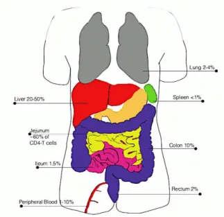
Overall, MAIT cells represent a large part of T cells though their frequency varies in different organs (Figure 4). They are mostly found in human liver (20-50% of T-cells), gastro-intestinal tract (3-5% of intestinal T cells), peripheral blood (1-10% of T cells) and lungs (2-4% of T cells)16.
Figure 4: Frequency and localization of MAIT cells16
D.
Effector functions
Upon stimulation, MAIT cells produce and secrete several cytokines, mostly TNFa, INFg and IL-17. MAIT cells exert cytotoxic functions as they are able to kill infected cells in a MR1-dependent manner through perforin and granzyme B secretion17,18. Activated MAIT cells can also trigger the activation of other immune cells such as B lymphocytes and dendritic cells, thus eliciting the recruitment of adaptive immune cells19.
II.
MAIT cell ligands and activation
A.
TCR-MR1 dependent modulation of MAIT cell activity
1. MHC class I-related (MR1) protein
The major histocompatibility complex (MHC) protects vertebrates by assuring the presentation of a wide array of antigenic peptides to T lymphocytes triggering an adaptive immune response. The MHC is subdivided into two classes (I and II) (Figure 5). Immunogenic peptide-MHC class I complexes are presented at the surface of nucleated cells to cytotoxic CD8+ T cells, while MHC class
like T cells like iNKT cells and MAIT cells. iNKT cells recognize the MHC analogue CD1d molecule that present lipidic and glycolipidic antigens (like a-GalCer) to the TCR while MAIT cells are restricted to MR1 that present small microbial antigens derived from several vitamin metabolism.
Figure 5: Different modes of antigen presentation to conventional and non-conventional T cells21
For humans, MR1 gene is ubiquitously expressed and is located on chromosome 1 near the CD1d gene. It is also highly conserved among mammalian species (90% sequence homology between mouse and human)22. Similarly to MHC class I, MR1 is constituted of three a domains (a1, a2 and a3), a transmembrane domain and an intracytoplasmic tail. To be fully functional, MR1 must bind both to a molecule of b2-microglobulin (b2m) and to its antigens.
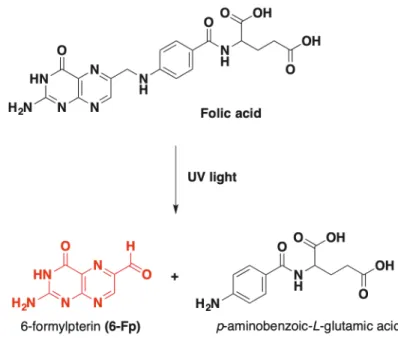
2. MAIT cell inhibitory ligands derived from folic acid (vitamin B9)
Identifying MAIT cell ligands was initially based on observing the MR1-mediated antimicrobial activity of MAIT cells against several strains of yeasts and bacteria (but not of viruses), meaning that MAIT cell ligands were produced by such micro-organisms23,24. Later, Kjer-Nielsen et al. found that a small amount of MR1 was folded in the presence of b2m and RPMI 1640, a common cell culture media, even in the absence of microbes25. RPMI medium contains many components including several vitamins, produced exclusively by yeasts and bacteria but not by animal cells. After having separately tested the different components of this culture media, they found that a significant amount of MR1 was folded in the presence of vitamin B9, also known as folic acid. Mass spectrometry analysis of the folded complex showed that it was actually 6-formylpterine (6-Fp), a photodegradation product of
vitamin B9 formed upon exposure to UV light that was bound to MR1 instead of folic acid (Figure 6). However, 6-Fp was not able to activate Jurkat.MAIT cell line (immortalized human CD4+ T lymphocyte cell line expressing human MAIT cell TCR chains) and the molecule inhibited the activation of MAIT cells by Salmonella tiphymurium supernatant.
Figure 6: Formation of 6-Fp MR1 ligand from photodegradation of folic acid26
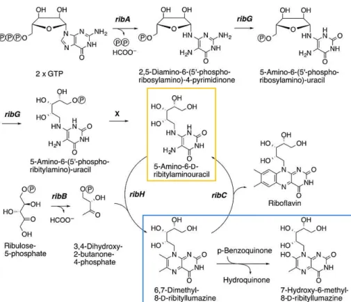
3. MAIT cell stimulatory ligands derived from the riboflavin (vitamin B2) pathway Bacterial culture of S. tiphymurium in vitamin B9 free media (to avoid any competition between stimulatory antigens and 6-Fp) allowed to isolate MR1 complex with the stimulatory antigen bound to the protein. Elution of the ligand followed by mass spectrometry analysis identified two ribityllumazine compounds derived from the riboflavin (vitamin B2) pathway as potential MAIT cell agonists: 7-hydroxy-6-methyl-8-D-ribityllumazine (RL-6-Me-7-OH) and 6,7-dimethyl-8-D-ribityllumazine (RL-6,7-diMe) (blue frame in Figure 7).
These two products were synthetized and tested for Jurkat.MAIT cell activation. Both compounds were active but with lower potency in comparison to S. tiphymurium supernatants. This meant that the chemical structure of the more potent MAIT cells agonist still was not elucidated.
Figure 7: Riboflavin and ribityllumazine biosynthesis pathways27
Three other molecules with similar chemical structure but distinct from the riboflavin pathway were then proposed as potential MAIT cell agonists: rRL-6-CH2OH, rRL-6-Me-7-OH and 5-OP-RU (with the latter initially thought to be too unstable in water) (Figure 8). rRL-6-CH2OH was synthetized and it potently activated MAIT cells but crystallographic analyses of the ternary complex (ligand bound to MR1 and the TCR) showed that it was actually a single ring molecule present in the binding pocket instead of the double lumazine rings, meaning rRL-6-CH2OH was not the potent antigen they were looking for.
Figure 8: Chemical structure of hypothetic MAIT cell agonists
N H N NH H N HO HO OH OH O O O rRL-6-Me-7-OH N H N NH H N HO HO OH OH O O rRL-6-CH2OH HO N HN NH H N HO HO OH OH O O 5-OP-RU O
A genetic approach was complementary used to help identify the ligands6. The riboflavin pathway relies on different genes called rib genes that are grouped in a same bacterial operon of Gram-positive bacteria (Figure 7). A set of individual rib gene deficient bacteria (Lactococcus lactis) was cultivated to assess the importance of the different biosynthetic intermediates for MAIT cell activation. While supernatants of ribB and ribH mutants did not induce lower MAIT cell activation than the wild-type strain, ribA and ribG mutants clearly did. This result pointed out that the molecule 5-A-RU (5-amino-6-D-ribitylaminouracil, yellow frame in Figure 7) is a key intermediate in the synthesis of MAIT cell stimulatory ligands. In the riboflavin pathway, 5-A-RU can react through a non-enzymatic reaction with 3,4-dihydroxy-2-butanone-4-phosphate to give the unstable intermediate 5-MOP-RU that spontaneously cyclizes to RL-6,7-DiMe (Figure 9). By analogy to this reaction, it was thought that 5-A-RU could react with other small endogenous metabolites such as glyoxal or methylglyoxal (derived from the glycolysis pathway) that would give respectively compounds 5-OE-RU and 5-OP-RU (the latter was discussed earlier). The two unstable pyrimidine adducts were synthetized and showed the highest potency to activate MAIT cells, confirming the elucidation of the chemical structure of the most potent MAIT cell antigens.
Figure 9: MAIT cell pyrimidine adduct antigen biosynthesis27
10c). 6-Fp also interacts with MR1 through Van der Waals interactions with Arg9, Arg94 and Ile96 residues. Most importantly, 6-Fp covalently binds to Lys43 of MR1 by forming an unusual imine (also commonly called Schiff base). This covalent bond is essential for the binding to MR1 as it will be described later. 6-Fp is deeply inserted inside MR1 cleft because of this covalent linkage, resulting in an inaccessibility to the MAIT cell TCR. Thus, even if the molecule can interact with Tyr95a by water mediated interaction, it is too weak to efficiently bind to the TCR and activate MAIT cells (Figure 10b).
Figure 10: Structural basis of MR1-binding and TCR recognition of 6-Fp (a) ternary structure of MR1 presenting 6-FP to the MAIT cell TCR; (b) contact between the MR1 bound antigen 6-FP and the TCR; (c)
interactions of 6-Fp with MR127
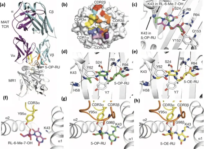
Like 6-Fp, pyrimidine adduct antigens (5-OP-RU and 5-OE-RU) form a Schiff base with Lys43 of MR1 but this time, the ribityl chain mediates strong interaction with the TCR (Figure 11c,d,e,g,h). Indeed, the four hydroxyls groups create a hydrogen bond network with both the TCR and MR1. The stimulatory ligands interact especially with Tyr95a of the TCR CDR3a loop and with diverse residues of MR1 (Arg9, Tyr152, Gln153). Ribityllumazine antigens finally have the same non-covalent interactions but are unable to form the Schiff base with Lys43 (Figure 11c,f). This is probably responsible for a different orientation of the molecule in the binding cleft, leading to a less efficient interaction with the TCR compared to 5-OP-RU and 5-OE-RU.
(c)
Figure 11: Structural basis of MR1-binding and TCR recognition of MAIT cell vitamin B2-derivative antigens. (a-c): MAIT TCR-MR1-antigen docking (a), MAIT TCR footprint on MR1 surface (b) and 5-OP-RU and RL-6-Me-7-OH overlay (c); (d-h): MR1 contacting 5-OP-RU (d) and 5-OE-RU (e), MAIT TCR contacting RL-6-Me-RL-6-Me-7-OH (f),
5-OP-RU (g) or 5-OE-RU (h)6
5. Antigen processing
To conclude on the mechanisms of antigen presentation and interaction with their targets, it is important to understand how the antigens are processed inside APCs to allow the presentation by MR1 to the TCR. In 2016, McWilliams et al. showed that the imine bond formed between Lys43 of MR1 and vitamin B-derivative antigens acted as a molecular switch allowing MR1 to egress the endoplasmic reticulum (ER) where it is sequestered at steady state28. This mechanism seems to rely on the neutralization of the positively charged amino group of Lys43 through the formation of the Schiff base (Lys43Ala mutants displaying a neutral alanine instead of a positively charged lysine spontaneously
(a) (b) (c)
(d) (e)
(f) (g)
Some questions still remain around how antigens reach the ER where MR1 resides at steady state. Indeed, MAIT cell antigens are exogenously produced by microbes, they are not produced by APCs themselves. They can be released either by microbes in the extracellular environment, in the lumen of phagosomes containing endocyted bacteria or they can come from the cytosol of cells infected by intracytosolic bacteria. In both cases, we do not yet know how they manage to reach the ER. We also don't know which cellular machinery is involved in this process (one hypothesis would involve transporters expressed at cell and/or endosome membranes).
Figure 12: MR1 trafficking and antigen processing (A) at steady state (absence of antigens); (B) in the presence of Vitamin B-derivative antigens30
B.
TCR-independent activation of MAIT cells
In addition to the TCR-dependent activation, MAIT cells can be activated in a TCR-independent manner (like iNKT cells) through innate inflammatory and antiviral cytokine stimulation (mostly IL-18, IL-12, IL-15 or IFN-a-b)1,31. These cytokines are produced by APCs through triggering of toll-like receptor (TLR) or other pattern recognition receptors (PRR) in response to viral infection. Thus, it is likely that MAIT cells can sense virus infections. The ability of MAIT cells to recognize such cytokines would also naturally extend their implication to non-infectious diseases such as inflammatory and auto-immune diseases as it will be discussed later.
C.
Summary on MAIT cell activation
To summarize, MAIT cells can be activated either in a TCR-dependent manner or through a TCR-independent mechanism relying on cytokines stimulation (Figure 13). TCR-independent activation of MAIT cells requires APCs to recognize TLR ligands, triggering the release of inflammatory cytokines (such as IL-12, IL-18). TCR-mediated activation of MAIT cells rely on the presentation of microbial antigens derived from the riboflavin biosynthesis pathway (5-OP-RU, 5-OE-RU). Antigen processing in APCs involves refolding of MR1 in the ER in the presence of these antigens that triggers trafficking of the complex to the cell membrane where it is recognized by the MAIT cell TCR. Cytokine-mediated co-stimulation can also occur. In all cases, MAIT cell activation lead to the secretion of pro-inflammatory cytokines such as IFNg or TNFa and to the development of a cytotoxic phenotype (similar to CD8+ cytotoxic T cells).
Figure 13: Summary of MAIT cell activation process32
Having described the main characteristics of MAIT cells and how MAIT cells can sense infectious diseases, we will now discuss MAIT cells' immune roles, especially for infectious diseases, and provide some insight around their potential use in immunotherapy.
III.
MAIT cells as potential therapeutic targets
A.
Protection against infectious diseases
1. Bacterial infections
Many studies were conducted to understand how MAIT cells contribute to protective immunity against bacterial infection in human and mice. So far, the most studied bacterial infection has been tuberculosis due to Mycobacterium tuberculosis (Mtb). The first evidence that MAIT cells could recognize Mycobacterium tuberculosis was made by Gold et al. in 2010 (before the elucidation of MAIT cell antigens) with highlight of a MAIT cell population able to detect bacterially infected human cells24. Other studies showed a decreased frequency of peripheral blood MAIT cells in patients infected by Mtb23,33–37. This phenomenon was observed with other bacteria such as Pseudomonas aeruginosa in cystic fibrosis38, Vibrio cholerae39, Shigella in vaccine clinical trials18, Helicobacter pylori40 and also in septic shocks41. MAIT cells were also able to accumulate in tissues infected by different bacteria (in human and mice)23,42,43. An important study demonstrated a critical role of MAIT cells in the response of mouse model lung infection with live Francisella talurensis44. MAIT cells could colonize and expand in the lungs at both early and intermediate stages and they still expanded after clearance of the bacteria. MAIT cells also produced pro-inflammatory cytokines INFg, TNFa and IL-17. Moreover, MR1 knockout mice showed higher bacterial burden correlated to a delay in the recruitment of INFg producing CD4+ and CD8+ T cells into the lungs. Another study in mice pointed out the protective effect of MAIT cells against pulmonary infection due to Legionella longbeachae45.
Altogether, these data suggest that MAIT cells can migrate from peripheral blood to infected tissues where they could contribute to host defense against infections through direct cytotoxicity, secretion of pro-inflammatory cytokines (IFNg, TNFa or IL-17) or other mediators that are yet to be discovered18,46.
2. Viral infections
Recent data suggest that MAIT cells can be activated and expanded in a TCR-independent manner in the course of diverse viral infections including hepatitis C, dengue and influenza virus47,48.
B.
Other roles of MAIT cells in non-infectious diseases
1. Auto-immune and inflammatory diseases
The involvement of MAIT cells in immunity is not restricted to infectious diseases since MAIT cells respond to various cytokine stimuli. MAIT cells are modulated in several auto-immune and inflammatory diseases such as multiple sclerosis, gastro-intestinal disorders (celiac disease and inflammatory bowel diseases), allergic diseases, asthma and metabolic diseases (type 1 and 2 diabetes)49,52. Like for microbial infection, a decrease of MAIT cell frequency in peripheral blood was often found, probably reflecting a migration to inflamed sites in the course of immune-mediated diseases53. However, there is no clear evidence of the protective or pathogenic role of MAIT cells in such diseases.
2. Cancer
MAIT cells' roles in malignant diseases have also been investigated (both for solid and hematological malignancies) but still remain poorly understood. Patients with cancer often showed a depletion of MAIT cell population in peripheral blood compared to healthy donors49,52,53. In parallel, MAIT cells could infiltrate various tumors and metastases but it is unclear whether this phenomenon contributes to host protection or to pathogenesis. Furthermore, an impairment of MAIT cell cytokine profile was described several times, notably a decreased of INF-g secretion and/or an increased production of cytokines supporting tumor growth like IL-17. A recent in vivo study on mice suggests a pro-tumorigenic effect of MAIT cells caused by the suppression of T and/or NK cells (partly due to IL-17 secretion) after interaction with MR1 molecules expressed on tumor cells54. Pre-treatment of tumor cells with 5-OP-RU was also associated with an increase of lung metastasis.
3. Graft-versus-host-disease (GvHD)
MAIT cells are preferentially localized in organs implicated in GvHD (such as liver) justifying the investigations related to this disease. MAIT cells are unlikely to induce alloreactive response and GvHD since they are not directed towards polymorphic MHC molecules55. Little data is available on this topic but one study showed that an early increase in the number of MAIT cells in peripheral blood after stem cell transplantation was associated with a reduced risk of GvHD53.
C.
Development of immunotherapies targeting MAIT cells
As depicted above, MAIT cells have important immune functions since they seem to be involved in the response to many diseases. MAIT cells could represent attractive targets for cancer immunotherapy because of their secretory capacity and their ability to recruit other immune cells. However, more investigation is required to assess the safety and the efficacy of this therapeutic strategy. If MAIT cells appeared to induce pro-tumorigenic effect as it was supposed in the study discussed above54, a direct inhibition of MR1 could be envisioned with inhibitory ligands. Another promising therapeutic option under study is the development of CAR (chimeric antigen receptor)-MAIT cells.
The antimicrobial activity of MAIT cells is without any doubt the best-documented immune role of MAIT cells. There is strong evidence that MAIT cells help fight several bacterial (and perhaps viral) diseases. Thus, they potentially represent an interesting therapeutic target for innovative antimicrobial immunotherapies. They could especially be a good target for new antimicrobial mucosal vaccines. Indeed, most existing vaccines are administered through the systemic route and they may not confer an effective and durable mucosal immunity56. Targeting MAIT cells with new mucosal vaccines could overcome this issue since this population of T cells is abundant at mucosal sites (lungs, gastro-intestinal tract…). We could consider directly targeting MAIT cells with such vaccines since they display a memory phenotype. Another option (perhaps more relevant) would be to harness MAIT cells as vaccine adjuvants in order to boost systemic vaccine efficacy.
It is clear that the feasibility of such vaccine would rely on the discovery of more stable MAIT cell agonists than the very unstable ones we have (5-OP-RU, 5-OE-RU). Having understood this critical element, several research teams deeply investigated MAIT cell antigens to unravel the molecular basis of interaction with MR1 and the MAIT cell TCR. A better knowledge of the structure-activity relationships of MAIT cell antigens would greatly help design new stable MAIT cell ligands. A review of all the research completed in this field is presented in the following and final section of this introductory chapter.
IV.
Overview of the research aiming to find new antigens of MAIT cells
A.
Synthesis of new MAIT cell competitive antagonists
Several analogues of 6-Fp were synthetized, the first being acetyl-6-formylpterin (Ac-6-Fp). It proved to be much more potent than 6-Fp to inhibit MAIT cell activation, presumably due to a higher chemical stability (Figure 14)57. Soudais et al. reported later the synthesis and biological evaluation of two other analogues of 6-Fp that they called Compound A (2-amino-4-hydroxy-6-formylpteridine dimethyl acetal) and Compound C (2-acetylamino-4-hydroxy-6-formylpteridine dimethyl acetal). While Compound A did not show inhibitory activity, Compound C showed a concentration-dependent activity similar to Ac-6-Fp for MR1 up-regulation (reflecting the binding affinity to MR1) and inhibition of MAIT cell activation.
Figure 14: Chemical structure of synthetic MAIT cell inhibitory ligands
B.
Analytical study and synthesis of new stable agonists of 5-OP-RU
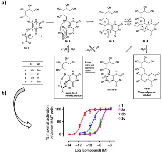
The a-iminocarbonyl moiety of pyrimidine adduct antigens is prone to hydrolysis and cyclisation to give thermodynamically stable but far less antigenic ribityllumazines. Mak et al. investigated the underlying mechanism causing this instability to overcome this problem. For this purpose, they studied the effect of the solvent on the formation and the stability of three different pyrimidine adduct derivatives (3a-c) synthetized through condensation reaction between 5-A-RU and small dicarbonyl metabolites: butane-2,3-dione (3a), glyoxal (5-OE-RU 3b) and methylglyoxal (5-OP-RU 3c). In PBS (pH 8.0, 15°C), 3a underwent degradation in less than five minutes while 3b had a half-life of 18 minutes. 3c (5-OP-RU) was more stable than the two other compounds with a half-life of 14 hours even if the stability decreased with lower pH and higher temperature. Under physiological conditions (PBS, pH 7.4, 37°C), the half-life of 3c was only 88 minutes. Among the three molecules, 5-OP-RU (3c)
N N HN N OCH3 OCH3 O H2N 2-amino-4-hydroxy-6-formylpteridinedimethylacetal (Compound A) N N HN N OCH3 OCH3 O N H O 2-acetylamino-4-hydroxy-6-formylpteridinedimethylacetal (Compound C) N N HN N O N H O H O 2-acetylamino-4-hydroxy-6-formylpteridin (Ac-6-Fp)
Then, the authors deeply studied the kinetic of formation of 5-OP-RU and the influence of the solvent used for the reaction. They showed that the condensation reaction between methylglyoxal and 5-A-RU in PBS (pH 7.4, 37°C) reached a maximum concentration of 5-OP-RU corresponding to only 1.1% conversion after 5 minutes. The same reaction done in DMSO led to total conversion of the starting material to 5-OP-RU after 2 days. Furthermore, the imine formed in DMSO was reasonably stable with more than 90% of the product remaining unchanged after 2 days at 22°C. A mechanism was proposed in which the reaction should be under thermodynamic control in water. Trans kinetic product 3 would rehydrate to give 6, followed by cyclisation via 7 (or cis 3) giving at the end thermodynamic product lumazines 4 (Figure 15a). In polar aprotic solvent DMSO, the reaction should be under kinetic control where trans 3 product should form faster than cis 3. Once formed, trans 3 is unlikely to isomerize because of a high energy barrier for the isomerization thus stabilizing the pyrimidine antigens.
Figure 15: Analytical study and biological evaluation of 3a-c; a) Study of the mechanisms of antigenic pyrimidine adducts synthesis in water or DMSO; b) biological evaluation of 3a-c (on Jurkat.MAIT cell line)58
The same study reported the design and synthesis of three potential stable analogues of 5-OP-RU. They synthetized two N-methylated products keeping either the a-iminocarbonyl group (9) or replacing the imine function by an alkene (10). The third molecule 11 contained an alkyl ribityl chain
b)
a)
lacking the reactive secondary amino group and the imine was also replaced by an alkene like for 10 (Figure 16).
Figure 16: Chemical structure of compounds 9-11
Surprisingly, 9 was even less stable than 5-OP-RU under physiological conditions while 10 was slightly more stable though it also rapidly formed quaternary ammonium cyclized species like 9. In contrast, compound 11 was completely stable. Biological evaluation on Jurkat.MAIT cells showed an important loss of potency for the three molecules in comparison to 5-OP-RU (EC50 1.6 pM). 11 (EC50 1.6 nM) and 9 (EC50 14 nM) displayed similar potencies (1000-fold less active than 5-OP-RU) while 10 was poorly active (EC50>10 µM).
C.
Identification of drugs and drug-like molecules able to modulate MAIT cell
activity
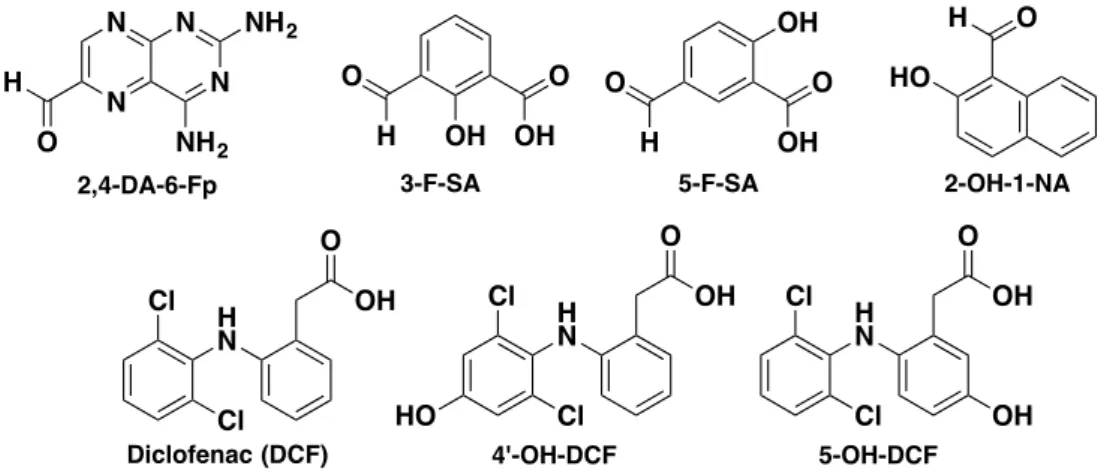
Another study identified several drugs and drug-like molecules able to modulate the functions of MAIT cells59. Multiple in silico screening were done to identify new MR1 binding ligands and MAIT cell agonists. 81 compounds of the in silico hits were biologically evaluated. Among them, several molecules could up-regulate MR1 and/or activate MAIT cells. Compounds 3-formaldehyde-salicylic acid (3-F-SA), 5-formaldehyde-salicylic acid (5-F-SA), 2-hydroxy-napthaldehyde (2-OH-1-NA) and 2,4-diamino-6-formylpteridine (2,4-DA-6-Fp, obtained from the photodegradation of antineoplastic drug aminopterin) were able to up-regulate MR1 of C1R.MR1 cells (C1R human lymphoblastoid cells overexpressing MR1). 3-F-SA and 2-OH-1-NA inhibited Jurkat.MAIT cells while 5-F-SA moderately activated specific clones of Jurkat.MAIT cells (depending on the nature of the TCRb chain).
The well-known and commonly used drug diclofenac (used for the treatment of inflammatory NH H N O O N O N HO HO OH OH 9 NH H N O O O N HO HO OH OH 10 NH H N O O O HO HO OH OH 11
Figure 17: Chemical structure of drugs and drug-like molecules able to modulate MAIT cell functions Interestingly, crystallographic data showed that all the inhibitory compounds bearing an aromatic aldehyde functional group were able to form a Schiff base with Lys43 of MR1 like 5-OP-RU whereas diclofenac and its metabolites did not form such chemical bond. The inhibitory compounds did not directly interact with the TCR in the MR1-TCR complex like 6-Fp, thus explaining their inhibitory effect. In contrast, diclofenac and its metabolites could make few interactions with the TCR but their mode of binding was different and less effective than the one of 5-OP-RU leading to a weak activation of Jurkat.MAIT cells. Nevertheless, this study proved that MR1 can present other compounds than pyrimidine adduct antigens and folic acid derivatives to the MAIT cell TCR. Thus, it offers the possibility of discovering new small molecules able to modulate MAIT cell activity. It finally gave some insight about the poorly understood contribution of the b chain to the TCR binding of the ligands, since DCF and 5-F-SA only activated one clone of Jurkat.MAIT cells bearing a specific TCRb chain (Jurkat.MAIT-A-F7 cell strain).
D.
Design of 5-OP-RU analogues to unravel the structural basis for the
recognition of MAIT cell antigens by MR1 and the TCR
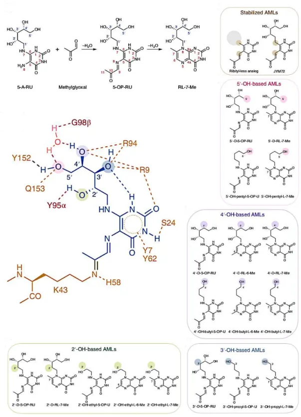
A large survey was recently published in which the authors studied structure-activity relationships (SAR) of twenty 5-OP-RU altered metabolites ligands (AMLs) compared to 5-OP-RU and its degradation product RL-7-Me60. Among these molecules, the chemical synthesis of deoxy AMLs (2’-D-5-OP-RU, 3’-(2’-D-5-OP-RU, 4’-D-5-OP-RU 5’-D-5-OP-RU) and their corresponding lumazine degradation products (2’-D-RL-7-Me, 3’-D-RL-7-Me, 4’-D-RL-7-Me, 5’-D-RL-7-Me, respectively) was previously described by Ler et al.61. They additionally synthetized series of pyrimidine and lumazine ligands bearing a monohydroxy ribityl chain (2’-OH-ethyl-5-OP-U, 2’-OH-ethyl-L-6-Me, 3’-OH-ethyl-5-OP-U…). Finally, a ribityl-less analogue and JYM72 (previously discussed as compound 1158) were also included in the study (Figure 18).
O H OH O OH O H O OH OH HO O H 2-OH-1-NA 3-F-SA 5-F-SA Cl H N O OH Cl H N O OH HO Cl H N O OH OH
Diclofenac (DCF) 4'-OH-DCF 5-OH-DCF
Cl Cl Cl N N N N H O NH2 NH2 2,4-DA-6-Fp
Table 1: Chemical stability, MR1 up-regulation, MAIT cell activity and binding affinity of AMLs60
The different pyrimidine adduct AMLs showed almost the same instability as 5-OP-RU while the ribityl-less analogue and JYM72 were highly stable (t1/2>100h in PBS, pH 7,4, 37°C). All the compounds could up-regulate MR1 with the ribityl-less analogue showing the best potency. Ribityllumazine analogues displayed the slower kinetic of up-regulation, most likely because they were unable to form a Schiff base with Lys43 of MR1. Unstable AMLs reached a maximum up-regulation after 4-8h while stable ribityl-less analogue and JYM72 continued to up-regulate MR1 after 24h. All these results suggest that concentration and time-dependent MR1 up-regulation rely on the stability of the ligands and on their ability to form a covalent bond with Lys43 of MR1. Thus, the ribityl chain does not seem to be a prerequisite for inducing strong MR1 up-regulation and it might be rather deleterious.
MAIT cell activation assay showed that 4’-D-5-OP-RU and 5’-D-5-OP-RU could still activate MAIT cells with strong potency close to the one of 5-OP-RU while 4’-OH-butyl-5-OP-U and 5’-OH-pentyl-5-OP-U did not activate MAIT cells. This means that 4’-OH and 5’-OH are probably not essentials for the binding to the MAIT cell TCR. On the contrary, 2’-OH and 3’-OH seem to be very important for MAIT cell activation since compounds lacking one of these hydroxyl groups (2’-D-5-OP-RU and 3’-D-5-OP-RU) were not active. Finally, the different lumazine compounds showed only moderate MAIT cell activation presumably due to the absence of covalent linkage to Lys43 though they maintained weak interaction with the TCR.
To finish, the authors gave some information about the structural basis explaining the structure-activity relationships of the AMLs by studying ternary structures of the ligands interacting with MR1 and the TCR. They affirmed that the 2’-OH and 5’-OH groups of 5-OP-RU formed an essential interaction triad with Tyr95 of the TCRa and Tyr152 of MR1. According to them, AMLs that perturbed
these interactions were subject to a dramatic loss of potency. The strong potency of 4’D-5-OP-RU and 5’-D-5-OP-RU was theoretically explained by conformational malleability and dynamic compensations inside the binding groove allowing to maintain this interaction triad. The moderate AMLs displayed an altered interaction triad causing a decrease of biological activity while the weak or non-agonistic AMLs like 4’-OH-butyl-5-OP-U and 5’-OH-pentyl-5-OP-U simply could not interact properly with the TCR because they lack 2’-OH and 3’-OH groups.
A second recent study described the synthesis and study of new glyco-analogues of 5-OP-RU and RL-6-Me-7-OH to give insight about the influence of the chemical changes on MAIT cell activation26,62. Several analogues with different absolute configuration, with 2’-OH deoxygenation or displaying longer ribityl chain were synthetized (Figure 19).
b. R = OH OH OH OH OH OH OH OH OH OH OH OH OH OH OH OH b. R = OH OH OH OH OH OH OH OH d.R = OH OH OH c. R = OH OH e. R = OH OH OH N N N HN R OH O O NH HN O O N H N O R a. R = OH OH OH OH a. R = OH OH OH OH OH OH OH OH g. R = OH OH h. R = OH OH OH f. R = OH OH OH OH 5-OP-RU RL-6-Me-7-OH 5a-h 3a-e d. R = c. R = e. R =
MAIT cell line). In contrast, all the glyco-analogues of 5-OP-RU were active at 10 µM concentration. 5d, 5f, 5g and 5h displayed a similar activity compared to 5-OP-RU while 5b, 5e and especially 5c (lacking 2'-OH) were less potent than 5-OP-RU. A virtual docking study was done to help explain these results. It showed that 5c and 5e probably did not interact with Tyr95 of the TCRa, thus explaining the low activity. The authors emitted the theory that the more the compounds could make interactions with Tyr95a residue, the more potent they were.
The same research groups recently published a second survey in which they synthesized several molecules by modulating the 6-aminoalkyl (ribityl) chain63. They produced three aminoalkyl derivatives with a single terminal alcohol (3b-d), a N-methylated analogue (3e) and three aminoalkyl products without any hydroxyl group (3f-h).
Figure 20: Chemical structure and biological evaluation of 6-alkylamino analogues of 5-OP-RU; 1b is Ac-6-Fp and 3a is 5-OP-RU (adapted from Braganza et al.)63
Biological evaluation of these molecules revealed a high potency of 3b and 3c at activating MAIT cell line 6C2 similarly to 5-OP-RU (10 µM concentration). The authors suggested that 3b is of high interest for the study of MAIT cells since it is as potent as 5-OP-RU and its synthesis is easier and faster compared to the synthesis of 5-OP-RU. However, these data are contradictory with the one of Awad
et al. (described above)60. They produced and tested the same monohydroxy analogue 3b but the biological evaluation showed almost no Jurkat.MAIT cell activation. The use of different biological models by the two research groups (different MR1 expressing cells and MAIT cells) could explain these divergent results. Thus, further investigation is required to confirm these observations.
HN NH O O N H OH N O n 3b n= 1 3c n= 2 3d n= 3 HN NH O O N H N O HN NH O O N H N O n 3f n= 1 3g n= 2 3h n= 3 3e
Research work
I.
Introduction
As described earlier, MAIT cells could become attractive targets for new immunotherapies against infectious diseases (mucosal vaccines) and presumably other pathologies like cancers. However, more investigation is required to validate this therapeutic approach and many questions around MAIT cell biology and ligands must be addressed.
The recent discovery of MAIT cell highly potent antigens (5-OP-RU and 5-OE-RU) was a major breakthrough in the field. The thorough investigation of MAIT cell antigens, at the interface of chemistry and biology, provided some valuable information, especially around the molecular basis governing their interaction with MR1 and the MAIT cell TCR. Despite all these discoveries, we still lack stable highly potent analogues of MAIT cell antigens. Today, it is one of the main hurdles limiting the efficient study of MAIT cells as potential therapeutic targets. Thus, we urgently need to find original research approaches in order to produce new potent antigen of MAIT cells.
We wanted to address this major issue by adopting two different medicinal chemistry approaches. The first one consisted in doing original pharmacomodulations on 5-OP-RU chemical structure (Figure 21). All our work was focused on 5-OP-RU (like most studies on MAIT cells) since it is the most potent MAIT cell antigen and it is also more stable than 5-OE-RU58. By doing pharmacomodulations of 5-OP-RU, we aimed at obtaining stable products with comparable activity to OP-RU. The second strategy relied on designing prodrugs of A-RU, the biosynthetic precursor of 5-OP-RU able to generate 5-5-OP-RU in situ after condensation with endogenous methylglyoxal. Our final objective was to develop new chemical probes that could help understand MAIT cell biology. For this Chemical Biology approach, we designed a unique clickable analogue of 5-OP-RU that can be used to track MAIT cell ligands inside biological tissues and cells.
Figure 21: Objectives of the research project
NH H N O O HN HO HO OH OH N O H 5-OP-RU NH H N O O HN HO HO OH OH N O 5-A-RU Cleavable linker N3 CuAAC Cu(I) Fluorescent probe 5-OP-RU HN NH N
3) Design and synthesis of new chemical tools for the study of MAIT cell biology
5-OP-RU Stabilization by
replacement of chemically unstable moieties
Carrier
2) Synthesis of 5-A-RU prodrugs with in situ formation of 5-OP-RU
+ Endogenous methylglyoxal Cleavage in cells 5-A-RU NH2
5-OP-RU
II.
Synthesis and study of stable analogues of 5-OP-RU
A.
Synthesis and chemical study of 5-A-RU and 5-OP-RU
1. Bibliographic review
5-OP-RU is unstable in physiological media and therefore very difficult to isolate for further use in biology. The molecule is commonly formed in situ prior to addition on cells by condensation between synthetic 5-A-RU and commercial methylglyoxal. Unlike 5-OP-RU, 5-A-RU was discovered many years ago and has been extensively used as a chemical intermediate for the synthesis of riboflavin and lumazine analogues. Several research groups have developed efficient synthesis routes towards 5-A-RU since the late 1950’s and all this precious work has been exploited to synthetize 5-OP-RU for the study of MAIT cells. All published chemical routes rely first on the synthesis of the D-ribitylamine tail, followed by its coupling to the uracil heterocycle and amine functionalization. The main syntheses of 5-A-RU are presented in this section.
The initial syntheses of 5-A-RU were described by Maley and Plaut64 and Winestock and Plaut65 (Scheme 1). 5-A-RU was used as an intermediate for the synthesis of 6,7-dimethyl-8-ribityllumazine. Their syntheses started from D-ribose and went through a D-ribityloxime intermediate, which was then reduced by hydrogenation with platinum oxide heterogeneous catalyst. D-ribitylamine reacted with 6-chlorouracil and position 5 of the molecule was functionalized by a nitroso group. Reduction of the nitroso with sodium dithionite afforded 5-A-RU, which was directly used without isolation for the last step of the synthesis.
Scheme 1: First synthesis of 5-A-RU by Plaut and co-workers. Reagents and conditions: NH2OH.HCl, NaOMe in
EtOH (yield not mentioned); (ii) H2, PtO2 in AcOH; (iii) 6-chlorouracil, 128°C, 44%; (iv) HNO2 in H2O, 72%; (v)
Na2S2O4 in H2O (product not isolated); (vi) biacetyl
5-A-RU was used for the synthesis of other molecules and several modifications of the initial synthesis were done keeping the same synthesis strategy (Scheme 2). Katagiri et al. proposed a different synthesis of the D-ribitylamine chain by reductive amination with ammoniac and sodium borohydride66. Other variations were performed by Wood and co-workers with the use 6-chloro-5-nitrouracil instead of 6-chlorouracil for the coupling reaction with D-ribitylamine in ethanol67,68. The nitro group at ortho position of the chloro allowed the reaction to proceed at room temperature in contrast to the synthesis of Plaut and co-workers that required strong heating at 128°C. Reduction of the nitro was done by hydrogenation either with platinum oxide or with palladium on charcoal (Pd/C). Later, Cushman et al. described hydrogenation of the nitroso precursor in aqueous HCl to get a more stable form of 5-A-RU as hydrochloride salts (instability issues will be discussed later)69.
O OH OH HO HO HO HO OH OH N HO HO OH OH NH2 HO (i) (ii) NH H N O O HN HO HO OH OH NH H N O O HN HO HO OH OH ON NH H N O O HN HO HO OH OH H2N (iii) (iv) (v) NH N O N HO HO OH OH N O (vi)
Scheme 2: Different modifications of the initial synthesis of 5-A-RU (by Plaut and co-workers) for the synthesis of lumazine derivatives. Modifications are highlighted in red
Relying on these published syntheses and chemical procedures, 5-A-RU as a precursor of 5-OP-RU could be efficiently synthesized. The initial syntheses of 5-OP-5-OP-RU were done by Corbett et al.6 and Mak et al.58 (research group that initially identified MAIT cell antigens). To make 5-OP-RU, they produced 5-A-RU precursor by adapting the synthesis of Plaut and co-workers. They synthetized the D-ribitylamine moiety by forming a D-ribitylbenzylamine intermediate instead of an oxime through the reaction of D-ribose with benzylamine, followed by hydrogenation reaction with platinum oxide catalyst (Scheme 3). Then, D-ribitylbenzylamine was reduced to D-ribitylamine by hydrogenation over Pd/c in water.
Scheme 3: Recent modifications of the synthesis of 5-A-RU to make 5-OP-RU. Modifications are highlighted in red.
Modification of this synthesis was recently done by Lange et al. that aimed to produce a prodrug of 5-A-RU70. They reduced D-ribitylbenzylamine by hydrogenation using Pd(OH)
2 instead of Pd/C (in a mixture of methanol and ammoniac) to afford D-ribitylamine in high yield (99%). The same authors also proposed an efficient coupling reaction with 6-chlorouracil using ethylene glycol as